Embed Size (px)
Citation preview

The immunomodulator AS101 abrogates DSS-induced Colitis
1
Multifunctional activity of a small tellurium redox immunomodulator compound, AS101, on DSS-
induced murine colitis.
Gilad Halpert*, Tom Eitan
*, Elena Voronov
†, Ron N. Apte
†, Lea Rath-Wolfson
‡, Michael Albeck
§,
Yona Kalechman* and Benjamin Sredni
*
*C.A.I.R. Institute, The Safdié AIDS and Immunology Research Center, The Mina & Everard Goodman
Faculty of Life Sciences, Bar-Ilan University, Ramat-Gan 52900, Israel. †The Shraga Segal Department of Microbiology and Immunology, and Faculty of Health Sciences and
Cancer Research Center, Ben-Gurion University of the Negev, Beer-Sheva 84105, Israel. ‡Department of Pathology, Rabin Medical Center, Golda Campus, Petah Tikva 49372, Israel, and Sackler
School of Medicine, Tel Aviv University, Tel Aviv, Israel. §Department of Chemistry, Faculty of Exact Sciences, Bar-Ilan University, Ramat-Gan 52900, Israel.
Running title: The immunomodulator AS101 abrogates DSS-induced colitis
Keywords: Inflammatory bowel diseases, inflammation, cytokine, intestine, innate immunity,
immunomodulator, AS101, DSS, Interleukin 17, Interleukin 1β.
Capsule:
1. Background: AS101, a novel small tellurium
compound, is a potent anti-inflammatory/anti-
apoptotic immunomodulator.
2. Results: AS101 prevents leukocyte migration
and protects the mucosal barrier in murine
colitis.
3. Conclusion: AS101 exerts multifunctional anti
inflammatory/anti-apoptotic activity in
experimental Inflammatory Bowel Diseases.
4. Significance: This is the first report showing
significant effect of tellurium-based compound
for the treatment of inflammatory bowel
diseases.
Abstract
Inflammatory bowel diseases (IBD) are a group
of idiopathic, chronic immune-mediated diseases
characterized by an aberrant immune response,
including imbalances of inflammatory cytokine
production and activated innate and adaptive
immunity. Selective blockade of leukocyte
migration into the gut is a promising strategy for
the treatment of IBD. This study explored the
effect of the immunomodulating tellurium
compound, ammonium trichloro (dioxoethylene-
o,o') tellurate (AS101) on DSS-induced murine
colitis. Both oral and i.p. administration of AS101
significantly reduced clinical manifestations of
IBD. Colonic inflammatory cytokine levels (IL-17,
IL-1β) were significantly downregulated by AS101
treatment, whereas IFN-γ was not affected.
Neutrophil and α4β7+ macrophage migration into
the tissue was inhibited by AS101 treatment.
Adhesion of Mesenteric Lymph Node (MLN) cells
to mucosal addressin cell adhesion molecule
(MAdCAM-1), the ligand for α4β7 integrin, was
blocked by AS101 treatment both in vitro and in
vivo. DSS-induced destruction of colonic epithelial
barrier/integrity was prevented by AS101, via
upregulation of colonic Glial-Derived
Neurotrophic Factor (GDNF), which was found
previously to regulate the intestinal epithelial
barrier through activation of the PI3K/AKT
pathway. Indeed, the upregulation of GDNF by
AS101 was associated with increased levels of
colonic pAKT and BCL-2, and decreased levels of
BAX. Furthermore, AS101 treatment reduced
colonic permeability to Evans Blue, and decreased
colonic TUNEL+ cells. Our data revealed
multifunctional activities of AS101 in the DSS-
induced colitis model via anti inflammatory and
anti apoptotic properties. We suggest that
treatment with the small, non toxic molecule,
AS101, may be an effective early therapeutic
approach for controlling human IBD.
Introduction
Crohn's disease (CD) and ulcerative colitis (UC)
are the two major forms of chronic inflammatory
bowel disease (IBD). Studies in humans and in
experimental models of IBD have revealed
impaired mucosal barrier function, activated innate
immunity, altered production of Th1 and Th2
http://www.jbc.org/cgi/doi/10.1074/jbc.M113.536664The latest version is at JBC Papers in Press. Published on April 24, 2014 as Manuscript M113.536664
Copyright 2014 by The American Society for Biochemistry and Molecular Biology, Inc.
by guest on March 24, 2018
http://ww
w.jbc.org/
Dow
nloaded from

The immunomodulator AS101 abrogates DSS-induced Colitis
2
cytokines, and the activation of CD4+ T cells in the
pathogenesis of IBD (1-3). T regulatory cells play
an essential role in the maintenance of intestinal
immune homeostasis and were also shown to be
capable of preventing and reversing established
colitis (4). The etiologies of these diseases remain
unclear. The most widely held hypothesis
regarding the pathogenesis of IBD is that aberrant
activation or defective downregulation of innate
immunity, and overly aggressive acquired (T cell)
immune responses to a subset of commensal
enteric bacteria develop in genetically susceptible
hosts, and environmental factors precipitate the
onset or reactivation of disease (1,2,5). Cytokines provide key signals in the intestinal
immune system, and were shown to have a central
role in the pathophysiology of IBD (6). It is
evident now that both innate cell populations (e.g.
γδ T cells, LTi-like cells, and
monocytes/macrophages) and adaptive T cells
(Th17 cells) can produce IL-17, which has a
crucial role in inflammatory and autoimmune
diseases (7-10). IL-17 was found to be elevated in
peripheral blood and intestinal tissue of IBD
patients (11,12). In addition to IL-17, IL-1 is
important in the pathogenesis of IBD due to its
pro-inflammatory activities. Peripheral blood
mononuclear cells obtained from patients with CD
were shown to produce high levels of IL-1 in vitro
compared with normal control cells (13), and
enhanced secretion of IL-1β was shown in colonic
biopsies from patients with inflammatory bowel
disease (12,14). Based on these findings, several
cytokine-based therapies have been developed and
tested for the treatment of IBD patients (6,15).
Macrophages have been implicated in the
pathogenesis of a variety of chronic and
autoimmune diseases including IBD. The
classically activated macrophages are key
producers of many cytokines (e.g. IL-1, IL-6 and
TNFα) and reactive metabolites of oxygen and
nitrogen (e.g. nitric oxide) that have been
implicated in the development of IBD (16,17).
Feeding mice with polymeric Dextran Sodium
Sulfate (DSS) in their drinking water induces acute
or chronic colitis characterized by bloody diarrhea,
ulceration, loss of body weight, and infiltration
with granulocytes or mononuclear cells, reflecting
IBD symptoms (18). This widely used murine
colitis model is helpful for studying the effect of
novel therapeutic agents for the management of
IBD (19).
Integrins are transmembrane cell adhesion
receptors composed of noncovalently associated α
and β subunits that bind to cell-surface ligands,
soluble ligands and extracellular matrix proteins
(20). These adhesive interactions are essential for
leukocyte recirculation, migration into
inflammatory sites, recognition of foreign
antigens, and survival and proliferation (21-23).
Circulating T cells that bear α4β7 integrins bind to
Mucosal Addressin Cell Adhesion Molecule
(MAdCAM), which is constitutively expressed on
high endothelial venules of Peyer's patches (PP)
and Mesenteric Lymph Nodes (MLN), as well as
on postcapillary venules of gut lamina propia (LP)
(2,24). MAdCAM-1 expression is upregulated on
inflamed venules in chronic inflammatory diseases
such as in IBD, diabetes, chronic relapsing
experimental autoimmune encephalomyelitis etc.
Of note, α4β7 integrin is also expressed on
stimulated monocytes and macrophages (25).
Monoclonal antibody to integrin-α4 (natalizumab),
which binds both integrin-α4β7 (the MAdCAM-1
ligand) and integrin-α4β1 (the Vascular Cell
Adhesion Molecule-1(VCAM-1) ligand), is
effective in treating Crohn’s disease (26), while a
humanized anti-integrin-α4β7 antibody has been
used to treat active ulcerative colitis (27).
The neurotrophin - Glial-Derived Neurotrophic
Factor (GDNF) has a strong effect on neurite
outgrowth and differentiation and is able to protect
neurons from apoptosis under various conditions.
Interestingly, it has been shown that ablation of
enteric glia leads to a fulminant hemorrhagic
jejunoileitis in mice (28), suggesting a central role
of the enteric glia in the maintenance of gut
mucosal integrity. GDNF is upregulated in IBD,
and has strong anti-apoptotic properties in colonic
epithelial cells via activation of MAPK and
PI3K/AKT pathways (29).
AS101, is a small non toxic tellurium-IV
compound (Fig. 1), currently being evaluated in
PhaseII clinical trials in psoriasis patients
(unpublished data) and in prevention of bone
marrow toxicity induced by chemotherapy in
cancer patients (30). This compound is a potent
immunomodulator (in-vitro and in-vivo) with a
variety of potential therapeutic applications (31-
33). Accumulated evidence suggests that much of
the biological activity of tellurium compounds is
by guest on March 24, 2018
http://ww
w.jbc.org/
Dow
nloaded from

The immunomodulator AS101 abrogates DSS-induced Colitis
3
directly related to their specific chemical
interactions with endogenous thiols. If the reacting
thiol is a cysteine residue, the reaction product
may alter the biological activity of the target
protein (34). The Te(IV)-thiol chemical bond may
lead to conformational change or disulfide bond
formation, possibly resulting in a loss of biological
activity, if the thiol residue is essential for a
particular function. Indeed, we demonstrated that
AS101 specifically inactivates cysteine proteases
while exhibiting no effect on the other families of
serine-, aspartic-, and metallo-proteases, in good
agreement with the predictions of its unique Te
(IV)-thiol chemistry. Furthermore, the proteolytic
activity of the inactivated cysteine proteases could
be recovered by reducing agents such as NaBH4,
further supporting the suggestion that the
inactivation process involves oxidation of the
catalytic thiol to a disulfide (34). In light of its
chemical activities, AS101 has been shown to
exert anti inflammatory and anti apoptotic effects
in different in vivo models (35-38), through
inhibition of caspase 1, 3 and 8 activities and by its
ability to induce GDNF production (38,39).
In the present study, we tested the effect of
AS101 on a DSS-induced murine colitis model.
Our findings revealed anti-inflammatory and anti-
apoptotic properties of AS101 through the
downregulation of colonic inflammatory cytokine
levels, prevention of inflammatory cell migration
into the tissue, and inhibition of colonic apoptotic
processes.
Experimental procedures
Mice
Male C57BL/6 mice, 8-12 weeks old, were
purchased from Harlan Laboratories (Jerusalem,
Israel). Mice were kept under specific pathogen-
free (SPF) environment and fed standard pellet diet
and tap water. Mice were allowed to acclimate 7
days before the experiments. Animal experiments
were performed in accordance with institutional
protocols, and experiments were approved by the
Institutional Animal Care and Use Committee.
Induction of DSS colitis
Mice were divided into groups with identical
average body weight. DSS (M.W. 36000-50000;
MP Biomedicals, LLC, Eschwege, Germany) was
added to tap water at a concentration of 3.5%
(w/v). Mice were given DSS solution for 7 days,
followed by 5 days of regular water. Fresh DSS
solution was prepared every other day.
AS101
AS101 was supplied in a solution of PBS, pH
7.4, and maintained at 4°C. Before use, AS101 was
diluted in PBS, and the indicated doses were used.
AS101 treatment
AS101 was administrated either by i.p.
injection or by oral administration. In the injection
treatment, mice were given DSS, and injected
daily i.p. with PBS [DSS+PBS] or with AS101
(10µgr/mouse per injection) either concomitantly
with the initiation of DSS supplementation
[DSS+AS(d0)] or 2 days later [DSS+AS(d2)].
Control mice received regular tap water only and
were injected daily with i.p. injections of PBS
[PBS]. For the oral treatment, mice were given
DSS, and were treated by oral administration of
PBS [DSS+PBS] or AS101 (100μgr per mouse)
given concomitantly with DSS [DSS+AS(d0)] by
18 gauge feeding needle. Control mice received
oral administration of PBS alone [PBS].
Assessment of DSS colitis severity
Mice were monitored for body weight, stool
consistency, rectal bleeding and fecal occult blood.
Fecal occult blood was examined by Hemoccult
test (Beckman Coulter Inc, Fullerton CA).
Bleeding score included the fecal occult blood test
and visible blood from the rectum and was graded
as 0 = negative, 2 = positive by Hemoccult, and 3
= visible blood. Fecal pellets were given a stool
consistency score of 0 = normal fecal pellet, 1 =
slightly loose stool, 2 = loose stool, and 3 =
diarrhea. At necropsy, the colons were cut out and
measured to determine colon length. 1 cm of the
distal colon was taken for histology or
immunohistochemical staining. Another 1-2 cm of
the distal colon was used for protein analysis.
Histology
Tissue sections from the distal colon were fixed
in 4% buffered formaldehyde. Paraffin-embedded
sections were stained with hematoxylin and eosin.
Histopathology scoring was performed by a
pathologist in a blinded fashion and graded
according to the following parameters: severity of
inflammation (0-3; none, slight, moderate, and
by guest on March 24, 2018
http://ww
w.jbc.org/
Dow
nloaded from

The immunomodulator AS101 abrogates DSS-induced Colitis
4
severe), depth of injury (0-3; none, mucosal,
mucosal and submucosal, and transmural) and
crypt damage (0-4; none, basal damage, severe
basal damage, only surface epithelium intact, and
loss of entire crypt and epithelium). The score of
all the parameters was summed to a maximum
score of 10 per mouse, and calculated as an
average histopathology score per group.
Histopathology scoring was performed on sections
prepared from the experiment testing AS101
treatment by i.p. injection.
Immunohistochemistry
Immunohistochemical detection of IL-17 and
CD4 was performed on paraffin-embedded slides
of distal colon sections. Deparaffinized sections
were incubated overnight (4oC) with rabbit
polyclonal antibody against human IL-17 (Santa
Cruz) or mouse monoclonal antibody against
mouse CD4 (Abcam, Cambridge). Subsequently,
incubation with secondary antibodies and substrate
addition was performed using a commercial
staining kit (Vectastain ABC Kit) following the
manufacturer's instructions. Sections were
counterstained with hematoxylin. For in situ
detection of apoptosis, the terminal
deoxynucleotidyltransferase mediated dUTP nick
end-labeling (TUNEL) method (Apoptag kit;
Milipore; USA) was used.
Immunofluorescence Paraffin tissue sections were deparaffinized and
rehydrated followed by heat-induced antigen
retrieval using boiling 0.01 M citrate buffer (pH
6.0) for 10 minutes in a microwave. The sections
were then blocked for 30 minutes using a blocking
buffer (1% BSA and 0.5% Triton in PBS). Double
staining was performed at 4°C overnight using a
mixture of mouse anti-CD68 (Abcam) and rabbit
anti-ITGB7 (Proteintech) primary antibodies.
Secondary antibodies were a mixture of Alexa 594
(anti-mouse) and Alexa 488 (anti-rabbit)
conjugates (Molecular Probes). Nuclei were
counterstained using Hoechst.
Tissue Myeloperoxidase (MPO) activity
MPO was measured in tissue from distal colon.
Samples were rinsed with cold PBS, blotted dry
and immediately frozen in liquid nitrogen.
Samples were stored at -80OC until assayed for
MPO activity using the ο-dianisidine method (40).
Tissue samples were thawed and weighed. The
samples were then suspended (5% wt/vol) in 50
mM potassium buffer phosphate (pH=6)
containing 0.5% hexadecyltrimethylammonium
bromide (0.1gr/20ml potassium phosphate), and
homogenized. Samples were sonicated for 30
seconds (15w) and then centrifuged at 13000 rpm
for 25 minutes at 40C. The reaction was initiated
by incubating the supernatant (20ul) at 200C for 20
minutes with 50 mM potassium phosphate buffer
(180 µl) containing 1mM ο-dianisidine
dihydrochloride and 5×10⁻⁴
% H2O2. The change in
the absorbance was read every 30 seconds by a
microplate reader (Bio-Rad). MPO activity was
expressed as the amount of enzyme necessary to
produce a 1 unit change in the absorbance per
minute per g tissue.
Quantification of cytokine and GDNF levels
An IL-17/IL-1β ELISA kit (R&D Systems) and
IFN-γ ELISA kit (Diaclone) were used for
quantitative cytokine measurements in mouse
colonic lysates. GDNF concentrations in colonic
lysates were measured by a commercial GDNF
ELISA kit according to the manufacturer’s
instructions (GDNF Emax ImmunoAssay System
Kit, Promega).
Protein isolation and western blotting
Colon tissues were suspended in ice-cold lysis
buffer containing 50 mM Tris (pH 7.5), 150 mM
NaCl, 10% glycerol, 1% TritonX, 1 mM EDTA, 1
mM PMSF, 1mM sodium vanadate, 0.1% protease
inhibitor cocktail (Calbiochem) for 30 min on ice,
and centrifuged at 13000 rpm for 20 min. Tissue
lysates were boiled for 5 min, electrophoresed on
SDS-PAGE, and membranes were incubated with
anti-Bax, anti-BCL2 (Santa-Cruz Biotechnology),
anti-pAKT (Cell Signaling), anti-iNOS (Santa-
Cruz Biotechnology) and anti-Actin (Sigma). Blots
were developed using horseradish peroxidase-
conjugated secondary antibodies and the ECL
detection system (Pierce).
Adhesion assay of MLN cells to MAdCAM-1
A 96-well plate was coated with 80ul/well of
5µgr/ml recombinant mouse MAdCAM-1 Fc
chimera (R&D systems). For the in vitro assay,
control wells were coated with 2.5% BSA (Sigma).
The plate was incubated overnight at 4OC. Then,
the wells were washed two times with 150-200µl
by guest on March 24, 2018
http://ww
w.jbc.org/
Dow
nloaded from

The immunomodulator AS101 abrogates DSS-induced Colitis
5
PBS and blocked with 80ul of 2.5% BSA for 1 h in
the incubator at 37OC. The wells were washed
again two times with 150-200µl PBS. Next,
0.5×10⁶ MLN cells/100µl from five DSS-treated
mice were seeded onto each well, and 0.1, 0.5 or 1
µgr/ml AS101 was added into MAdCAM-1-coated
wells. For the in vivo experiment, 0.5×10⁶ MLN
cells/100µl from each group (n=5) were seeded on
the MAdCAM-1-coated wells. After 2 h, the wells
were washed twice with 150-200µl PBS to remove
the unbound cells. Then, each well was loaded
with 100µl RPMI + 50µl XTT solution (Biological
Industries) and the plate incubated over night in
the incubator at 37C. Then, the plate was read in a
microplate reader (Bio-Rad) at a wavelength of
450 nm.
Measurement of colonic mucosal permeability
Colonic mucosal permeability was assayed
using a modification of the method described by
Kitajima et al (41). The mice were sacrificed and
the colon of each mouse was removed. The
dissected colon was opened longitudinally and
stained with 0.1 ml of 0.02% (w/v) Evans blue
(Sigma) in PBS. After 120 min of exposure to
Evans blue, the colon was rinsed three times in 6
mM acetylcysteine to remove any unabsorbed dye.
The colon was then dried, weighed and incubated
with 1 ml of formamide for 12h to elute the Evans
blue. Colorimetric measurements of the solvent
were performed in a microplate reader (Bio-Rad)
at a wavelength of 655 nm, and permeability was
calculated as μg Evans blue/mg of colonic tissue
based on the standard curve of Evans blue in
formamide.
Isolation of lamina propia mononuclear cells
from the colon
The cells from intestinal lamina propria (LP)
were isolated as described previously (42) with
some modifications. Briefly, the colons were
flushed with PBS/BSA buffer, cut into small
pieces and incubated twice in PBS/FCS buffer
containing 5mM EDTA at 370C for 15 minutes in
a shaking incubator. Then, the LP cells were
isolated by digesting intestinal tissue with
collagenase type IV (Sigma-Aldrich, Israel) in
RPMI/FCS/15mM Hepes buffer for 1h at 370C in a
shaking incubator. After 1h, the released LP cells
were passed through a cell strainer (100µm) and
keep in PBS/BSA/EDTA buffer at 40C. Then, the
cells were further purified using Percoll (GE
Healthcare Life Sciences), counted and stained
with relevant antibodies for subsequent flow
cytometry analysis.
Flow cytometry analysis
Tregs LP lymphocytes were detected using the
following antibodies: anti-mouse CD3-Pacific
Blue, anti-mouse CD4-FITC, anti-mouse CD25-PE
and anti mouse/Rat Foxp3-APC (eBioscience).
Briefly, LP cells were pre-blocked with anti-mouse
CD16/CD32 FC blocker for 20 min at 40C, washed
with FACS buffer and stained with anti-mouse
indicated antibodies for 30 min at 40C. The cells
were washed again and incubated overnight with
Fixation/Permeabilization reagent (eBioscience).
Then cells were washed with permeabilization
buffer and stained with anti-mouse/rat Foxp3
antibody for 30 min at 40C. Cells were analyzed
using flow cytometry (FACSCantoII, Becton
Dickinson). Datasets were analyzed using FlowJo
software (Tree Star, Inc.). Tregs were designed as
CD3+
CD4+ CD25
+ Foxp3
+ cells.
Statistical analysis
Results are expressed as mean ± S.E. p<0.05
was considered statistically significant.
Differences in clinical symptoms between groups
were analyzed using ANOVA repeated measures.
Differences between groups in colon length,
cytokine levels, histopathology score, adhesion of
MLN, and colonic permeability to Evans blue were
analyzed using one-way ANOVA test. Differences
between groups in IL-1β levels, MPO activity and
GDNF levels were analyzed using Student’s t-test.
Results
AS101 reduces clinical symptoms of DSS-
induced murine colitis
Initially, we tested the effect of AS101 on
clinical symptoms of diseased mice, reflecting
manifestations of human IBD. As the severity of
clinical symptoms in our protocol (e.g. intestinal
bleeding) start to increased 2 days after DSS
administration, together with the fact that the onset
of the inflammatory and apoptotic process in the
colon of DSS-treated mice starts 24h after DSS
administration (43,44) – we decided to check the
effect of AS101 compound - given 2 days after
DSS administration.
by guest on March 24, 2018
http://ww
w.jbc.org/
Dow
nloaded from

The immunomodulator AS101 abrogates DSS-induced Colitis
6
During the first 5 days of DSS administration,
there were no differences in the total average body
weight between the groups. Then, the average
body weight of the DSS+PBS group started to
decline significantly from day 6 until the end of
the experiment (p<0.01). AS101, given
concomitantly with DSS or starting 2 days later,
significantly restored the decreased body weight
seen in DSS + PBS-treated mice (Fig. 2A).
Furthermore, both AS101- treated groups revealed
a significantly reduced bleeding score (Fig. 2B)
and stool consistency score (Fig. 2C) vs. the DSS
+ PBS group over the course of the experiment.
Colon length, which was greatly reduced in
diseased mice, was significantly normalized in
DSS+AS101-treated groups (Fig. 2D, E).
Importantly, in the oral treatment experiment, we
found the similar efficacy in reducing clinical
symptoms (Fig. 3). AS101, given concomitantly
with DSS, significantly normalized the decreased
body weight seen in DSS + PBS-treated mice (Fig.
3A) and reduced bleeding score vs. the DSS + PBS
group (Fig. 3B). Colonic shortening induced by
DSS was also significantly restored by AS101
treatment (Fig. 3C, D). These results demonstrate
that AS101 reduces disease manifestations of DSS-
induced murine colitis.
The anti-inflammatory role of AS101 on DSS-
induced murine colitis
We next examined the immunomodulatory
effect of AS101 on this model. The pro-
inflammatory cytokines IL-17, IFN-γ and IL-1β,
which have a central role in the pathophysiology of
IBD (6,11-14,45-49) were measured in the colon.
The DSS + PBS group exhibited elevated levels of
IL-17 as compared to control mice. Treatment with
AS101, either at or after disease onset,
significantly reduced colon IL-17 levels (Fig. 4A).
To validate IL-17 inhibition upon AS101
treatment, we performed immunohistochemical
analysis on paraffin-embedded tissue sections. As
shown in Figure 4B, IL-17 was significantly
upregulated in DSS + PBS treated mice as
compared to control mice. The colons of
DSS+AS101-treated mice exhibited normal levels
of IL-17, similar to those in control mice.
Additionally, colonic IL-1β was significantly
reduced by oral treatment of AS101 as compared
to diseased mice (Fig. 4C). IFN-γ was upregulated
in the DSS + PBS group as compared to the
control mice, though AS101 treatment, either at or
after disease onset, did not significantly affect its
level (data not shown).
Histopathological examination of the distal
colon, at day 12, revealed granulated tissue
exhibiting destruction of the crypt structure in
DSS+PBS-treated mice. Furthermore, massive
mononuclear cell (lymphocytes and macrophages)
infiltration into the mucosa and submucosa was
found. In comparison, the colons of DSS+AS101-
treated mice (either by injection or oral treatment)
exhibited normal appearance as in control mice
(Fig. 5 A,B). Histopathology score, which
includes severity of inflammation and tissue
damage examination, was found to be significantly
higher in DSS+PBS group versus the control mice.
In comparison, the average histopathology score of
DSS+AS101-treated mice was significantly
reduced, and similar to that of control animals
(Fig. 5C).
Histological features after 7 days of DSS
administration showed destruction of the crypt
structure and massive neutrophil infiltration into
the mucosa and submucosa of DSS+PBS-treated
mice. In comparison, the colons of DSS+AS101-
treated mice exhibited a normal appearance (Fig.
6A). To confirm neutrophil infiltration into the
colon, we examined colonic MPO activity, which
directly reflects the level of neutrophil infiltration
in tissues (40). High levels of MPO activity were
measured in DSS + PBS-treated mice. MPO
activity in AS101-treated mice was significantly
reduced to normal levels, similar to those observed
in control mice (Fig. 6B).
Recently, we found in our lab that AS101
inhibits the activity of VLA-4 (α4β1 integrin) by
redox modulation based on its Te-thiol interaction.
Considering the clinical importance of blockade of
the gut homing receptor α4β7 in human IBD
patients (27), we wished to examine whether
AS101 may prevent the migration of α4β7+ innate
cells into the tissue of DSS-treated mice.
Immunofluorescence double staining using anti-
CD68 and anti-α4β7 antibodies indicated increased
co-localization of α4β7+ and CD68
+ cells in the
colons of DSS-treated mice in comparison to
control mice. However, AS101 treatment inhibited
the infiltration of α4β7+
CD68+ cells into the colon
of DSS-treated mice (Fig. 7A).
Expression of MAdCAM-1 (the ligand for α4β7
integrin) is restricted to gut-associated lymphoid
by guest on March 24, 2018
http://ww
w.jbc.org/
Dow
nloaded from

The immunomodulator AS101 abrogates DSS-induced Colitis
7
tissues, and its expression dramatically increases
in IBD (24,50). In order to determine the effect of
AS101 on the functional activity of α4β7 integrin
or its ligand, MAdCAM-1, we isolated MLN cells
from DSS-treated mice and incubated them on
MAdCAM-coated plates. As shown in Figure 8A,
MLN cells from DSS-treated mice revealed
significantly increased adhesion to MAdCAM-1
vs. BSA. In comparison, the addition of 0.5 or 1
µgr/ml of AS101 in vitro significantly reduced the
adhesion of MLN cells to MAdCAM-1. To verify
this property of AS101 in vivo, we isolated MLN
cells from all groups at the end of the experiment
(day 10), and found that MLN cells from DSS-
treated group revealed increased adhesion to
MAdCAM-1 vs. the control mice. In comparison,
MLN cells from AS101- treated group revealed the
same adhesion to MAdCAM-1 as the control (Fig.
8B). These results demonstrated the ability of
AS101 to interrupt the interaction between α4β7
and MAdCAM-1, and therefore to block
diapedesis of innate inflammatory effectors cells
from gut-associated lymphoid tissue or the blood
into the tissue itself.
Several reports implicated reactive oxygen and
nitrogen metabolites in the initiation and/or
progression of IBD. Macrophages, together with
neutrophils, may contribute to intestinal damage
by releasing reactive metabolites of oxygen and
nitrogen (51). As shown in Figure 7B, high levels
of inducible nitric oxide synthase (iNOS) were
detected in the colon of DSS-treated mice as
compared to the control. In comparison, the iNOS
level of AS101-treated mice was reduced to
normal, as in the control mice.
Interestingly, we found increased infiltration of
CD4 positive cells into the mucosa of DSS-treated
mice, 7 days after DSS administration which
represents the acute phase of colitis. In
comparison, the DSS+AS101 treated-groups
contained basal levels of CD4 positive cells that
were similar to those of the control group (Fig.
9A). In order to find the effect of AS101 on
colonic T regulatory cells we isolated colonic
Lamina Propia Lymphocyte (LPL) and found that
AS101 treatment increase the percentage of
CD4+CD25
+Foxp3
+ T regulatory population vs. the
DSS+PBS-treated group (Figure 9B).
Collectively, these results indicate anti
inflammatory activity of AS101 in this model, as
manifested by the downregulation of colonic
inflammatory cytokine levels and prevention of
inflammatory cell migration into the tissue.
Colonic epithelial barrier function is preserved
by AS101 treatment
The neurotrophin, GDNF, was shown to have a
central role in protecting mucosal barrier function
in experimental IBD and in IBD patients via up
regulation of PI3K/AKT pathway (29,52).
Previously, we showed that AS101 induces GDNF
production both in vitro and in vivo (38,39). Here,
we show that the DSS-treated group revealed
significant downregulation of colonic GDNF
relative to the PBS group. In comparison, AS101
treatment, starting 2 days after initiation of disease,
significantly increased colonic GDNF levels vs.
the DSS+PBS group (Fig. 10A). This result was
correlated with upregulation of colonic pAKT and
BCL-2 and downregulation of BAX following
AS101 treatment versus the DSS+PBS group (Fig.
10B).
To validate the anti apoptotic effect of AS101,
we performed a TUNEL assay and found increased
apoptotic cells in the surface epithelium of DSS-
treated mice. In comparison, AS101-treated groups
revealed almost no staining, similar to the control
group (Fig. 10C). To verify AS101-mediated
protection of mucosal integrity, we used the
colonic Evans Blue staining assay and found
significantly increased colonic permeability to
Evans Blue in the DSS-treated group relative to the
control group. However, i.p. administration of
AS101 significantly normalized the increased
colonic permeability seen in DSS-treated mice
(Fig. 10D). These results demonstrate the
contribution of AS101 to reducing apoptosis in the
colon and in maintaining epithelial
barrier/integrity.
Discussion
The gut specific homing receptor, α4β7
integrin, and its ligand, MAdCAM-1, have
emerged as anti-adhesion therapeutic targets in
IBD because of their central role in leukocyte
trafficking from blood to the intestinal tissue
(15,24,27). In rodents and humans, MAdCAM-1 is
constitutively expressed by mucosal endothelial
cells of PPs, MLNs, and LP of the small and large
intestine (50). Its expression is upregulated on
inflamed venules in chronic inflammatory diseases
such as in IBD, diabetes, chronic relapsing
by guest on March 24, 2018
http://ww
w.jbc.org/
Dow
nloaded from
The immunomodulator AS101 abrogates DSS-induced Colitis
8
experimental autoimmune encephalomyelitis, and
other pathologies. Monoclonal antibody to
integrin-α4 (natalizumab), which binds both
intergrin-α4β7 (the MAdCAM1 ligand) and
integrin-α4β1 (the VCAM-1 ligand), is effective in
treating Crohn’s disease (26), while a humanized
anti-integrin-α4β7 antibody has been used to treat
active ulcerative colitis (27). Therefore,
interrupting the α4β7-MAdCAM-1 interaction in
the MLN upon AS101 treatment (both in vitro and
in vivo) and the ability of AS101 to block the
migration of α4β7+ inflammatory cells into the
colon further demonstrates the clinical importance
of this interaction.
Accumulating data have suggested the role of
IL-17/Th17 in inflammatory and autoimmune
diseases in general, and in IBD in particular
(10,47,48,53). IL-17, as well as Th17 cells, were
both found to be elevated in peripheral blood and
intestinal tissue of IBD patients (11,12). The
activation of the IL-23/IL-17 axis was found to
play a fundamental role in the pathogenesis of IBD
(47). Experimental IBD revealed that during innate
immune-mediated intestinal inflammation,
increased IL-17 expression is observed in
neutrophils and monocytic cells isolated from the
intestinal lamina propia (54) . Of note, in human
IBD patients, the increased expression of IL-17 in
the inflamed mucosa was attributed to both CD68+
monocytes/macrophages and T cells (11). We
assume that the reduction of colonic IL-17 by
AS101 treatment may be a consequence of the
inhibition of the migration of CD68+ or CD4
+ (like
Th17) cells into the colon of DSS-treated mice by
AS101.
The DSS colitis model is particularly useful to
study the contribution of innate immune
mechanism of colitis. Interestingly, we found
enhanced CD4+ cells in the lamina propia of the
diseased mice, 7 days after DSS administration,
which represents the acute colitis phase. Our result
correlates with recent studies indicating that CD4+
T cells become activated in the periphery in the
DSS-induced colitis model and home to the colon
during the first 3 to 7 days of DSS administration
(55,56). Furthermore, Hall et al. showed that
adaptive immune populations such as CD3+ and B
cells are also activated in the late acute phase of
DSS-induced colitis (43). We show that AS101
treatment, starting either at or after disease onset,
prevents the infiltration of CD4+ cells into the
colon (Fig. 9). This inhibition of CD4+ T cell
migration into the colon by AS101 treatment
probably prevents the aggravation of the disease
by the adaptive immunity system as seen in
AS101-treated groups, 12 days after DSS
administration, which represents the progression to
chronic inflammation phase. As T- and B-cell
deficient C.B-17scid or Rag1−/−
mice also develop
severe colitis (57), the adaptive immune system
does not play a major part, at least in the acute
phase in this model, and appears to be a secondary
response, enhancing the inflammatory process.
Therefore, we assume that the main activity of
AS101 in our model is mediated through the
blocking of innate cell (macrophages and
neutrophils) migration into the colon, and the
consequent prevention of the
initiation/development of the inflammatory
process.
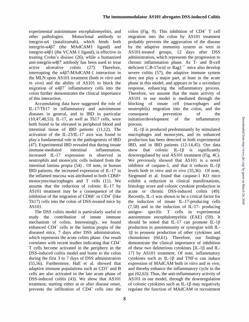
IL-1β is produced predominantly by stimulated
macrophages and monocytes, and its enhanced
production has been detected in both experimental
IBD, and in IBD patients (12-14,45). Our data
show that colonic IL-1β is significantly
downregulated by oral AS101 treatment (Fig. 4C).
We previously showed that AS101 is a novel
inhibitor of caspase-1, and that it reduces IL-1β
levels both in vitro and in vivo (35,36). Of note,
Siegmund et al. found that caspase-1 KO mice
exhibit a reduction in clinical manifestations,
histology score and colonic cytokine production in
acute or chronic DSS-induced colitis (49).
Recently, IL-1 was shown to be a critical factor for
the induction of innate IL-17-producing cells
(7,58) and in the induction of IL-17- producing
antigen- specific T cells in experimental
autoimmune encephalomyelitis (EAE) (59). It
should be noted that IL-17 can promote IL-1β
production in autoimmunity or synergize with IL-
1β to promote production of other cytokines and
chemokines (60,61). Therefore, our findings
demonstrate the clinical importance of inhibition
of these two deleterious cytokines [IL-1β and IL-
17] by AS101 treatment. Of note, inflammatory
cytokines such as IL-1β and TNF-α can induce
expression of MAdCAM both in vitro and in vivo,
and thereby enhance the inflammatory cycle in the
gut (62,63). Thus, the anti-inflammatory activity of
AS101 in our model, through the downregulation
of colonic cytokines such as IL-1β may negatively
regulate the function of MAdCAM in recruitment
by guest on March 24, 2018
http://ww
w.jbc.org/
Dow
nloaded from

The immunomodulator AS101 abrogates DSS-induced Colitis
9
of leukocytes into the intestine. Recently it was
found that IL-1β attenuates CD4+CD25
+FoxP3
+
regulatory T cell function (64). In light of this, the
downregulation of IL-1β by oral AS101
administration may increase the frequency of
colonic CD4+CD25
+FoxP3
+ regulatory T cells in
AS101-treated group as shown in Fig 9B.
We also showed reduced MPO levels in the
colon in response to AS101. These results suggest
that AS101 reduces the degree of neutrophil
infiltration in the tissue. IL-17 has been shown to
induce chemokines (CXCL-8, CXCL-1) and
growth factors (G-CSF, GM-CSF) leading to
augmented neutrophil accumulation as well as
granulopoiesis (53). Thus, IL-17 inhibition (Fig.
4A, B) by AS101 may explain the blockade of
neutrophil accumulation in the colon.
Collectively, it appears that AS101 mediates its
anti-inflammatory activity by attenuating innate
immune function by preventing of colonic
diapedesis of innate cells (neutrophils and
macrophages) and by downregulating
inflammatory cytokines (IL-1β and IL-17).
It is now becoming evident that
aberrant epithelial barrier function plays a central
role in the pathophysiology of IBD (2,5). With
increasing evidence of the involvement of the
innate immune system and the intestinal
epithelium in IBD, the therapeutic paradigm is also
shifting from mere immunosuppression to the
reinforcement of the intestinal barrier (65). In light
of this, the finding that ablation of enteric glia
leads to a fulminant hemorrhagic jejunoileitis (28)
and that enterocolitis is also inducible by
autoimmune targeting of glial cells (66), led to the
understanding of the complex network of
neurotrophins and neurofactors such as GDNF as
an important players in the maintenance of
mucosal integrity. GDNF was found to be
upregulated in both experimental IBD and in IBD
patients. GDNF has strong anti-apoptotic activities
in colonic epithelial cells, which depends on
activation of MAPK and PI3K/AKT pathways
(29,52). One form of epithelial cell injury in
inflamed colonic mucosa in UC is reported to
involve apoptosis of these cells (67). DSS-induced
colitis as a widely used model of UC has been
shown to cause marked apoptosis of colonic
epithelial cells and upregulation of proapoptotic
proteins like Fas/FasL and Bax (68). Our results
show that the diseased mice revealed reduced
levels of colonic GDNF vs. control animals. We
assume that this reduction is probably due to the
strong DSS-induced destruction of the colonic
tissue as seen in the histological staining of
DSS+PBS group (Fig. 5A,B). Importantly, AS101,
which has been previously shown by us to induce
GDNF production both in vitro and in vivo (38,39),
significantly increased colonic GDNF levels as
compared to the diseased mice. This increase in
GDNF levels was accompanied by up regulation of
colonic pAKT and BCL-2 and down regulation of
BAX, indicating the anti apoptotic properties of
GDNF. Furthermore, TUNEL staining revealed
almost no staining in the AS101-treated groups as
compared to the DSS+PBS group (Fig. 10C).
Together with the finding that AS101 normalized
the increased colonic permeability seen in DSS-
treated mice, these results demonstrate the
contribution of AS101 to reducing the apoptosis
process in the colon and in protection of mucosal
barrier integrity as seen in histological sections of
DSS+AS101-treated groups, compared to DSS +
PBS mice (Fig. 5A, B and Fig. 6A).
Most of the current medical treatments for IBD
rely on non-specific, anti inflammatory and
immunosuppressive drugs. These treatments can
cause severe side effects, including cytotoxicity
and increased susceptibility to opportunistic
infections (15,69). Acute, subchronic and chronic
toxicity studies of AS101 were conducted in rats
and dogs and demonstrated that AS101 has a wide
safety margin with severe toxicity observed only at
doses 60–120 times higher than the equivalent
doses proposed for clinical studies (70). At
present, no severe adverse toxicological effects
have been seen in the course of clinical trials
(70,71).
Our results reveal multifunctional effects of the
non toxic, tellurium compound, AS101 in the
DSS-induced colitis model via anti inflammatory
and anti-apoptotic activities, through the
downregulation of colonic cytokine levels [IL-17
and IL-1β] and by the blockade of leukocyte
(neutrophils and macrophages) migration into the
colon. AS101 mediates these activities at least in
part by blocking the α4β7- MAdCAM-1
interaction. Furthermore, AS101 treatment protects
epithelial barrier function by induction of the anti-
apoptotic pathway, GDNF/AKT.
Several trials have shown that early treatment
of Crohn's disease with immunomodulators and
by guest on March 24, 2018
http://ww
w.jbc.org/
Dow
nloaded from

The immunomodulator AS101 abrogates DSS-induced Colitis
11
anti-TNFα agents leads to a superior clinical
outcome, including healing of the mucosa,
compared with standard therapy alone (72,73).
Therefore, we suggest that the non toxic,
tellurium compound AS101, may be an effective
early treatment for the management of IBD
patients.
by guest on March 24, 2018
http://ww
w.jbc.org/
Dow
nloaded from

The immunomodulator AS101 abrogates DSS-induced Colitis
11
References
1. Podolsky, D. K. (2002) Inflammatory bowel disease. N Engl J Med 347, 417-429
2. Sartor, R. B. (2006) Mechanisms of disease: pathogenesis of Crohn's disease and ulcerative
colitis. Nat Clin Pract Gastroenterol Hepatol 3, 390-407
3. Blumberg, R. S., Saubermann, L. J., and Strober, W. (1999) Animal models of mucosal
inflammation and their relation to human inflammatory bowel disease. Curr Opin Immunol 11,
648-656
4. Mottet, C., Uhlig, H. H., and Powrie, F. (2003) Cutting edge: cure of colitis by CD4+CD25+
regulatory T cells. J Immunol 170, 3939-3943
5. Strober, W., Fuss, I., and Mannon, P. (2007) The fundamental basis of inflammatory bowel
disease. J Clin Invest 117, 514-521
6. Sanchez-Munoz, F., Dominguez-Lopez, A., and Yamamoto-Furusho, J. K. (2008) Role of
cytokines in inflammatory bowel disease. World J Gastroenterol 14, 4280-4288
7. Cua, D. J., and Tato, C. M. (2010) Innate IL-17-producing cells: the sentinels of the immune
system. Nat Rev Immunol 10, 479-489
8. Kanai, T., Mikami, Y., Sujino, T., Hisamatsu, T., and Hibi, T. (2012) RORgammat-dependent IL-
17A-producing cells in the pathogenesis of intestinal inflammation. Mucosal Immunol 5, 240-247
9. Mangan, P. R., Harrington, L. E., O'Quinn, D. B., Helms, W. S., Bullard, D. C., Elson, C. O.,
Hatton, R. D., Wahl, S. M., Schoeb, T. R., and Weaver, C. T. (2006) Transforming growth factor-
beta induces development of the T(H)17 lineage. Nature 441, 231-234
10. Steinman, L. (2010) Mixed results with modulation of TH-17 cells in human autoimmune
diseases. Nat Immunol 11, 41-44
11. Fujino, S., Andoh, A., Bamba, S., Ogawa, A., Hata, K., Araki, Y., Bamba, T., and Fujiyama, Y.
(2003) Increased expression of interleukin 17 in inflammatory bowel disease. Gut 52, 65-70
12. Eastaff-Leung, N., Mabarrack, N., Barbour, A., Cummins, A., and Barry, S. (2010) Foxp3+
regulatory T cells, Th17 effector cells, and cytokine environment in inflammatory bowel disease.
J Clin Immunol 30, 80-89
13. Satsangi, J., Wolstencroft, R. A., Cason, J., Ainley, C. C., Dumonde, D. C., and Thompson, R. P.
(1987) Interleukin 1 in Crohn's disease. Clin Exp Immunol 67, 594-605
14. Reinecker, H. C., Steffen, M., Witthoeft, T., Pflueger, I., Schreiber, S., MacDermott, R. P., and
Raedler, A. (1993) Enhanced secretion of tumour necrosis factor-alpha, IL-6, and IL-1 beta by
isolated lamina propria mononuclear cells from patients with ulcerative colitis and Crohn's
disease. Clin Exp Immunol 94, 174-181
15. Rutgeerts, P., Vermeire, S., and Van Assche, G. (2009) Biological therapies for inflammatory
bowel diseases. Gastroenterology 136, 1182-1197
16. Mosser, D. M., and Edwards, J. P. (2008) Exploring the full spectrum of macrophage activation.
Nat Rev Immunol 8, 958-969
17. Wynn, T. A., Chawla, A., and Pollard, J. W. (2013) Macrophage biology in development,
homeostasis and disease. Nature 496, 445-455
18. Okayasu, I., Hatakeyama, S., Yamada, M., Ohkusa, T., Inagaki, Y., and Nakaya, R. (1990) A
novel method in the induction of reliable experimental acute and chronic ulcerative colitis in mice.
Gastroenterology 98, 694-702
19. Wirtz, S., and Neurath, M. F. (2007) Mouse models of inflammatory bowel disease. Adv Drug
Deliv Rev 59, 1073-1083
20. Hynes, R. O. (1992) Integrins: versatility, modulation, and signaling in cell adhesion. Cell 69, 11-
25
21. Hynes, R. O. (2002) Integrins: bidirectional, allosteric signaling machines. Cell 110, 673-687
22. Kinashi, T. (2005) Intracellular signalling controlling integrin activation in lymphocytes. Nat Rev
Immunol 5, 546-559
by guest on March 24, 2018
http://ww
w.jbc.org/
Dow
nloaded from

The immunomodulator AS101 abrogates DSS-induced Colitis
12
23. Pribila, J. T., Quale, A. C., Mueller, K. L., and Shimizu, Y. (2004) Integrins and T cell-mediated
immunity. Annu Rev Immunol 22, 157-180
24. Gorfu, G., Rivera-Nieves, J., and Ley, K. (2009) Role of beta7 integrins in intestinal lymphocyte
homing and retention. Curr Mol Med 9, 836-850
25. Yang, Y., Harrison, J. E., Print, C. G., Lehnert, K., Sammar, M., Lazarovits, A., and Krissansen,
G. W. (1996) Interaction of monocytoid cells with the mucosal addressin MAdCAM-1 via the
integrins VLA-4 and LPAM-1. Immunol Cell Biol 74, 383-393
26. Ghosh, S., Goldin, E., Gordon, F. H., Malchow, H. A., Rask-Madsen, J., Rutgeerts, P., Vyhnalek,
P., Zadorova, Z., Palmer, T., and Donoghue, S. (2003) Natalizumab for active Crohn's disease. N
Engl J Med 348, 24-32
27. Feagan, B. G., Greenberg, G. R., Wild, G., Fedorak, R. N., Pare, P., McDonald, J. W., Dube, R.,
Cohen, A., Steinhart, A. H., Landau, S., Aguzzi, R. A., Fox, I. H., and Vandervoort, M. K. (2005)
Treatment of ulcerative colitis with a humanized antibody to the alpha4beta7 integrin. N Engl J
Med 352, 2499-2507
28. Bush, T. G., Savidge, T. C., Freeman, T. C., Cox, H. J., Campbell, E. A., Mucke, L., Johnson, M.
H., and Sofroniew, M. V. (1998) Fulminant jejuno-ileitis following ablation of enteric glia in adult
transgenic mice. Cell 93, 189-201
29. Steinkamp, M., Geerling, I., Seufferlein, T., von Boyen, G., Egger, B., Grossmann, J., Ludwig, L.,
Adler, G., and Reinshagen, M. (2003) Glial-derived neurotrophic factor regulates apoptosis in
colonic epithelial cells. Gastroenterology 124, 1748-1757
30. Sredni, B., Albeck, M., Tichler, T., Shani, A., Shapira, J., Bruderman, I., Catane, R., Kaufman, B.,
and Kalechman, Y. (1995) Bone marrow-sparing and prevention of alopecia by AS101 in non-
small-cell lung cancer patients treated with carboplatin and etoposide. J Clin Oncol 13, 2342-2353
31. Sredni, B., Caspi, R. R., Klein, A., Kalechman, Y., Danziger, Y., Ben Ya'akov, M., Tamari, T.,
Shalit, F., and Albeck, M. (1987) A new immunomodulating compound (AS-101) with potential
therapeutic application. Nature 330, 173-176
32. Sredni, B., Xu, R. H., Albeck, M., Gafter, U., Gal, R., Shani, A., Tichler, T., Shapira, J.,
Bruderman, I., Catane, R., Kaufman, B., Whisnant, J. K., Mettinger, K. L., and Kalechman, Y.
(1996) The protective role of the immunomodulator AS101 against chemotherapy-induced
alopecia studies on human and animal models. Int J Cancer 65, 97-103
33. Sredni, B., Gal, R., Cohen, I. J., Dazard, J. E., Givol, D., Gafter, U., Motro, B., Eliyahu, S.,
Albeck, M., Lander, H. M., and Kalechman, Y. (2004) Hair growth induction by the Tellurium
immunomodulator AS101: association with delayed terminal differentiation of follicular
keratinocytes and ras-dependent up-regulation of KGF expression. FASEB J 18, 400-402
34. Albeck A, W. H., Sredni B, Albeck M. Tellurium compounds: Selective inhibition of cysteine
proteases and model reaction with thiols. Inorganic Chemistry 1998, 37:1704-1712. .
35. Brodsky, M., Yosef, S., Galit, R., Albeck, M., Longo, D. L., Albeck, A., and Sredni, B. (2007)
The synthetic tellurium compound, AS101, is a novel inhibitor of IL-1beta converting enzyme. J
Interferon Cytokine Res 27, 453-462
36. Brodsky, M., Hirsh, S., Albeck, M., and Sredni, B. (2009) Resolution of inflammation-related
apoptotic processes by the synthetic tellurium compound, AS101 following liver injury. J Hepatol
51, 491-503
37. Brodsky, M., Halpert, G., Albeck, M., and Sredni, B. (2010) The anti-inflammatory effects of the
tellurium redox modulating compound, AS101, are associated with regulation of NFkappaB
signaling pathway and nitric oxide induction in macrophages. J Inflamm (Lond) 7, 3
38. Sredni, B., Geffen-Aricha, R., Duan, W., Albeck, M., Shalit, F., Lander, H. M., Kinor, N., Sagi,
O., Albeck, A., Yosef, S., Brodsky, M., Sredni-Kenigsbuch, D., Sonino, T., Longo, D. L.,
Mattson, M. P., and Yadid, G. (2007) Multifunctional tellurium molecule protects and restores
dopaminergic neurons in Parkinson's disease models. FASEB J 21, 1870-1883
39. Okun, E., Saida, H., Albeck, M., Sredni, B., and Avtalion, R. R. (2006) Upregulation of carp
GDNF mRNA by the immunomodulator AS101. Dev Comp Immunol 30, 441-446
by guest on March 24, 2018
http://ww
w.jbc.org/
Dow
nloaded from

The immunomodulator AS101 abrogates DSS-induced Colitis
13
40. Krawisz, J. E., Sharon, P., and Stenson, W. F. (1984) Quantitative assay for acute intestinal
inflammation based on myeloperoxidase activity. Assessment of inflammation in rat and hamster
models. Gastroenterology 87, 1344-1350
41. Kitajima, S., Takuma, S., and Morimoto, M. (1999) Changes in colonic mucosal permeability in
mouse colitis induced with dextran sulfate sodium. Exp Anim 48, 137-143
42. Weigmann, B., Tubbe, I., Seidel, D., Nicolaev, A., Becker, C., and Neurath, M. F. (2007)
Isolation and subsequent analysis of murine lamina propria mononuclear cells from colonic tissue.
Nat Protoc 2, 2307-2311
43. Hall, L. J., Faivre, E., Quinlan, A., Shanahan, F., Nally, K., and Melgar, S. (2011) Induction and
activation of adaptive immune populations during acute and chronic phases of a murine model of
experimental colitis. Dig Dis Sci 56, 79-89
44. Qiu, W., Wu, B., Wang, X., Buchanan, M. E., Regueiro, M. D., Hartman, D. J., Schoen, R. E.,
Yu, J., and Zhang, L. (2011) PUMA-mediated intestinal epithelial apoptosis contributes to
ulcerative colitis in humans and mice. J Clin Invest 121, 1722-1732
45. Acosta-Rodriguez, E. V., Napolitani, G., Lanzavecchia, A., and Sallusto, F. (2007) Interleukins
1beta and 6 but not transforming growth factor-beta are essential for the differentiation of
interleukin 17-producing human T helper cells. Nat Immunol 8, 942-949
46. Fuss, I. J., Neurath, M., Boirivant, M., Klein, J. S., de la Motte, C., Strong, S. A., Fiocchi, C., and
Strober, W. (1996) Disparate CD4+ lamina propria (LP) lymphokine secretion profiles in
inflammatory bowel disease. Crohn's disease LP cells manifest increased secretion of IFN-
gamma, whereas ulcerative colitis LP cells manifest increased secretion of IL-5. J Immunol 157,
1261-1270
47. Holtta, V., Klemetti, P., Sipponen, T., Westerholm-Ormio, M., Kociubinski, G., Salo, H.,
Rasanen, L., Kolho, K. L., Farkkila, M., Savilahti, E., and Vaarala, O. (2008) IL-23/IL-17
immunity as a hallmark of Crohn's disease. Inflamm Bowel Dis 14, 1175-1184
48. Kleinschek, M. A., Boniface, K., Sadekova, S., Grein, J., Murphy, E. E., Turner, S. P., Raskin, L.,
Desai, B., Faubion, W. A., de Waal Malefyt, R., Pierce, R. H., McClanahan, T., and Kastelein, R.
A. (2009) Circulating and gut-resident human Th17 cells express CD161 and promote intestinal
inflammation. J Exp Med 206, 525-534
49. Siegmund, B., Lehr, H. A., Fantuzzi, G., and Dinarello, C. A. (2001) IL-1 beta -converting
enzyme (caspase-1) in intestinal inflammation. Proc Natl Acad Sci U S A 98, 13249-13254
50. Briskin, M., Winsor-Hines, D., Shyjan, A., Cochran, N., Bloom, S., Wilson, J., McEvoy, L. M.,
Butcher, E. C., Kassam, N., Mackay, C. R., Newman, W., and Ringler, D. J. (1997) Human
mucosal addressin cell adhesion molecule-1 is preferentially expressed in intestinal tract and
associated lymphoid tissue. Am J Pathol 151, 97-110
51. Krieglstein, C. F., Cerwinka, W. H., Laroux, F. S., Salter, J. W., Russell, J. M., Schuermann, G.,
Grisham, M. B., Ross, C. R., and Granger, D. N. (2001) Regulation of murine intestinal
inflammation by reactive metabolites of oxygen and nitrogen: divergent roles of superoxide and
nitric oxide. J Exp Med 194, 1207-1218
52. Zhang, D. K., He, F. Q., Li, T. K., Pang, X. H., Cui de, J., Xie, Q., Huang, X. L., and Gan, H. T.
(2010) Glial-derived neurotrophic factor regulates intestinal epithelial barrier function and
inflammation and is therapeutic for murine colitis. J Pathol 222, 213-222
53. Kolls, J. K., and Linden, A. (2004) Interleukin-17 family members and inflammation. Immunity
21, 467-476
54. Hue, S., Ahern, P., Buonocore, S., Kullberg, M. C., Cua, D. J., McKenzie, B. S., Powrie, F., and
Maloy, K. J. (2006) Interleukin-23 drives innate and T cell-mediated intestinal inflammation. J
Exp Med 203, 2473-2483
55. Sund, M., Xu, L. L., Rahman, A., Qian, B. F., Hammarstrom, M. L., and Danielsson, A. (2005)
Reduced susceptibility to dextran sulphate sodium-induced colitis in the interleukin-2
heterozygous (IL-2) mouse. Immunology 114, 554-564
by guest on March 24, 2018
http://ww
w.jbc.org/
Dow
nloaded from

The immunomodulator AS101 abrogates DSS-induced Colitis
14
56. Da Silva, A. P., Pollett, A., Rittling, S. R., Denhardt, D. T., Sodek, J., and Zohar, R. (2006)
Exacerbated tissue destruction in DSS-induced acute colitis of OPN-null mice is associated with
downregulation of TNF-alpha expression and non-programmed cell death. J Cell Physiol 208,
629-639
57. Dieleman, L. A., Ridwan, B. U., Tennyson, G. S., Beagley, K. W., Bucy, R. P., and Elson, C. O.
(1994) Dextran sulfate sodium-induced colitis occurs in severe combined immunodeficient mice.
Gastroenterology 107, 1643-1652
58. Sutton, C. E., Mielke, L. A., and Mills, K. H. (2012) IL-17-producing gammadelta T cells and
innate lymphoid cells. Eur J Immunol 42, 2221-2231
59. Sutton, C., Brereton, C., Keogh, B., Mills, K. H., and Lavelle, E. C. (2006) A crucial role for
interleukin (IL)-1 in the induction of IL-17-producing T cells that mediate autoimmune
encephalomyelitis. J Exp Med 203, 1685-1691
60. Koenders, M. I., Lubberts, E., Oppers-Walgreen, B., van den Bersselaar, L., Helsen, M. M., Di
Padova, F. E., Boots, A. M., Gram, H., Joosten, L. A., and van den Berg, W. B. (2005) Blocking
of interleukin-17 during reactivation of experimental arthritis prevents joint inflammation and
bone erosion by decreasing RANKL and interleukin-1. Am J Pathol 167, 141-149
61. Koenders, M. I., Kolls, J. K., Oppers-Walgreen, B., van den Bersselaar, L., Joosten, L. A., Schurr,
J. R., Schwarzenberger, P., van den Berg, W. B., and Lubberts, E. (2005) Interleukin-17 receptor
deficiency results in impaired synovial expression of interleukin-1 and matrix metalloproteinases
3, 9, and 13 and prevents cartilage destruction during chronic reactivated streptococcal cell wall-
induced arthritis. Arthritis Rheum 52, 3239-3247
62. Ando, T., Langley, R. R., Wang, Y., Jordan, P. A., Minagar, A., Alexander, J. S., and Jennings,
M. H. (2007) Inflammatory cytokines induce MAdCAM-1 in murine hepatic endothelial cells and
mediate alpha-4 beta-7 integrin dependent lymphocyte endothelial adhesion in vitro. BMC Physiol
7, 10
63. Connor, E. M., Eppihimer, M. J., Morise, Z., Granger, D. N., and Grisham, M. B. (1999)
Expression of mucosal addressin cell adhesion molecule-1 (MAdCAM-1) in acute and chronic
inflammation. J Leukoc Biol 65, 349-355
64. O'Sullivan, B. J., Thomas, H. E., Pai, S., Santamaria, P., Iwakura, Y., Steptoe, R. J., Kay, T. W.,
and Thomas, R. (2006) IL-1 beta breaks tolerance through expansion of CD25+ effector T cells. J
Immunol 176, 7278-7287
65. Peyrin-Biroulet, L., Desreumaux, P., Sandborn, W. J., and Colombel, J. F. (2008) Crohn's disease:
beyond antagonists of tumour necrosis factor. Lancet 372, 67-81
66. Cornet, A., Savidge, T. C., Cabarrocas, J., Deng, W. L., Colombel, J. F., Lassmann, H.,
Desreumaux, P., and Liblau, R. S. (2001) Enterocolitis induced by autoimmune targeting of
enteric glial cells: a possible mechanism in Crohn's disease? Proc Natl Acad Sci U S A 98, 13306-
13311
67. Iwamoto, M., Koji, T., Makiyama, K., Kobayashi, N., and Nakane, P. K. (1996) Apoptosis of
crypt epithelial cells in ulcerative colitis. J Pathol 180, 152-159
68. Vetuschi, A., Latella, G., Sferra, R., Caprilli, R., and Gaudio, E. (2002) Increased proliferation
and apoptosis of colonic epithelial cells in dextran sulfate sodium-induced colitis in rats. Dig Dis
Sci 47, 1447-1457
69. Grimm, M. C. (2009) New and emerging therapies for inflammatory bowel diseases. J
Gastroenterol Hepatol 24 Suppl 3, S69-74
70. Nyska, A., Waner, T., Pirak, M., Albeck, M., and Sredni, B. (1989) Toxicity study in rats of a
tellurium based immunomodulating drug, AS-101: a potential drug for AIDS and cancer patients.
Arch Toxicol 63, 386-393
71. Sredni B, C. R., Shani A, Gezin A, Levi E, Schlezinger M, et al. (1990) Phase I study of AS101
(anorganotellurium compound) in patients with advanced malignancies., In: Rubinstein E, Adam
D, editors. Recent advances in chemotherapy. Jerusalem: E. Lewin-Epstein;
by guest on March 24, 2018
http://ww
w.jbc.org/
Dow
nloaded from

The immunomodulator AS101 abrogates DSS-induced Colitis
15
72. D'Haens, G. R. (2010) Top-down therapy for IBD: rationale and requisite evidence. Nat Rev
Gastroenterol Hepatol 7, 86-92
73. D'Haens, G., Baert, F., van Assche, G., Caenepeel, P., Vergauwe, P., Tuynman, H., De Vos, M.,
van Deventer, S., Stitt, L., Donner, A., Vermeire, S., Van de Mierop, F. J., Coche, J. C., van der
Woude, J., Ochsenkühn, T., van Bodegraven, A. A., Van Hootegem, P. P., Lambrecht, G. L.,
Mana, F., Rutgeerts, P., Feagan, B. G., Hommes, D., Group, B. I. B. D. R., and Club, N.-H. G.
(2008) Early combined immunosuppression or conventional management in patients with newly
diagnosed Crohn's disease: an open randomised trial. Lancet 371, 660-667
by guest on March 24, 2018
http://ww
w.jbc.org/
Dow
nloaded from

The immunomodulator AS101 abrogates DSS-induced Colitis
16
Footnotes
This work was partly supported by the Dr. Tovi Comet-Walerstein Cancer Research Chair, by The Dave
and Florence Muskovitz Chair in Cancer Research, and the Jaime Lusinchi Research Institute in Applied
Sciences.
1To whom correspondence may be addressed : Prof. Benjamin Sredni. The Mina & Everard Goodman
Faculty of Life Sciences, C.A.I.R. Institute, Bar-Ilan University, Ramat-Gan 52900, Israel. Tel.: 972-3-
5318605; Fax: 972-3-738-4060. Email address: [email protected]
2Abbreviations used: IBD, inflammatory bowel diseases; DSS, dextran sodium sulfate; MPO,
myeloperoxidase; CD, Crohn's disease; UC, ulcerative colitis; TUNEL, terminal
deoxynucleotidyltransferase -mediated dUTP nick end labeling; MAdCAM, mucosal addressin cell
adhesion molecule; GDNF, glial-derived neurotrophic factor; iNOS, inducible nitric oxide synthase.
by guest on March 24, 2018
http://ww
w.jbc.org/
Dow
nloaded from

The immunomodulator AS101 abrogates DSS-induced Colitis
17
Figure legends
Figure 1. The chemical structure of AS101.
Figure 2. AS101 injection reduces clinical symptoms of DSS-induced murine colitis.
Colitis was induced by addition of 3.5% DSS to the drinking water of C57Bl/6 mice. AS101
(10µgr/mouse) was injected daily i.p. from day 0 [DSS+AS(d0)], or from 2 days after DSS administration
[DSS+AS(d2)]. A. AS101 restores decreased body weight induced by colitis. Total average body weight
of each group was monitored. * p < 0.01 decrease vs. other groups (analyzed from day 6 to day 11 by
ANOVA Repeated Measures). B. AS101 reduces intestinal bleeding induced by colitis. Intestinal bleeding
was scored by Hemoccult test for the presence of occult blood and by visual observation of rectal
bleeding. * p < 0.001 increase vs. other groups (analyzed by ANOVA Repeated Measures). C. AS101
reverses colitis-induced elevation of stool morphology score. * p < 0.001 increase vs. other groups
(analyzed by ANOVA Repeated Measures). D, E. AS101 restores colon length shortened by colitis.
Colons were removed and measured at necropsy (day 12). * p < 0.001 increase vs. DSS + PBS group
(analyzed by one-way ANOVA test). Results shown are mean ± SE [n=4 for PBS; n=8 for DSS+PBS; n=6
for DSS+AS(d0); n=10 for DSS+AS(d2)]. For colon length, n=7 for PBS; n=18 for DSS+PBS; n=17 for
DSS+AS(d0); n=10 for DSS+AS(d2).
Figure 3. Oral administration of AS101 reduces clinical symptoms of DSS-induced murine colitis.
Colitis was induced by addition of 3.5% DSS to the drinking water of C57Bl/6 mice. Mice were treated
orally with daily administration of AS101 (100µgr per mouse), starting at day 0 [DSS+AS(d0)]. Results
shown are mean ± SE of 10 mice per group. A. AS101 restores decreased body weight induced by colitis.
Total body weight average of each group was monitored. * p < 0.01 decrease vs. other groups (analyzed
by ANOVA Repeated Measures). B. AS101 reduces intestinal bleeding induced by colitis. Intestinal
bleeding was scored by Hemoccult test for the presence of occult blood and by visual observation of rectal
bleeding. * p < 0.05 increase vs. other groups (analyzed by ANOVA Repeated Measures). C, D. AS101
normalizes colon length shortened by colitis. Colons were removed and measured at necropsy (day 12). *
p < 0.001 increase vs. DSS + PBS group (analyzed by one-way ANOVA test).
Figure 4. Treatment with AS101 reduces colonic levels of inflammatory cytokines.
Colitis was induced by addition of 3.5% DSS to the drinking water of C57Bl/6 mice. AS101
(10µgr/mouse per injection) was injected daily i.p. from day 0 [DSS+AS(d0)], or from 2 days after DSS
administration [DSS+AS(d2)]. At necropsy (day 12), 1-2 cm of the distal colon was removed and
homogenized. Cytokine levels were measured by ELISA. Results shown are mean ± SE. A. Treatment
with AS101 either at or after disease onset significantly reduced colon IL-17 levels. * p < 0.01 decrease
vs. DSS + PBS group (analyzed by one-way ANOVA test). [n=7 for PBS; n=12 for DSS+PBS or
DSS+AS(d0); n=10 for DSS+AS(d2)]. B. Immunohistochemical detection of IL-17 levels in distal colon.
At necropsy (day 7), 1-2 cm of the distal colon was removed and fixed in 4% buffered formaldehyde.
Immunohistochemical analysis was performed in paraffin-embedded slides. Pictures shown are
representative of three mice per group. Scale bars indicate 80μm. C. Colonic IL-1β is significantly reduced
by oral administration of AS101. * p < 0.02 decrease vs. DSS+PBS group (analyzed by Student’s t-test).
Results shown are mean ± SE of 4 mice per group.
Figure 5. Histopathological examination of the distal colon.
Colitis was induced by addition of 3.5% DSS to the drinking water of C57Bl/6 mice. At necropsy (day
12), 1-2 cm of the distal colon was removed and fixed in 4% buffered formaldehyde. Paraffin-embedded
sections were stained with hematoxylin and eosin. A. Histopathological examination of colons from
AS101 injection treatment. AS101 (10µgr/mouse per injection) was injected i.p. daily from day 0 or
starting 2 days after DSS administration. Pictures shown are representative of 3 mice per group. Scale bars
by guest on March 24, 2018
http://ww
w.jbc.org/
Dow
nloaded from

The immunomodulator AS101 abrogates DSS-induced Colitis
18
indicate 80μm. B. Histopathological examination of colons following oral AS101 treatment. Mice were
treated orally with daily administration of AS101 (100µgr per mouse) starting at day 0. Pictures shown are
representative of 3 mice per group. Scale bars indicate 160μm C. AS101 treatment (injection) significantly
reduces histopathology score. Histopathology score was determined as described in Materials and
Methods. * p < 0.01 decrease vs. DSS + PBS group. Results shown are mean ± SE [n=2 for PBS or
DSS+AS(d2), n=7 for DSS+PBS, n=8 for DSS+AS(d0)].
Figure 6. AS101 prevents neutrophil migration into the colon.
Colitis was induced by addition of 3.5% DSS to the drinking water of C57Bl/6 mice. AS101
(10µgr/mouse per injection) was injected from day 0, or starting 2 days after DSS administration. At
necropsy (day 7), 2 cm of the distal colon was removed and cut longitudinally: One section was fixed in
4% buffered formaldehyde, and stained with hematoxylin and eosin. The other section was kept at -80C
until assay for MPO activity, as described in Materials and Methods. A. Histopathological examination of
the distal colon. Pictures shown are representative of 3 mice per group. Scale bars indicate 160μm. B.
MPO activity in the distal colon. * p< 0.05 decrease vs. DSS+PBS group (analyzed by Students t-test).
Results shown are mean ± SE [n=8 for PBS; n=7 for DSS+PBS; n=8 for DSS+AS(d0); n=9 for
DSS+AS(d2)].
Figure 7. AS101 prevents the migration of α4β7+ macrophages into the colon.
A. Colitis was induced by addition of 3.5% DSS to the drinking water of C57Bl/6 mice. AS101
(10µgr/mouse per injection) was injected daily starting from day 0. At necropsy (day 12), 1-2 cm of the
distal colon was removed and fixed in 4% buffered formaldehyde. Paraffin tissue sections were prepared,
and slides were stained with anti-CD68 (red), anti-α4β7 (green) and Hoechst (blue). White circles indicate
cells of ~20μm diameter (estimated diameter of macrophages) which were double-stained for CD68 and
α4β7 (yellow). Representative picture are shown (magnification ×40). Scale bars indicate 40μm. B.
Downregulation of colonic iNOS levels by AS101 treatment. Proteins in colonic tissue homogenates were
separated by SDS-PAGE and subjected to immunoblotting with antibodies to iNOS and actin-HRP as a
control (n=5 for PBS; n=8 for DSS+PBS or DSS+AS(d0) groups).
Figure 8. AS101 prevents the adhesion of MLN cells to MAdCAM-1 both in vitro and in vivo. Colitis
was induced by addition of 2% DSS to the drinking water of C57Bl/6 mice. A. MLN cells from DSS+PBS
group (n=5) were seeded for 2h on MAdCAM-1 or 2.5% BSA-coated wells. 0.1-1 µgr/ml AS101 was
added into MAdCAM-1-coated wells. After 2 h, the wells were washed, and XTT assay were performed
to evaluate the bound cells. # p < 0.01 increase vs. BSA group, * p< 0.05 decrease vs. Madcam group
analyzed by one way ANOVA test. Results shown are mean ± SE. B. AS101 (10µgr/mouse) was injected
daily from day 0. At day 10, MLN cells were isolated from all groups (n=5) and seeded for 2h on
MAdCAM-1- coated plate. After 2 hr, the wells were washed and XTT assay was performed to quantitate
the bound cells. # p < 0.01 increase vs. PBS group, * p< 0.01 decrease vs. DSS+PBS group analyzed by
one way ANOVA test. Results shown are mean ± SE.
Figure 9. AS101 prevents the migration of CD4+ cells into the colon and increases colonic
CD4+CD25
+Foxp3
+ T regulatory cells.
A. Colitis was induced by addition of 3.5% DSS to the drinking water of C57Bl/6 mice. AS101
(10µgr/mouse per injection) was injected daily i.p. from day 0 [DSS+AS(d0)], or from 2 days after DSS
administration [DSS+AS(d2)]. At necropsy (day 7), 1-2 cm of the distal colon was removed and fixed in
4% buffered formaldehyde. Immunohistochemical analysis was performed in paraffin-embedded slides.
Pictures shown are representative of 3 mice per group. Scale bars indicate 160μm. B. Lamina Propia
Lymphocytes (LPL) cells were isolated from the colons of treated groups and stained for
CD4+CD25
+Foxp3
+ T regulatory cells as described in Experimental procedures. Dot plots of each group
showing CD25+FOXP3
+ LPL cells (Plots are gated on CD3
+CD4
+ LPL cells). Each plot is representative
of 3 experiments.
by guest on March 24, 2018
http://ww
w.jbc.org/
Dow
nloaded from

The immunomodulator AS101 abrogates DSS-induced Colitis
19
Figure 10. AS101 treatment restores DSS-induced destruction of colonic epithelial barrier integrity.
Colitis was induced by addition of 3.5% DSS to the drinking water of C57Bl/6 mice. At necropsy (day
12), 1-2 cm of the distal colon was removed and homogenized. A. AS101 treatment increases GDNF
levels. GDNF was measured in colonic lysates. # p<0.05 increases vs. DSS+PBS group, * p<0.02 increase
vs. DSS+PBS group (analyzed by Student’s t-test). Results shown are mean ± SE [n=3 for PBS; n=6 for
DSS+PBS; and n=9 for DSS+AS(d2)]. B. Upregulation of pAKT and BCL-2 and downregulation of Bax
upon AS101 treatment. Proteins in tissue homogenates were separated by SDS-PAGE and subjected to
immunoblotting with antibodies to BCL2, pAKT, Bax, and actin-HRP as a control (n=4 in each group). C.
AS101 decreases colonic TUNEL positive cells. At necropsy (day 7), 1-2 cm of the distal colon was
removed and fixed in 4% buffered formaldehyde. Pictures shown are representative of 3 mice per group.
Scale bars indicate 80μm. D. AS101 normalizes the DSS-induced permeability of colonic epithelial cells.
Colonic permeability to Evans Blue was performed as described in the Materials and Methods. # p< 0.01
increase vs. PBS group, * p< 0.01 decrease vs. DSS+PBS group analyzed by one way ANOVA. Results
shown are mean ± SE [n=5 for PBS; n=7 for DSS+PBS; and n=8 for DSS+AS(d0)].
by guest on March 24, 2018
http://ww
w.jbc.org/
Dow
nloaded from

The immunomodulator AS101 abrogates DSS-induced Colitis
21
by guest on March 24, 2018
http://ww
w.jbc.org/
Dow
nloaded from

The immunomodulator AS101 abrogates DSS-induced Colitis
21
by guest on March 24, 2018
http://ww
w.jbc.org/
Dow
nloaded from

The immunomodulator AS101 abrogates DSS-induced Colitis
22
by guest on March 24, 2018
http://ww
w.jbc.org/
Dow
nloaded from

The immunomodulator AS101 abrogates DSS-induced Colitis
23
by guest on March 24, 2018
http://ww
w.jbc.org/
Dow
nloaded from

The immunomodulator AS101 abrogates DSS-induced Colitis
24
by guest on March 24, 2018
http://ww
w.jbc.org/
Dow
nloaded from

The immunomodulator AS101 abrogates DSS-induced Colitis
25
by guest on March 24, 2018
http://ww
w.jbc.org/
Dow
nloaded from

The immunomodulator AS101 abrogates DSS-induced Colitis
26
by guest on March 24, 2018
http://ww
w.jbc.org/
Dow
nloaded from

The immunomodulator AS101 abrogates DSS-induced Colitis
27
by guest on March 24, 2018
http://ww
w.jbc.org/
Dow
nloaded from

The immunomodulator AS101 abrogates DSS-induced Colitis
28
by guest on March 24, 2018
http://ww
w.jbc.org/
Dow
nloaded from

The immunomodulator AS101 abrogates DSS-induced Colitis
29
by guest on March 24, 2018
http://ww
w.jbc.org/
Dow
nloaded from

Albeck, Yona Kalechman and Benjamin SredniGilad Halpert, Tom Eitan, Elena Voronov, Ron N. Apte, Lea Rath-Wolfson, Michael
AS101, on DSS-induced murine colitisMultifunctional activity of a small tellurium redox immunomodulator compound,
published online April 24, 2014J. Biol. Chem.
10.1074/jbc.M113.536664Access the most updated version of this article at doi:
Alerts:
When a correction for this article is posted•
When this article is cited•
to choose from all of JBC's e-mail alertsClick here
by guest on March 24, 2018
http://ww
w.jbc.org/
Dow
nloaded from