Embed Size (px)
Citation preview
1
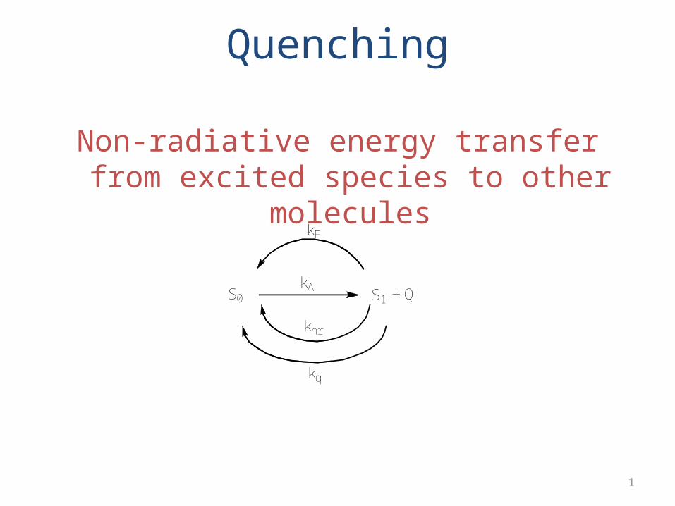
Quenching
Non-radiative energy transfer from excited species to other molecules
S0 S1kA
kF
knr
+ Q
kq
2
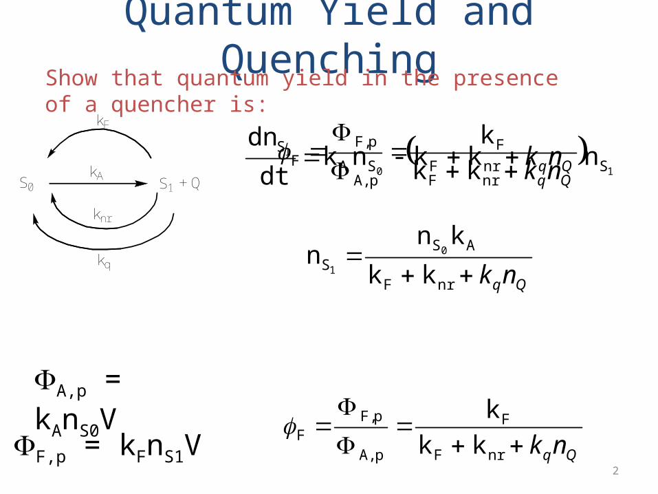
Quantum Yield and Quenching
S0 S1kA
kF
knr
+ Q
kq
10
1
SnrFSAS nk k - nk
dt
dnQqnkQqnk
nrF
F
pA,
pF,F k k
k
Qqnk
nrF
ASS k k
kn n 0
1
Show that quantum yield in the presence of a quencher is:
FA,p = kAnS0V
FF,p = kFnS1V Qqnk
nrF
F
pA,
pF,F k k
k

3
Dynamic Quenching/Collisional QuenchingRequires contact between quencher and excited lumophore during collision (temperature and viscosity dependent). Luminescence lifetime drops with increasing quencher concentration.
QqnrF
QqnrF
f
of nK
kk
nkkk
1
Since fluorescence emission is directly proportional to quantum yield:
QqnKF
F10
Stern-Volmer Equation
4
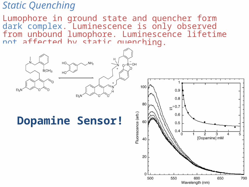
Static QuenchingLumophore in ground state and quencher form dark complex. Luminescence is only observed from unbound lumophore. Luminescence lifetime not affected by static quenching.
Dopamine Sensor!
5
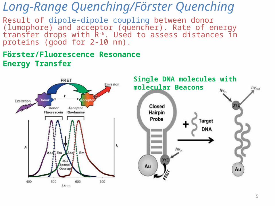
Long-Range Quenching/Förster QuenchingResult of dipole-dipole coupling between donor (lumophore) and acceptor (quencher). Rate of energy transfer drops with R-6. Used to assess distances in proteins (good for 2-10 nm).
Förster/Fluorescence Resonance Energy Transfer
Single DNA molecules with molecular Beacons
6
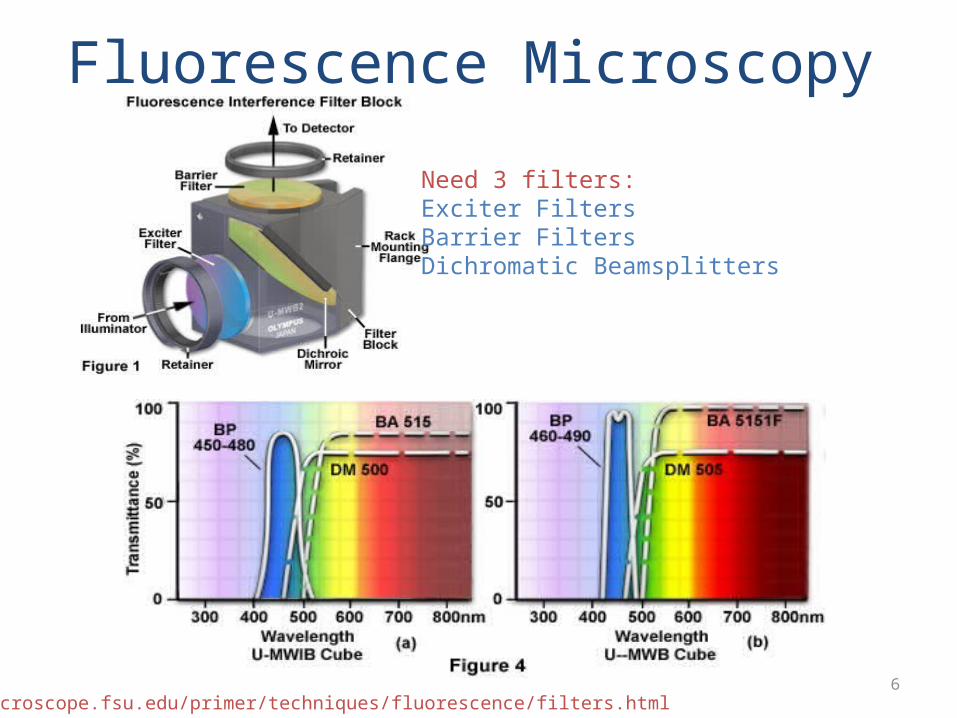
Fluorescence Microscopy
Need 3 filters:Exciter FiltersBarrier FiltersDichromatic Beamsplitters
http://microscope.fsu.edu/primer/techniques/fluorescence/filters.html
7
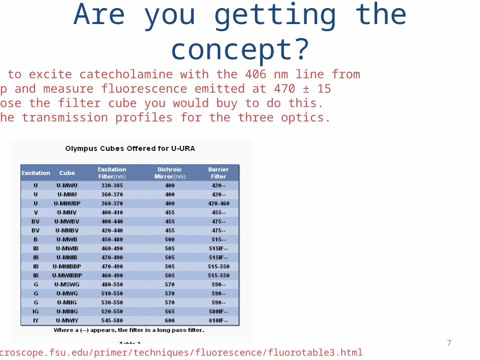
Are you getting the concept?You plan to excite catecholamine with the 406 nm line froma Hg lamp and measure fluorescence emitted at 470 ± 15nm. Choose the filter cube you would buy to do this.Sketch the transmission profiles for the three optics.
http://microscope.fsu.edu/primer/techniques/fluorescence/fluorotable3.html

8
Fluorescence Microscopy Objectives
Image intensity is a function of the objective numericalaperture and magnification:
2
4
)(
)( mag
NAI obj
Fabricated with low fluorescence glass/quartz with anti-reflection coatings
http://micro.magnet.fsu.edu/primer/techniques/fluorescence/anatomy/fluoromicroanatomy.html

9
Fluorescence Microscopy Detectors
No spatial resolution required: PMT or photodiodeSpatial resolution required: CCD
http://micro.magnet.fsu.edu/primer/digitalimaging/digitalimagingdetectors.html
10
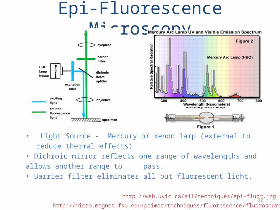
Epi-Fluorescence Microscopy
• Light Source - Mercury or xenon lamp (external to reduce thermal effects)• Dichroic mirror reflects one range of wavelengths and allows another range to pass.• Barrier filter eliminates all but fluorescent light.
http://micro.magnet.fsu.edu/primer/techniques/fluorescence/fluorosources.html
http://web.uvic.ca/ail/techniques/epi-fluor.jpg

11
Special Fluorescence Techniques
TIRF
http://microscopy.fsu.edu/primer/techniques/fluorescence/tirf/tirfintro.html
LIF
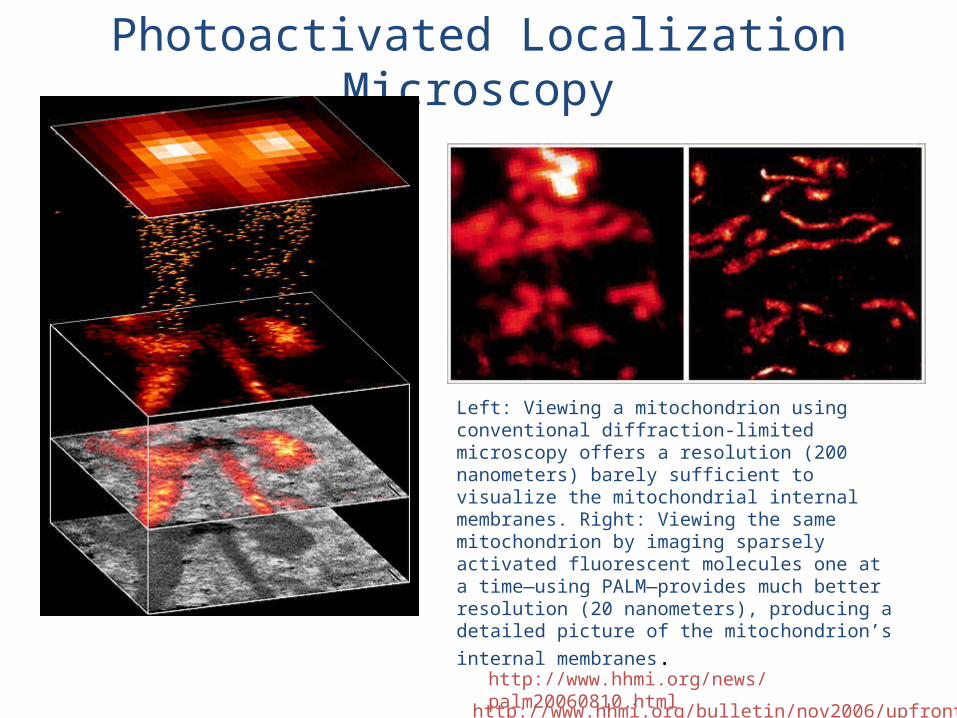
Photoactivated Localization Microscopy
http://www.hhmi.org/bulletin/nov2006/upfront/image.html
Left: Viewing a mitochondrion using conventional diffraction-limited microscopy offers a resolution (200 nanometers) barely sufficient to visualize the mitochondrial internal membranes. Right: Viewing the same mitochondrion by imaging sparsely activated fluorescent molecules one at a time—using PALM—provides much better resolution (20 nanometers), producing a detailed picture of the mitochondrion’s internal membranes.
http://www.hhmi.org/news/palm20060810.html