Embed Size (px)
Citation preview

FACTA UNIVERSITATIS Series: Physics, Chemistry and Technology Vol. 6, No 1, 2008, pp. 127 - 139 DOI: 10.2298/FUPCT0801127P
QUANTUM-CHEMICAL CALCULATIONS OF THE PRODUCTS AND ENERGIES OF ELECTRON INDUCED IONIZATION OF
2-FURANMETHANOL, TETRAHYDRO-AND 3-FURANOL UDC 539.2
P. Papp1, P. Mach2, J. Urban2, Š. Matejčík1 1Department of Experimental Physics,
2Department of Nuclear Physics and Biophysics, Faculty of Mathematics, Physics and Informatics, Comenius University,
Mlynská dolina, 842 48 Bratislava, Slovakia
Abstract. A theoretical treatment is used to perform conformational studies of title compounds, which was previously successfully used within our group for characterization of fragmentation patterns of some bio-molecules. Now we present studies of electron impact ionization of 2 Furanmethanol, Tetrahydro (C5H10O2) and 3-Furanol, Tetrahydro (C4H8O2), both as important models for more complicated compounds like nucleic acids. In this paper geometry of the neutral and cationic conformers of these two molecules was optimized on the DFT level with B3LYP functional, and ionization energies were estimated. DFT calculated results are supplemented with G3MP2 calculations, and a set of higher-level ab initio methods were empirically corrected to obtain more reliable results.
Key words: ab initio, DFT, geometry, optimization, ionization, 2 furanmethanol, tetrahydro-, 3 furanol, tetrahydro-
INTRODUCTION
Furanose structured alcohols, 2-Furanmethanol, Tetrahydro- (or tetrahydrofurfuryl alcohol - THFA) and 3-Furanol, Tetrahydro- (or 3-hydroxy-tetrahydrofuran - 3HTHF), reported in this article are considered as models for more complicated biological compounds as deoxynucleosides, deoxynucleotides or finally nucleic acids (see Fig.1). Electron impact ionization of both THFA and 3HTHF was previously studied using a crossed electron/molecule beam apparatus equipped with a trochoidal electron monochromator and quadrupole mass spectrometer [1] at the laboratory in Bratislava. One serious drawback of all measurements with our crossbeam experiment is inability to identify chemical structure of formed fragments. Thus, to supplement the experimental
Received July 14, 2008

P. PAPP, P. MACH, J. URBAN, Š. MATEJČÍK 128
work we decided to perform conformational studies of both molecules, for their neutral and especially cationic states, to estimate the theoretical value of ionization energies and find the most stable geometrical conformers (these results can be used for fragmentation studies, to be published [2]). As mentioned before both THFA [3] and 3HTHF [4] represent important models of larger biological compounds. This fact is important also from quantum-chemical point of view. Using smaller models allows us to perform more accurate quantum-chemical calculations of their optimal geometries, ground state energies, conformational studies and other qualitative characteristics. It is evident that with standard quantum chemical methods we are able to perform these types of calculations on restricted structures like THFA (Fig.1-c) and 3HTHF (Fig.1-d) instead of larger biological compounds with nitrogen bases, CH2OH and phosphate groups substituted. For these larger structures it is impossible to achieve accurate results at the same level.
In this paper we present detailed conformational studies for both parent molecules and positive ions. It is important to note that there is strong coupling between side-group (-OH or –CH2OH) orientation and ring puckering. As a result, some conformers, expected from simple structural models, disappear. For their cationic states ring opening or partial dissociation of the side-group can be typical. The later result, fragmentation of the neutral CH2OH, seems to be in good agreement with experiment as the most abundant cationic fragment in mass spectrum of THFA is m/Z=71 and in mass spectrum of 3HTHTF is m/Z=57 (more details in [2]). For most stable conformers, adiabatic ionization energies are calculated using G3MP2 method.
R
O
HOH
HH
HH
HO
R
O
HOH
HH
HH
OP-O
O-
O
HO
HH
HH
HH
HOH
O
HOH
HH
H
H
H
a) b)
d)c)
Fig. 1. Structures of biological compounds: a) deoxynucleoside, where R should be replaced by nitrogenous base (adenine, guanine, thymine, uracil or cytosine), b) deoxynucleotide, where R is the same as in previous, c) 2-Furanmethanol, Tetrahydro- (THFA) and d) 3-Furanol, Tetrahydro- (3HTHF).

Quantum-Chemical Calculations of the Products and Energies of Electron Induced Ionization ... 129
CALCULATIONS
Quantum-chemical calculations were performed using the GAUSSIAN 03 program package [5]. Geometries of THFA and 3HTHF conformers were taken from previous works of Borisenko [3] and Berthier [4] respectively. In both cases only the six energetically lowest conformers were considered, while in the case of 3HTHF only six conformers are reported in [4]. Geometries of all these conformers considered in our work were re-optimized at DFT [6,7,8,9] level of theory, using a B3LYP [10] functional together with 6-311+G(2d,2p) basis set [11,12,13]. One dimensional potential energy surface (PES) scans of 3HTHF were performed on MP2/6-31G(1d) [14,15,16,17,18] level of theory. This bases set (6-31G(1d)) is a standard 6-31G* basis [13,19,20,21,22,23,24,25,26,27,28] with modified polarization exponent of carbon (ζC=0.7 as reported in the work of Berthier [4]). This PES scan was also performed with B3LYP/6-31G(1d). Finally, G3MP2 method [29] was used to calculate more reliable results of relative energies of stable conformers found when one would like to compare them to experiment. The B3LYP geometries were used as the input geometries for G3MP2, these are consecutively re-optimized with HF and later MP2 methods and thermal and empiric corrections are added to the resulting total energy.
RESULTS AND DISCUSSION
Till now two recent theoretical papers dealt with conformational studies of neutral THFA [3] and 3HTHF [4]. They reported twelve different conformers of THFA at HF/6-311++G**. From these six most stable were singled out for more detailed study at MP2/6-311++G** and compared to results of electron diffraction experiment. These six neutral THFA conformers were used as a starting point for our research. For 3HTHF six different conformers were reported at MP2/6-31G(1d) and DFT (BP86/6-31G(1d)) levels of theory. However recent experimental work (positron scattering) on 3HTHF [30] results in existence of only two neutral conformers with distinguishable total dipole moments. We re-optimized for both molecules the six energetically lowest conformers with B3LYP and G3MP2 and compared our results to previously published values. These results are discussed in the respective sections. For all these stable conformers of neutral species their cationic states were also optimized at the same theoretical levels. To our knowledge, no previous conformational studies dealing with cations of THFA and 3HTHF were published.
Tetrahydrofurfuryl Alcohol (THFA)
Neutral THFA
The six energetically lowest conformers of THFA labeled from A to F in Fig.2 were selected from [3] and re-optimized with B3LYP/6-311+G(2d,2p) level of theory. The structural features of these conformers are described in more details in [3]; we just mention that these noticeable features are two ring types and the relative orientation of the side chain group CH2OH to its ring. All low energy conformers share the same
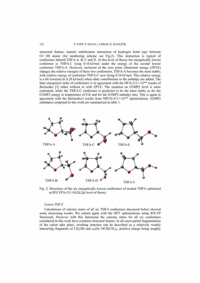
P. PAPP, P. MACH, J. URBAN, Š. MATEJČÍK 130
structural feature, namely stabilization interaction of hydrogen bond type between O1…H8 atoms (for numbering scheme see Fig.2). This interaction is typical of conformers labeled THFA-A, B, C and D. At this level of theory the energetically lowest conformer is THFA-C lying 0.16 kJ/mol under the energy of the second lowest conformer THFA-A. However, inclusion of the zero point vibrational energy (ZPVE) changes the relative energies of these two conformers, THFA-A becomes the most stable; with relative energy of conformer THFA-C now being 0.34 kJ/mol. This relative energy is a bit lowered (to 0.26 kJ/mol) when other contributions to the enthalpy are added. The later energetical order of conformers is in agreement with the HF/6-311++G** results of Borisenko [3] either without or with ZPVE. The situation on G3MP2 level is more consistent, while the THFA-C conformer is predicted to be the most stable as for the G3MP2 energy at temperature of 0 K and for the G3MP2 enthalpy also. This is again in agreement with the Borisenko's results from MP2/6-311++G** optimizations. G3MP2 enthalpies computed in this work are summarized in table 1.
THFA-A THFA-C THFA-E
THFA-B THFA-D THFA-F Fig. 2. Structures of the six energetically lowest conformers of neutral THFA optimized
at B3LYP/6-311+G(2d,2p) level of theory.
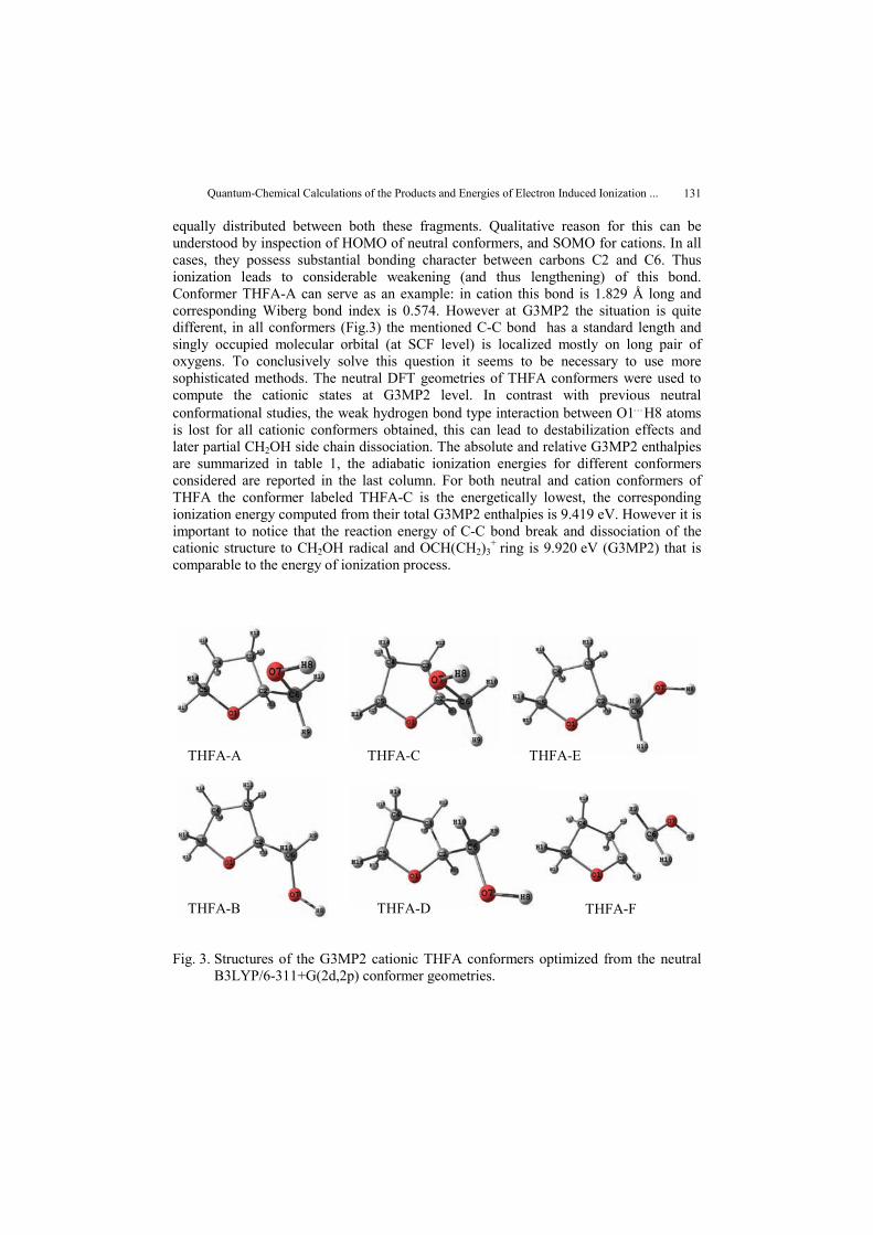
Cation THFA+ Calculations of cationic states of all six THFA conformers discussed before showed
some interesting results. We started again with the DFT optimizations using B3LYP functional. However with this functional the cationic states for all six conformers considered in this work have common structural feature. In all cases partial fragmentation of the cation take place, resulting structure can be described as a relatively weakly interacting fragments of CH2OH and cyclic OCH(CH2)3, positive charge being roughly
Quantum-Chemical Calculations of the Products and Energies of Electron Induced Ionization ... 131
equally distributed between both these fragments. Qualitative reason for this can be understood by inspection of HOMO of neutral conformers, and SOMO for cations. In all cases, they possess substantial bonding character between carbons C2 and C6. Thus ionization leads to considerable weakening (and thus lengthening) of this bond. Conformer THFA-A can serve as an example: in cation this bond is 1.829 Å long and corresponding Wiberg bond index is 0.574. However at G3MP2 the situation is quite different, in all conformers (Fig.3) the mentioned C-C bond has a standard length and singly occupied molecular orbital (at SCF level) is localized mostly on long pair of oxygens. To conclusively solve this question it seems to be necessary to use more sophisticated methods. The neutral DFT geometries of THFA conformers were used to compute the cationic states at G3MP2 level. In contrast with previous neutral conformational studies, the weak hydrogen bond type interaction between O1…H8 atoms is lost for all cationic conformers obtained, this can lead to destabilization effects and later partial CH2OH side chain dissociation. The absolute and relative G3MP2 enthalpies are summarized in table 1, the adiabatic ionization energies for different conformers considered are reported in the last column. For both neutral and cation conformers of THFA the conformer labeled THFA-C is the energetically lowest, the corresponding ionization energy computed from their total G3MP2 enthalpies is 9.419 eV. However it is important to notice that the reaction energy of C-C bond break and dissociation of the cationic structure to CH2OH radical and OCH(CH2)3
+ ring is 9.920 eV (G3MP2) that is
comparable to the energy of ionization process.
THFA-A THFA-C THFA-E
THFA-B THFA-D THFA-F
Fig. 3. Structures of the G3MP2 cationic THFA conformers optimized from the neutral B3LYP/6-311+G(2d,2p) conformer geometries.

P. PAPP, P. MACH, J. URBAN, Š. MATEJČÍK 132
Table 1.The absolute [Hartree] and relative [kJ/mol] enthalpies from G3MP2 calculations for the neutral and cationic THFA conformers. AIE in [eV] are the corresponding
adiabatic ionization energies
neutral cation
Conformers H [Hartree]
ΔH [kJ/mol]
H [Hartree]
ΔH [kJ/mol]
AIE [eV]
THFA-A -346.432349 0.51 -346.086269 0.34 9.417 THFA-B -346.431984 1.47 -346.082735 9.62 9.504 THFA-C -346.432544 0.00 -346.086397 0.00 9.419 THFA-D -346.431854 1.81 -346.082791 9.47 9.498 THFA-E -346.428848 9.70 -346.081044 14.05 9.464 THFA-F -346.429227 8.71 fragmented - -
3-hydroxy-tetrahydrofuran (3HTHF)
Neutral 3HTHF Previous work of Berthier [4] reports six different neutral conformers of 3HTHF at
MP2/6-31G(1d), three are with axial orientation of the OH group (actually, the orientation of C2-O6 bond) relatively to the plane of the O5 oxygen and its neighboring carbons C1 and C4 (3HTHF conformers labeled A, B and C on Fig. 4), while the remaining three are with equatorial orientation of the OH group to the same plane (3HTHF conformers labeled D, E and F). However at DFT level using the BP86 functional they reported only two different equatorial conformers, the third collapsed to one of the three axial conformers.
3HTHF-A 3HTHF-B 3HTHF-C
3HTHF-D 3HTHF-E 3HTHF-F
Fig. 4. Structures of the six energetically lowest conformers of neutral 3HTHF optimized at MP2/6-31G(1d) level of theory [4], first row conformers are with axially oriented side chain group while the second row conformers are equatorial.

Quantum-Chemical Calculations of the Products and Energies of Electron Induced Ionization ... 133
First, we tried to reproduce these conformers at B3LYP/6-311+G(2d,2p) and G3MP2 levels, to select the energetically lowest conformers for further fragmentation studies. Optimization at B3LYP level found only four distinct conformers. The loss of one equatorial conformer using DFT was observed previously with BP86/6-31G(1d) [4], however with B3LYP/6-311+G(2d,2p) we lost another equatorial conformer. Thus we obtained three axial conformers (labeled 3HTHF-A, B and C) and only one equatorial conformer (3HTHF-F) with B3LYP, these are on Fig.5.
3HTHF-A 3HTHF-B 3HTHF-C
3HTHF-D 3HTHF-E 3HTHF-F
Fig. 5. Structures of the B3LYP/6-311+G(2d,2p) re-optimized conformers of neutral 3HTHF, notice that all are with axially oriented side chain except conformer F that is equatorial. It is also visible that now conformers C and D also B and E are pairs of enantiomers on this level, contrary to MP2/6-31G(1d). (compare Fig.4)
Positron scattering study of 3HTHF [30] suggests an existence of two different
conformers of this molecule in gas-phase, with dipole moments 1.7442 D and 2.8772 D and dipole polarizabilities 50.6779 au and 50.9782 au. Calculated properties for our two energetically lowest conformers 3HTHF-A and 3HTHF-B are in good agreement with these values, the corresponding B3LYP/6-311+G(2d,2p) dipole moments are 1.6793 D and 2.7072 D and dipole polarizabilities are 55.303 au and 55.534 au. However our stable conformer 3HTHF-C has the B3LYP/6-311+G(2d,2p) total dipole moment 1.7710 D and dipole polarizability 55.401 au, that is practically indistinguishable from conformer 3HTHFA-A.
Although we found a good agreement with previous works and localized the geometry of energetically lowest conformer, the loss of two equatorial conformers remains still unsolved. Therefore we performed a one dimensional potential energy surface (PES) scan with MP2 and B3LYP methods using the same basis set 6-31G(1d) as in [4]. The crucial conformational differences appear in orientation of the -OH side chain

P. PAPP, P. MACH, J. URBAN, Š. MATEJČÍK 134
group relatively to the plane of neighboring atoms labeled C1, O5 and C4. We optimized the geometry for several orientations of this side chain group. Dihedral angle characterizing the relative position of atoms C3-C2-O6-H14 was varied with step of 5 degrees, for each point of the PES all other geometrical parameters were optimized. As the starting geometry for PES scan with axial orientation of -OH group the geometry of conformer 3HTHF-A was selected dih(C3-C2-O6-H14)=68.750 deg), while for the equatorial conformers the 3HTHF-F (dih(C3-C2-O6-H14)=303.475 deg) was the starting point. All PES scans performed in this study are plotted in Fig.6, I – from MP2/6-31G(1d), II, III and IV – from B3LYP/6-31G(1d) optimizations.
-50 0 50 100 150 200 250 300 350 400-2
0
2
4
6
8
10
12
14
16
18
20
22
24
Rel
ativ
e En
ergy
[kJ/
mol
]
Dihedral Angle C-C-O-H [deg]
axial equatorial
-50 0 50 100 150 200 250 300 350 400-2
0
2
4
6
8
10
12
14
16
18
Rel
ativ
e En
ergy
[kJ/
mol
]
Dihedral Angle C-C-O-H [deg]
axial equatorial
-50 0 50 100 150 200 250 300 350 400-2
0
2
4
6
8
10
12
14
16
18
Rel
ativ
e En
ergy
[kJ/
mol
]
Dihedral Angle C-C-O-H [deg]
axial equatorial equatorial - forward equatorial - backward
-50 0 50 100 150 200 250 300 350 400-2
0
2
4
6
8
10
12
14
16
18
Rel
ativ
e En
ergy
[kJ/
mol
]
Dihedral Angle C-C-O-H [deg]
axial equatorial equatorial - forward equatorial - backward
E D F
A BC
I II
IVIII
E D F
A B C
Fig. 6. PES scans for 3HTHF with axially and equatorially oriented -OH side chain group
relatively to the plane of C1, O5 and C4 atoms. I – performed with MP2/6-31G(1d) method, II, III and IV – performed with B3LYP/6-31G(1d) method. Relative energies in kJ/mol are plotted as functions of the dihedral angle dih(C3-C2-O6-H14) characterizing the orientation of the OH side chain group.
At MP2 6-31G(1d) level it is easy to distinguish the axial and equatorial conformers
reported in [4]. Three lowest conformers are axial, lying in the local minima of the respective curve on Fig.6-I, our computed relative stabilities are also comparable to the values derived from the total energies in [4]. The global minimum is conformer 3HTHF-A followed by axial conformers B and C. Then there is a gap of 7.11 kJ/mol

Quantum-Chemical Calculations of the Products and Energies of Electron Induced Ionization ... 135
followed by three energetically close equatorial conformers labeled in order of relative energies with D, E and F (Fig.6-I). With B3LYP/6-31G(1d) the situation changes a bit for the shape of the axial curve (Fig.6-II, open squares). The order of conformers remains still the same however the transition barriers between these conformers and the relative energies decrease a few, as an effect of using of another method. Significant changes occur when one performs a PES scan of equatorial conformers (Fig.6-II, full circles) with B3LYP/6-31G(1d).
Increasing the dihedral angle from the starting geometry of conformer 3HTHF-F we go through the first barrier to a local minimum of conformer 3HTHF-E. However the second barrier that led to conformer 3HTHF-D at MP2, is not complete, the curve is broken and leads to a new local minimum. The last point of this PES ends approximately 8.3 kJ/mol lower in energy compared to the energy of the starting point (conformer 3HTHF-F). This structure is energetically comparable to the global minimum - conformer 3HTHF-A. The whole curve is broken in two points and is not smooth, therefore we performed another two scans. First is continuation from the last geometry of the previous scan (Fig.6-III, right-facing triangles, labeled as equatorial-forward) and a second one that was run varying the dihedral angle in opposite direction from the starting geometry of 3HTHF-F conformer (Fig.6-III, left-facing triangles, labeled as equatorial-backward). One could see on Fig.6-III that the backward scan completes a curve that can be compared to a previous equatorial curve at MP2 with three local minima; however, at dihedral angle about 115 degrees the curve is broken and continues into a new minimum at 66.555 degrees, energetically between the corresponding axial and equatorial conformers. Finally this backward scan finishes in the starting point of forward scan. This curve of continuous equatorial-forward scan copies three different curves, first the equatorial-backward curve, than the axial curve and finally the broken equatorial curve. The final picture is that we have a new PES that crosses the axial curve at 180 degrees. However rotating the values of dihedral angle of the axial curve over 180 degrees, as one can set the positive value of the dihedral angle counted clockwise or counterclockwise, and re-plotting the axial curve causes that all these broken equatorial parts of PES, forward and backward scans seem to be as a part of our axial PES scan only (Fig.6-IV). This implicates that we have three axial conformers, one equatorial conformer and two equatorial conformers that are unstable at B3LYP. With a larger bases set (B3LYP/6-311+G(2d,2p), Fig.5) we obtained 3 axial and only 1 equatorial conformers. One could notice from Fig.Error! Reference source not found. that at this level (B3LYP/6-311+G(2d,2p)) conformers 3HTHF-D and 3HTHF-C are enantiomers as also conformers 3HTHF-B and 3HTHF-E, but not at B3LYP/6-31G(1d) (Fig.6-IV).
All these calculations do not change the fact, that conformer 3HTHF-A is the energetically most favorable and should be consider as the starting geometry for fragmentation reaction studies. Conformers 3HTHF-D and E are separated by low barrier. In this region the B3LYP potential energy curves of axial and equatorial scans are very close thus allowing interconversion of equatorial to axial conformers. The first part of table 2 contains the absolute and relative enthalpies of 3HTHF conformers re-computed with G3MP2.

P. PAPP, P. MACH, J. URBAN, Š. MATEJČÍK 136
Cation 3HTHF+ After we finished the geometrical studies of neutral conformers of 3HTHF we can try
to compute their corresponding cationic conformers. We took the geometries of the B3LYP/6-311+G(2d,2p) neutral conformers (3 axial and one equatorial) and let them optimize with positive charge. We obtained only three different positively charged conformers of 3HTHF, their geometries are on Fig.7. Two of them, 3HTHF-A and F have practically C2-C1 bond broken. As in the already mentioned case of THFA conformers, C1C2 distance is substantially larger at B3LYP level than at MP2 level, used on G3MP2 method (for illustration, in conformer 3HTHF-A this distance is 2.178 Å at B3LYP/6-311+G(2d,2p), but only 1.871 Å at MP2(full)/6-31G*. According to our previous definition of axial and equatorial conformers it is worth to note that cationic 3HTHF-A conformer is axial as its neutral counterpart. On the other hand the cationic 3HTHF-F conformer is equatorial. The structure of cationic conformer labeled 3HTHF-B is comparable to the neutral structure of conformer 3HTHF-C, the furan ring remains closed. From the absolute (in units of Hartree) or relative (in units of kJ/mol) enthalpies in table 2 it is evident that starting from the neutral geometry of conformer 3HTHF-C we obtain the same cationic structure and energy as previously discussed, so we have a same conformer optimized. However the energetically most stable conformer is one with ring opened structure and is labeled 3HTHF-A. Finally the adiabatic ionization energy taking the most stable conformers for neutral and cationic 3HTHF (labeled A for both states) at G3MP2 level is 9.480 eV.
3HTHF-A 3HTHF-B 3HTHF-F Fig. 7. Structures of the G3MP2 cationic 3HTHF conformers, conformer C is not plotted as
it converged to B, there exist also conformers D and E but are energetically to high.
Table 2. The absolute [Hartree] and relative [kJ/mol] enthalpies from G3MP2 calculations for the neutral and cationic 3HTHF conformers. AIE in [eV] are the
corresponding adiabatic ionization energies.
neutral cation
Conformers H [Hartree]
ΔH [kJ/mol]
H [Hartree]
ΔH [kJ/mol]
AIE [eV]
3HTHF-A -307.195612 0.00 -306.847213 0.00 9.480 3HTHF-B -307.194773 2.20 -306.844398 7.39 9.534 3HTHF-C -307.194778 2.19 -306.844393 7.40 9.534 3HTHF-F -307.192097 9.23 -306.843731 9.14 9.480

Quantum-Chemical Calculations of the Products and Energies of Electron Induced Ionization ... 137
CONCLUSIONS
Conformational studies of neutral and cationic molecules tetrahydrofurfuryl alcohol labeled THFA and 3-hydroxy-tetrahydrofuran labeled 3HTHF were carried out in this paper using quantum-chemical methods. Both molecules were selected as reasonable models of larger biological compounds for experimental electron impact ionization and the related fragmentation reaction studies by means of mass spectrometry [2].
Two different theoretical approaches were applied: the density functional theory with B3LYP functional and the complex energy calculation method G3MP2. Conformational studies of six energetically lowest THFA conformers selected from [3] did not show any surprising result for neutral molecules, however in cationic species the side chain group is no more stabilized via O1…H8 interaction that can lead to partial dissociation of the side chain group. This appeared in all six conformers at DFT level, in contrast with G3MP2 where five conformers exist with standard C-C bond length. Both for neutral and cationic G3MP2 optimizations the conformer THFA-C appeared to be the energetically most favorable (see Fig.2 and 3), the corresponding adiabatic ionization energy is 9.419 eV (table 1). However this value is comparable to the C-C bond dissociation that leads to the separation of radical side chain from the (cationic) ring. This dissociation enthalpy, calculated from the G3MP2 enthalpies is 9.920 eV.
Same methods were used for three axial and three equatorial conformers of neutral 3HTHF selected from [4]. These studies had to be extended with one dimensional potential energy surface (PES) scan varying the dihedral angle C3-C2-O6-H14 that characterizes the relative position of the side chain hydroxyl group to the plane of furan ring atoms C1-O5-C4. Comparing the MP2/6-31G(d) and B3LYP/6-31G(d) scans (Fig.6) we conclude, that there should exist three axial conformers of 3HTHF as reported in [3] but only one equatorial conformer. The transition barrier between conformers 3HTHF-D and E is low and close to the axial PES and therefore these two conformers can interconverse to axial 3HTHF-C and B respectively (for geometries see Fig.5). Finally, the cationic conformers were re-computed, only three of them were found to be stable (Fig.7). Ring opening occurred for the most stable cationic 3HTHF-A (axial) and cationic 3HTHF-F (equatorial) that is higher in energy about 9.142 kJ/mol. The corresponding adiabatic ionization energy (table 2) is evaluated from neutral and cationic 3HTHF-A to be 9.480 eV. No other dissociations occurred that implicates at least one cationic 3HTHF conformer should be detected in experiment of mass spectrometry.
Acknowledgements: This research was supported by the Slovak Research and Development Agency, Project No. APVT-20-007504. PM and JU would like also to acknowledge to VEGA grant 1/3040/06 and PP to ESF project JPD 3 BA - 2005/1-034. Collective of co-authors would like to thank Dr. A. R. Milosavljević (Institute of Physics in Belgrade, Serbia) and to our PhD. students D. Kubala and J. Kočíšek (Comenius University in Bratislava, Slovakia) for experimental measurements.

P. PAPP, P. MACH, J. URBAN, Š. MATEJČÍK 138
REFERENCES 1. Š. Matejčík, V. Foltín, M. Stano, and J. D. Skalný, Int. J. Mass Spect. 223, 9-19 (2003) 2. A. R. Milosavljević, J. Kočišek, P. Papp, D. Kubala, B. P. Marinković and Š. Matejčík, to be published in
Phys. Chem. Chem. Phys. 3. K. B. Borisenko, S. Samdal, I. F. Shishkov, and L. V. Vilkov, J. Mol. Struct. 448, 29-41 (1998) 4. G. Berthier, B. Cadioli, E. Gallinella, A. Aamouche, and M. Ghomi, J. Mol. Struct . (Theochem) 390, 11-
21 (1997) 5. M. J. Frisch, G. W. Trucks, H. B. Schlegel, G. E. Scuseria, M. A. Robb, J. R. Cheeseman, J. A.
Montgomery, Jr., T. Vreven, K. N. Kudin, J. C. Burant, J. M. Millam, S. S. Iyengar, J. Tomasi, V. Barone, B. Mennucci, M. Cossi, G. Scalmani, N. Rega, G. A. Petersson, H. Nakatsuji, M. Hada, M. Ehara, K. Toyota, R. Fukuda, J. Hasegawa, M. Ishida, T. Nakajima, Y. Honda, O. Kitao, H. Nakai, M. Klene, X. Li, J. E. Knox, H. P. Hratchian, J. B. Cross, V. Bakken, C. Adamo, J. Jaramillo, R. Gomperts, R. E. Stratmann, O. Yazyev, A. J. Austin, R. Cammi, C. Pomelli, J. W. Ochterski, P. Y. Ayala, K. Morokuma, G. A. Voth, P. Salvador, J. J. Dannenberg, V. G. Zakrzewski, S. Dapprich, A. D. Daniels, M. C. Strain, O. Farkas, D. K. Malick, A. D. Rabuck, K. Raghavachari, J. B. Foresman, J. V. Ortiz, Q. Cui, A. G. Baboul, S. Clifford, J. Cioslowski, B. B. Stefanov, G. Liu, A. Liashenko, P. Piskorz, I. Komaromi, R. L. Martin, D. J. Fox, T. Keith, M. A. Al-Laham, C. Y. Peng, A. Nanayakkara, M. Challacombe, P. M. W. Gill, B. Johnson, W. Chen, M. W. Wong, C. Gonzalez, and J. A. Pople, Gaussian, Inc., Wallingford CT, 2004
6. P. Hohenberg and W. Kohn, Phys. Rev. 136, B864 (1964) 7. W. Kohn and L. J. Sham, Phys. Rev. 140, A1133 (1965) 8. The Challenge of d and f Electrons, edited by D. R. Salahub and M. C. Zerner, ACS, Washington, D.C., 1989 9. R. G. Parr and W. Yang, Density-functional theory of atoms and molecules, edited by Oxford Univ.
Press, Oxford, 1989 10. A. D. Becke, J. Chem. Phys. 98, 5648-5652 (1993) 11. A. D. McLean and G. S. Chandler, J. Chem. Phys. 72, 5639-5648 (1980) 12. R. Krishnan, J. S. Binkley, R. Seeger, and J. A. Pople, J. Chem. Phys. 72, 650-654 (1980) 13. T. Clark, J. Chandrasekhar, G. W. Spitznagel, and P. v. R. Schleyer, J. Comp. Chem. 4, 294-301 (1983) 14. M. Head-Gordon, J. A. Pople, and M. J. Frisch, Chem. Phys. Lett. 153, 503-506 (1988) 15. M. J. Frisch, M. Head-Gordon, and J. A. Pople, Chem. Phys. Lett. 166, 275-280 (1990) 16. M. J. Frisch, M. Head-Gordon, and J. A. Pople, Chem. Phys. Lett. 166, 281-289 (1990) 17. M. Head-Gordon and T. Head-Gordon, Chem. Phys. Lett. 220, 122-128 (1994) 18. S. Saebo and J. Almlof, Chem. Phys. Lett. 154, 83-89 (1989) 19. R. Ditchfield, W. J. Hehre, and J. A. Pople, J. Chem. Phys. 54, 724-728 (1971) 20. W. J. Hehre, R. Ditchfield, and J. A. Pople, J. Chem. Phys. 56, 2257-2261 (1972) 21. P. C. Hariharan and J. A. Pople, Mol. Phys. 27, 209-214 (1974) 22. M. S. Gordon, Chem. Phys. Lett. 76, 163-168 (1980) 23. P. C. Hariharan and J. A. Pople, Theo. Chim. Acta 28, 213-222 (1973) 24. J.-P. Blaudeau, M. P. McGrath, L. A. Curtiss, and L. Radom, J. Chem. Phys. 107, 5016-5021 (1997) 25. M. M. Francl, W. J. Pietro, W. J. Hehre, J. S. Binkley, D. J. DeFrees, J. A. Pople, and M. S. Gordon, J.
Chem. Phys. 77, 3654-3665 (1982) 26. R. C. Binning Jr. and L. A. Curtiss, J. Comp. Chem. 11, 1206-1216 (1990) 27. V. A. Rassolov, J. A. Pople, M. A. Ratner, and T. L. Windus, J. Chem. Phys. 109, 1223-1229 (1998) 28. V. A. Rassolov, M. A. Ratner, J. A. Pople, P. C. Redfern, and L. A. Curtiss, J. Comp. Chem. 22, 976-984
(2001) 29. L. A. Curtiss, P. C. Redfern, K. Raghavachari, V. Rassolov, and J. A. Pople, J. Chem. Phys. 110, 4703-
4709 (1999) 30. A. Zecca, L. Chiari, A. Sarkar and M. J. Brunger, J. Phys. B: At. Mol. Opt. Phys. 41, 085201 (5pp) (2008)

Quantum-Chemical Calculations of the Products and Energies of Electron Induced Ionization ... 139
KVANTNO-HEMIJSKI PRORAČUNI PODUKATA I ENERGIJA PRI ELEKTRONIMA INDUKOVANOJ JONIZACIJI
2-FURANMETHANOL, TETRAHYDRO-I 3-FURANOL, TETRAHYDRO MOLEKULA
P. Papp, P. Mach, J. Urban, Š. Matejčík
Studija molekula navedenih u naslovu rada je urađena pomoću teorijskog postupka koji je ranije uspešno korišćen za karakterizaciju fragmentacije nekih biomolekula. Ovde razmatramo elektronima indukovanu jonizaciju 2 Furanmethanol, Tetrahydro (C5H10O2) i 3-Furanol, Tetrahydro (C4H8O2) molekula, koji predstavljaju modele kompleksnijih jedinjenja kao što su nukleinske kiseline. Geometrija neutralnih i katjonskih konformera ova dva molekula je optimizovana u okviru DFT modela sa B3LYP funkcijama, pri čemu su procenjene i energije jonizacije. DFT rezultati su prošireni G3MP2 proračunima, zasnovanog na skupu ab initio metoda višeg nivoa, empirijski korigovanih kako bi se dobili realni rezultati.