Embed Size (px)
Citation preview

1
Public Summary Document
Application No. 1356 - Melanoma surveillance photography - total body photography and digital dermoscopy
Applicants: Australasian College of Dermatologists Melanoma Research Group
Date of MSAC consideration: MSAC 71st Meeting, 23 November 2017
Context for decision: MSAC makes its advice in accordance with its Terms of Reference, visit the MSAC website
1. Purpose of application
An application requesting Medicare Benefits Schedule (MBS) listing of melanoma surveillance photography (MSP), involving follow-up total body photography (TBP) and digital dermoscopy (DD) for patients at high-risk (population 1) and very high-risk (population 2) for melanoma was received from the Australian College of Dermatologists by the Department of Health. The Melanoma Research Group was accepted as a co-applicant in May 2017.
The applicant advised that the proposed eligible population for the application was changed to the very high risk population only, to be more in line with the Australian based evidence and modelling.
2. MSAC’s advice to the Minister
After considering the strength of the available evidence in relation to comparative safety, clinical effectiveness and cost effectiveness, MSAC did not support public funding of melanoma surveillance photography (MSP). MSAC acknowledged that detection of melanoma was an important issue for Australia but considered that the incremental patient-relevant clinical benefit of MSP compared with current practice had not been sufficiently established and was highly uncertain.
MSAC advised that any resubmission would need to be considered by ESC.
3. Summary of consideration and rationale for MSAC’s advice
MSAC noted that this was a submission for melanoma surveillance photography (MSP), involving total body photography (TBP) and digital dermoscopy (DD) for patients based on the degree of risk of developing melanoma (either high-risk or very high-risk). This service is to be conducted in a specialist dermatologist setting.

2
MSAC noted that this procedure would be performed as an adjunct to measures employed in current clinical practice, which are clinical examination and dermoscopy as clinically indicated (6–12 monthly). MSAC noted that current practice involves self- and GP examination, TBP and dermoscopy in the specialist setting with rapid follow-up of suspicious lesions. Given this, and a lack of robust data, MSAC remained uncertain around how the proposed intervention adds value to current practice, and the clinical need for the procedure.
MSAC noted that if the practice of TBP and DD is currently in widespread use, it could be considered to be part of a dermatologist’s ‘toolkit’, particularly for DD. However, MSAC noted the absence of information in the submission regarding the proportion of dermatology practices that currently perform the proposed intervention. MSAC also noted that what is considered standard care may be different in urban as opposed to rural practices, and that there may also be differences in standard care with respect to public versus private care. MSAC advised that the applicant should provide information on the variability in standard practice to support the submission.
MSAC noted that three relatively small retrospective cohort studies of MSP in a tertiary care setting were provided as evidence to support the use of MSP in high-risk populations (Salerni G et al 2011; Salerni G et al 2014; Mintsoulis and Beecker 2016). MSAC noted that patient populations in each arm were not comparable due to the high heterogeneity in known patient and prognostic factors (i.e. age and baseline melanoma risk factors), and the use of patient groups in specialised versus community settings. In particular, MSAC noted the lack of clarity around levels of care received by patients in the comparison groups (apart from the intervention). MSAC considered that this introduced uncertainty around:
whether the proposed MBS items match those evaluated in the studies; if the comparator (current care) referred to in the submission matches what was
evaluated in the studies provided to support the application; given that it is a 2-step process what the incremental value of each step was; whether the populations in the studies are comparable to or match the eligible
Australian population; and the likelihood that the reported effect was due to differences in the disease spectrum
of the included patients in those who received TBP and DD compared with those who did not instead of the intervention itself.
MSAC noted that data from the included studies suggested the intervention resulted in an increased detection rate of invasive melanoma of between 30% and 42%. MSAC queried whether such high rates of failure to detect melanoma were an accurate reflection of clinical practice.
MSAC noted that the extrapolation of imprecise estimates of effect from small studies to large numbers of Australians at risk of melanoma as proposed may be problematic.
MSAC noted that the model in the economic evaluation used an indirect comparison of a prospective single-arm study (MSP in a specialist clinic at a Sydney tertiary hospital, Royal Prince Alfred Hospital [RPAH], among patients at very-high risk of melanoma) and population linked data (matched patients from ‘The 45 and Up Study’, assumed to receive usual care). MSAC noted that the use of data from specialist versus community settings raised the same issues regarding the lack of comparability between study populations as were raised for establishing clinical effectiveness.
MSAC agreed with the comments by ESC that as patients in the control group in the published model (Watts CG et al 2017), upon which the submission model was based, were treated by both dermatologists and GPs in an unreported ratio, outcomes may be a function of

3
clinician expertise. MSAC agreed that as such the cost-effectiveness may not reflect the technology component alone.
MSAC noted that in the Watts et al model, the cost of MSP estimated that 36% of patients would be seen by a dermatologist and 64% of patients would be seen by a GP (estimated probable treatment related to follow-up). MSAC noted that this was very different to the proposed model, where targeted patients are managed by dermatologists and the follow-up TBP and DD is also supervised by dermatologists. MSAC noted that this was also relevant to the issue of quantifying the proportion of the degree of improvement due to clinician expertise.
MSAC noted that the cost-effectiveness results were highly sensitive to the probability of excision in standard care, with standard care dominant (less costly and improved outcomes) when probability of an excision in standard care was low, whereas MSP was dominant when the probability of an excision in standard care was high. MSAC noted (cited from Watts CG et al 2017) that in order for standard care to be the cost-effective strategy, the probability of an excision in standard care (p = 0.32) would need to be less than half the rate used in the base case (p = 0.64). MSAC queried whether the optimal policy, therefore, may be to address melanoma excision rates rather than funding costly technology. MSAC concluded that although the model had demonstrated cost-effectiveness, the results were questionable due to the sensitivity of the results to changes in the excision probability and the uncertainty around the comparator used.
MSAC agreed, as noted by ESC, that the utilities adopted from the published model mirrored resource implications as they are based on excision rates and not quality of life with or without MSP. MSAC also noted that utility values used in the submission model were based on utilities for the excision of non-melanoma lesions (Chen SC et al 2004) with uncharacterised disease severity. This introduced further uncertainty as the impact of disease severity (as determined by the thickness of each lesion, known as Breslow thickness) on measured utilities was unknown.
MSAC also noted that the fees for the annual cost of MSP used in the model were anomalous ($155, as opposed to $884 used in the Watts et al model).
MSAC noted that inappropriate excision, whereby any suspicious lesion was removed despite the low probability of the lesion being melanotic, and the consequent psychological effect, are difficult elements to capture in any economic analysis but are important considerations.
MSAC noted, as per the ESC advice, that the financial implications of the model were uncertain due to the unclear derivation of the at-risk estimates in the Australian population and the reliance on unreferenced ‘clinical advice’.
MSAC noted the high patient numbers estimated in terms of uptake of the technology, and the subsequent cost to the MBS ($30 million in year 1 and increasing to approximately $137 million total costs by year 5), which assumed no leakage into community settings.
MSAC noted the substantial out-of-pocket fees for the procedure, and queried whether introduction of MBS subsidies in a concentrated market such as dermatology, would lead to a fall in out-of-pocket costs. MSAC queried whether the consequences would be an increase in screening of people who are already adequately screened. MSAC noted that a parallel could be drawn with this potential scenario and cervical screening, where the problem was the never-screened population, and that the proposed MSP intervention would not address the problem of the never-screened melanoma population.
MSAC noted the concern raised by PASC that there is currently no concept of a ‘melanographer’, with no associated training or quality assurance, and that this concern was

4
ongoing. MSAC considered that despite the applicant’s claim of non-inferiority of safety, there were safety implications for patients undergoing MSP by a ‘melanographer’ as this represents a change in the expertise of the practitioner undertaking the intervention. MSAC noted that there was no evidence provided by the applicant for either the training of nurses to the role of ‘melanographer’, or for an altered safety profile.
MSAC also queried the applicant’s claim of relevance to the rural population, noting that the model in the submission was based on a specialist practice located in a major capital city, and that insufficient evidence was provided as to the claim that rural populations would benefit given the concentration of specialist centres in large urban areas and consequent limited access for rural patients.
MSAC advised that required evidence of effectiveness of the intervention could take the form of:
ideally, a randomised clinical trial with two comparable groups; or a diagnostic test accuracy study – a cross-sectional study comparing the intervention
with standard care in the comparable or the same group of patients (paired but blinded data), with a linked data approach to downstream treatment and outcome consequences.
MSAC acknowledged that melanoma is a particularly important issue for Australia. MSAC considered referral to the Medical Research Future Fund (MRFF) as a possible avenue for further funding of research using this intervention.
4. Background
MSAC has not previously considered this application.
5. Prerequisites to implementation of any funding advice
There are currently several commercially available hardware and software packages for MSP in Australia which are approved by the Therapeutic Goods Administration (TGA).
6. Proposal for public funding
The proposed medical service would be implemented in addition to current clinical practice. Ideally patients at high and very high risk of melanoma perform their own self-examination at home, have a spouse or relative/friend look at inaccessible places such as the back regularly, see their GP regularly and are under the care of a dermatologist. Those who would benefit from MSP would have their TBP and DD images in addition to the above measures routinely done as a baseline, and repeated as per the attending dermatologist’s recommendations.
The applicant noted that the wording of the proposed Medicare item numbers was to reflect that the image capture may be performed by a melanographer as a task substitution for the Dermatologist. A melanographer is a registered nurse with further training in MSP, specifically the capturing and storage of images of lesions on the skin.
A private fee for comprehensive MSP, initial or long-term follow-up study in Australia is $300-450 per person per year. A current fee schedule for Molemap Australia quotes $449 for an initial study (1 hour) and $329 for follow-up (Molemap 2017). The fee for a similar service in the USA is $US400-450.
The proposed MBS fee for TBP is $100.00, and the fee for DD ranges from $117.65 to $205.90, depending on the number of lesions photographed. The suggested fee for short-term
5
follow-up has been costed at $70. The proposed MBS fee does not cover the total cost of MSP and patients would likely still incur out of pocket costs.
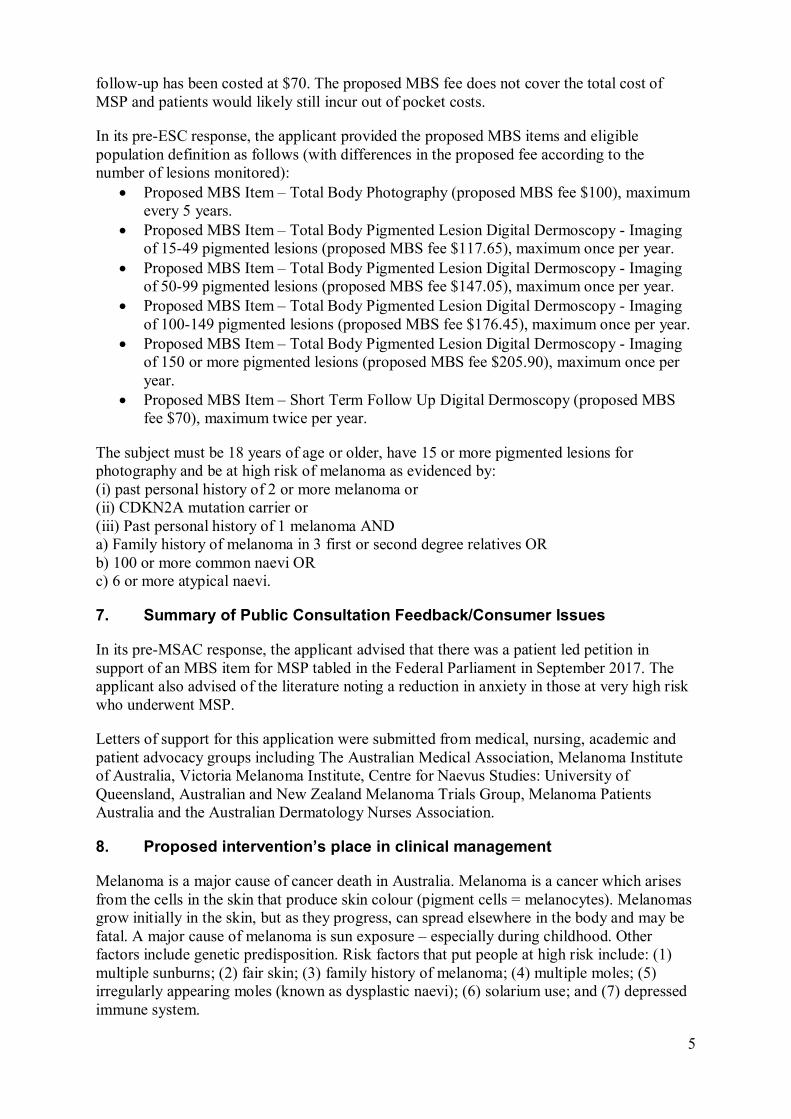
In its pre-ESC response, the applicant provided the proposed MBS items and eligible population definition as follows (with differences in the proposed fee according to the number of lesions monitored):
Proposed MBS Item – Total Body Photography (proposed MBS fee $100), maximum every 5 years.
Proposed MBS Item – Total Body Pigmented Lesion Digital Dermoscopy - Imaging of 15-49 pigmented lesions (proposed MBS fee $117.65), maximum once per year.
Proposed MBS Item – Total Body Pigmented Lesion Digital Dermoscopy - Imaging of 50-99 pigmented lesions (proposed MBS fee $147.05), maximum once per year.
Proposed MBS Item – Total Body Pigmented Lesion Digital Dermoscopy - Imaging of 100-149 pigmented lesions (proposed MBS fee $176.45), maximum once per year.
Proposed MBS Item – Total Body Pigmented Lesion Digital Dermoscopy - Imaging of 150 or more pigmented lesions (proposed MBS fee $205.90), maximum once per year.
Proposed MBS Item – Short Term Follow Up Digital Dermoscopy (proposed MBS fee $70), maximum twice per year.
The subject must be 18 years of age or older, have 15 or more pigmented lesions for photography and be at high risk of melanoma as evidenced by: (i) past personal history of 2 or more melanoma or (ii) CDKN2A mutation carrier or (iii) Past personal history of 1 melanoma AND a) Family history of melanoma in 3 first or second degree relatives OR b) 100 or more common naevi OR c) 6 or more atypical naevi.
7. Summary of Public Consultation Feedback/Consumer Issues
In its pre-MSAC response, the applicant advised that there was a patient led petition in support of an MBS item for MSP tabled in the Federal Parliament in September 2017. The applicant also advised of the literature noting a reduction in anxiety in those at very high risk who underwent MSP.
Letters of support for this application were submitted from medical, nursing, academic and patient advocacy groups including The Australian Medical Association, Melanoma Institute of Australia, Victoria Melanoma Institute, Centre for Naevus Studies: University of Queensland, Australian and New Zealand Melanoma Trials Group, Melanoma Patients Australia and the Australian Dermatology Nurses Association.
8. Proposed intervention’s place in clinical management
Melanoma is a major cause of cancer death in Australia. Melanoma is a cancer which arises from the cells in the skin that produce skin colour (pigment cells = melanocytes). Melanomas grow initially in the skin, but as they progress, can spread elsewhere in the body and may be fatal. A major cause of melanoma is sun exposure – especially during childhood. Other factors include genetic predisposition. Risk factors that put people at high risk include: (1) multiple sunburns; (2) fair skin; (3) family history of melanoma; (4) multiple moles; (5) irregularly appearing moles (known as dysplastic naevi); (6) solarium use; and (7) depressed immune system.

6
Photography of body regions (total body photography) allows future comparison to look for new pigmented lesions which may be melanoma. Close up high resolution photographs of existing moles and freckles (digital dermoscopy) can be used for future comparison to look for the early changes of melanoma arising in existing moles and freckles. This new digital/computer based technology assists in early detection of melanoma.
Based on the proposed clinical management algorithm, MSP is intended as an addition to standard clinical assessments; to allow for the comparison of melanocytic naevi over time. Patients are initially assessed as either having a high, or very high, level of risk of developing melanoma by either a GP or by both a GP and a dermatologist. Patients at high-risk and very-high-risk of melanoma are then photographed using TBP, followed by macro-imaging with TBP at close range and DD (short-term and long-term).
Suspicious naevi are then excised for biopsy, and patients may undergo further treatment as per current clinical management, with the nature of the treatment dependent on the extent of tumour invasion (i.e. Breslow thickness used to inform the American Joint Commission on Cancer AJCC staging criteria). Surveillance is to continue following lesion identification.
The proposed services are an addition to current clinical management of high-risk and very high risk patients for melanoma with the aim to diagnose melanoma at an early stage. The more advanced the melanoma is at the time of diagnosis (measured by Breslow thickness in mm) the more extensive the surgery, further assessment, treatment, morbidity, mortality and cost. Dermatologic consultation, skin excisions (including biopsy), and pathology costs are funded by the MBS, but surveillance including the use of digital dermoscopy is not funded under the MBS.
Currently, a patient at risk of melanoma is presented to a GP (MBS item 23 or 26). The patient may not seek further follow-up with a GP or dermatologist, or may continue with GP follow-up only (1 or 2 visits per year), or the GP may refer the patient to a dermatologist for further examination (MBS item 104).
The patient may then either continue with follow-up visits with the dermatologist (1 or 2 per year; MBS item 105), continue with follow-up with the GP (1 or 2 visits per year; MBS item 23 or 26), or continue follow-up with both the GP and dermatologist (1 or 2 visits per year; MBS items 23, 36 and 105).
The current clinical algorithm pathway is shown in black in Figure 1. The additional MSP program proposed in this assessment is shown in dotted red.

7
Figure 1 Clinical management algorithm for the proposed MSP relative to current clinical practice

8
9. Comparator
The comparator is standard care without MSP conducted by a GP or dermatologist, or self-examination. This may include:
Self-examination at home without the use of comparative photographic images GP clinical examination without access to photography (TBP, DD) for real time
comparison Dermatologist clinical examination (including hand-held dermoscopy) without access
to TBP/DD for real-time comparison
Many high-risk patients do not examine themselves and in fact do not visit their GP, let alone see a dermatologist. They might not be identified as high-risk by their GPs, or if they are identified as high-risk they might not offered a dermatologist consultation by the GP.
MSP is intended to be added to standard-of-care, which is the nominated main comparator. The application’s clinical evidence for standard-of- care included dermatologist clinical examination (with hand-held dermoscopy).
10. Comparative safety
The studies included in the application did not present any data regarding the safety of the intervention.
The application claimed that MSP using TBP and DD is a safe service. This was attributed to the non-invasive nature of TBP and DD.
The critique considered that this was reasonable; however, no comparative data was available on the safety of including MSP with standard-of-care for both patient populations.
11. Comparative effectiveness
High-risk melanoma (population 1) The application provided a naïve comparison of treatment efficacy of MSP versus standard-of-care (without MSP), using the mean Breslow Index (Table 1) and the frequency of in-situ: invasive melanoma (Table 2).
Table 1 Mean Breslow Index (mm) in high-risk melanoma (population 1)
Study MSP SoC P-value
Salerni et al (2011), mean (mm) ± SE 0.54 ± 0.02 1.71 a ± 0.25 - <0.001
Salerni et al (2014), mean (mm) 0.52 0.77 b 1.43 c <0.05
Mintsoulis and Beecker (2016), mean (mm) 0.03 0.34 - 0.02
MSP = melanoma surveillance photography; SE = standard error; w/o = without a Considered intermediate tumour (1 mm-4mm) b Dermatologist consultation c Melanoma Routine care (with handheld dermoscopy)
The application stated that the mean Breslow index was significantly lower in patients with melanomas in the MSP group (Range across studies: 0.03 mm – 0.54 mm) compared with standard-of-care groups (Range across studies: 0.34 – 1.71) (P<0.05 across studies). However, for Salerni et al (2011), the application stated that the incremental effectiveness was likely biased towards MSP, due to the higher baseline risk of melanoma. The critique considered that this was appropriate.

9
Table 2 Frequency of in-situ and invasive melanomas Study Melanoma type MSP SoC P-value
Salerni et al (2011) In-situ (n, %) 35 (70%) 46 (27.8%) - <0.001
Invasive (n, %) 15 (30%) 119 (72.2%) - <0.001
In-situ : invasive ratio 2.33 :1 0.38 :1 - -
Salerni et al (2014) In-situ (n, %) 7 (58.3%) 8 (22.9%) a 26 (50.0%) b <0.05
Invasive (n, %) 5 (41.7%) 27 (77.1%) a 26 (50.0%) b <0.005 c
In-situ : invasive 1.40 :1 0.29 :1 a 1:1b -
Mintsoulis and Beecker (2016)
In-situ (n, %) 13 (93%) 171 (62%) - 0.02
Invasive (n, %) 1 (7%) 106 (38%) - NR
In-situ : invasive 13 :1 1.61 :1 - -
MSP = melanoma surveillance photography; NR = not reported; SoC = standard-of-care. a Melanoma consultation b Melanoma routine control c Not specified which comparator this belongs to
Bold = significant
The application stated that the mean in-situ: invasive melanoma ratio was significantly higher in patients with melanomas in the MSP group (Range: 1.40: 1 to 13: 1 across studies) compared with standard-of-care groups (Range: 0.29: 1 to 1.61: 1 across studies) (P < 0.05 for all studies).
The reason the application stated that the effectiveness of MSP (relative to standard care) could not be estimated was due to the:
high risk of bias from low quality evidence; patient populations were not comparable, due to the high heterogeneity in known
patient and prognostic factors (i.e. age and baseline melanoma risk factors), and limited reporting of baseline data; and
small patient numbers (n: 9-40) that received MSP across all studies.
In addition, the application stated that included statistical results did not adjust for potential confounders, and therefore the naïve comparisons were less meaningful.
Population at very-high-risk (population 2) The application did not present any clinical evidence or make a clinical claim for the patient population at a very-high-risk of melanoma. Using MSP, the median Breslow thickness of all primary melanomas was in situ (in situ to 0.6mm), which accounted for 51% of melanomas detected. However, no comparative results for standard-of-care could be extracted from the economic evaluation performed in Watts et al (2017).
Clinical Claim The applicant claimed that the successful listing of MSP for the target population and specialist setting will lead to earlier detection of melanoma and subsequent improvement in prognosis, as well as reduced requirement for sentinel lymph node biopsy, treatment of metastatic disease, less aggressive skin surgery, and fewer benign lesion excisions.
On the basis of the benefits and harms reported in the evidence base, the application proposed that, relative to standard of care (without MSP), MSP has non-inferior safety and uncertain effectiveness for patients considered at a high risk of developing melanomas.
The critique considered that the clinical claim for non-inferior safety and uncertain effectiveness was reasonable.

10
12. Economic evaluation
One cost-effectiveness analysis comparing MSP in a specialist dermatology setting with standard care in the community was identified in the economic literature search (Watts, Cust et al. 2017). Results from this study indicated that MSP is both more effective and less costly than standard care in patients at very high risk of melanoma.
The model by Watts et al. (2017) was replicated for this assessment. Three changes were made to the model:
1) MSP cost was amended to reflect our proposed MBS items fee, 2) the cost of new cancer treatments were added, and 3) the 2013 MBS items were updated to include 2017 costs.
Table 3 Summary of the economic evaluation
Perspective Australian Health System
Comparator Standard care in the community
Type of economic evaluation Cost-effectiveness evaluation
Sources of evidence Indirect comparison of a prospective single-arm study and population-based linked data
Time horizon 10 years in the model base case
Outcomes QALYs
Methods used to generate results Markov model
Health states Patient presents for surveillance, dead from other causes, dead due to melanoma, recurrence of melanoma, stage III disease, stage IV disease
Cycle length 12 months
Discount rate 5%
Software packages used TreeAgePro 2017
QALY: quality adjusted life year
The clinical inputs and model structure were unchanged. There is some uncertainty regarding the effectiveness inputs in the model, as the sources used to derive the utilities for melanoma by stage and probability of excisions are not published.
As per the published model, the replicated model showed that MSP in patients at very high risk of melanoma is more effective and less costly than standard care. The overall costs and effectiveness, and incremental costs and effectiveness as calculated for the intervention and comparator in the model are shown in Table 4.
Table 4 Base case cost-effectiveness results (10-year time horizon) MSP Standard care Incremental
Cost ($) 7,172.63 15,558.83 8,386.19
Effectiveness (QALY) 7.53 7.32 0.21
QALY: quality-adjusted life year; MSP: melanoma surveillance photography
Results from a one-way sensitivity analysis indicate that MSP is less costly and more effective than standard care up to a threshold of $1100 annual MSP cost per patient, which is a higher cost than the annual estimate per patient (based on the proposed MBS items fees) of $155 included in the model. It is important to note these results were obtained from a model of MSP in a population at very high risk of melanoma attending a highly specialised clinic within an urban centre, and these results might not be generalisable to the high-risk population across the country.

11
The model was most sensitive to variations in the probability of excisions in standard care, with a slight increase in the incremental cost-effectiveness ratio (ICER) as the probability of excision in standard care was changed from 0.8 to 0.1. However, none of the variables had a substantial impact on the ICER. A potential increase in the cost of MSP from $155 to $1000 did not significantly alter the cost-effectiveness of the intervention. Other key drivers of the economic model were the probability of early detection of melanoma (in-situ and stage I) by MSP, treatment costs for melanoma stage III and IV, and the probability of benign excisions in standard care.
13. Financial/budgetary impacts
An epidemiological approach was used to estimate the financial implications of the introduction of MSP in high-risk and very high-risk patients. The calculation of the number of patients who would be eligible to receive MSP was based on data from the Australian Institute of Health and Welfare (AIHW) (Australian Institute of Health and Welfare 2017, Australian Institute of Health and Welfare 2017) and the Australian Bureau of Statistics (Australian Bureau of Statistics 2017).
Two scenarios were estimated to evaluate low and moderate utilisation of the proposed service in both populations, as 100% participation of the total eligible population in screening and monitoring services is not realistic. In the moderate utilisation scenario, an increase in MSP uptake from 20% to 60% over 5 years was assumed, and 90% compliance with follow-up MSP every year. Table 6 presents the estimated number or individuals at high risk and very high risk at 100% participation and moderate utilisation of MSP.
Table 5 Number of eligible individuals at high-risk and very high-risk of melanoma in the Australian population; 100% and moderate utilisation of MSP Population 2018 2019 2020 2021 2022
Total eligible (n); High risk
787,650 798,378 809,106 819,835 830,563
Total eligible (n), very high risk
40,958 41,068 41,179 41,289 41,399
Moderate utilisation (n); High risk
157,530 334,031 490,658 606,181 680,192
Moderate utilisation (n); Very high risk
8,192 17,235 25,089 30,680 34,044
Moderate utilisation: uptake 20% in 2018, increasing by 10% every year, and reaching 60% in 2022, 90% compliance;
The total costs for the high-risk and very high-risk populations given a moderate utilisation, including an annual GP referral visit are presented below in Table 6.

12
Table 6 Total costs ($) to the MBS associated with MSP in high-risk and very high-risk patients; moderate utilisation
Population Cost per patient
2018 2019 2020 2021 2022
High risk
GP ($37.05) + MSP ($155)
$30,253,619.01 $64,150,713.75 $94,230,898.45 $116,416,976.07 $130,630,860.53
Very high risk
GP ($37.05) + MSP ($155)
$1,573,186.59 $3,310,053.80 $4,818,365.30 $5,892,122.39 $6,538,092.15
Moderate utilisation: uptake 20% in 2018, increasing by 10% every year, and reaching 60% in 2022, 90% compliance GP: general practitioner; MSP: melanoma surveillance photography3816
There is a high level of uncertainty regarding the approximate number of individuals at high risk and very high risk of melanoma in Australia; therefore, these estimates are only approximate. Access and utilisation of these medical services may be lower than these projections indicate, especially in rural areas.
14. Key issues from ESC for MSAC
ESC noted that the submission requests new MBS item numbers for melanoma surveillance photography (MSP), involving total body photography (TBP) and digital dermoscopy (DD) for patients based on the degree of risk of developing melanoma (either high-risk or very high-risk). This service is to be conducted in a dermatologist specialist setting and involves the use of TBP in conjunction with DD as an adjunct to measures employed in current clinical practice. The submission claims that MSP allows for the improved detection of changes indicative of the development of melanoma over time compared with naked-eye clinical examinations or standard DD alone. Furthermore, repeated follow-up TBP and DD allows for the comparison of images of suspicious lesions stored on a database over time.
ESC noted that the proposed item descriptors make reference to a registered nurse’s eligibility to perform the service. ESC noted the concern raised by PASC that there is currently no concept of a ‘melanographer’, with no associated training or quality assurance, and that this concern was ongoing. ESC noted that the role of the nurse and any training requirements may need to be referenced in the explanatory notes or appropriate guidelines but that as nurses cannot claim MBS benefits, it is likely that there is no authority to specify any requirements for a non-MBS claiming provider in the descriptors.
ESC noted that the fee is intended to cover the entire procedure, which may take up to 2 hours, but that as this did not include the additional report-writing time, the fee would most likely increase.
ESC noted that there was considerable overlap between the populations for high risk and very high risk as set out in the item descriptors.
ESC noted that if MSP is accepted as standard of care in high risk groups as claimed in the cited consensus-based NHMRC/Australian Cancer Network guidelines (2008) it should already be part of the clinicians’ tool kit. ESC queried why an item was being requested to cover what is standard of care.
ESC noted the lack of high quality evidence available to support the submission and that three retrospective cohort studies of MSP in a tertiary care setting versus standard care in patients at high risk of melanoma were provided as evidence to support the use of MSP in high-risk populations (Salerni G et al 2011; Salerni G et al 2014; Mintsoulis and Beecker
13
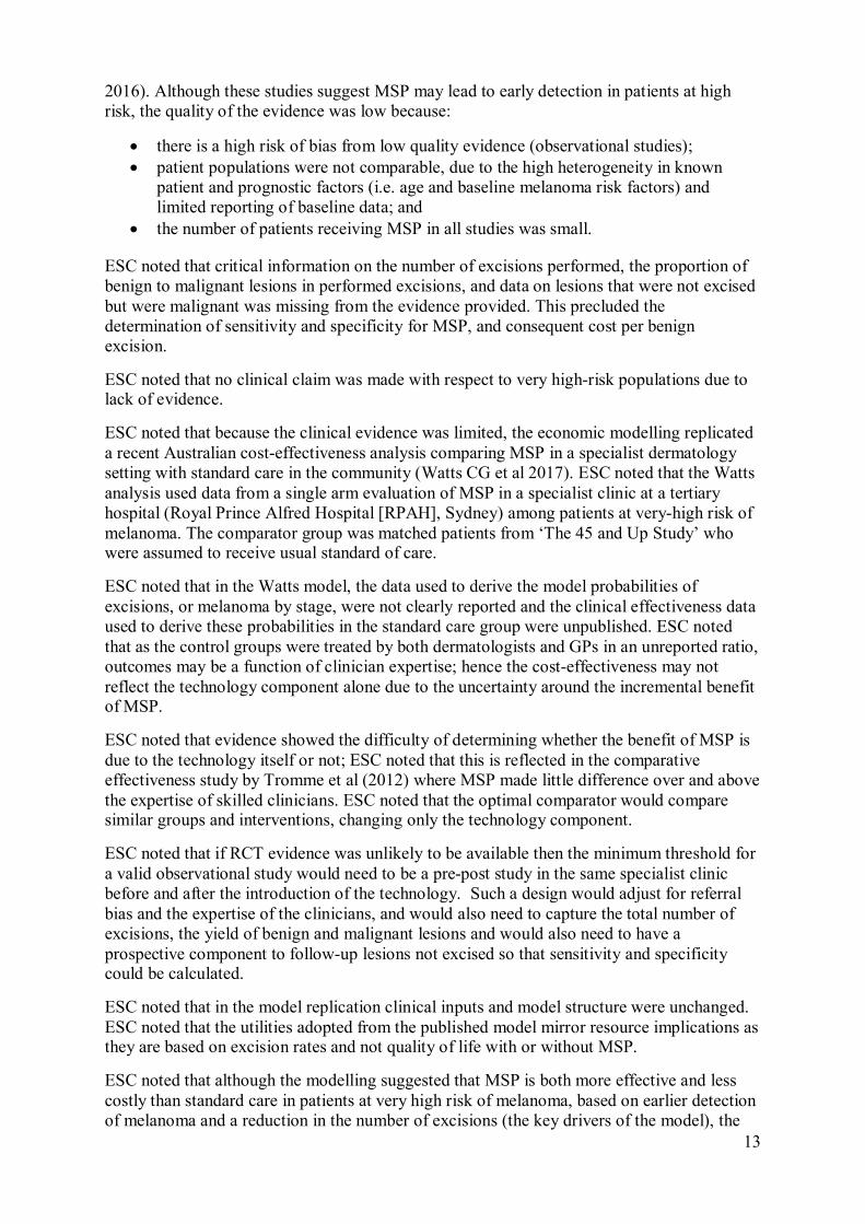
2016). Although these studies suggest MSP may lead to early detection in patients at high risk, the quality of the evidence was low because:
there is a high risk of bias from low quality evidence (observational studies); patient populations were not comparable, due to the high heterogeneity in known
patient and prognostic factors (i.e. age and baseline melanoma risk factors) and limited reporting of baseline data; and
the number of patients receiving MSP in all studies was small.
ESC noted that critical information on the number of excisions performed, the proportion of benign to malignant lesions in performed excisions, and data on lesions that were not excised but were malignant was missing from the evidence provided. This precluded the determination of sensitivity and specificity for MSP, and consequent cost per benign excision.
ESC noted that no clinical claim was made with respect to very high-risk populations due to lack of evidence.
ESC noted that because the clinical evidence was limited, the economic modelling replicated a recent Australian cost-effectiveness analysis comparing MSP in a specialist dermatology setting with standard care in the community (Watts CG et al 2017). ESC noted that the Watts analysis used data from a single arm evaluation of MSP in a specialist clinic at a tertiary hospital (Royal Prince Alfred Hospital [RPAH], Sydney) among patients at very-high risk of melanoma. The comparator group was matched patients from ‘The 45 and Up Study’ who were assumed to receive usual standard of care.
ESC noted that in the Watts model, the data used to derive the model probabilities of excisions, or melanoma by stage, were not clearly reported and the clinical effectiveness data used to derive these probabilities in the standard care group were unpublished. ESC noted that as the control groups were treated by both dermatologists and GPs in an unreported ratio, outcomes may be a function of clinician expertise; hence the cost-effectiveness may not reflect the technology component alone due to the uncertainty around the incremental benefit of MSP.
ESC noted that evidence showed the difficulty of determining whether the benefit of MSP is due to the technology itself or not; ESC noted that this is reflected in the comparative effectiveness study by Tromme et al (2012) where MSP made little difference over and above the expertise of skilled clinicians. ESC noted that the optimal comparator would compare similar groups and interventions, changing only the technology component.
ESC noted that if RCT evidence was unlikely to be available then the minimum threshold for a valid observational study would need to be a pre-post study in the same specialist clinic before and after the introduction of the technology. Such a design would adjust for referral bias and the expertise of the clinicians, and would also need to capture the total number of excisions, the yield of benign and malignant lesions and would also need to have a prospective component to follow-up lesions not excised so that sensitivity and specificity could be calculated.
ESC noted that in the model replication clinical inputs and model structure were unchanged. ESC noted that the utilities adopted from the published model mirror resource implications as they are based on excision rates and not quality of life with or without MSP.
ESC noted that although the modelling suggested that MSP is both more effective and less costly than standard care in patients at very high risk of melanoma, based on earlier detection of melanoma and a reduction in the number of excisions (the key drivers of the model), the
14
limitations of the data introduced a high degree of uncertainty around the true effect of the intervention.
ESC agreed that the submission had not established the cost-effectiveness of MSP in a well-defined population group. ESC also noted that there was insufficient evidence to assess cost-effectiveness for the high-risk population because the data that contributed to the economic model was all from patients at very high risk of melanoma.
ESC noted that the total annual cost per patient undergoing MSP was estimated to be $155 in the model (calculated at 85% of the scheduled fee based on the proposed MBS items for this intervention). ESC noted that this was considerably less than the cost of $884 used in the published model, which had included the cost of salaries, procedures, and training.
ESC noted that it was unlikely the proposed MBS fee would cover the total cost of MSP and patients would likely still incur out-of-pocket costs. ESC noted that this introduced potential issues regarding the availability of the technology for disadvantaged groups and hence equity in service provision.
ESC also queried the absence of subgroup analyses. ESC noted that although earlier diagnosis and fewer excisions would improve quality of life, there may be other issues not captured by the published studies.
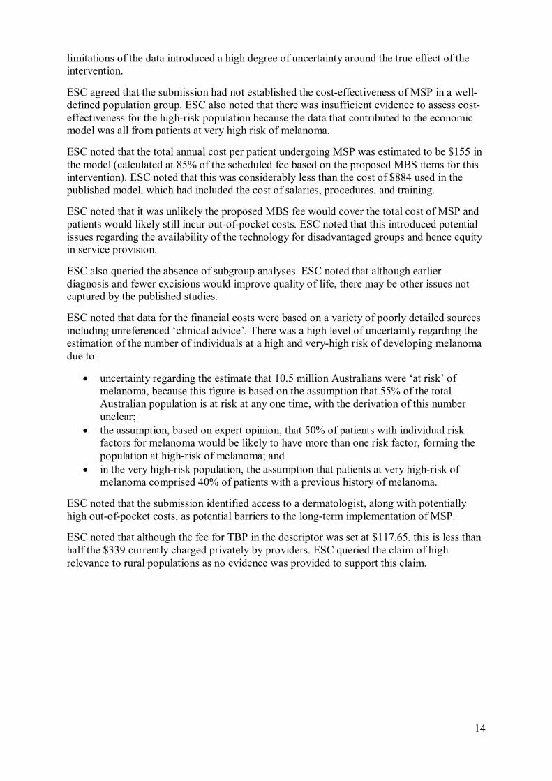
ESC noted that data for the financial costs were based on a variety of poorly detailed sources including unreferenced ‘clinical advice’. There was a high level of uncertainty regarding the estimation of the number of individuals at a high and very-high risk of developing melanoma due to:
uncertainty regarding the estimate that 10.5 million Australians were ‘at risk’ of melanoma, because this figure is based on the assumption that 55% of the total Australian population is at risk at any one time, with the derivation of this number unclear;
the assumption, based on expert opinion, that 50% of patients with individual risk factors for melanoma would be likely to have more than one risk factor, forming the population at high-risk of melanoma; and
in the very high-risk population, the assumption that patients at very high-risk of melanoma comprised 40% of patients with a previous history of melanoma.
ESC noted that the submission identified access to a dermatologist, along with potentially high out-of-pocket costs, as potential barriers to the long-term implementation of MSP.
ESC noted that although the fee for TBP in the descriptor was set at $117.65, this is less than half the $339 currently charged privately by providers. ESC queried the claim of high relevance to rural populations as no evidence was provided to support this claim.
15
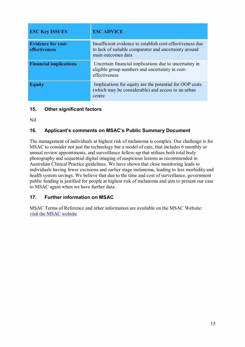
ESC Key ISSUES ESC ADVICE
Evidence for cost-effectiveness
Insufficient evidence to establish cost-effectiveness due to lack of suitable comparator and uncertainty around main outcomes data
Financial implications Uncertain financial implications due to uncertainty in eligible group numbers and uncertainty in cost-effectiveness
Equity Implications for equity are the potential for OOP costs (which may be considerable) and access to an urban centre
15. Other significant factors
Nil
16. Applicant’s comments on MSAC’s Public Summary Document
The management of individuals at highest risk of melanoma is complex. Our challenge is for MSAC to consider not just the technology but a model of care, that includes 6 monthly or annual review appointments, and surveillance follow-up that utilises both total body photography and sequential digital imaging of suspicious lesions as recommended in Australian Clinical Practice guidelines. We have shown that close monitoring leads to individuals having fewer excisions and earlier stage melanoma, leading to less morbidity and health system savings. We believe that due to the time and cost of surveillance, government public funding is justified for people at highest risk of melanoma and aim to present our case to MSAC again when we have further data.
17. Further information on MSAC
MSAC Terms of Reference and other information are available on the MSAC Website: visit the MSAC website