Embed Size (px)
Citation preview
+
PTSD and Abnormal N-acetylaspartate Levels in
Hippocampus and Anterior Cingulate Cortex
Kimberly Sitter

+What is PTSD? (DSM-V)
Criterion A: stressor
Criterion B: intrusive symptoms
Criterion C: avoidance
Criterion D: negative alterations in cognitions and mood
Criterion E: alterations in arousal and reactivity
Criterion F: Persistence (in Criteria B, C, D, E) for >1 month
Criterion G: functional significance
+Who has PTSD?
Veterans
Children (extended in DSM-V)
Rape Victims
Survivors of Natural Disasters
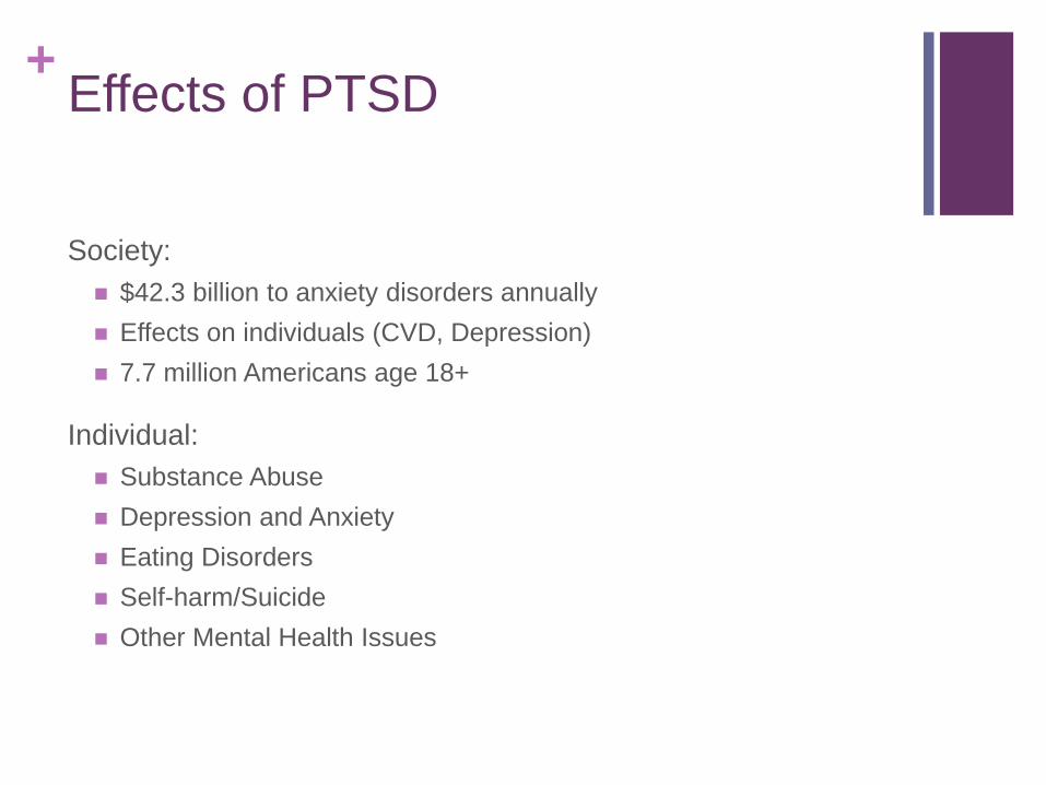
+Effects of PTSD
Society:
$42.3 billion to anxiety disorders annually
Effects on individuals (CVD, Depression)
7.7 million Americans age 18+
Individual:
Substance Abuse
Depression and Anxiety
Eating Disorders
Self-harm/Suicide
Other Mental Health Issues
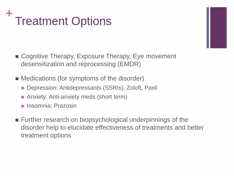
+Treatment Options
Cognitive Therapy, Exposure Therapy, Eye movement
desensitization and reprocessing (EMDR)
Medications (for symptoms of the disorder)
Depression: Antidepressants (SSRIs), Zoloft, Paxil
Anxiety: Anti-anxiety meds (short term)
Insomnia: Prazosin
Further research on biopsychological underpinnings of the
disorder help to elucidate effectiveness of treatments and better
treatment options

+Brain Abnormalities of PTSD
Volumetric reduction of brain structures (hippocampus,
amygdala, anterior cingulate cortex)
Effects: Working Memory, Fear condition/extinction
Higher Glucocorticoids (BDNF) and increased cell sensitization
Effects: deficits in short-term memory loss
Decreased levels NAA

+Role of Anterior Cingulate Cortex
and Hippocampus in PTSD
Anterior Cingulate Cortex:
ACC and medial prefrontal cortex postulated to modulate amygdala
during anxiety
Abnormal relay of information to hippocampus, amygdala, and
sensory cortex plays a role in failure of extinction
Hippocampus:
vital in declarative memory and organizes episodic memories
Low Hippocampal volume associated with severe re-experience
symptoms in PTSD subjects
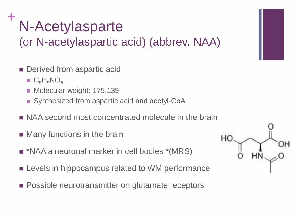
+N-Acetylasparte(or N-acetylaspartic acid) (abbrev. NAA)
Derived from aspartic acid
C6H9NO5
Molecular weight: 175.139
Synthesized from aspartic acid and acetyl-CoA
NAA second most concentrated molecule in the brain
Many functions in the brain
*NAA a neuronal marker in cell bodies *(MRS)
Levels in hippocampus related to WM performance
Possible neurotransmitter on glutamate receptors
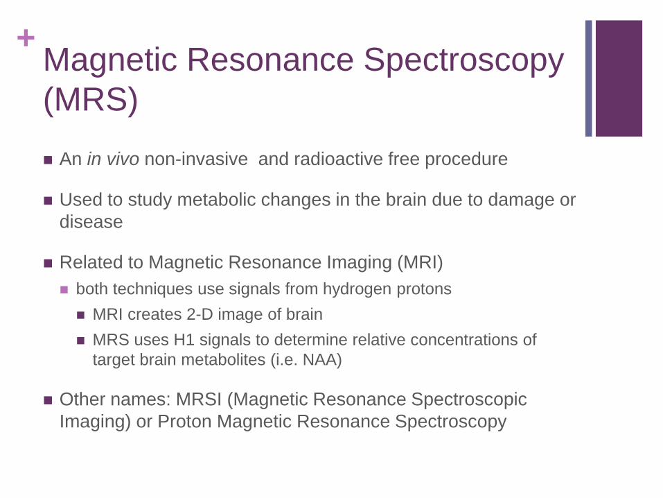
+Magnetic Resonance Spectroscopy
(MRS)
An in vivo non-invasive and radioactive free procedure
Used to study metabolic changes in the brain due to damage or
disease
Related to Magnetic Resonance Imaging (MRI)
both techniques use signals from hydrogen protons
MRI creates 2-D image of brain
MRS uses H1 signals to determine relative concentrations of
target brain metabolites (i.e. NAA)
Other names: MRSI (Magnetic Resonance Spectroscopic
Imaging) or Proton Magnetic Resonance Spectroscopy

+Study 1: Decreased N-acetyl-aspartate levels in anterior
cingulate and hippocampus in subjects with post-traumatic stress
disorder: a proton magnetic resonance spectroscopy study
Authors: Ham, B., Chey, J., Yoon, S.J., Sung, Y., Jeong, D.,
Kim, S.J., Sim, M.E., Choi, N., Choi, I., Renshaw, P.F., Lyoo,
I.K.
Purpose: study relationship of NAA levels in the brain with
clinical presentations of PTSD in subjects
Hypothesis: find decreased NAA levels in ACC and
hippocampus in PTSD subjects, and level of NAA decrease
would correlate with severity of symptoms
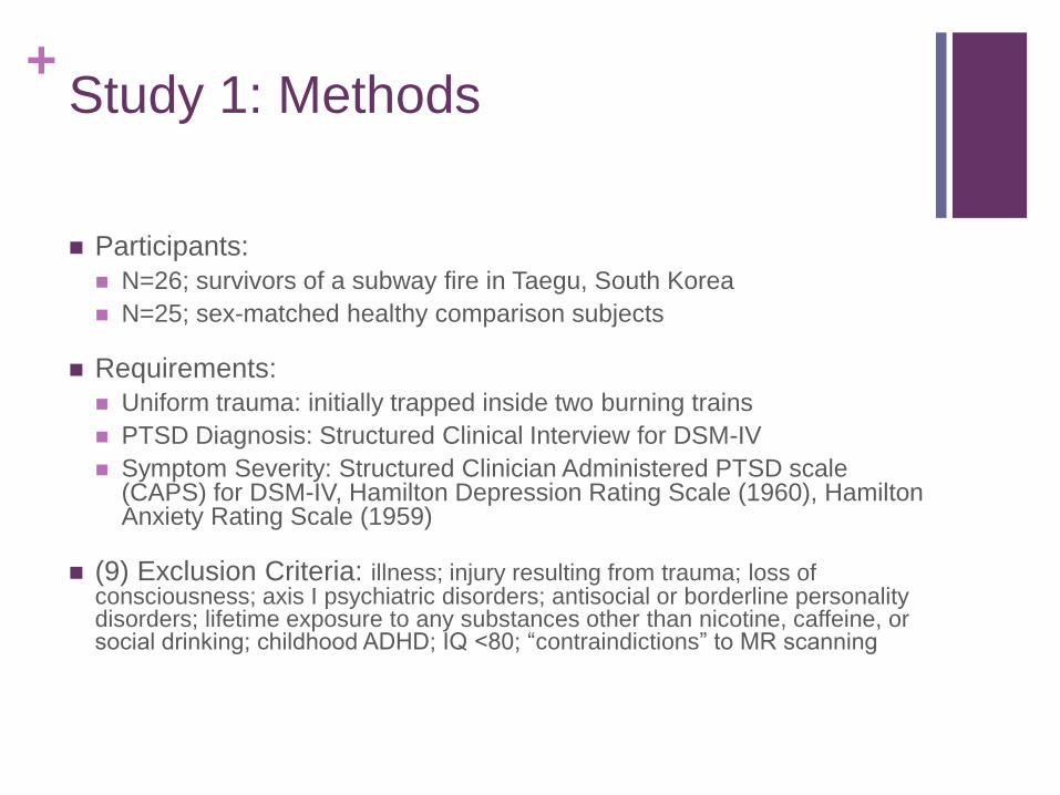
+Study 1: Methods
Participants:
N=26; survivors of a subway fire in Taegu, South Korea
N=25; sex-matched healthy comparison subjects
Requirements:
Uniform trauma: initially trapped inside two burning trains
PTSD Diagnosis: Structured Clinical Interview for DSM-IV
Symptom Severity: Structured Clinician Administered PTSD scale (CAPS) for DSM-IV, Hamilton Depression Rating Scale (1960), Hamilton Anxiety Rating Scale (1959)
(9) Exclusion Criteria: illness; injury resulting from trauma; loss of consciousness; axis I psychiatric disorders; antisocial or borderline personality disorders; lifetime exposure to any substances other than nicotine, caffeine, or social drinking; childhood ADHD; IQ <80; “contraindictions” to MR scanning
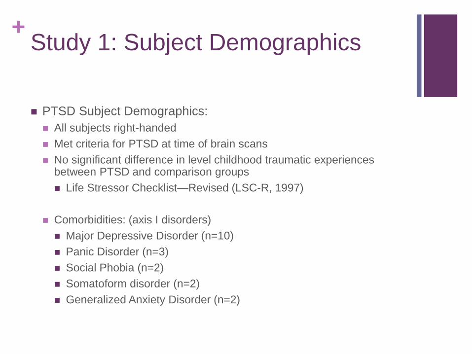
+Study 1: Subject Demographics
PTSD Subject Demographics:
All subjects right-handed
Met criteria for PTSD at time of brain scans
No significant difference in level childhood traumatic experiences between PTSD and comparison groups
Life Stressor Checklist—Revised (LSC-R, 1997)
Comorbidities: (axis I disorders)
Major Depressive Disorder (n=10)
Panic Disorder (n=3)
Social Phobia (n=2)
Somatoform disorder (n=2)
Generalized Anxiety Disorder (n=2)

+Study 1: Placement of ACC and
Bilateral Hippocampus
All scans performed 15.0 ± 1.1 months post fire accident
MRI: 3-Tesla whole-body imaging system (GE VH/1, USA)
Brain voxels of interest (VOIs)

+Study 1: Results
Significantly decreased NAA concentration in ACC and
hippocampus in PTSD subjects
NAA levels of ACC and hippocampus negatively correlated with
re-experience symptom scores in PTSD subjects
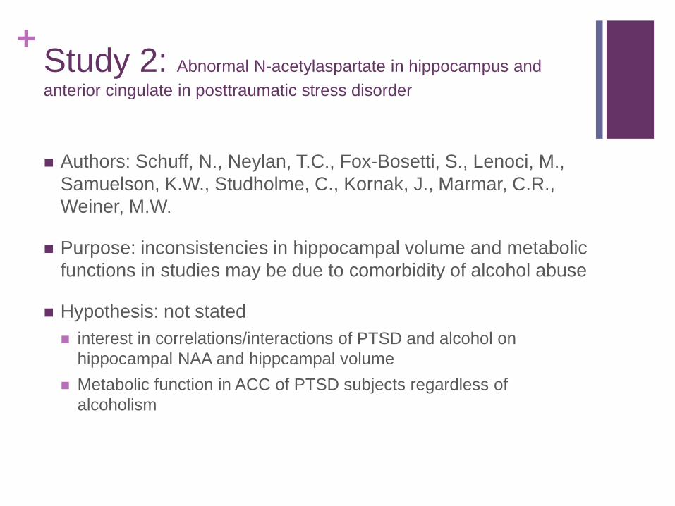
+Study 2: Abnormal N-acetylaspartate in hippocampus and
anterior cingulate in posttraumatic stress disorder
Authors: Schuff, N., Neylan, T.C., Fox-Bosetti, S., Lenoci, M.,
Samuelson, K.W., Studholme, C., Kornak, J., Marmar, C.R.,
Weiner, M.W.
Purpose: inconsistencies in hippocampal volume and metabolic
functions in studies may be due to comorbidity of alcohol abuse
Hypothesis: not stated
interest in correlations/interactions of PTSD and alcohol on
hippocampal NAA and hippcampal volume
Metabolic function in ACC of PTSD subjects regardless of
alcoholism

+Study 2: Method
Participants:
N=55; subjects with PTSD
N=49; subjects without PTSD
4 Groups:
N=28; PTSD with alcohol abuse
N=27; PTSD without alcohol abuse
N=23; No PTSD with alcohol abuse
N=26; No PTSD without alcohol
Subject recruitment:
From outpatient mental health clinic of San Francisco Veterans Affairs Center and from the community by advertisement

+Study 2: Criteria
PTSD Measures (interviews):
Clinician Administered PTSD Scale (CAPS)
Tested for inter-rater reliability
Structured Clinical Interview for DSM-IV Diagnosis (SCID) (Diagnose alcohol or drug abuse, or dependence)
Interview of Life Stressor Checklist—Revised (LSC-R, 1996)
Alcohol Abuse Measures:
PTSD with alcohol: Veterans with alcohol abuse in past 5 years
PTSD without alcohol: Veterans with no history alcohol abuse
Operational definition alcohol consumption:
Total cumulate drinks/year over past 5 years
Current alcohol consumption as cumulate drinks month prior study

+Study 2: Exclusion Criteria
Exclusions:
Veterans with past but not current PTSD or subsyndromal PTSD
Psychotic disorder or bipolar disorder
Drug abuse or Dependence
Neurological Illness, head trauma with loss of consciousness,
medical disorders affecting brain function
MRI scanning exclusion criteria
MRIs reviewed for exclusionary neuropathological conditions
(brain tumors, lesions, vessel disease)

+Study 2: MRI and MRS Acquisition
1.5 T Vision MR system (Siemens Inc., Iselin, NJ)
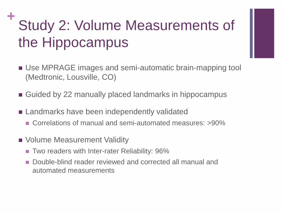
+Study 2: Volume Measurements of
the Hippocampus
Use MPRAGE images and semi-automatic brain-mapping tool
(Medtronic, Lousville, CO)
Guided by 22 manually placed landmarks in hippocampus
Landmarks have been independently validated
Correlations of manual and semi-automated measures: >90%
Volume Measurement Validity
Two readers with Inter-rater Reliability: 96%
Double-blind reader reviewed and corrected all manual and
automated measurements

+Study 2: Results
Brain Volumes:
PTSD alone not significantly associated smaller volumes of left or right hippocampus
Strong association increasing age and smaller hippocampal volume (left and right)
Trend of smaller hippocampi with childhood trauma
Still no significant effect on hippocampal volume when adjusting for the most severe symptoms in subjects or just comparing men
No significant effect of PTSD with long-term alcohol-drinking behavior on hippocampal volume
However, trend for smaller frontal lobe white matter in PTSD

+Study 2: Results (cont.d)
Brain Metabolites:
PTSD associated with reduced NAA concentration in hippocampus
PTSD associated with reduced NAA concentration in ACC
Long-term alcohol consumption did not alter these results
No significant effect of childhood trauma or lifetime or current
depression on metabolite concentrations
No significant effect of psychiatric medications on metabolites
No other significant metabolite abnormalities found in PTSD outside
of ACC
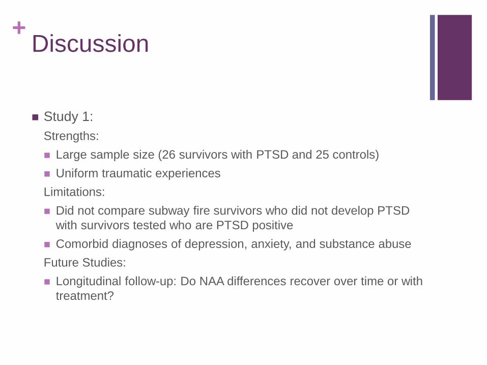
+Discussion
Study 1:
Strengths:
Large sample size (26 survivors with PTSD and 25 controls)
Uniform traumatic experiences
Limitations:
Did not compare subway fire survivors who did not develop PTSD
with survivors tested who are PTSD positive
Comorbid diagnoses of depression, anxiety, and substance abuse
Future Studies:
Longitudinal follow-up: Do NAA differences recover over time or with
treatment?
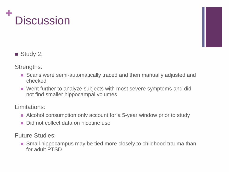
+Discussion
Study 2:
Strengths:
Scans were semi-automatically traced and then manually adjusted and checked
Went further to analyze subjects with most severe symptoms and did not find smaller hippocampal volumes
Limitations:
Alcohol consumption only account for a 5-year window prior to study
Did not collect data on nicotine use
Future Studies:
Small hippocampus may be tied more closely to childhood trauma than for adult PTSD

+Discussion
Findings in BOTH studies:
PTSD associated with reduced NAA in hippocampus in absence of
reduced hippocampal volume
PTSD associated with reduced NAA concentration in ACC
PTSD associated reduced NAA/Cr ratio in hippocampus
Explains PTSD symptoms: abnormalities in cognition, emotion
regulation, fear conditioning and fear extinction (hyper-active
response to trauma cues), re-experience
Metabolite measures are a more sensitive marker for neuronal
abnormality in PTSD than volumetric measures of the brain