Embed Size (px)
Citation preview

Protozoa of the Mountain Marmot, Marmota flaviventer Audubon &Bachman, 1841Author(s): J. Russel GabelSource: Transactions of the American Microscopical Society, Vol. 80, No. 1 (Jan., 1961), pp. 43-53Published by: Wiley on behalf of American Microscopical SocietyStable URL: http://www.jstor.org/stable/3223705 .
Accessed: 29/06/2014 08:46
Your use of the JSTOR archive indicates your acceptance of the Terms & Conditions of Use, available at .http://www.jstor.org/page/info/about/policies/terms.jsp
.JSTOR is a not-for-profit service that helps scholars, researchers, and students discover, use, and build upon a wide range ofcontent in a trusted digital archive. We use information technology and tools to increase productivity and facilitate new formsof scholarship. For more information about JSTOR, please contact [email protected].
.
Wiley and American Microscopical Society are collaborating with JSTOR to digitize, preserve and extendaccess to Transactions of the American Microscopical Society.
http://www.jstor.org
This content downloaded from 151.227.68.60 on Sun, 29 Jun 2014 08:46:19 AMAll use subject to JSTOR Terms and Conditions

HISTOCHEMISTRY OF COLPODA HISTOCHEMISTRY OF COLPODA
LITERATURE CITED ALLEN, R. J. L. and BOURNE, G. H. 1943. Some experiments on the microscopical
demonstration of zymohexase in animal tissues. Jour. Exper. Biol., 20: 61-64. BLES, E. J. 1929. Arcella. A study in cell physiology. Quart. Jour. Microsc.
Sci., 72: 527-648. GLICK, D. 1949. Techniques of Histo- and Cytochemistry. Interscience Pub. Co.,
New York. GOMORI, G. 1941. Distribution of acid phosphatase in tissues under normal and
pathological conditions. Arch. Path., 32: 189-199. 1946. The distribution of lipase in tissues and sections under normal and patho-
logical conditions. Ibid., 41: 121-129. 1951. Alkaline phosphatase of cell nuclei. Jour. Lab. Clin. Med., 37: 526-531.
HUNTER, N. W. 1957. Intracellular localization of some hydrolases, and iron and
copper porphyrin enzymes of Opalina carolinensis. Trans. Amer. Microsc. Soc., 76: 36-45.
HUNTER, N. W. and HUNTER, ROY, JR. 1957. Enzyme systems of Colpoda cucullus. I. Oxidation of certain krebs cycle intermediates. Jour. Cell. Comp. Physiol., 50: 341-346.
LAIDLAW, G. F. and BLACKBERG, S. N. 1932. Melanoma studies. II. A simplified technique for dopa reaction. Amer. Jour. Path., 8: 491-498.
MCJUNKIN, F. A. 1922. Peroxydase staining with benzidin in paraffin sections of human tissue. Anat. Rec., 24: 67-77.
OSTER, K. A. and SCHLOSSMAN, N. C. 1942. Histochemical demonstration of amine oxidase in the kidney. Jour. Cell. Comp. Physiol., 2: 373-378.
PROTOZOA OF THE MOUNTAIN MARMOT, MARMOTA FLA VIVENTER AUDUBON & BACHMAN, 1841
J. RUSSEL GABEL
Division Science, Mathematics and Engineering, San Francisco State College, San Francisco 27, California
INTRODUCTION
Examination of material from the caecum of the mountain marmot, Marmota flaviventer Audubon and Bachman 1841, has resulted in the identification of eight species of Protozoa. No material was available for study of blood parasites of this host although Trypanosoma was reported by Dias (1938) in M. flaviventer nosophora Howell 1915. A complete survey in the future of M. flaviventer involving examination of many more individuals, and other organs and tissues is indicated. Such an investigation should result in identification of more parasites of the intestinal tract and other parts of this host.
MATERIALS AND METHODS
The caecal contents of the marmot were fixed in Hollande's fluid and stained with Heidenhain's iron-haematoxylin and the Bodian protargol techniques. No fresh material was available for study.
43 43
This content downloaded from 151.227.68.60 on Sun, 29 Jun 2014 08:46:19 AMAll use subject to JSTOR Terms and Conditions

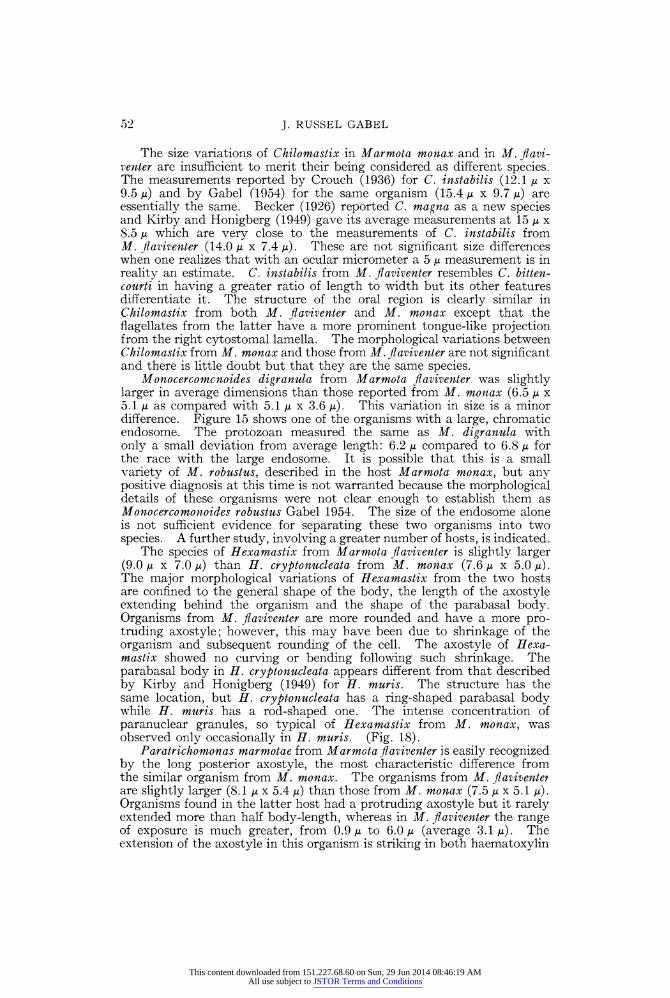
J. RUSSEL GABEL
All protozoa measurements were made with a calibrated eyepiece micrometer. Twenty-five normally shaped organisms of each species were taken at random for measurement, except as noted for Octomitus pulcher (Becker 1926) Gabel 1954, of which only ten were found. The extremes of the size range are stated as well as the computed averages of all dimensions. The illustrations were drawn at 4000 X with the aid of a camera lucida, except figure 7 which was enlarged to 8000 X.
RESULTS
Chilomastix instabilis Crouch 1936 (Figure 6)
The anterior end is often more pointed than most species of Chilomastix thus the animal is broadest at the middle of the body (Fig. 6). The average length is 14.1 ,u (10.3-18.9) and the average width is 7.4 u/ (6.0- 11.2), so the organism appears slender because the length is usually twice the width. There is no significant variation in over-all dimensions of organisms stained by Heidenhain's or the protargol method.
Briefly described, the organism has three anterior flagella which are from Y2 to 2 body-length and a fourth flagellum within the oral pouch. This pouch, on the oral surface, extends beyond the middle of the body. It is bordered by cytostomal fibers, the right one looping around the cytostome, at the posterior end of the pouch, before turning inward to disappear in the cytoplasm near the posterior end of the animal. Right and left cytostomal fibers and lamellae are present. There is a slight tongue-like projection into the oral pouch from the right cytostomal lamella. A nucleus, in the anterior quarter of the body, has a large and deeply stained endosome within a well differentiated nuclear membrane.
The morphological characters of C. instabilis from Marmota flaviventer are typical for the species described by Crouch (1936) and Gabel (1954). Minor structural variations are not sufficient to differentiate the organisms from the two hosts. In addition to variation in shape, the organisms from M. flaviventer have a less prominent tongue-like projection of the right cytostomal lamella than that described in C. instabilis from M. monax Linnaeus.
Monocercomonoides digranula (Crouch 1933) Gabel 1954 (Figures 13, 14)
This flagellate is rounded anteriorly, the posterior end having a sharp point where the chromatic axostyle protrudes. The inverted teardrop- shaped organism has its greatest breadth in the anterior 1/ to 13 of the cell. Average measurements are: length 6.5 / (5.2-8.6), width 5.1 , (3.4-8.6). Three of the four flagella are usually directed forward; the fourth one is normally in a trailing position. They are of equal thickness and greater than body length; protargol preparations show each flagellum to have a terminal filament.
A thin, chromatic axostyle passes from anterior to posterior and is surrounded by a narrow chromatic ring as it leaves the body. A funis lies under the dorsal surface of the cell and a pelta appears to lie across the anterior end of the animal. There is a prominent, chromatic endosome within the anteriorly placed nucleus. The two blepharoplasts, from which the flagella and axostyle arise, are connected by a posteriorly arched fiber and have small fibers extending anteriorly and posteriorly from them to support the pelta (Figs. 13, 14).
44
This content downloaded from 151.227.68.60 on Sun, 29 Jun 2014 08:46:19 AMAll use subject to JSTOR Terms and Conditions

PROTOZOA IN MARMOTA
1. Paratrichomonas marmotae-Heidenhain's haematoxylin. 2. P. marmotae-Heidenhain's haematoxylin. 3. P. marmotae-Bodian protargol. 4. Hexamitus teres-Bodian protargol.
45
This content downloaded from 151.227.68.60 on Sun, 29 Jun 2014 08:46:19 AMAll use subject to JSTOR Terms and Conditions

J. RUSSEL GABEL
Hexamastix muris (Wenrich 1924) (Figures 16-20)
Hexamastix muris is usually relatively large and stout and was quite numerous in Marmota flaviventer. Its shape is often more elongate than the drawings indicate, being more ellipsoidal (less rounded). The effects of fixatives and stains on the protozoan often cause distortion. The average dimensions are: length 9.0 ,u (6.0-12.0) and width 7.2 t, (4.3-10.3); the axostyle protrudes an average of 2.7 u (1.7-4.3) behind the organism. That there is a considerable range in size of the protozoan can be seen from figures 17 and 18.
Wenrich (1924) states there are six flagella, five directed anteriorly and one trailing. The anterior flagella are of unequal length, fused at the base, with the shortest one breaking free from the others first. Protargol preparations show that the tips of the flagella have knob-like swellings (Figs. 16, 20). The nucleus is round to oval and near the anterior end. The axostyle varies from hyaline to granular, has a club-shaped capitulum (Fig. 18), and tapers sharply to a needle-like point at its posterior end. A crescent-shaped pelta forms a chromatic arch across the apex of the cell anterior to the nucleus (Figs. 16, 19, 20). A short, rod-like parabasal body (Fig. 19) lies close to the nucleus or along its dorsal surface and in contact with the nuclear membrane, being connected to the blepharoplast by a fine rhizoplast. The structure of the parabasal body resembles that described by Kirby and Honigberg (1949) for Hexamastix muris from Citellus Oken 1816, and the close similarity of structure between the organisms in M. fiaviventer and Citellus leads to the conclusion that this protozoan is most likely H. muris. This was erroneously reported as H. cryptonucleata by Gabel (1954a).
Paratrichomonas marmotae (Crouch 1933) Gabel 1954 (Figures 1-3)
The shape of this organism is usually oval with a very long axostyle extending behind the cell; however, shrinkage of the organism during fixation and staining may account for this condition. Exclusive of the axostyle, the average length is 8.1 / (5.2-11.2) and the average width is 5.4 u (5.2-7.7).
There are three anterior flagella of equal diameter but unequal length, the tips of which are found to be swollen in protargol preparations (Fig. 3). One of the anterior flagella is usually shorter than the other two. An undulating membrane extends 34 the body length, has from two to five undulations and is bordered by a thickened posterior flagellum which decreases sharply in diameter (to the same thickness as the anterior flagella) as it becomes a trailing fiber. A thin chromatic costa lies under the undulating membrane and no discrete subcostal granules can be dis- tinguished. The costa is usually about % body-length but sometimes extends to the posterior end (Fig. 2). A granular axostyle protrudes from the posterior end of the body an average distance of 3.1 ,u (0.7-6.0). It may taper gradually (Fig. 1) or narrow rapidly to a needle-like point (Fig. 3). The anterior end of the axostyle is broadened into a spoon- shaped capitulum. An endosome lies within the anteriorly placed nucleus. Protargol preparations show a ring-shaped parabasal body, usually pressed against the nuclear membrane, and lying dorso-laterally to the nucleus (Fig. 3). A crescent-shaped pelta lies at the apex of the cell (Fig. 3).
46
This content downloaded from 151.227.68.60 on Sun, 29 Jun 2014 08:46:19 AMAll use subject to JSTOR Terms and Conditions

PROTOZOA IN MARMOTA
7
5. H. teres-Heidenhain's haematoxylin. 6. Chilomastix instabilis-Heidenhain's haematoxylin. 7. Octomitus pulcher-Heidenhain's haematoxylin, anterior end, 8000X. 8. 0. pulcher-Heidenhain's haematoxylin. 9. Tritrichomonas minuta-Heidenhain's haematoxylin.
47
This content downloaded from 151.227.68.60 on Sun, 29 Jun 2014 08:46:19 AMAll use subject to JSTOR Terms and Conditions

J. RUSSEL GABEL
Tritrichomonas minuta (Wenrich 1924) (Figures 9-12)
This organism is bean-shaped, the ventral edge flattened or concave, the anterior end is slightly rounded and the posterior end tapers where the axostyle leaves the body. Its average dimensions are: length 6.8 y (5.2-8.6) and width 3.3 u (0.3-3.8). The minuteness of the organism makes detailed study difficult, but certain structures are visible.
There are three anterior flagella of unequal length, the longer two slightly more than body length (average 8.6 u) and the third, shorter one averages 4.9 tu. All three are of equal thickness and protargol preparations show knob-like tips on each flagellum (Fig. 12). The fiber bordering the undulating membrane is about twice the thickness of a flagellum but it has an abrupt reduction to the diameter of the others as it becomes a free, trailing flagellum. It usually extends 6 Iu beyond the cell. A round, chromatic rod forms a supporting structure for the narrow undulat- ing membrane as these two organelles spiral to the posterior end (Figs. 10, 12). Beneath the costa is a single row of subcostal granules (Figs. 9, 11).
The axostyle is a hyaline rod tapering sharply to a point and pro- truding about 1.3 , (0.7-1.7) beyond the body. There is a ring around the axostyle where it emerges from the cell (Figs. 10, 11) and a group of para-axostylar granules along its anterior half (Fig. 9). The nucleus lies in the anterior part of the cell and its endosome is obscure. A rhizoplast connects the nucleus to the blepharoplast (Figs. 9, 12). A small, chromatic, rod-like parabasal body lies dorsally along the anterior half of the nucleus (Fig. 12) and a thin rhizoplast is sometimes visible connecting the parabasal apparatus with the blepharoplast. A pelta, shaped like a crescent with a dark border, lies along the anterior edge of the protozoan (Fig. 12). The inner area of the pelta stains less intensely with gold chloride than does its border.
The small size of this organism makes a detailed analysis of its mor- phology difficult, however it follows the morphological pattern of the Tritrichomonas muris group. There are no observable incra-axostylar granules as described in T. wenrichi, (Gabel, 1954) nor can one distinguish any distinct grouping of cytoplasmic granules other than those mentioned as lying beneath the costa. The organism is sometimes parasitized by a species of Sphaerita (Fig. 10).
Hexamitus teres Kirby and Honigberg 1949 (Figures 4, 5)
This protozoan has an oval shape with its posterior end slightly broader than the anterior because of the lateral spreading of the axostyles. Measurements gave an average length of 8.4 , (6.9-11.0) and a width of 6.7 ,u (5.2-8.6). There are usually eight flagella but sometimes an organism is seen with fewer than the normal number (Fig. 5). Two nuclei, each containing a prominent deep-staining endosome, are situated at the extreme anterior end of the cell. The twin axostyles are distinct morphological features of this species particularly their trumpet-like posterior ends (Fig. 5). It has not been possible to determine whether the two trailing flagella are within the axostyles or whether they lie in juxtaposition to the outer surfaces. At the anterior end of the cell is a chromatic, cap-like structure which resembles the trichomonad pelta
48
This content downloaded from 151.227.68.60 on Sun, 29 Jun 2014 08:46:19 AMAll use subject to JSTOR Terms and Conditions

PROTOZOA IN MARMOTA
10. T. minuta-Heidenhain's haematoxylin, Sphaerita in vacuole. 11. T. minuta-Heidenhain's haematoxylin. 12. T. minuta--Bodian protargol. 13. MIonocercomonoides digranula-Bodian protargol. 14. M. digranula-Heidenhain's haematoxylin. 15. Monocercomonoides sp.-Heidenhain's haematoxylin. 16. Hexamastix muris-Bodian protargol. 17. H. muris-Heidenhain's haematoxylin.
49
This content downloaded from 151.227.68.60 on Sun, 29 Jun 2014 08:46:19 AMAll use subject to JSTOR Terms and Conditions

J. RUSSEL GABEL
except that this one stains with iron-haematoxylin as well as with protargol. It serves a possible supportive function for the nuclei.
Octomitus pulcher (Becker 1926) Gabel 1954 (Figures 7, 8)
This protozoan was rare in the population studied and only ten normal specimens were found which could be used for measurements. It is oval with a pointed posterior end, while the anterior end bulges where the twin nuclei are located (Fig. 8). Average dimensions are: length 8.0 A. (6.9-8.6) and width 4.5 ,A (3.4-6.9).
There are eight flagella, the six anterior ones having swollen bases which are absent from the two trailing flagella. The nuclei have a minute endosome close to or against the anterior part of the karyotheca (Fig. 7). Passing posteriorly between the nuclei are fused axostyles. Protargol preparations show the outside portion of the axostyles to be chromatic while the inner part is more hyaline. The cytoplasm has chromatic bodies ranging from 0.5 IA to 2.1 / scattered through it.
Entam6eba marmotae Crouch 1936 (Figure 21)
Entamoeba marmotae is generally rounded in fixed and stained prepara- tions, but oval or elongate specimens have been observed. The average size is 17.5 t (10.3-22.4). The nucleus varies in size from 3.4 / to 8.6 It with an average diameter of 4.8 I. It has an eccentric endosome with granular rays extending from it to the karyotheca. The cytoplasm has many bacterial food inclusions within its vacuoles. No inclusions of blood cells or body cells of the host were observed; however, these amoebae are not averse to devouring other protozoa present in the caecum. Several examples have been observed of the associated flagellates enclosed within food vacuoles, thus adding animal protein to the amoeba's normal vege- tarian diet. No cysts were observed.
DISCUSSION Six of the eight species of protozoa from Marmota flaviventer were
described from M. monax. Neither Tritrichomonas minuta nor Hexamastix muris were found in the latter host. It is notable that some of the pro- tozoa found in M. flaiiventer are larger than those reported from M. monax even though there are no significant morphological characters to merit designating them as new species. Monocercomonoides, Paratrichomonas and Entamoeba had greater average sizes in M. flaviventer. The principal morphological variations in the protozoa of the two hosts are restricted to four of the eight organisms.
The taxonomy of Chilomastix from rodent hosts is still in a rather confused condition. Alexeieff (1911) created the new genus, and there has resulted a profusion of species designations, such as: C. intestinalis, C. instabilis, C. magna, C. bittencourti, C. cuniculi, C. caprae, C. rosenbuschi, C. wenrichi, C. muris, C. oblonga, and C. mesnili. Many of these are the result of finding Chilomastix in a host previously unreported and using the criterion of absolute host specificity as a species trait. Transfaunation experiments (Saxe, 1954) with many symbiotic protozoa, are accumulating evidence contrary to this concept. In many instances, great emphasis has been placed upon measurement of the organism with little consideration of its morphology in determining new species.
This content downloaded from 151.227.68.60 on Sun, 29 Jun 2014 08:46:19 AMAll use subject to JSTOR Terms and Conditions

PROTOZOA IN MARMIOTA
21
18. H. muris-Heidenhain's haematoxylin. 19. H. muris-Bodian protargol, anterior end. 20. H. muris-Bodian protargol. 21. Entamoeba marmotae-Heidenhain's haematoxylin.
51
This content downloaded from 151.227.68.60 on Sun, 29 Jun 2014 08:46:19 AMAll use subject to JSTOR Terms and Conditions
J. RUSSEL GABEL
The size variations of Chilomastix in Marmota monax and in M. flavi- venter are insufficient to merit their being considered as different species. The measurements reported by Crouch (1936) for C. instabilis (12.1 , x 9.5 ,u) and by Gabel (1954) for the same organism (15.4 u x 9.7 ,u) are essentially the same. Becker (1926) reported C. magna as a new species and Kirby and Honigberg (1949) gave its average measurements at 15 ,u x 8.5 u which are very close to the measurements of C. instabilis from M. flaviventer (14.0 / x 7.4 u). These are not significant size differences when one realizes that with an ocular micrometer a 5 ,u measurement is in reality an estimate. C. instabilis from M. flaviventer resembles C. bitten- courti in having a greater ratio of length to width but its other features differentiate it. The structure of the oral region is clearly similar in Chilomastix from both M. flaviventer and M. monax except that the flagellates from the latter have a more prominent tongue-like projection from the right cytostomal lamella. The morphological variations between Chilomastix from M. monax and those from M. flaviventer are not significant and there is little doubt but that they are the same species.
Monocercomcnoides digranula from Marmota flaviventer was slightly larger in average dimensions than those reported from M. monax (6.5 Au x 5.1 I as compared with 5.1 u x 3.6 ,u). This variation in size is a minor difference. Figure 15 shows one of the organisms with a large, chromatic endosome. The protozoan measured the same as M. digranula with only a small deviation from average length: 6.2 /u compared to 6.8 / for the race with the large endosome. It is possible that this is a small variety of M. robustus, described in the host Marmota monax, but any positive diagnosis at this time is not warranted because the morphological details of these organisms were not clear enough to establish them as Monocercomonoides robustus Gabel 1954. The size of the endosome alone is not sufficient evidence for separating these two organisms into two species. A further study, involving a greater number of hosts, is indicated.
The species of Hexamastix from Marmota fiaviventer is slightly larger (9.0 Ia x 7.0 Ia) than H. cryptonucleata from M. monax (7.6 , x 5.0 /). The major morphological variations of Hexamastix from the two hosts are confined to the general shape of the body, the length of the axostyle extending behind the organism and the shape of the parabasal body. Organisms from M. fiaviventer are more rounded and have a more pro- truding axostyle; however, this may have been due to shrinkage of the organism and subsequent rounding of the cell. The axostyle of Hexa- mastix showed no curving or bending following such shrinkage. The parabasal body in H. cryptonucleata appears different from that described by Kirby and Honigberg (1949) for H. muris. The structure has the same location, but H. cryptonucleata has a ring-shaped parabasal body while H. muris has a rod-shaped one. The intense concentration of paranuclear granules, so typical of Hexamastix from M. monax, was observed only occasionally in H. muris. (Fig. 18).
Paratrichomonas marmotae from Marmota flaviventer is easily recognized by the long posterior axostyle, the most characteristic difference from the similar organism from M. monax. The organisms from M. flaviventet are slightly larger (8.1 , x 5.4 /u) than those from M. monax (7.5 ,u x 5.1 Iu). Organisms found in the latter host had a protruding axostyle but it rarely extended more than half body-length, whereas in M. fiaviventer the range of exposure is much greater, from 0.9 I, to 6.0 Iu (average 3.1 ,u). The extension of the axostyle in this organism is striking in both haematoxylin
52
This content downloaded from 151.227.68.60 on Sun, 29 Jun 2014 08:46:19 AMAll use subject to JSTOR Terms and Conditions

PROTOZOA IN MARMO TA
and protargol preparations. There are no structural features in Hexa- mitus, Octomitus or Entamoeba from M. flaviventer to distinguish them from those described from M. monax.
That eight genera, Chilomastix, Monocercomonoides, Hexamastix, Paratrichomonas, Tritrichomonas, Hexamitus, Octomitus and Entamceba should occur in such closely related hosts is not unusual. Even though the two hosts now live in different environments and do not normally come into contact with each other, it is not surprising that they should harbor similar protozoan genera, six species of which prove to be the same. It is not unreasonable to suppose that the protozoa invaded the lower intestine of Marmota before the two host species separated into different ecological niches. The close similarity of the protozoan fauna and the bacterial flora (the latter were observed to be quite alike in both hosts), may be taken as an indication that these two hosts once lived on such intimate terms that they shared their protozoa. It is likewise reasonable to assume that the slight variations which occur in the structure of these protozoa probably occurred after the two hosts moved into separate environments.
SUMMARY
Eight species of protozoa are reported from the mountain marmot, Marmota flaviventer. The organisms found in the host are: Chilomastix instabilis Crouch 1936, Monocercomonoides digranula (Crouch 1933) Gabel 1954, Hexamastix muris (Wenrich 1924), Paratrichomonas marmotae (Crouch 1933) Gabel 1954, Tritrichomonas minuta (Wenrich 1924), Hexamitus teres Kirby and Honigberg 1949, Octomitus pulcher (Becker 1926) Gabel 1954, and Entamoeba marmotae Crouch 1936. A comparison is made in respect to the morphological variations from the protozoans reported from Marmota monax Linnaeus.
LITERATURE CITED
ALEXEIEFF, A. 1911. Notes sur les flagelles. Arch. Zool. Exper. et Gen., S6r. 5, 6: 491-527.
BECKER, E. R. 1926. The flagellate fauna of the caecum of the striped ground squirrel, Citellus tridecemlineatus with special reference to Chilomastix magna sp. nov. Biol. Bull., 51: 287-298.
CROUCH, H. B. 1933. Four new species of Trichomonas from the woodchuck (Mar- mota monax Linn.). Jour. Parasit., 19: 293-301.
1936. The animal parasites of the woodchuck (Marmota monax) with special reference to protozoa. Iowa State Coll. Jour. Sci., 11: 48-50.
DIAS, E. 1938. Tr.panosoma de um roedor, Marmota flaviventris nosophora, achado em Montana, Estados Unidos. Libro Jubil. Prof. L. Travassos.: 141-143.
GABEL, J. R. 1954. The morphology and taxonomy of the intestinal protozoa of the American woodchuck, Marmota monax Linnaeus. Jour. Morph., 94: 473-550.
1954a. Protozoa from the mountain marmot, Marmota flaviventris. (Abstract). Jour. Protoz. 1, Suppl. 1: 1.
KIRBY, H. and HONIGBERG, B. 1949. Flagellates of the caecum of ground squirrels. Univ. Calif. Pub. in Zool., 53: 315-366.
SAXE, L. R. 1954. Transfaunation studies on the host specificity of the enteric protozoa of rodents. Jour. Protoz., 1: 220-230.
WENRICH, D. H. 1924. Trichomonad flagellates in the caecum of rats and mice. Anat. Rec., 29: 118.
53
This content downloaded from 151.227.68.60 on Sun, 29 Jun 2014 08:46:19 AMAll use subject to JSTOR Terms and Conditions