Embed Size (px)
Citation preview

cells
Review
Proteostasis Disturbances and Inflammation inNeurodegenerative Diseases
Tuuli-Maria Sonninen 1, Gundars Goldsteins 1, Nihay Laham-Karam 1 , Jari Koistinaho 1,2
and Šárka Lehtonen 1,2,*1 A.I. Virtanen Institute for Molecular Sciences, University of Eastern Finland, Kuopio, Neulaniementie 2,
70211 Kuopio, Finland; [email protected] (T.-M.S.); [email protected] (G.G.);[email protected] (N.L.-K.); [email protected] (J.K.)
2 Neuroscience Center, University of Helsinki, Haartmaninkatu 8, 00014 Helsinki, Finland* Correspondence: [email protected]
Received: 31 August 2020; Accepted: 24 September 2020; Published: 28 September 2020�����������������
Abstract: Protein homeostasis (proteostasis) disturbances and inflammation are evident in normalaging and some age-related neurodegenerative diseases. While the proteostasis network maintainsthe integrity of intracellular and extracellular functional proteins, inflammation is a biologicalresponse to harmful stimuli. Cellular stress conditions can cause protein damage, thus exacerbatingprotein misfolding and leading to an eventual overload of the degradation system. The regulationof proteostasis network is particularly important in postmitotic neurons due to their limitedregenerative capacity. Therefore, maintaining balanced protein synthesis, handling unfolding,refolding, and degrading misfolded proteins are essential to preserve all cellular functions inthe central nervous sysytem. Failing proteostasis may trigger inflammatory responses in glial cells,and the consequent release of inflammatory mediators may lead to disturbances in proteostasis.Here, we review the mechanisms of proteostasis and inflammatory response, emphasizing theirrole in the pathological hallmarks of neurodegenerative diseases such as Alzheimer’s disease,Parkinson’s disease, and amyotrophic lateral sclerosis. Furthermore, we discuss the interplaybetween proteostatic stress and excessive immune response that activates inflammation and leads todysfunctional proteostasis.
Keywords: neuroinflammation; immunoproteosome; ER stress; protein misfolding; ROS;pro-inflammatory cytokines; lipid peroxidation; advanced glycation end-products
1. Inflammation Produces Proteostatic Dysfunction
Inflammation and the disruption of proteostasis manifest upon normal aging and in someage-related neurodegenerative diseases. The neuroinflammation is considered as a beneficialphysiological response within the brain or spinal cord, promoting the clearance of neuronal debris andassisting in tissue repair. However, uncontrolled and sustained inflammatory signaling can contributeto a variety of chronic inflammatory diseases. In the central nervous system (CNS), brain residentmicroglia and astrocytes are the primary sources of inflammation. Under pathological conditions,these glial cells facilitate the events that promote a neurotoxic environment [1,2]. Since neurons areprimarily non-dividing cells and have a limited regenerative capacity, excessive neuronal death in theCNS has consequences on the motor, cognitive, and memory functions that are typically seen in patientsof Parkinson’s disease (PD) and Alzheimer’s disease (AD), respectively. Therefore, inflammation hasbeen considered as a contributor to neurodegeneration, together with glial activation and peripheralimmune infiltration [3,4].
Cells 2020, 9, 2183; doi:10.3390/cells9102183 www.mdpi.com/journal/cells

Cells 2020, 9, 2183 2 of 23
Proteostasis i.e., protein homeostasis, is the process of maintaining the intracellular andextracellular functional proteins. Proteostasis ensures the proper folding of newly synthesizedproteins by mechanisms including the regulation of protein translation as well as the unfolding,refolding, and degradation of misfolded proteins. This process is essential, as 33–35% of newlysynthesized proteins are prone to misfolding [5]. The control of proteostasis in postmitotic neuronsbecomes especially important with aging when the selection of proteostasis machineries are reduced,causing an accumulation of damaged proteins and organelles [6]. The two main protein degradationsystems are the ubiquitin–proteasome system (UPS), which is responsible for the degradation of bothfunctional and dysfunctional short-lived proteins coupled with ubiquitin molecules [7,8], and theautophagy-lysosomal system that degrades long-lived proteins, large aggregates of proteins, cellularcomponents and organelles through the lysosomal compartment [9].
In this part of the review, we describe how inflammation causes proteostasis disturbances throughthe induction of reactive oxygen species (ROS) or reactive nitrogen species (RNS), leading first to theoxidative modification of proteins followed by protein misfolding. We also describe how subsequentdysregulation in the endoplasmic reticulum (ER), UPS, and autophagy leads to proteostatic dysfunctionin neurodegenerative diseases (Figure 1).
Cells 2020, 9, x FOR PEER REVIEW 3 of 22
cerebrospinal fluid of patients with ALS [19]. HNE was also found to colocalize with huntingtin inclusions in the striatal neurons, and HNE adducts are present in the caudate and putamen of HD brains [20].
Figure 1. The extracellular and intracellular proteostasis in neurodegenerative diseases. Scheme representing proteostatic dysfunction in neurodegenerative diseases and the associated link between proteostasis and the inflammatory response. ND, neurodegenerative diseases; NF-kB, nuclear factor kappa-light-chain-enhancer of activated B cells; ERdj3, endoplasmic reticulum DnaJ homologue; TREM2, triggering receptor expressed on myeloid cells 2.
1.2.2. Advanced Glycation End-Products
The elevated ROS production can also lead to the formation of advanced glycation end-products (AGEs). Protein glycation is a process in which monosaccharides modify free amino groups of proteins. During this reaction, various intermediate compounds and eventually AGEs are formed. In AD, glycation plays a key role in the formation of amyloid protein, and high levels of AGEs have been observed in fractions of brain plaques. Furthermore, immunohistochemical stainings have demonstrated the presence of AGEs in neurofibrillary tangles and senile plaques [21]. In PD, the glycation of α-SYN is one of the important factors leading to aggregation and LB formation [22]. AGEs are colocalized with α-SYN and accelerate the aggregation process [23]. Glycation was also detected in the spinal cord and brain of ALS patients, and further related to ALS, increased levels of AGEs have been found in the presence of the copper–zinc superoxide dismutase (Cu, Zn-SOD-1) mutation that causes ALS [24]. These results suggest that glycation is responsible for the oxidative stress that culminates in neurodegenerative diseases.
1.2.3. Reactive Nitrogen Species
In addition to ROS, reactive nitrogen species (RNS) are able to contribute to oxidative stress. RNS is derived from nitric oxide (•NO) and superoxide (O2•−) produced via the enzymatic activity
Degradation by the autophagy-lysosomal pathway
Degradation by the ubiquitin-proteasome pathway
Folded
Misfolded
Plasma membrane
Misfolded
Aggregated
AggregatedFolded
ERdj3 ↑
ERdj3
Phagocytosis
ROS/RNSReactive carbonyls
ER stress and UPR
GliaTLR2TLR4
TREM2
ROS/RNSPro-inflammatory Cytokines
iNOS ↑NOX ↑ NFkB ↑
- ERAD dysfunction caused by downregulation of membralin in AD
- PSEN1 reduces IRE1 function in AD- The interaction of phosphorylated tau with ERAD in AD- IRE1/XBP1 signaling exacerbates AD pathogenesis- Excessive protein oxidative folding in PD- Accumulated α-SYN impairs protein trafficking from ER
to Golgi - PDI-dependent NOX activation and cell death in ALS
upon UPR- Mutant SOD1 impairs the ERAD machinery in ALS- VAPB variant reduces activity of IRE1 and ATF6
Immune responses induced by proteostasis dysfunction
- ER stress induces production of immunogenic lipids, CREBH and promotion of NF-kB signaling
- Microglia driven propagation of tau pathology in AD
- TREM2/mTOR dependent amyloid clearance in AD
- α-SYN -mediated TLR2/TLR4/NLRP3 activation in PD
- ER stress causes NOX activation and ROS increase by excessive protein oxidative refolding
- Induction of immunoproteasome in ND
- Overwhelmed UPS causes dysfunction in ND
- Accumulated tau impedes the clearance of ubiquitinated proteins in AD
- Mutant LRRK2 impairs autophagy leading to the accumulation of α-SYN
- Mutant PINK1 promotes selective autophagy of mitochondria in PD
- Aggregated α-SYN blocks the function of lysosome and autophagosome proteins
- Mutant GBA1 and ATP13A2 impair lysosomal activity and disrupt the autophagy process in PD
Intracellular proteostasis
Extracellular proteostasis
Figure 1. The extracellular and intracellular proteostasis in neurodegenerative diseases. Schemerepresenting proteostatic dysfunction in neurodegenerative diseases and the associated link betweenproteostasis and the inflammatory response. ND, neurodegenerative diseases; NF-kB, nuclear factorkappa-light-chain-enhancer of activated B cells; ERdj3, endoplasmic reticulum DnaJ homologue;TREM2, triggering receptor expressed on myeloid cells 2.
1.1. Inflammation Induces Oxidative Stress
Inflammation is a protective response of a multicellular organism to injury. The function ofinflammation is to localize, eliminate, and remove harmful stimuli and to recover damaged tissues.There has been evidence that ROS are involved in the initiation and progression of the inflammatory

Cells 2020, 9, 2183 3 of 23
response [10]. ROS have important physiological functions such as the oxidation of cysteines, which isa necessary step in forming disulfide bonds into proteins [11]. However, excessive ROS productioncan cause oxidative stress, which is defined as disequilibrium between ROS production and the abilityto detoxify the reactive oxygen intermediates. The extreme production and release of ROS havebeen proposed as a general pathological mechanism in all major chronic neurodegenerative diseases,including AD, PD, amyotrophic lateral sclerosis (ALS), Hungtington’s disease (HD), and multiplesclerosis (MS). Since oxidative stress can induce cell death and promote inflammation [12], cells havea battery of antioxidizing molecules and enzymes to prevent ROS accumulation. In a healthy state,mediators of oxidative stress and inflammation are in balance with the counteracting detoxifyingand anti-inflammatory molecules. This balance is disturbed in some pathological states, and it isshifted toward the oxidative stress and pro-inflammatory direction, leading to DNA and proteindamage, inflammation, and neuronal cell death. The accumulation of ROS under oxidative stressconditions results in the induction of protein oxidative modifications, including lipid peroxidation andglycoxidation reactions.
1.2. ROS and RNS Cause Protein Oxidative Modification Leading to Protein Misfolding
1.2.1. Lipid Peroxidation
ROS can cause protein oxidative modification and lipid peroxidation at the cellular level, resultingin the generation of 4-hydroxy-2-nenotal (HNE). Due to its high reactivity, HNE forms protein adductsthat cause protein misfolding and disturbances in the protein function. Moreover, HNE can inducecarbonyl stress and deplete the antioxidant capacity of the cells. Evidence for lipid peroxidationhas been found in AD, PD, ALS, and HD. In AD, elevated levels of HNE have been confirmed inpatients [13] and found to target multiple proteins and enzymes. HNE affects the enzymes involved inthe elimination of amyloid β-protein (Aβ), which are key enzymes of energy metabolism includingaldolase, enolase, aconitase, and ATP synthase [14], as well as enzymes involved in antioxidant defense,such as superoxide dismutase, heme oxygenase, and peroxiredoxins [15]. In PD, the formation ofHNE-alpha-synuclein adduct increases the oligomerization potential, thus triggering alpha-synuclein(α-SYN) aggregation [16]. Also, Lewy bodies (LB) stain positively for HNE in PD brains [17]. Besides thedirect effect with α-SYN, HNE binds to the dopamine transporter and inhibits dopamine uptake,enhancing the progression of PD [18]. HNE levels are elevated in the cerebrospinal fluid of patientswith ALS [19]. HNE was also found to colocalize with huntingtin inclusions in the striatal neurons,and HNE adducts are present in the caudate and putamen of HD brains [20].
1.2.2. Advanced Glycation End-Products
The elevated ROS production can also lead to the formation of advanced glycation end-products(AGEs). Protein glycation is a process in which monosaccharides modify free amino groups ofproteins. During this reaction, various intermediate compounds and eventually AGEs are formed.In AD, glycation plays a key role in the formation of amyloid protein, and high levels of AGEshave been observed in fractions of brain plaques. Furthermore, immunohistochemical stainingshave demonstrated the presence of AGEs in neurofibrillary tangles and senile plaques [21]. In PD,the glycation of α-SYN is one of the important factors leading to aggregation and LB formation [22].AGEs are colocalized with α-SYN and accelerate the aggregation process [23]. Glycation was alsodetected in the spinal cord and brain of ALS patients, and further related to ALS, increased levelsof AGEs have been found in the presence of the copper–zinc superoxide dismutase (Cu, Zn-SOD-1)mutation that causes ALS [24]. These results suggest that glycation is responsible for the oxidativestress that culminates in neurodegenerative diseases.

Cells 2020, 9, 2183 4 of 23
1.2.3. Reactive Nitrogen Species
In addition to ROS, reactive nitrogen species (RNS) are able to contribute to oxidative stress.RNS is derived from nitric oxide (•NO) and superoxide (O2•
−) produced via the enzymatic activity ofinducible nitric oxide synthase 2 (NOS2) and NADPH oxidase. RNS acts together with ROS to damagethe cells and cause nitrosative stress. Among the RNS, especially the highly reactive peroxynitrite,(ONOO−) is known to induce lipid peroxidation and cause DNA damage [25]. RNS generation alsomodifies cysteine residues in proteins through S-nitrosylation or nitrotyrosination. The latter hasbeen described in several neurodegenerative diseases linked to oxidative stress. NO production hasbeen directly associated with neuroinflammation, especially with the inflammatory glial response(either astrocyte or microglia) [26]. NO-induced glial activation has a detrimental effect on neurons inAD, PD, and MS [27]. Susceptibility to NO and ONOO− depends on the intracellular antioxidants andstress resistance signaling pathways. High levels of NO metabolites were also detected in post-mortembrains from patients with ALS along with protein damage caused by oxidation [28,29].
1.3. Misfolded Proteins Promote ER Stress
One of the typical pathological hallmarks of many neurodegenerative diseases is the accumulationof misfolded proteins within the ER of neurons and glia. ER serves many functions, includingfolding and correcting the folding of newly synthesized proteins, the disposal of misfolded proteins,and trafficking proteins to the Golgi apparatus. The disturbance and imbalance between the load onthe ER functions and its capacity lead to ER stress. ER stress triggers the unfolded protein response(UPR) in the ER in order to return the ER to its normal physiological balance [30]. The activation ofUPR turns on a mechanism that allows cells to deal with the accumulated unfolded proteins [31].While moderate stress enhances cellular protection by altering the transcriptome and proteome of thecell, prolonged ER stress disrupts the protective mechanism of the UPR [32]. Then, the inability torestore ER functions induces cell death via apoptosis and exacerbates neuroinflammation.
Recent research indicates a profound interplay between the ER and oxidative stress, which ismediated by ROS and derived reactive carbonyls, converging at the redox imbalance between areducing environment in the cytosol and an oxidative ER, respectively [33,34]. ER stress may be both atrigger and a consequence of chronic inflammation. Chronic inflammation is often associated withdiseases that arise because of primary misfolding mutations and ER stress. Similarly, ER stress andactivation of the UPR are features of many chronic inflammatory and autoimmune diseases [35–37].Next, we describe how dysregulation in the ER, UPS, and autophagy leads to proteostatic deficit inneurodegenerative diseases.
1.4. Dysfunction of Cellular Proteostasis in Neurodegenerative Diseases
ER is a key contributor to proteostasis and UPR controls proteostasis. The UPR and ER-associateddegradation (ERAD) interacts in a coordinated manner with the UPS and autophagy–lysosomal systemto alleviate protein misfolding or its consequences. It is generally accepted that proteostasis deficitsare linked to various neurodegenerative diseases, including AD, PD, and ALS disorders that arecharacterized by neuronal loss in different regions of the CNS. Even though all these diseases havedifferent clinical outcomes, they all feature the accumulation of protease-resistant misfolded andaggregated pathological proteins.
1.4.1. Proteostasis in Alzheimer’s Disease
AD is the most common cause of dementia characterized by progressive cognitive and memorydecline. The neuropathology includes the extracellular deposition of Aβ in the hippocampus and cortexas well as the formation of intracellular neurofibrillary tangles consisting of hyperphosphorylatedtau protein. The Aβ peptides are derived from the amyloid precursor protein (APP) cleaved bybeta-secretase and gamma-secretase to yield Aβ. Mutations in APP and PSEN 1 and 2 genes account

Cells 2020, 9, 2183 5 of 23
for about 5% of all AD cases, while the remaining cases are sporadic. For these patients, the risk isdetermined by a combination of genetic and environmental risk factors as well as aging.
Typically, the production of Aβ is counterbalanced by its elimination via processes includingproteolytic degradation, cell-mediated clearance, or clearance from the brain into the peripheralblood circulation through passive and active transport. Several enzymes including neprilysin,insulin-degrading enzyme (IDE), endothelin-converting enzyme (ECE), and angiotensin-convertingenzyme (ACE) have been reported to be capable of degrading Aβ [38,39]. Additionally, the UPS servesas a major regulator of Aβ accumulation in neuronal cells, either by decreasing the production of Aβ
or promoting its proteolytic degradation [40]. Therefore, dysregulation in the UPS and/or an inabilityto clear Aβ deposits completely leads to Aβ accumulation in neurons’ cytoplasm, facilitating Aβ
plaques formation. Furthermore, the ER protein membralin, which is an essential component of theERAD complex mediating the degradation of ER luminal and membrane substrates, was shown tobe downregulated in AD, suggesting a critical role for ERAD in AD pathogenesis [41]. Moreover,UPR activation has been demonstrated to correlate with the neuropathology (Braak stages) of ADand with the phosphorylation of ER stress transducer inositol-requiring enzyme-1α (IRE1α) [42].IRE1 controls the expression of transcription factor XBP1. Interestingly, the polymorphism of the XBP1promoter was suggested to be a risk factor to the development of AD. Furthermore, PSEN1 inhibitsIRE1α function [43,44]. Tau, another characteristic protein associated with AD, is an ultrastructuralprotein that can be degraded by both autophagy and UPS based on its conformation; for example,hypoacetylated tau is preferentially degraded by UPS. On the other hand, soluble and phosphorylatedtau can interact with ERAD components and result in the activation of the UPR [45]. The accumulatedtau impedes the clearance of ubiquitinated proteins from the ER and causes an ER stress response [45].
1.4.2. Proteostasis in Parkinson’s Disease
PD is the second most prevalent neurodegenerative disease that mainly affects the motor system.PD is characterized by a gradual loss of dopaminergic (DA) neurons in the substantia nigra parcompacta and the presence of inclusions known as LB and Lewy neurites. Alpha-SYN fibrils are themain component found in these inclusions located either in neuronal cell bodies or neuronal dendritesand axons. The progressive accumulation of α-SYN can be linked to the disruption of the UPS [46]and different types of autophagy [47,48]. Pathologically, α-SYN can affect the functions of severalorganelles, including ER, Golgi, proteasomes, lysosomes, and mitochondria. The aggregated formof α-SYN can bind to lysosomal membrane proteins and block their function [47]. It can also inhibitcertain enzymatic activity domains of proteasomes [46] and the expression of proteins relevant toautophagosome assembly [48]. It leads to the inefficient removal of aggregated proteins due to theimpairment in macroautophagy. Mutant α-SYN accumulates in the ER, where it can impair proteintrafficking from ER to Golgi by interaction with Ras-related protein Rab-1A [49].
While both UPS and autophagy can clear α-SYN, the main pathway for its degradation appears tobe lysosomal [50,51]. α-SYN can be degraded by macroautophagy and chaperone-mediated autophagy(CMA) depending on the structure of the aggregate and possible mutations in genes associated withPD [52]. Small soluble forms of α-SYN are degraded by CMA. Still, in the pathological condition,the burden shifts to macroautophagy. Yet, both pathways can compensate for each other. Misfoldedα-SYN undergoes alternatively refolding in the ER. However, excessive refolding upregulates proteindisulfide isomerase (PDI) reduction. PDI is a chaperone that assists oxidative refolding by formingdisulfide bonds in proteins [53]. The re-oxidation of PDI is linked to an increase of hydrogen peroxide(H2O2), causing the release of cytoplasmic calcium from the ER through the dysregulation of inositoltrisphosphate receptor. Released calcium may activate calpain and eventually lead to apoptosis.We have previously shown that PDI’s pharmacological inhibition by bacitracin prevents ER redoximbalance and downstream pro-apoptotic events [34]. Importantly, with age, lysosomal functionality isfound to be dramatically impaired, and this could be one of the contributing factors to α-SYN pathology.

Cells 2020, 9, 2183 6 of 23
Several genes relevant for the onset of PD are involved in or interact with the autophagy–lysosomalsystem, including mutations in the GBA1 and ATP13A2 (PARK9) genes [54]. GBA1 encodes thelysosomal hydrolase Gcase and PARK9 encodes the lysosomal ATPase. When these genes are mutated,they impair lysosomal activity and disrupt the autophagy process. In addition to the mutationsin genes coding for lysosomal components, other PD-associated mutations have been implicatedin the process of autophagy. Among those are mutations in the gene encoding vacuolar proteinsorting-associated protein 35 (VPS35) responsible for endosomal–lysosomal trafficking and mutationsin Parkin (PARK2), PINK1 (PARK6), DJ-1 (PARK7), and Fbxo7 (PARK15), which have been linked to theprocess of mitophagy involving the degradation of dysfunctional mitochondria by autophagy [54,55].PINK1 interacts with Parkin and promotes the selective autophagy of damaged mitochondria [55].Moreover, mutated LRRK2 (PARK8) impairs CMA, leading to the accumulation of α-SYN [56] as wellan increased phosphorylation of leucyl-tRNA synthetase impairing autophagy [57].
1.4.3. Proteostasis in Amyotrophic Lateral Sclerosis
ALS is characterized by the progressive damage of motor neurons, causing loss of musclecontrol. Pathologically, the nuclear TAR DNA-binding protein 43 (TDP-43) was identified as a keycomponent of the insoluble and ubiquitinated inclusions in ALS patients’ brains [58,59]. TDP-43protein deposition in cytoplasm occurs concomitantly with the depletion of its native form from thenucleus [58]. The cytosolic aggregates are known to be toxic. In addition, they can recruit nuclearTDP-43 and thus contribute to nuclear loss-of-function. As a consequence of the combination of thenuclear loss-of-function and cytosolic gain-of-function of TDP-43, motor neurons gradually degeneratein the brain and the spinal cord of patients with ALS. The accumulation of cytosolic TDP-43 is turnedover mainly by UPS even though both degradation pathways—UPS and the autophagy–lysosomalsystem—are active. The crosstalk exists between the two clearance systems [60], and the inhibition ofone clearance pathway renders the remaining one more effective [61]. Furthermore, many mutationsassociated with ALS affect genes involved in UPS or autophagy-mediated degradation [40].
The ubiquitinated inclusions of TDP-43 are the major features of pathological TDP-43. The E3ubiquitin ligase (Parkin) ubiquitinates TDP-43 via the ubiquitin lysines, K-48 and K-63 [62].While K-48-linked polyubiquitin chains of TDP-43 are degraded by UPS, K-63-linked polyubiquitinchains of TDP-43 undergo autophagic removal. As suggested by recent data [63], autophagy can havea dual role in TDP-43-associated toxicity; it can either accelerate or slow down the disease progression.The vacuolar fusion machinery and the endo-lysosomal pathways are essential for the TDP-43 clearanceand cell survival. Defective endocytosis caused by abnormal levels of TDP-43 has been detected inthe frontal cortex tissue of an ALS patient [64]. Impaired endocytosis leads to an increase in TDP-43aggregation, whereas enhancing endocytosis can reverse TDP-43 toxicity and spare motor neurons [64].
ALS patients with mutations in superoxide dismutase 1 (SOD1) or RNA-binding protein FUS(FUS) are negative for ubiquitinated inclusions of TDP-43 but immunoreactive for mutant aggregatedCu/Zn SOD1 and fused in sarcoma protein (FUS), respectively [65,66]. Likewise, TDP-43, mutantFUS demonstrates abnormal cytoplasmic redistribution and aggregation [65]. In C9orf72-related ALS,TDP-43 proteinopathy is present, but additional inclusions devoid of TDP-43 are p62/sequestosome-1and ubiquitin-positive [67].
As already mentioned, PDI assists protein refolding in PD. In ALS, PDI has been reported tobe upregulated in the spinal cords of sporadic ALS patients [68]. Furthermore, PDI co-localizestogether with TDP-43 [69] and SOD1 [68]. PDI is usually seen as a beneficial molecule. Nevertheless,recent studies have demonstrated that misfolded protein accumulation increases PDI levels, promotingthe cell death cascade [34,70]. Concomitantly with these findings, our lab has shown that UPR maylead to the activation of PDI-dependent NADPH oxidase (NOX) and thus contribute to neurotoxicityin ALS [71]. The accumulated mutant SOD1 can also impair the ERAD machinery by interacting withERAD components and, therefore, induce ER stress by altering protein trafficking [72]. ER stress canalso be induced by the interaction of ALS-linked vesicle-associated membrane protein (VAPB) variant

Cells 2020, 9, 2183 7 of 23
with either IRE1α/XBP1 [73,74] or activating transcription factor 6 (ATF6) [75]. Both IRE1α/XBP1 andATF6 are ER stress sensor proteins, and their activities are reduced upon the interaction of VAP proteinwith the ER stress signaling system.
2. Immune Responses Induced by Dysfunctional Proteostasis in Neurodegenerative Diseases
The cellular stress response is a major regulator of the proteostasis network in various scenariosof induced imbalance in proteostasis. A growing body of evidence indicates that immune reactionsare induced by proteostatic stress, and excessive inflammation may contribute to dysfunctionalproteostasis [76]. Here, we review the interplay between the immune response and proteostasis,particularly in the context of neurodegenerative diseases.
Tight crosstalk between ER stress and immune responses has been demonstrated in several studies.First of all, immunogenic lipids are produced upon ER stress in antigen-presenting cells (APC) thatcause the activation of natural killer T-cells (NKT cells) [77]. The indispensable factors here are UPRmediators, IRE1α, and protein kinase R-like ER kinase (PERK). Secondly, ER stress may trigger anacute inflammatory response through regulated intramembrane proteolysis of ER membrane-anchoredtranscription factor cyclic adenosine monophosphate-responsive element-binding protein H (CREBH),which is required for the activation of acute-phase response genes [78]. Moreover, a key regulator ofthe inflammatory response, nuclear factor kappa-light-chain-enhancer of activated B cells (NF-kB),becomes activated upon ER stress through the interaction of IRE1α with TNF receptor asssociatedfactor 2 (TRAF2) [79,80]. In parallel, the ER stress-induced PERK-translation initiation factor 2α (eIF2α)signaling pathway suppresses protein synthesis, which results in an increased ratio of NF-κB to IκB(inhibitor of nuclear factor kappa B) and the promotion of NF-κB-dependent transcription [81]. On theother hand, dysfunctional proteostasis affects cells involved in both innate and adaptive immunity.The resulting misfolded protein accumulation may promote increased pro-inflammatory cytokineproduction [82] as well as contribute to the development of immune senescence [83].
2.1. Immune Response in Alzheimer’s Disease
In AD, the hallmark of dysfunctional extracellular proteostasis is Aβ deposition. The Aβ plaquescause microglial inflammatory activation, migration, and phagocytosis [84,85]. While the primaryimmune response results in the clearance of Aβ, sustained microglial activation produces reactivemicrogliosis, causing the exacerbation of AD pathology. This is associated with the decreased microglialcapability to phagocytose Aβ and pro-inflammatory cytokine release [86]. Another hallmark of ADpathology, tauopathy, has been demonstrated to spread between the brain cells by direct secretion,ectosomal, and exosomal mechanisms [87]. In tauopathy, a key role is played by microglia, which maypropagate tau-related pathology through exosome release [88].
Among multiple factors supporting microglial fitness to maintain extracellular proteostasis,a remarkable role is played by the triggering receptor expressed on myeloid cell 2 (TREM2).TREM2 signaling is essential for microglial activation and survival, particularly for Aβ depositiondetermined responses [89]. Recently, a link between TREM2 signaling and microglia metabolic activityrelying on mammalian target of rapamycin (mTOR) signaling was described, indicating the importanceof balanced energy metabolism in proper microglial function.
2.2. Immune Response in Parkinson’s Disease
Alpha-SYN mediated neuroinflammation is evident in PD neurodegeneration [90]. The formationof α-SYN fibrils that is facilitated by mutations results in the deposition of protein inclusions andconsecutive microglial activation [91]. Remarkably, the inoculation of preformed α-SYN fibrils leads toLB pathology due to spreading through intercellular transmission [92]. The fibrillar form α-SYN thatis produced and released by neuronal cells binds Toll-like receptor 2 (TLR2) and activates microglialinflammatory responses [93], leading to neurotoxicity. Besides activated TLR2-induced inflammation,activation of the NLR family pyrin domain containing 3 (NLRP3) has been demonstrated to result in

Cells 2020, 9, 2183 8 of 23
the formation of inflammasome [94]. Altogether, these findings suggest that non-cell-autonomousneurotoxic effects of α-SYN are mediated primarily by glial pro-inflammatory activation [95].
It has also been demonstrated that α-SYN-induced phagocytic activity releases pro-inflammatorycytokines and ROS in microglia by activating TLR4 receptor [96]. However, astrocytic α-SYN uptakewas not dependent on TLR4 expression. Pro-inflammatory factors released from activated microgliainduce also neuronal major histocompatibility complex class-I (MHC-I) expression, which may triggerthe antigenic response and DA neuron death mediated by cytotoxic T cells [97].
2.3. Immune Response in Amyotrophic Lateral Sclerosis
In the mutant SOD1-expressing rodent models of ALS and in patients with sporadic ALS,UPR-related molecules such as stress sensor kinases, chaperones, and apoptotic mediators are inducedat disease onset and end stage, which indicates the presence of disturbed proteostasis in the disease [71].
In an ALS rat model, SOD1 carrying G93A mutation is destabilized and aggregates, causingmitochondrial dysfunction and increased ROS production. The disulfide-reduced SOD1 is increasedwith the course of the disease. In contrast, the ER-resident chaperone PDI, which is capable ofre-oxidizing disulfide bonds between cysteine residues of SOD1, increases upon progression of thedisease [98].
Importantly, the early upregulation of PDI in the microglia of transgenic mutant SOD1 micecoincides with the expression of a UPR marker: growth arrest and DNA damage-inducible protein(GADD34) in the spinal cord glia [72]. The impact of the UPR-induced upregulation of PDI during theER stress still remains a controversial matter, since, besides the adaptive function of increased proteinrefolding, PDI may also generate the production of ROS through NOX activation or contribute tohydroperoxide generation [34,99]. The ROS species released in turn provide support for the neurotoxicand inflammatory activation of microglia.
2.4. NLRP3 Inflammasome in Neurodegenerative Diseases
Inflammasomes are multiprotein complexes normally located in the CNS [100]. They are expressedin neurons, microglia, astrocytes, macrophages, and endothelial cells. Inflammasomes are part ofthe innate immune system and recognize pathogen-associated molecular patterns (PAMPs) anddanger-associated molecular patterns (DAMPs) [101]. The assembly of the inflammasome resultsin the activation of caspase-1 and the subsequent release of pro-inflammatory cytokines IL-1β andIL-18 as well as the induction of pyroptosis [102]. The activation of inflammasomes has been reportedin several neurodegenerative diseases, especially the nucleotide-binding oligomerization domainleucine-rich repeat and pyrin domaincontaining (NLRP) 3 inflammasome is now widely investigated.Activation of the NLRP3 inflammasome is a two-step process. The first step primes the inflammasomeand requires activation of the the NF-κB pathway to upregulate the expression of NLRP3, caspase-1,and prointerleukin-1β (pro-IL-1β) through the stimulation of TLRs [103,104]. After priming, severalstimuli such as ionic flux, extracellular ATP, ROS, and lysosomal rupture can activate the NLRP3complex [105].
The role and activation of inflammasomes have been studied in several neurodegenerativediseases, including AD, PD, and ALS. Human and animal studies have shown that abnormal proteinaggregation of Aβ, α-SYN, or SOD1 can activate microglia, induce IL-1β release, and activate theNLRP3 pathway (reviewed in [106,107]). In AD, Aβ can activate the NLRP3 in microglia to produceIL-1β through TLR4 [108]. Additionally, higher levels of NLRP3, caspase-1, IL-1β, and IL-18 weredetected in peripheral blood mononuclear cells (PBMCs) from AD patients [109]. In AD transgenic mice,Aβ treatment induced a high level of caspase-1 and IL-1β in the brain tissue [110–112], whereas theinhibition of either NLRP3 or caspase-1 in an AD mouse model increased the clearance of Aβ bymicroglia, reduced the Aβ deposition, and improved cognitive impairment [112,113]. The dysfunctionof the blood–brain barrier and the release of pro-inflammatory cytokines from endothelial cells arelinked to AD. Recently, a study showed that Aβ can activate the NLRP3 inflammasome and the
Cells 2020, 9, 2183 9 of 23
production of IL-1β and IL-18 in endothelial cells [114]. Furthermore, inhibition of the NLRP3 increasedthe endothelial properties and survival, suggesting a role of NLRP3 in blood–brain barrier dysfunctionin AD.
NLRP3, caspase-1, and IL-1β were increased in PD patients’ PBMCs and plasma when comparedto age-matched healthy controls [115–117]. In addition, increased levels of IL-1b and IL-18 levels havebeen detected in the cerebrospinal fluid of PD patients [118]. High mRNA and protein expressionlevels of NLRP3 inflammasome components were found in several PD animal models [119,120].Several studies have now linked the α-SYN and NLRP3 inflammasome activation in PD. Increasedplasma levels of α-SYN and IL-1β in PD patients have been shown to correlate with the motor severityin PD patients [115]. Moreover, the fibrillar form of α-SYN induced NLRP3–caspase-1 complexactivation and the release of IL-1β in PBMCs, monocytes, microglia, and astrocytes [117,121]. Recently,the role of α-SYN and NLRP3 activation in astrocytes was demonstrated [121]. Mouse astrocytestreated with oligomerized α-SYN increased the expression levels of NLRP3, caspase-1, and IL-1β,indicating an important role for astrocytes in NLRP3-related neuroinflammation in PD.
The activation of the inflammasome and upregulation of NLRP3 and its components, caspase-1 andIL-1β, have been reported in ALS patients and ALS mouse models, suggesting a role of inflammasomesin ALS [122–125]. In the mouse SOD1G93A model, higher levels of caspase-1 and IL-1β in microgliacontributed to the disease progression [126]. Additionally, lipopolysaccharide (LPS) can activatecaspase-1 and lead to an increased release of IL-1β in SOD1G93A mice [126]. In addition to microglia,studies suggest a critical role of astrocyte NLRP3 inflammasomes in ALS. Increased levels of NLRP3,ASC, caspase-1, and IL-18 were found in post-mortem spinal cord tissue, and astrocytes were identifiedas the main NLRP3 inflammasome-expressing cell type [127].
3. Immunoproteasome and Neuroinflammation in Neurodegenerative Diseases
3.1. Structure and Function of Immunoproteasome
Proper protein turnover, including protein translation and degradation, is crucial for cell signalingand especially for removing damaged, misfolded, or oxidized proteins. The autophagy–lysosomalpathway and UPS degrade proteins. The degradation of proteins through UPS is divided into two distinctsteps. In the first step, the target protein is conjugated to multiple ubiquitin units by the coordinatedactivation of ubiquitin-activating (E1), conjugating (E2), and ligase (E3) enzymes, in an ATP-dependentmanner. In the second part, the 26S proteasome complex recognizes the polyubiquitin chain anddegrades the protein into peptides. The recognition is carried out by the regulatory 19S complex,which also aids the substrate entry to the proteolytic site by binding, deubiquitylating, and unfoldingubiquitylated proteins [128]. The catalytic 20S complex is hollow, cylindrical, and composed of twoouter and inner rings [129]. The outer rings are composed of seven α-subunits that bind and aidthe substrate translocation into the catalytic core. The proteolytic site is located in the inner rings.The standard proteasome has seven unique β-subunits, and three of these have catalytic activity.They include β1, β2, and β5 subunits with caspase-like, trypsin-like, or chymotrypsin-like activity,respectively. In addition to the S19 complex, other regulatory proteasome activators (PA) exist,including 11S (PA28), PI31, and PA200, which can alter and enhance the proteasome function.
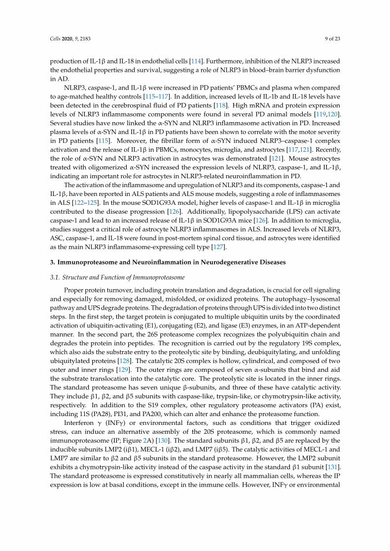
Interferon γ (INFγ) or environmental factors, such as conditions that trigger oxidizedstress, can induce an alternative assembly of the 20S proteasome, which is commonly namedimmunoproteasome (IP; Figure 2A) [130]. The standard subunits β1, β2, and β5 are replaced by theinducible subunits LMP2 (iβ1), MECL-1 (iβ2), and LMP7 (iβ5). The catalytic activities of MECL-1 andLMP7 are similar to β2 and β5 subunits in the standard proteasome. However, the LMP2 subunitexhibits a chymotrypsin-like activity instead of the caspase activity in the standard β1 subunit [131].The standard proteasome is expressed constitutively in nearly all mammalian cells, whereas the IPexpression is low at basal conditions, except in the immune cells. However, INFγ or environmental

Cells 2020, 9, 2183 10 of 23
factors can drastically increase the assembly of IP also in nonimmune cells. Additionally, INFγ caninduce PA28αβ expression, which increases the activities of the beta subunits.
Cells 2020, 9, x FOR PEER REVIEW 10 of 22
The most studied function of the IP is related to the immune function and antigen presentation. IP is capable of generating antigen peptides, which are first complexed to MHC-I in the ER and then exposed on the plasma membrane to be presented to CD8+ T lymphocytes [132]. The changes in the β subunits in the IP lead to increased overall chymotrypsin activity, which aids the generation of antigen peptides with hydrophobic C-termini and improves their fit into the groove of MHC-I molecules [133–135]. This increases the repertoire of peptides generated for MHC presentation. The assembly of IP in response to INFγ is faster than in a standard proteasome, which helps expanding the peptide pool needed for efficient immune responses [136]. The halftime of IP is shorter, which prevents persistent immune activation.
Figure 2. Immunoproteosome and its function in neurodegenerative diseases. (A) Immunoproteosome is formed from the constitutive proteosome upon inflammatory stimuli by the incorporation of specific β subunits and the co-production of proteosomal activator 11S PA28αβ. (B) Evidence for the increased function of immunoproteosome in neurodegenerative diseases.
In addition to the role in immune functions, the IP has a role in responding to various stress factors. During oxidative stress, the IP is efficient, and in some cases, it is even better in selectively degrading oxidized proteins than the standard proteasome [137,138]. Both the selectivity and activity of the IP can be increased by binding with the 11S (PA28) regulator. Increased levels of H2O2 cause protein oxidation, while increased levels of H2O2 cause protein oxidation, but the IP and the 11S regulator together with the standard proteasome help maintain the homeostasis during H2O2-induced oxidative stress. Even low levels of H2O2 without any protein damage have been shown to increase the synthesis of IP, 11S, and standard proteasome [137–141]. This may help the cells to preadapt to a potential increase in oxidative stress and be more prepared to degrade higher levels of oxidized proteins. A low level of nitric oxide is an essential factor in regulating the vascular tone, but, again, high concentrations can cause oxidative damage [142]. High levels of nitric oxide upregulate IP and help cells cope with increased protein damage [143–145]. Cells that are naturally exposed to higher levels of nitric oxide, including endothelial cells, express correspondingly higher levels of IP. IP’s expression and activity are essential for cell survival in the environment with high NO levels.
3.2. Immunoproteasome Function in CNS
LMP2 (β1i), chymotrypsin-like activity MECL-1 (β2i)LMP7 (β5i) αβ
Main functions:- Antigen processing- Degradation of aggregation prone proteins
20S immunoproteasome
Gain of function in ND:
- AD: increased gene and protein expressions of LMP7 and MECL-1 subunits in hippocampus and IP activity in neurons and plaque-associated glial cells
- PD and LBD: increased LMP7 subunits expression in neurons and glial cells in SN and VTA
- ALS: elevated expression of immuno-proteasome subunits in microglia and astrocytes in the spinal cord
- HD: increased levels of LMP7 and LMP2 in neurons and glia in the cortex and striatum
- MS: increased expression of immuno-proteasome subunits in the cortex and white matter plaques in neurons and glia
+INFγ
peptides
11S PA28αβ
polyUb-proteins
Ub
α-ring
β-ringsα-ring
A B
Figure 2. Immunoproteosome and its function in neurodegenerative diseases. (A) Immunoproteosomeis formed from the constitutive proteosome upon inflammatory stimuli by the incorporation of specificβ subunits and the co-production of proteosomal activator 11S PA28αβ. (B) Evidence for the increasedfunction of immunoproteosome in neurodegenerative diseases.
The most studied function of the IP is related to the immune function and antigen presentation. IP iscapable of generating antigen peptides, which are first complexed to MHC-I in the ER and then exposedon the plasma membrane to be presented to CD8+ T lymphocytes [132]. The changes in the β subunitsin the IP lead to increased overall chymotrypsin activity, which aids the generation of antigen peptideswith hydrophobic C-termini and improves their fit into the groove of MHC-I molecules [133–135].This increases the repertoire of peptides generated for MHC presentation. The assembly of IP inresponse to INFγ is faster than in a standard proteasome, which helps expanding the peptide poolneeded for efficient immune responses [136]. The halftime of IP is shorter, which prevents persistentimmune activation.
In addition to the role in immune functions, the IP has a role in responding to various stress factors.During oxidative stress, the IP is efficient, and in some cases, it is even better in selectively degradingoxidized proteins than the standard proteasome [137,138]. Both the selectivity and activity of the IPcan be increased by binding with the 11S (PA28) regulator. Increased levels of H2O2 cause proteinoxidation, while increased levels of H2O2 cause protein oxidation, but the IP and the 11S regulatortogether with the standard proteasome help maintain the homeostasis during H2O2-induced oxidativestress. Even low levels of H2O2 without any protein damage have been shown to increase the synthesisof IP, 11S, and standard proteasome [137–141]. This may help the cells to preadapt to a potentialincrease in oxidative stress and be more prepared to degrade higher levels of oxidized proteins. A lowlevel of nitric oxide is an essential factor in regulating the vascular tone, but, again, high concentrationscan cause oxidative damage [142]. High levels of nitric oxide upregulate IP and help cells cope withincreased protein damage [143–145]. Cells that are naturally exposed to higher levels of nitric oxide,including endothelial cells, express correspondingly higher levels of IP. IP’s expression and activity areessential for cell survival in the environment with high NO levels.

Cells 2020, 9, 2183 11 of 23
3.2. Immunoproteasome Function in CNS
Previously, the CNS has been seen as an immune-privileged area because of the immunosuppressiveenvironment and the absence of dendric cells. However, currently, the evidence supports the ideathat the CNS is not isolated but is actively communicating with the immune system. Nowadays,neuroinflammation is seen as a complex interplay between the CNS and systemic cells. In the CNS,neurons and glia (microglia, astrocytes, and oligodendrocytes) continuously express low amounts ofIP, which suggests that IP has a role in maintaining homeostasis in the CNS [146,147]. In addition,pro-inflammatory cytokines, including INFγ and TNFα, or oxidative stress generally induce theexpression of the IP and the disassembly of the standard proteasome [148–151]. This is thoughtto enhance protein degradation and allow the cells to cope with the protein overload. The IP candegrade aggregation-prone proteins at the same or even higher rate and efficacy than the standardproteasome [152,153]. The expression of MHC-I in the CNS has functions beyond antigen presentation.MHC-I expression in neurons has been linked to early neuronal development, synaptic plasticity,axonal regeneration, memory, and reward [154–157]. Nevertheless, neurons and glia can act asprofessional APC and thereby prolong inflammation/oxidized stress. Consequently, the increased IPmay make the CNS cells more vulnerable to auto-immune damage [147].
3.3. The Role of Immunoproteasome in Neurodegenerative Diseases
Several studies have shown increased IP activity in various neurodegenerative diseases, includingAD, PD, ALS, HD, and MS (Figure 2B). The increased expression and activity of the IP might bebeneficial in the early stages of neurodegeneration by compensating for the protein overload anddecreased function of the standard proteasome. However, the persistent overactivity of the IP canenhance the neuroinflammation and lead to neuronal cell death.
3.3.1. Immunoproteosome in Alzheimer Disease
Several human post-mortem studies and experimental models have shown changes in the IPfunction and activity in AD. Decreased gene expression of standard b5 subunit and increased gene andprotein expressions of LMP7 (β5i) and MECL-1 subunits have been observed in the hippocampus ofAD brains [158,159]. In addition, the activities of the IP subunits LMP7 (β5i), MECL-1 (β2i), and LMP2(β1i) are increased in the hippocampus of AD brains, which also correlates with the tau pathology [158].Overall, the expression of IP in AD patients has been reported to be elevated compared to non-dementedelderly, while in the young brain, the expression may be barely detectable [160].
Increased IP expression has also been detected in several AD animal models. The IP activitywas primarily raised in the AD mice cortex and amplified gene and protein expressions in neuronsand glia surrounding the amyloid-beta plaques [158]. Decreased levels of standard β5 subunit andincreased LMP2 (β1i) and MECL-1 (β2i) levels and trypsin activity were detected in AD mice [161].The increased gene and protein expressions of LMP7 (β5i) and LMP2 (β1i) subunits also correlatedwith age and amyloid-beta pathology in AD mice [162].
3.3.2. Immunoproteosome in Parkinson’s Disease
Increased levels and activity of LMP7 (β5i) subunits were found from post-mortem brains of PDand dementia with LB patients. Notably, the increase was detected in both neurons and glial cells inthe substantia nigra area. In contrast, the increase was seen only in glial cells in the less vulnerableventral tegmental area [163]. Increased activity of the LMP7 (β5i) subunit was also demonstrated in anexperimental model using 6-hydroxydopamine (6-OHDA). The neurotoxin, 6-OHDA, upregulated theLMP7 subunit in DA neurons, both in vitro and in vivo studies [164].

Cells 2020, 9, 2183 12 of 23
3.3.3. Immunoproteosome in Amyotrophic Lateral Sclerosis
Although evidence of the relation between the IP and ALS is missing from human studies,several experimental animal models of ALS have shown increased IP function. Proteasome activitywas raised in the spinal cord of SOD1 G93A transgenic mice. The standard proteasome subunits 7and 5 were expressed constitutively, but a marked increase in IP subunits LMP2 (β1i), MECL-1 (β2i),and LMP7 (β5i) were found. Additionally, the induction of IP subunits occurred mainly in microgliaand astrocytes [165]. Other studies have also shown similar initiation of the IP in the spinal cord withSOD1 G93A transgenic mice, although the standard proteasome activity was found decreased. [166,167].A study by Puttaparthi et al. showed increased proteasome activity and induction of the IP in the spinalcord of SOD1 G93A transgenic mice. Additionally, mice lacking the LMP2 (G93A SOD1/LMP2−/−)subunit did not exhibit a change in the motor function decline, suggesting that IP function does notalter the SOD1-induced behavioral phenotype [168].
3.3.4. Immunoproteosome in Huntington Disease
Increased levels of LMP7 (β5i) and LMP2 (β1i) were detected in the cortex and the striatum ofHD patients’ brain compared to age-matched controls [169]. The increase was related to decreasedlevels of the corresponding subunits of the standard proteasome. Moreover, the induction of the IPsubunits in neurons was associated with neurodegeneration. Similarly, the IP subunits LMP7 (β5i) andLMP2 (β1i) were increased in neurons and glia in the striatum and cortex of HD mice [169].
3.3.5. Immunoproteosome in Multiple Sclerosis
The IP and PA28ab regulator’s expression was detected in MS patients but was not seen in youngcontrols. In addition, LMP2 (β1i) and PA28αβ were detected in the cortex and the white matter plaquesin neurons and glia [170]. In the experimental model of MS, MOG-EAE mice (myelin oligodendrocyteglycoprotein experimental autoimmune encephalomyelitis), overall peptidase proteasome activityduring the acute phase of EAE correlated with increased levels of LMP2 (β1i), MECL-1 (β2i), and LMP7(β5i) subunits in neurons and glia. These findings were opposite in the chronic phase [171,172].Similarly, the amount and activity of the IP subunits were increased in MOG-EAE rats [173]. In myelinBasic Peptide (MBP)-EAE mice, LMP2 (β1i) and LMP7 (β5i) subunits were increased, and LMP2 (β1i)was dominantly expressed in oligodendrocytes, whereas LMP7 (β5i) was mainly in brain-infiltratinglymphocytes [174].
3.4. Immunoproteasome Inhibitors in Neurodegenerative Diseases
The increasing evidence of the role of IP in inflammation and neurodegenerative diseases hasraised the interest to target the IP for therapy. Several IP inhibitors have been developed for autoimmunediseases and some cancers [175]. Currently, KZR-616 inhibitor from Kezar Life Sciences is being testedin a clinical trial for Systemic Lupus Erythematosus with and without Nephritis (ClinicalTrials.govIdentifier: NCT03393013). While the IP inhibitors have beneficial effects in immune-related diseases,the preclinical results from neurodegenerative diseases have been contradictory. The impact of IPinhibition seems to be disease and context-dependent. While for AD and MS, the inhibition could bebeneficial based on the current findings, for PD and ALS, the obtained results from animal modelsshowed the opposite.
In the AD mice models, IP inhibition improved mainly cognitive functions. Trasngenic APP-PS1mice crossed with mice deficient for the IP subunit LMP7 (β5i) resulted in impaired IP function.These LMP7 (β5i)-deficient mice did not show significant Aβ pathology; however, microglia showedaltered cytokine responses. The altered cytokine profile was associated with improved Aβ-associatedcognitive deficits typically observed in APP-PS1 mice [165]. In another study, dual inhibition of IPsubunits LMP2 (β1i) and cP catalytic subunit Y with YU102 ameliorated the cognitive effects in the

Cells 2020, 9, 2183 13 of 23
AD mouse model [176]. The inhibition did not affect the Aβ deposition but suppressed the cytokinesecretion from microglia cells.
In different experimental models of MS, proteasome inhibitors proved to be efficient to someextent [173,177]. The impact of IP inhibitor ONX 0914 was studied in two different mouse models ofMS. ONX 0914 attenuated the disease progression in MOG35–55 and PLP139–151-induced-EAE [178].The isolation of lymphocytes from the spinal cord revealed a substantial reduction of cytokine-producingCD4 cells in treated mice. These results suggest that IP inhibitors may have a potential for treatingMS patients.
However, the fact that the IP inhibition could also have adverse effects was demonstrated in PDand ALS rodent models. While 6-OHDA upregulated the LMP7 (β5i) subunit in DA neurons, inhibitionof the IP increased 6-OHDA-induced neurotoxicity both in vitro and in vivo [164]. A similar effect wasseen in SH-SY5Y cells exposed to rotenone [179]. Knockdown of the ib1 subunit resulted in increasedα-SYN accumulation, the degradation of tyrosine hydroxylase, the release of ROS, an increased levelof malondialdehyde, and a decreased level of glutathione, and it also promoted apoptosis in SH-SY5Ycells after rotenone treatment. On the other hand, Oxyphylla A, the LMP7 (β5i) subunit activator,promoted α-SYN degradation in the cellular PD model [180]. Altogether, these results demonstrate thepotential neuroprotective role of the IP in PD.
Our laboratory studies have shown a potential neuroprotective effect of the IP in ALS whentreating G93A-SOD1 transgenic mice with pyrrolidine dithiocarbamate (PDTC), which is an inhibitorof NF-κB [181]. The PDTC treatment completely blocked the IP expression and significantly decreasedthe survival of the mice. The exposure did not affect the standard proteasome. These results suggestedthat the IP may help the nervous system to cope with the harmful effects of SOD1-G93A mutation.
4. Conclusions
Cellular viability and functions are dependent on not only adequate protein production but alsoan efficient degradation of excess, damaged, and misfolded proteins. Thus, disturbances in cellularprotein homeostasis or proteostasis can be detrimental to the cell. Here, we have reviewed the currentresearch related to the relationship between proteostasis disturbances and inflammatory response inneurodegenerative diseases. A growing body of evidence shows that protein misfolding, aggregation,and aberrant modifications can lead to excessive immune responses causing neuroinflammation,which is associated with neurodegenerative diseases.
On the other hand, reactive glial cells in the CNS play an important role in deleteriousnon-cell-autonomous mechanisms leading to the loss of proteostasis. This vicious feed-forward loop mayhave a critical impact on neuronal viability. Particularly, failures in the clearance of aggregated proteinshave been associated with aging and neurodegenerative diseases such as AD, PD, and ALS, especiallyin gene mutations connected with cellular proteostasis. Furthermore, excessive immune responsesthat initiate inflammation and lead to dysfunctional proteostasis are evident in AD, PD, and ALS.Further elucidation of the individual steps of proteostasis and inflammation, especially in human-basedmodels, will provide a better understanding of the cellular processes and open a window/the way forthe development of novel pharmacological strategies for neurodegenerative diseases.
Funding: This work was funded through grants to S.L. and J.K. from the Finnish Parkinson Foundation (S.L.),Joint Programme for Neurodegenerative Disease (JPND) research co-funded by the EU Research, InnovationProgramme Horizon 2020 through the ERA-NET co-fund scheme (J.K.) and by The Olav Thon Foundation (J.K.).
Conflicts of Interest: The authors declare no conflict of interest. The funders had no role in the design of thestudy; in the collection, analyses, or interpretation of data; in the writing of the manuscript, or in the decision topublish the results.

Cells 2020, 9, 2183 14 of 23
References
1. Sofroniew, M.V. Astrocyte barriers to neurotoxic inflammation. Nat. Rev. Neurosci. 2015, 16, 249–263.[CrossRef] [PubMed]
2. Prinz, M.; Priller, J. Microglia and brain macrophages in the molecular age: From origin to neuropsychiatricdisease. Nat. Rev. Neurosci. 2014, 15, 300–312. [CrossRef] [PubMed]
3. DiSabato, D.J.; Quan, N.; Godbout, J.P. Neuroinflammation: The devil is in the details. J. Neurochem. 2016,139, 136–153. [CrossRef] [PubMed]
4. Lee, K.M.; MacLean, A.G. New advances on glial activation in health and disease. World J. Virol. 2015, 4,42–55. [CrossRef]
5. Mymrikov, E.V.; Daake, M.; Richter, B.; Haslbeck, M.; Buchner, J. The Chaperone Activity and SubstrateSpectrum of Human Small Heat Shock Proteins. J. Biol. Chem. 2017, 292, 672–684. [CrossRef]
6. Labbadia, J.; Morimoto, R.I. The Biology of Proteostasis in Aging and Disease. Annu. Rev. Biochem. 2015, 84,435–464. [CrossRef]
7. Pickart, C.M. Mechanisms underlying ubiquitination. Annu. Rev. Biochem. 2001, 70, 503–533. [CrossRef]8. Glickman, M.H.; Ciechanover, A. The ubiquitin-proteasome proteolytic pathway: Destruction for the sake of
construction. Physiol. Rev. 2002, 82, 373–428. [CrossRef]9. Parzych, K.R.; Klionsky, D.J. An overview of autophagy: Morphology, mechanism, and regulation.
Antioxid. Redox Signal. 2014, 20, 460–473. [CrossRef]10. Chelombitko, M.A. Role of Reactive Oxygen Species in Inflammation: A Minireview. Mosc. Univ. Biol.
Sci. Bull. 2018, 73, 199–202. [CrossRef]11. Hsieh, H.-L.; Yang, C.-M. Role of Redox Signaling in Neuroinflammation and Neurodegenerative Diseases.
BioMed Res. Int. 2013. [CrossRef] [PubMed]12. Haddad, J.J. Oxygen-sensitive pro-inflammatory cytokines, apoptosis signaling and redox-responsive
transcription factors in development and pathophysiology. Cytokines Cell. Mol. Ther. 2002, 7, 1–14. [CrossRef][PubMed]
13. Markesbery, W.R.; Lovell, M.A. Four-hydroxynonenal, a product of lipid peroxidation, is increased in thebrain in Alzheimer’s disease. Neurobiol. Aging 1998, 19, 33–36. [CrossRef]
14. Sultana, R.; Boyd-Kimball, D.; Cai, J.; Pierce, W.M.; Klein, J.B.; Merchant, M.; Butterfield, D.A. Proteomicsanalysis of the Alzheimer’s disease hippocampal proteome. J. Alzheimer’s Dis. 2007, 11, 153–164. [CrossRef][PubMed]
15. Sultana, R.; Perluigi, M.; Butterfield, D.A. Lipid peroxidation triggers neurodegeneration: A redox proteomicsview into the Alzheimer disease brain. Free Radic. Biol. Med. 2013, 62, 157–169. [CrossRef] [PubMed]
16. Xiang, W.; Schlachetzki, J.C.M.; Helling, S.; Bussmann, J.C.; Berlinghof, M.; Schäffer, T.E.; Marcus, K.;Winkler, J.; Klucken, J.; Becker, C.-M. Oxidative stress-induced posttranslational modifications ofalpha-synuclein: Specific modification of alpha-synuclein by 4-hydroxy-2-nonenal increases dopaminergictoxicity. Mol. Cell. Neurosci. 2013, 54, 71–83. [CrossRef]
17. Anderson, G.; Maes, M. Neurodegeneration in Parkinson’s disease: Interactions of oxidative stress,tryptophan catabolites and depression with mitochondria and sirtuins. Mol. Neurobiol. 2014, 49, 771–783.[CrossRef]
18. Morel, P.; Tallineau, C.; Pontcharraud, R.; Piriou, A.; Huguet, F. Effects of 4-hydroxynonenal, a lipidperoxidation product, on dopamine transport and Na+/K+ ATPase in rat striatal synaptosomes. Neurochem. Int.1998, 33, 531–540. [CrossRef]
19. Zarkovic, K. 4-hydroxynonenal and neurodegenerative diseases. Mol. Asp. Med. 2003, 24, 293–303.[CrossRef]
20. Lee, J.; Kosaras, B.; Del Signore, S.J.; Cormier, K.; McKee, A.; Ratan, R.R.; Kowall, N.W.; Ryu, H. Modulationof lipid peroxidation and mitochondrial function improves neuropathology in Huntington’s disease mice.Acta Neuropathol. 2011, 121, 487–498. [CrossRef]
21. Vitek, M.P.; Bhattacharya, K.; Glendening, J.M.; Stopa, E.; Vlassara, H.; Bucala, R.; Manogue, K.; Cerami, A.Advanced glycation end products contribute to amyloidosis in Alzheimer disease. Proc. Natl. Acad. Sci. USA1994, 91, 4766–4770. [CrossRef] [PubMed]

Cells 2020, 9, 2183 15 of 23
22. Guerrero, E.; Vasudevaraju, P.; Hegde, M.L.; Britton, G.B.; Rao, K.S. Recent advances in α-synuclein functions,advanced glycation, and toxicity: Implications for Parkinson’s disease. Mol. Neurobiol. 2013, 47, 525–536.[CrossRef] [PubMed]
23. Padmaraju, V.; Bhaskar, J.J.; Prasada Rao, U.J.S.; Salimath, P.V.; Rao, K.S. Role of advanced glycation onaggregation and DNA binding properties of α-synuclein. J. Alzheimer’s Dis. 2011, 24, 211–221. [CrossRef][PubMed]
24. Shibata, N.; Hirano, A.; Hedley-Whyte, E.T.; Dal Canto, M.C.; Nagai, R.; Uchida, K.; Horiuchi, S.;Kawaguchi, M.; Yamamoto, T.; Kobayashi, M. Selective formation of certain advanced glycation end productsin spinal cord astrocytes of humans and mice with superoxide dismutase-1 mutation. Acta Neuropathol. 2002,104, 171–178. [CrossRef] [PubMed]
25. Ischiropoulos, H.; Beckman, J.S. Oxidative stress and nitration in neurodegeneration: Cause, effect, orassociation? J. Clin. Investig. 2003, 111, 163–169. [CrossRef]
26. Jekabsone, A.; Neher, J.J.; Borutaite, V.; Brown, G.C. Nitric oxide from neuronal nitric oxide synthase sensitisesneurons to hypoxia-induced death via competitive inhibition of cytochrome oxidase. J. Neurochem. 2007, 103,346–356. [CrossRef]
27. Duncan, A.J.; Heales, S.J.R. Nitric oxide and neurological disorders. Mol. Asp. Med. 2005, 26, 67–96.[CrossRef]
28. Boll, M.-C.; Alcaraz-Zubeldia, M.; Montes, S.; Murillo-Bonilla, L.; Rios, C. Raised nitrate concentration andlow SOD activity in the CSF of sporadic ALS patients. Neurochem. Res. 2003, 28, 699–703. [CrossRef]
29. Agar, J.; Durham, H. Relevance of oxidative injury in the pathogenesis of motor neuron diseases.Amyotroph. Lateral Scler. Other Mot. Neuron Disord. 2003, 4, 232–242. [CrossRef]
30. Kaufman, R.J. Stress signaling from the lumen of the endoplasmic reticulum: Coordination of genetranscriptional and translational controls. Genes Dev. 1999, 13, 1211–1233. [CrossRef]
31. Doyle, K.M.; Kennedy, D.; Gorman, A.M.; Gupta, S.; Healy, S.J.M.; Samali, A. Unfolded proteinsand endoplasmic reticulum stress in neurodegenerative disorders. J. Cell. Mol. Med. 2011, 15, 2025–2039.[CrossRef] [PubMed]
32. Hetz, C.; Mollereau, B. Disturbance of endoplasmic reticulum proteostasis in neurodegenerative diseases.Nat. Rev. Neurosci. 2014, 15, 233–249. [CrossRef] [PubMed]
33. Cao, S.S.; Kaufman, R.J. Endoplasmic reticulum stress and oxidative stress in cell fate decision and humandisease. Antioxid. Redox Signal. 2014, 21, 396–413. [CrossRef] [PubMed]
34. Lehtonen, Š.; Jaronen, M.; Vehviläinen, P.; Lakso, M.; Rudgalvyte, M.; Keksa-Goldsteine, V.; Wong, G.;Courtney, M.J.; Koistinaho, J.; Goldsteins, G. Inhibition of Excessive Oxidative Protein Folding Is Protectivein MPP+ Toxicity-Induced Parkinson’s Disease Models. Antioxid. Redox Signal. 2016, 25, 485–497. [CrossRef]
35. Hotamisligil, G.S. Endoplasmic reticulum stress and the inflammatory basis of metabolic disease. Cell 2010,140, 900–917. [CrossRef]
36. Matus, S.; Glimcher, L.H.; Hetz, C. Protein folding stress in neurodegenerative diseases: A glimpse into theER. Curr. Opin. Cell Biol. 2011, 23, 239–252. [CrossRef]
37. McGuckin, M.A.; Eri, R.D.; Das, I.; Lourie, R.; Florin, T.H. ER stress and the unfolded protein response inintestinal inflammation. Am. J. Physiol. Gastrointest. Liver Physiol. 2010, 298, G820–G832. [CrossRef]
38. Pivovarova, O.; Höhn, A.; Grune, T.; Pfeiffer, A.F.H.; Rudovich, N. Insulin-degrading enzyme:New therapeutic target for diabetes and Alzheimer’s disease? Ann. Med. 2016, 48, 614–624. [CrossRef]
39. Eckman, E.A.; Reed, D.K.; Eckman, C.B. Degradation of the Alzheimer’s Amyloid β Peptide byEndothelin-converting Enzyme. J. Biol. Chem. 2001, 276, 24540–24548. [CrossRef]
40. Cirulli, E.T.; Lasseigne, B.N.; Petrovski, S.; Sapp, P.C.; Dion, P.A.; Leblond, C.S.; Couthouis, J.; Lu, Y.-F.;Wang, Q.; Krueger, B.J.; et al. Exome sequencing in amyotrophic lateral sclerosis identifies risk genes andpathways. Science 2015, 347, 1436–1441. [CrossRef]
41. Zhu, B.; Jiang, L.; Huang, T.; Zhao, Y.; Liu, T.; Zhong, Y.; Li, X.; Campos, A.; Pomeroy, K.; Masliah, E.; et al.ER-associated degradation regulates Alzheimer’s amyloid pathology and memory function by modulatingγ-secretase activity. Nat. Commun. 2017, 8, 1472. [CrossRef] [PubMed]
42. Duran-Aniotz, C.; Cornejo, V.H.; Espinoza, S.; Ardiles, Á.O.; Medinas, D.B.; Salazar, C.; Foley, A.; Gajardo, I.;Thielen, P.; Iwawaki, T.; et al. IRE1 signaling exacerbates Alzheimer’s disease pathogenesis. Acta Neuropathol.2017, 134, 489–506. [CrossRef] [PubMed]
Cells 2020, 9, 2183 16 of 23
43. Katayama, T.; Imaizumi, K.; Sato, N.; Miyoshi, K.; Kudo, T.; Hitomi, J.; Morihara, T.; Yoneda, T.; Gomi, F.;Mori, Y.; et al. Presenilin-1 mutations downregulate the signalling pathway of the unfolded-protein response.Nat. Cell Biol. 1999, 1, 479–485. [CrossRef] [PubMed]
44. Niwa, M.; Sidrauski, C.; Kaufman, R.J.; Walter, P. A Role for Presenilin-1 in Nuclear Accumulation of Ire1Fragments and Induction of the Mammalian Unfolded Protein Response. Cell. 1999, 99, 691–702. [CrossRef]
45. Abisambra, J.F.; Jinwal, U.K.; Blair, L.J.; O’Leary, J.C.; Li, Q.; Brady, S.; Wang, L.; Guidi, C.E.; Zhang, B.;Nordhues, B.A.; et al. Tau accumulation activates the unfolded protein response by impairing endoplasmicreticulum-associated degradation. J. Neurosci. 2013, 33, 9498–9507. [CrossRef]
46. Lindersson, E.; Beedholm, R.; Højrup, P.; Moos, T.; Gai, W.; Hendil, K.B.; Jensen, P.H. Proteasomal inhibitionby alpha-synuclein filaments and oligomers. J. Biol. Chem. 2004, 279, 12924–12934. [CrossRef]
47. Malkus, K.A.; Ischiropoulos, H. Regional deficiencies in chaperone-mediated autophagy underlie α-synucleinaggregation and neurodegeneration. Neurobiol. Dis. 2012, 46, 732–744. [CrossRef] [PubMed]
48. Winslow, A.R.; Chen, C.-W.; Corrochano, S.; Acevedo-Arozena, A.; Gordon, D.E.; Peden, A.A.; Lichtenberg, M.;Menzies, F.M.; Ravikumar, B.; Imarisio, S.; et al. α-Synuclein impairs macroautophagy: Implications forParkinson’s disease. J. Cell Biol. 2010, 190, 1023–1037. [CrossRef]
49. Cooper, A.A.; Gitler, A.D.; Cashikar, A.; Haynes, C.M.; Hill, K.J.; Bhullar, B.; Liu, K.; Xu, K.; Strathearn, K.E.;Liu, F.; et al. Alpha-synuclein blocks ER-Golgi traffic and Rab1 rescues neuron loss in Parkinson’s models.Science 2006, 313, 324–328. [CrossRef]
50. Vogiatzi, T.; Xilouri, M.; Vekrellis, K.; Stefanis, L. Wild type alpha-synuclein is degraded by chaperone-mediated autophagy and macroautophagy in neuronal cells. J. Biol. Chem. 2008, 283, 23542–23556. [CrossRef]
51. Webb, J.L.; Ravikumar, B.; Atkins, J.; Skepper, J.N.; Rubinsztein, D.C. Alpha-Synuclein is degraded by bothautophagy and the proteasome. J. Biol. Chem. 2003, 278, 25009–25013. [CrossRef] [PubMed]
52. Lehtonen, Š.; Sonninen, T.-M.; Wojciechowski, S.; Goldsteins, G.; Koistinaho, J. Dysfunction of CellularProteostasis in Parkinson’s Disease. Front. Neurosci. 2019, 13, 457. [CrossRef] [PubMed]
53. Rao, R.V.; Bredesen, D.E. Misfolded proteins, endoplasmic reticulum stress and neurodegeneration. Curr. Opin.Cell Biol. 2004, 16, 653–662. [CrossRef] [PubMed]
54. Gan-Or, Z.; Dion, P.A.; Rouleau, G.A. Genetic perspective on the role of the autophagy-lysosome pathway inParkinson disease. Autophagy 2015, 11, 1443–1457. [CrossRef]
55. Burchell, V.S.; Nelson, D.E.; Sanchez-Martinez, A.; Delgado-Camprubi, M.; Ivatt, R.M.; Pogson, J.H.;Randle, S.J.; Wray, S.; Lewis, P.A.; Houlden, H.; et al. The Parkinson’s disease-linked proteins Fbxo7 andParkin interact to mediate mitophagy. Nat. Neurosci. 2013, 16, 1257–1265. [CrossRef]
56. Orenstein, S.J.; Kuo, S.-H.; Tasset, I.; Arias, E.; Koga, H.; Fernandez-Carasa, I.; Cortes, E.; Honig, L.S.;Dauer, W.; Consiglio, A.; et al. Interplay of LRRK2 with chaperone-mediated autophagy. Nat. Neurosci. 2013,16, 394–406. [CrossRef]
57. Ho, D.H.; Kim, H.; Nam, D.; Sim, H.; Kim, J.; Kim, H.G.; Son, I.; Seol, W. LRRK2 impairs autophagy bymediating phosphorylation of leucyl-tRNA synthetase. Cell Biochem. Funct. 2018, 36, 431–442. [CrossRef]
58. Neumann, M.; Sampathu, D.M.; Kwong, L.K.; Truax, A.C.; Micsenyi, M.C.; Chou, T.T.; Bruce, J.; Schuck, T.;Grossman, M.; Clark, C.M.; et al. Ubiquitinated TDP-43 in frontotemporal lobar degeneration and amyotrophiclateral sclerosis. Science 2006, 314, 130–133. [CrossRef]
59. Arai, T.; Hasegawa, M.; Akiyama, H.; Ikeda, K.; Nonaka, T.; Mori, H.; Mann, D.; Tsuchiya, K.; Yoshida, M.;Hashizume, Y.; et al. TDP-43 is a component of ubiquitin-positive tau-negative inclusions in frontotemporallobar degeneration and amyotrophic lateral sclerosis. Biochem. Biophys. Res. Commun. 2006, 351, 602–611.[CrossRef]
60. Cascella, R.; Fani, G.; Capitini, C.; Rusmini, P.; Poletti, A.; Cecchi, C.; Chiti, F. Quantitative assessment ofthe degradation of aggregated TDP-43 mediated by the ubiquitin proteasome system and macroautophagy.FASEB J. 2017, 31, 5609–5624. [CrossRef]
61. Wang, X.J.; Yu, J.; Wong, S.H.; Cheng, A.S.L.; Chan, F.K.L.; Ng, S.S.M.; Cho, C.H.; Sung, J.J.Y.; Wu, W.K.K.A novel crosstalk between two major protein degradation systems: Regulation of proteasomal activity byautophagy. Autophagy 2013, 9, 1500–1508. [CrossRef] [PubMed]
62. Scotter, E.L.; Vance, C.; Nishimura, A.L.; Lee, Y.-B.; Chen, H.-J.; Urwin, H.; Sardone, V.; Mitchell, J.C.;Rogelj, B.; Rubinsztein, D.C.; et al. Differential roles of the ubiquitin proteasome system and autophagy in theclearance of soluble and aggregated TDP-43 species. J. Cell Sci. 2014, 127, 1263–1278. [CrossRef] [PubMed]

Cells 2020, 9, 2183 17 of 23
63. Barmada, S.J.; Serio, A.; Arjun, A.; Bilican, B.; Daub, A.; Ando, D.M.; Tsvetkov, A.; Pleiss, M.; Li, X.;Peisach, D.; et al. Autophagy induction enhances TDP43 turnover and survival in neuronal ALS models.Nat. Chem. Biol. 2014, 10, 677–685. [CrossRef] [PubMed]
64. Liu, G.; Coyne, A.N.; Pei, F.; Vaughan, S.; Chaung, M.; Zarnescu, D.C.; Buchan, J.R. Endocytosis regulatesTDP-43 toxicity and turnover. Nat. Commun. 2017, 8, 2092. [CrossRef] [PubMed]
65. Kwiatkowski, T.J.; Bosco, D.A.; Leclerc, A.L.; Tamrazian, E.; Vanderburg, C.R.; Russ, C.; Davis, A.; Gilchrist, J.;Kasarskis, E.J.; Munsat, T.; et al. Mutations in the FUS/TLS gene on chromosome 16 cause familial amyotrophiclateral sclerosis. Science 2009, 323, 1205–1208. [CrossRef] [PubMed]
66. Mackenzie, I.R.A.; Bigio, E.H.; Ince, P.G.; Geser, F.; Neumann, M.; Cairns, N.J.; Kwong, L.K.; Forman, M.S.;Ravits, J.; Stewart, H.; et al. Pathological TDP-43 distinguishes sporadic amyotrophic lateral sclerosis fromamyotrophic lateral sclerosis with SOD1 mutations. Ann. Neurol. 2007, 61, 427–434. [CrossRef]
67. Al-Sarraj, S.; King, A.; Troakes, C.; Smith, B.; Maekawa, S.; Bodi, I.; Rogelj, B.; Al-Chalabi, A.; Hortobágyi, T.;Shaw, C.E. p62 positive, TDP-43 negative, neuronal cytoplasmic and intranuclear inclusions in the cerebellumand hippocampus define the pathology of C9orf72-linked FTLD and MND/ALS. Acta Neuropathol. 2011, 122,691–702. [CrossRef]
68. Atkin, J.D.; Farg, M.A.; Walker, A.K.; McLean, C.; Tomas, D.; Horne, M.K. Endoplasmic reticulum stress andinduction of the unfolded protein response in human sporadic amyotrophic lateral sclerosis. Neurobiol. Dis.2008, 30, 400–407. [CrossRef]
69. Honjo, Y.; Kaneko, S.; Ito, H.; Horibe, T.; Nagashima, M.; Nakamura, M.; Fujita, K.; Takahashi, R.; Kusaka, H.;Kawakami, K. Protein disulfide isomerase-immunopositive inclusions in patients with amyotrophic lateralsclerosis. Amyotroph. Lateral Scler. 2011, 12, 444–450. [CrossRef]
70. Hoffstrom, B.G.; Kaplan, A.; Letso, R.; Schmid, R.S.; Turmel, G.J.; Lo, D.C.; Stockwell, B.R. Inhibitors ofprotein disulfide isomerase suppress apoptosis induced by misfolded proteins. Nat. Chem. Biol. 2010, 6,900–906. [CrossRef]
71. Jaronen, M.; Vehviläinen, P.; Malm, T.; Keksa-Goldsteine, V.; Pollari, E.; Valonen, P.; Koistinaho, J.;Goldsteins, G. Protein disulfide isomerase in ALS mouse glia links protein misfolding with NADPHoxidase-catalyzed superoxide production. Hum. Mol. Genet. 2013, 22, 646–655. [CrossRef] [PubMed]
72. Nishitoh, H.; Kadowaki, H.; Nagai, A.; Maruyama, T.; Yokota, T.; Fukutomi, H.; Noguchi, T.; Matsuzawa, A.;Takeda, K.; Ichijo, H. ALS-linked mutant SOD1 induces ER stress- and ASK1-dependent motor neuron deathby targeting Derlin-1. Genes Dev. 2008, 22, 1451–1464. [CrossRef] [PubMed]
73. Kanekura, K.; Nishimoto, I.; Aiso, S.; Matsuoka, M. Characterization of amyotrophic lateral sclerosis-linkedP56S mutation of vesicle-associated membrane protein-associated protein B (VAPB/ALS8). J. Biol. Chem.2006, 281, 30223–30233. [CrossRef] [PubMed]
74. Suzuki, H.; Kanekura, K.; Levine, T.P.; Kohno, K.; Olkkonen, V.M.; Aiso, S.; Matsuoka, M. ALS-linkedP56S-VAPB, an aggregated loss-of-function mutant of VAPB, predisposes motor neurons to ER stress-relateddeath by inducing aggregation of co-expressed wild-type VAPB. J. Neurochem. 2009, 108, 973–985. [CrossRef][PubMed]
75. Gkogkas, C.; Middleton, S.; Kremer, A.M.; Wardrope, C.; Hannah, M.; Gillingwater, T.H.; Skehel, P.VAPB interacts with and modulates the activity of ATF6. Hum. Mol. Genet. 2008, 17, 1517–1526. [CrossRef]
76. Miles, J.; Scherz-Shouval, R.; van Oosten-Hawle, P. Expanding the Organismal Proteostasis Network:Linking Systemic Stress Signaling with the Innate Immune Response. Trends Biochem. Sci. 2019, 44, 927–942.[CrossRef]
77. Govindarajan, S.; Verheugen, E.; Venken, K.; Gaublomme, D.; Maelegheer, M.; Cloots, E.; Gysens, F.;De Geest, B.G.; Cheng, T.; Moody, D.B.; et al. ER stress in antigen-presenting cells promotes NKT cellactivation through endogenous neutral lipids. EMBO Rep. 2020, 21, e48927. [CrossRef]
78. Zhang, K. Endoplasmic reticulum stress activates cleavage of CREBH to induce a systemic inflammatoryresponse. Cell 2006, 124, 587–599. [CrossRef]
79. Hu, P.; Han, Z.; Couvillon, A.D.; Kaufman, R.J.; Exton, J.H. Autocrine Tumor Necrosis Factor Alpha LinksEndoplasmic Reticulum Stress to the Membrane Death Receptor Pathway through IRE1α-Mediated NF-κBActivation and Down-Regulation of TRAF2 Expression. Mol. Cell. Biol. 2006, 26, 3071–3084. [CrossRef]
80. Grootjans, J.; Kaser, A.; Kaufman, R.J.; Blumberg, R.S. The unfolded protein response in immunity andinflammation. Nat. Rev. Immunol. 2016, 16, 469–484. [CrossRef]

Cells 2020, 9, 2183 18 of 23
81. Tam, A.B.; Mercado, E.L.; Hoffmann, A.; Niwa, M. ER Stress Activates NF-κB by Integrating Functions ofBasal IKK Activity, IRE1 and PERK. PLoS ONE 2012, 7, e45078. [CrossRef] [PubMed]
82. Smith, J.A. Regulation of cytokine production by the unfolded protein response; Implications for infectionand autoimmunity. Front. Immunol. 2018, 9, 422. [CrossRef] [PubMed]
83. Ponnappan, S.; Ponnappan, U. Aging and immune function: Molecular mechanisms to interventions.Antioxid. Redox Signal. 2011, 14, 1551–1585. [CrossRef] [PubMed]
84. Simard, A.R.; Soulet, D.; Gowing, G.; Julien, J.P.; Rivest, S. Bone marrow-derived microglia play a critical rolein restricting senile plaque formation in Alzheimer’s disease. Neuron 2006, 49, 489–502. [CrossRef] [PubMed]
85. Malm, T.M.; Koistinaho, M.; Pärepalo, M.; Vatanen, T.; Ooka, A.; Karlsson, S.; Koistinaho, J. Bone-marrow-derived cells contribute to the recruitment of microglial cells in response to β-amyloid deposition in APP/PS1double transgenic Alzheimer mice. Neurobiol. Dis. 2005, 18, 134–142. [CrossRef]
86. Hickman, S.E.; Allison, E.K.; El Khoury, J. Microglial dysfunction and defectiveβ-amyloid clearance pathwaysin aging alzheimer’s disease mice. J. Neurosci. 2008, 28, 8354–8360. [CrossRef]
87. Brunello, C.A.; Merezhko, M.; Uronen, R.L.; Huttunen, H.J. Mechanisms of secretion and spreading ofpathological tau protein. Cell. Mol. Life Sci. 2020, 77, 1721–1744. [CrossRef]
88. Asai, H.; Ikezu, S.; Tsunoda, S.; Medalla, M.; Luebke, J.; Haydar, T.; Wolozin, B.; Butovsky, O.; Kügler, S.;Ikezu, T. Depletion of microglia and inhibition of exosome synthesis halt tau propagation. Nat. Neurosci.2015, 18, 1584–1593. [CrossRef]
89. Zhou, Y.; Ulland, T.K.; Colonna, M. TREM2-dependent effects on microglia in Alzheimer’s Disease.Front. Aging Neurosci. 2018, 10, 202. [CrossRef]
90. Gelders, G.; Baekelandt, V.; Van der Perren, A. Linking neuroinflammation and neurodegeneration inparkinson’s disease. J. Immunol. Res. 2018, 2018. [CrossRef]
91. Harms, A.S.; Delic, V.; Thome, A.D.; Bryant, N.; Liu, Z.; Chandra, S.; Jurkuvenaite, A.; West, A.B. α-Synucleinfibrils recruit peripheral immune cells in the rat brain prior to neurodegeneration. Acta Neuropathol. Commun.2017, 5, 85. [CrossRef] [PubMed]
92. Luk, K.C.; Kehm, V.; Carroll, J.; Zhang, B.; O’Brien, P.; Trojanowski, J.Q.; Lee, V.M.Y. Pathological α-synucleintransmission initiates Parkinson-like neurodegeneration in nontransgenic mice. Science 2012, 338, 949–953.[CrossRef] [PubMed]
93. Kim, C.; Ho, D.H.; Suk, J.E.; You, S.; Michael, S.; Kang, J.; Lee, S.J.; Masliah, E.; Hwang, D.; Lee, H.J.; et al.Neuron-released oligomeric α-synuclein is an endogenous agonist of TLR2 for paracrine activation ofmicroglia. Nat. Commun. 2013, 4, 1–12. [CrossRef]
94. Gustot, A.; Gallea, J.I.; Sarroukh, R.; Celej, M.S.; Ruysschaert, J.M.; Raussens, V. Amyloid fibrils are themolecular trigger of inflammation in Parkinson’s disease. Biochem. J. 2015, 471, 323–333. [CrossRef] [PubMed]
95. Kim, C.; Lee, H.J.; Masliah, E.; Lee, S.J. Non-cell-autonomous Neurotoxicity of α-synuclein through microglialtoll-like receptor 2. Exp. Neurobiol. 2016, 25, 113–119. [CrossRef] [PubMed]
96. Fellner, L.; Irschick, R.; Schanda, K.; Reindl, M.; Klimaschewski, L.; Poewe, W.; Wenning, G.K.; Stefanova, N.Toll-like receptor 4 is required for α-synuclein dependent activation of microglia and astroglia. GLIA 2013,61, 349–360. [CrossRef] [PubMed]
97. Cebrián, C.; Zucca, F.A.; Mauri, P.; Steinbeck, J.A.; Studer, L.; Scherzer, C.R.; Kanter, E.; Budhu, S.;Mandelbaum, J.; Vonsattel, J.P.; et al. MHC-I expression renders catecholaminergic neurons susceptible toT-cell-mediated degeneration. Nat. Commun. 2014, 5, 1–14. [CrossRef] [PubMed]
98. Ahtoniemi, T.; Jaronen, M.; Keksa-Goldsteine, V.; Goldsteins, G.; Koistinaho, J. Mutant SOD1 from spinalcord of G93A rats is destabilized and binds to inner mitochondrial membrane. Neurobiol. Dis. 2008, 32,479–485. [CrossRef]
99. Jaronen, M.; Goldsteins, G.; Koistinaho, J. ER stress and unfolded protein response in amyotrophic lateralsclerosis—a controversial role of protein disulphide isomerase. Front. Cell. Neurosci. 2014, 8, 402. [CrossRef]
100. Heneka, M.T.; McManus, R.M.; Latz, E. Inflammasome signalling in brain function and neurodegenerativedisease. Nat. Rev. Neurosci. 2018, 19, 610–621. [CrossRef]
101. Martinon, F.; Burns, K.; Tschopp, J. The inflammasome: A molecular platform triggering activation ofinflammatory caspases and processing of proIL-beta. Cell 2002, 10, 417–426.
102. Broz, P.; Dixit, V.M. Inflammasomes: Mechanism of assembly, regulation and signalling. Nat. Rev. Immunol.2016, 16, 407–420. [CrossRef] [PubMed]

Cells 2020, 9, 2183 19 of 23
103. Qiao, Y.; Wang, P.; Qi, J.; Zhang, L.; Gao, C. TLR-induced NF-κB activation regulates NLRP3 expression inmurine macrophages. FEBS Lett. 2012, 586, 1022–1026. [CrossRef] [PubMed]
104. Toma, C.; Higa, N.; Koizumi, Y.; Nakasone, N.; Ogura, Y.; McCoy, A.J.; Franchi, L.; Uematsu, S.;Sagara, J.; Taniguchi, S.; et al. Pathogenic Vibrio activate NLRP3 inflammasome via cytotoxins andTLR/nucleotide-binding oligomerization domain-mediated NF-kappa B signaling. J. Immunol. 2010, 184,5287–5297. [CrossRef]
105. Kelley, N.; Jeltema, D.; Duan, Y.; He, Y. The NLRP3 Inflammasome: An Overview of Mechanisms ofActivation and Regulation. Int. J. Mol. Sci. 2019, 20, 3328. [CrossRef]
106. Voet, S.; Srinivasan, S.; Lamkanfi, M.; van Loo, G. Inflammasomes in neuroinflammatory and neurodegenerativediseases. Embo Mol. Med. 2019, 11, e10248. [CrossRef]
107. Guan, Y.; Han, F. Key Mechanisms and Potential Targets of the NLRP3 Inflammasome in NeurodegenerativeDiseases. Neuroscience 2020, 14, 37.
108. Liu, Y.; Dai, Y.; Li, Q.; Chen, C.; Chen, H.; Song, Y.; Hua, F.; Zhang, Z. Beta-amyloid activates NLRP3inflammasome via TLR4 in mouse microglia. Neurosci. Lett. 2020, 736, 135279. [CrossRef]
109. Saresella, M.; La Rosa, F.; Piancone, F.; Zoppis, M.; Marventano, I.; Calabrese, E.; Rainone, V.; Nemni, R.;Mancuso, R.; Clerici, M. The NLRP3 and NLRP1 inflammasomes are activated in Alzheimer’s disease.Mol. Neurodegener. 2016, 11, 23. [CrossRef]
110. Lue, L.F.; Rydel, R.; Brigham, E.F.; Yang, L.B.; Hampel, H.; Murphy, G.M.; Brachova, L.; Yan, S.D.; Walker, D.G.;Shen, Y.; et al. Inflammatory repertoire of Alzheimer’s disease and nondemented elderly microglia in vitro.Glia 2001, 35, 72–79. [CrossRef]
111. Halle, A.; Hornung, V.; Petzold, G.C.; Stewart, C.R.; Monks, B.G.; Reinheckel, T.; Fitzgerald, K.A.; Latz, E.;Moore, K.J.; Golenbock, D.T. The NALP3 inflammasome is involved in the innate immune response toamyloid-beta. Nat. Immunol. 2008, 9, 857–865. [CrossRef] [PubMed]
112. Heneka, M.T.; Kummer, M.P.; Stutz, A.; Delekate, A.; Schwartz, S.; Vieira-Saecker, A.; Griep, A.; Axt, D.;Remus, A.; Tzeng, T.-C.; et al. NLRP3 is activated in Alzheimer’s disease and contributes to pathology inAPP/PS1 mice. Nature 2013, 493, 674–678. [CrossRef] [PubMed]
113. Dempsey, C.; Rubio Araiz, A.; Bryson, K.J.; Finucane, O.; Larkin, C.; Mills, E.L.; Robertson, A.B.; Cooper, M.A.;O’Neill, L.J.; Lynch, M.A. Inhibiting the NLRP3 inflammasome with MCC950 promotes non-phlogisticclearance of amyloid-β and cognitive function in APP/PS1 mice. Brain. Behav. Immun. 2017, 61, 306–316.[CrossRef] [PubMed]
114. Xiong, R.; Zhou, X.-G.; Tang, Y.; Wu, J.-M.; Sun, Y.-S.; Teng, J.-F.; Pan, R.; Law, B.Y.-K.; Zhao, Y.; Qiu, W.-Q.;et al. Lychee seed polyphenol protects the blood-brain barrier through inhibiting Aβ(25-35)-induced NLRP3inflammasome activation via the AMPK/mTOR/ULK1-mediated autophagy in bEnd.3 cells and APP/PS1mice. Phytother. Res. Ptr 2020. [CrossRef]
115. Fan, Z.; Pan, Y.-T.; Zhang, Z.-Y.; Yang, H.; Yu, S.-Y.; Zheng, Y.; Ma, J.-H.; Wang, X.-M. Systemic activation ofNLRP3 inflammasome and plasma α-synuclein levels are correlated with motor severity and progression inParkinson’s disease. J. Neuroinflammation 2020, 17, 11. [CrossRef]
116. Zhou, Y.; Lu, M.; Du, R.-H.; Qiao, C.; Jiang, C.-Y.; Zhang, K.-Z.; Ding, J.-H.; Hu, G. MicroRNA-7 targetsNod-like receptor protein 3 inflammasome to modulate neuroinflammation in the pathogenesis of Parkinson’sdisease. Mol. Neurodegener. 2016, 11, 28. [CrossRef]
117. Chatterjee, K.; Roy, A.; Banerjee, R.; Choudhury, S.; Mondal, B.; Halder, S.; Basu, P.; Shubham, S.; Dey, S.;Kumar, H. Inflammasome and α-synuclein in Parkinson’s disease: A cross-sectional study. J. Neuroimmunol.2020, 338, 577089. [CrossRef]
118. Zhang, P.; Shao, X.-Y.; Qi, G.-J.; Chen, Q.; Bu, L.-L.; Chen, L.-J.; Shi, J.; Ming, J.; Tian, B. Cdk5-DependentActivation of Neuronal Inflammasomes in Parkinson’s Disease. Mov. Disord. Off. Movement Disorders. 2016,31, 366–376. [CrossRef]
119. Mao, Z.; Liu, C.; Ji, S.; Yang, Q.; Ye, H.; Han, H.; Xue, Z. The NLRP3 Inflammasome is Involved in thePathogenesis of Parkinson’s Disease in Rats. Neurochem. Res. 2017, 42, 1104–1115. [CrossRef]
120. Qiao, C.; Zhang, Q.; Jiang, Q.; Zhang, T.; Chen, M.; Fan, Y.; Ding, J.; Lu, M.; Hu, G. Inhibition of the hepaticNlrp3 protects dopaminergic neurons via attenuating systemic inflammation in a MPTP/p mouse model ofParkinson’s disease. J. Neuroinflammation. 2018, 15, 193. [CrossRef]

Cells 2020, 9, 2183 20 of 23
121. Wang, X.; Chi, J.; Huang, D.; Ding, L.; Zhao, X.; Jiang, L.; Yu, Y.; Gao, F. α-synuclein promotes progressionof Parkinson’s disease by upregulating autophagy signaling pathway to activate NLRP3 inflammasome.Exp. Ther. Med. 2020, 19, 931–938. [CrossRef] [PubMed]
122. Iłzecka, J.; Stelmasiak, Z.; Dobosz, B. Interleukin-1beta converting enzyme/Caspase-1 (ICE/Caspase-1) andsoluble APO-1/Fas/CD 95 receptor in amyotrophic lateral sclerosis patients. Acta Neurol. Scand. 2001, 103,255–258. [CrossRef] [PubMed]
123. Meissner, F.; Molawi, K.; Zychlinsky, A. Mutant superoxide dismutase 1-induced IL-1beta accelerates ALSpathogenesis. Proc. Natl. Acad. Sci. USA 2010, 107, 13046–13050. [CrossRef] [PubMed]
124. Bellezza, I.; Grottelli, S.; Costanzi, E.; Scarpelli, P.; Pigna, E.; Morozzi, G.; Mezzasoma, L.; Peirce, M.J.;Moresi, V.; Adamo, S.; et al. Peroxynitrite Activates the NLRP3 Inflammasome Cascade in SOD1(G93A)Mouse Model of Amyotrophic Lateral Sclerosis. Mol. Neurobiol. 2018, 55, 2350–2361. [CrossRef] [PubMed]
125. Deora, V.; Lee, J.D.; Albornoz, E.A.; McAlary, L.; Jagaraj, C.J.; Robertson, A.A.B.; Atkin, J.D.; Cooper, M.A.;Schroder, K.; Yerbury, J.J.; et al. The microglial NLRP3 inflammasome is activated by amyotrophic lateralsclerosis proteins. Glia 2020, 68, 407–421. [CrossRef]
126. Alexianu, M.E.; Kozovska, M.; Appel, S.H. Immune reactivity in a mouse model of familial ALS correlateswith disease progression. Neurology 2001, 57, 1282–1289. [CrossRef] [PubMed]
127. Johann, S.; Heitzer, M.; Kanagaratnam, M.; Goswami, A.; Rizo, T.; Weis, J.; Troost, D.; Beyer, C. NLRP3inflammasome is expressed by astrocytes in the SOD1 mouse model of ALS and in human sporadic ALSpatients. Glia 2015, 63, 2260–2273. [CrossRef]
128. Liu, C.-W.; Jacobson, A.D. Functions of the 19S complex in proteasomal degradation. Trends Biochem. Sci.2013, 38, 103–110. [CrossRef]
129. Raynes, R.; Pomatto, L.C.D.; Davies, K.J.A. Degradation of oxidized proteins by the proteasome:Distinguishing between the 20S, 26S, and immunoproteasome proteolytic pathways. Mol. Asp. Med.2016, 50, 41–55. [CrossRef]
130. Griffin, T.A.; Nandi, D.; Cruz, M.; Fehling, H.J.; Kaer, L.V.; Monaco, J.J.; Colbert, R.A. Immunoproteasomeassembly: Cooperative incorporation of interferon gamma (IFN-gamma)-inducible subunits. J. Exp. Med.1998, 187, 97–104. [CrossRef]
131. Ferrington, D.A.; Gregerson, D.S. Immunoproteasomes: Structure, Function, and Antigen Presentation.Prog. Mol. Biol. Transl. Sci. 2012, 109, 75–112. [PubMed]
132. Rock, K.L.; York, I.A.; Saric, T.; Goldberg, A.L. Protein degradation and the generation of MHC classI-presented peptides. Adv. Immunol. 2002, 80, 1–70. [PubMed]
133. Cardozo, C.; Kohanski, R.A. Altered Properties of the Branched Chain Amino Acid-preferring ActivityContribute to Increased Cleavages after Branched Chain Residues by the “Immunoproteasome”. J. Biol. Chem.1998, 273, 16764–16770. [CrossRef] [PubMed]
134. Lei, B.; Abdul Hameed, M.D.M.; Hamza, A.; Wehenkel, M.; Muzyka, J.L.; Yao, X.-J.; Kim, K.-B.; Zhan, C.-G.Molecular basis of the selectivity of the immunoproteasome catalytic subunit LMP2-specific inhibitor revealedby molecular modeling and dynamics simulations. J. Phys. Chem. B. 2010, 114, 12333–12339. [CrossRef]
135. Driscoll, J.; Brown, M.G.; Finley, D.; Monaco, J.J. MHC-linked LMP gene products specifically alter peptidaseactivities of the proteasome. Nature 1993, 365, 262–264. [CrossRef]
136. Heink, S.; Ludwig, D.; Kloetzel, P.-M.; Krüger, E. IFN-gamma-induced immune adaptation of the proteasomesystem is an accelerated and transient response. Proc. Natl. Acad. Sci. USA 2005, 102, 9241–9246. [CrossRef][PubMed]
137. Pickering, A.M.; Koop, A.L.; Teoh, C.Y.; Ermak, G.; Grune, T.; Davies, K.J.A. The immunoproteasome, the 20Sproteasome and the PA28αβ proteasome regulator are oxidative-stress-adaptive proteolytic complexes.Biochem. J. 2010, 432, 585–594. [CrossRef]
138. Pickering, A.M.; Davies, K.J.A. Differential Roles of Proteasome and Immunoproteasome Regulators Pa28αβ,Pa28γ and Pa200 in the Degradation of Oxidized Proteins. Arch. Biochem. Biophys 2012, 523, 181–190.[CrossRef]
139. Pickering, A.M.; Linder, R.A.; Zhang, H.; Forman, H.J.; Davies, K.J.A. Nrf2-dependent Induction ofProteasome and Pa28αβ Regulator Are Required for Adaptation to Oxidative Stress. J. Biol. Chem. 2012, 287,10021–10031. [CrossRef]

Cells 2020, 9, 2183 21 of 23
140. Pickering, A.M.; Staab, T.A.; Tower, J.; Sieburth, D.; Davies, K.J.A. A conserved role for the 20S proteasomeand Nrf2 transcription factor in oxidative stress adaptation in mammals, Caenorhabditis elegans andDrosophila melanogaster. J. Exp. Biol. 2013, 216, 543–553. [CrossRef]
141. Pickering, A.M.; Vojtovich, L.; Tower, J.A.; Davies, K.J. Oxidative stress adaptation with acute, chronic,and repeated stress. Free Radic. Biol. Med. 2013, 55, 109–118. [CrossRef] [PubMed]
142. PACHER, P.; BECKMAN, J.S.; LIAUDET, L. Nitric Oxide and Peroxynitrite in Health and Disease. Physiol. Rev.2007, 87, 315–424. [CrossRef] [PubMed]
143. Thomas, S.; Kotamraju, S.; Zielonka, J.; Harder, D.R.; Kalyanaraman, B. Hydrogen peroxide induces nitricoxide and proteosome activity in endothelial cells: A bell-shaped signaling response. Free Radic. Biol. Med.2007, 42, 1049–1061. [CrossRef] [PubMed]
144. Kotamraju, S.; Matalon, S.; Matsunaga, T.; Shang, T.; Hickman-Davis, J.M.; Kalyanaraman, B. Upregulationof immunoproteasomes by nitric oxide: Potential antioxidative mechanism in endothelial cells. Free Radic.Biol. Med. 2006, 40, 1034–1044. [CrossRef]
145. Kotamraju, S.; Kalivendi, S.; Shang, T.; Kalyanaraman, B. Nitric oxide, proteasomal function, and ironhomeostasis–implications in aging and neurodegenerative diseases. Methods Enzymol. 2005, 396, 526–534.
146. Piccinini, M.; Mostert, M.; Croce, S.; Baldovino, S.; Papotti, M.; Rinaudo, M.T. Interferon-γ-inducible subunitsare incorporated in human brain 20S proteasome. J. Neuroimmunol. 2003, 135, 135–140. [CrossRef]
147. Limanaqi, F.; Biagioni, F.; Gaglione, A.; Busceti, C.L.; Fornai, F.A. Sentinel in the Crosstalk Between theNervous and Immune System: The (Immuno)-Proteasome. Front. Immunol. 2019, 10, 628. [CrossRef]
148. Kimura, H.; Caturegli, P.; Takahashi, M.; Suzuki, K. New Insights into the Function of the Immunoproteasomein Immune and Nonimmune. Cells 2015, 2015, 541984. [CrossRef]
149. Krüger, E.; Kloetzel, P.-M. Immunoproteasomes at the interface of innate and adaptive immune responses:Two faces of one enzyme. Curr. Opin. Immunol. 2012, 24, 77–83. [CrossRef]
150. Huber, E.M.; Basler, M.; Schwab, R.; Heinemeyer, W.; Kirk, C.J.; Groettrup, M.; Groll, M. Immuno- andConstitutive Proteasome Crystal Structures Reveal Differences in Substrate and Inhibitor Specificity. Cell2012, 148, 727–738. [CrossRef]
151. Hallermalm, K.; Seki, K.; Wei, C.; Castelli, C.; Rivoltini, L.; Kiessling, R.; Levitskaya, J. Tumor necrosisfactor-alpha induces coordinated changes in major histocompatibility class I presentation pathway, resultingin increased stability of class I complexes at the cell surface. Blood 2001, 98, 1108–1115. [CrossRef] [PubMed]
152. Seifert, U.; Bialy, L.P.; Ebstein, F.; Bech-Otschir, D.; Voigt, A.; Schröter, F.; Prozorovski, T.; Lange, N.; Steffen, J.;Rieger, M.; et al. Immunoproteasomes preserve protein homeostasis upon interferon-induced oxidativestress. Cell 2010, 142, 613–624. [CrossRef] [PubMed]
153. Nathan, J.A.; Spinnenhirn, V.; Schmidtke, G.; Basler, M.; Groettrup, M.; Goldberg, A.L. Immuno- andconstitutive proteasomes do not differ in their abilities to degrade ubiquitinated proteins. Cell 2013, 152,1184–1194. [CrossRef] [PubMed]
154. Cebrián, C.; Loike, J.D.; Sulzer, D. Neuronal MHC-I expression and its implications in synaptic function,axonal regeneration and Parkinson’s and other brain diseases. Front. Neuroanatomy. 2014, 8, 114. [CrossRef][PubMed]
155. Lazarczyk, M.J.; Kemmler, J.E.; Eyford, B.A.; Short, J.A.; Varghese, M.; Sowa, A.; Dickstein, D.R.; Yuk, F.J.;Puri, R.; Biron, K.E.; et al. Major Histocompatibility Complex. class I proteins are critical for maintainingneuronal structural complexity in the aging brain. Sci. Rep. 2016, 6, 26199. [CrossRef] [PubMed]
156. Edamura, M.; Murakami, G.; Meng, H.; Itakura, M.; Shigemoto, R.; Fukuda, A.; Nakahara, D. FunctionalDeficiency of MHC Class I Enhances LTP and Abolishes LTD in the Nucleus Accumbens of Mice. PLoS ONE2014, 9. [CrossRef]
157. Murakami, G.; Edamura, M.; Furukawa, T.; Kawasaki, H.; Kosugi, I.; Fukuda, A.; Iwashita, T.; Nakahara, D.MHC class I in dopaminergic neurons suppresses relapse to reward seeking. Sci. Adv. 2018, 4, eaap7388.[CrossRef]
158. Orre, M.; Kamphuis, W.; Dooves, S.; Kooijman, L.; Chan, E.T.; Kirk, C.J.; Dimayuga Smith, V.; Koot, S.;Mamber, C.; Jansen, A.H.; et al. Reactive glia show increased immunoproteasome activity in Alzheimer’sdisease. Brain. 2013, 136, 1415–1431. [CrossRef]
159. Nijholt, D.T.; de Graaf, T.R.; van Haastert, E.S.; Oliveira, A.O.; Berkers, C.R.; Zwart, R.; Ovaa, H.; Baas, F.;Hoozemans, J.J.M.; Scheper, W. Endoplasmic reticulum stress activates autophagy but not the proteasome inneuronal cells: Implications for Alzheimer’s disease. Cell Death Differ. 2011, 18, 1071–1081. [CrossRef]

Cells 2020, 9, 2183 22 of 23
160. Mishto, M.; Bellavista, E.; Santoro, A.; Stolzing, A.; Ligorio, C.; Nacmias, B.; Spazzafumo, L.; Chiappelli, M.;Licastro, F.; Sorbi, S.; et al. Immunoproteasome and LMP2 polymorphism in aged and Alzheimer’s diseasebrains. Neurobiol. Aging 2006, 27, 54–66. [CrossRef]
161. Aso, E.; Lomoio, S.; López-González, I.; Joda, L.; Carmona, M.; Fernández-Yagüe, N.; Moreno, J.; Juvés, S.;Pujol, A.; Pamplona, R.; et al. Amyloid generation and dysfunctional immunoproteasome activationwith disease progression in animal model of familial Alzheimer’s disease. Brain Pathol. 2012, 22, 636–653.[CrossRef] [PubMed]
162. Wagner, L.K.; Gilling, K.E.; Schormann, E.; Kloetzel, P.M.; Heppner, F.L.; Krüger, E.; Prokop, S.Immunoproteasome deficiency alters microglial cytokine response and improves cognitive deficits inAlzheimer’s disease-like APPPS1 mice. Acta Neuropathol. Commun. 2017, 5, 52. [CrossRef] [PubMed]
163. Ugras, S.; Daniels, M.J.; Fazelinia, H.; Gould, N.S.; Yocum, A.K.; Luk, K.C.; Luna, E.; Ding, H.; McKennan, C.;Seeholzer, S.; et al. Induction of the Immunoproteasome Subunit Lmp7 Links Proteostasis and Immunity inα-Synuclein Aggregation Disorders. EBioMedicine 2018, 31, 307–319. [CrossRef] [PubMed]
164. Mo, M.-S.; Li, G.-H.; Sun, C.-C.; Huang, S.-X.; Wei, L.; Zhang, L.-M.; Zhou, M.-M.; Wu, Z.-H.; Guo, W.-Y.;Yang, X.-L.; et al. Dopaminergic neurons show increased low-molecular-mass protein 7 activity induced by6-hydroxydopamine in vitro and in vivo. Transl. Neurodegener. 2018, 7, 19. [CrossRef] [PubMed]
165. Puttaparthi, K.; Elliott, J.L. Non-neuronal induction of immunoproteasome subunits in an ALS model:Possible mediation by cytokines. Exp. Neurol. 2005, 196, 441–451. [CrossRef]
166. Cheroni, C.; Peviani, M.; Cascio, P.; Debiasi, S.; Monti, C.; Bendotti, C. Accumulation of human SOD1 andubiquitinated deposits in the spinal cord of SOD1G93A mice during motor neuron disease progressioncorrelates with a decrease of proteasome. Neurobiol. Dis. 2005, 18, 509–522. [CrossRef]
167. Cheroni, C.; Marino, M.; Tortarolo, M.; Veglianese, P.; De Biasi, S.; Fontana, E.; Zuccarello, L.V.; Maynard, C.J.;Dantuma, N.P.; Bendotti, C. Functional alterations of the ubiquitin-proteasome system in motor neurons of amouse model of familial amyotrophic lateral sclerosis. Hum. Mol. Genet. 2009, 18, 82–96. [CrossRef]
168. Puttaparthi, K.; Van Kaer, L.; Elliott, J.L. Assessing the role of immuno-proteasomes in a mouse model offamilial. ALS. Exp. Neurol. 2007, 206, 53–58. [CrossRef]
169. Díaz-Hernández, M.; Hernández, F.; Martín-Aparicio, E.; Gómez-Ramos, P.; Morán, M.A.; Castaño, J.G.;Ferrer, I.; Avila, J.; Lucas, J.J. Neuronal induction of the immunoproteasome in Huntington’s disease.Neuroscience. 2003, 23, 11653–11661. [CrossRef]
170. Mishto, M.; Bellavista, E.; Ligorio, C.; Textoris-Taube, K.; Santoro, A.; Giordano, M.; D’Alfonso, S.; Listì, F.;Nacmias, B.; Cellini, E.; et al. Immunoproteasome LMP2 60HH variant alters MBP epitope generationand reduces the risk to develop multiple sclerosis in Italian female population. PLoS ONE 2010, 5, e9287.[CrossRef]
171. Zheng, J.; Bizzozero, O.A. Reduced proteasomal activity contributes to the accumulation of carbonylatedproteins in chronic experimental autoimmune encephalomyelitis. J. Neurochem. 2010, 115, 1556–1567.[CrossRef] [PubMed]
172. Zheng, J.; Dasgupta, A.; Bizzozero, O.A. Changes in 20S subunit composition are largely responsible foraltered proteasomal activities in experimental autoimmune encephalomyelitis. J. Neurochem. 2012, 121,486–494. [CrossRef]
173. Fissolo, N.; Kraus, M.; Reich, M.; Ayturan, M.; Overkleeft, H.; Driessen, C.; Weissert, R. Dual inhibitionof proteasomal and lysosomal proteolysis ameliorates autoimmune central nervous system inflammation.Eur. J. Immunol. 2008, 38, 2401–2411. [CrossRef] [PubMed]
174. Belogurov, A.; Kuzina, E.; Kudriaeva, A.; Kononikhin, A.; Kovalchuk, S.; Surina, Y.; Smirnov, I.; Lomakin, Y.;Bacheva, A.; Stepanov, A.; et al. Ubiquitin-independent proteosomal degradation of myelin basic proteincontributes to development of neurodegenerative autoimmunity. FASEB J. 2015, 29, 1901–1913. [PubMed]
175. Xi, J.; Zhuang, R.; Kong, L.; He, R.; Zhu, H.; Zhang, J. Immunoproteasome-selective inhibitors: An overviewof recent developments as potential drugs for hematologic malignancies and autoimmune diseases. Eur. J.Med. Chem. 2019, 182, 111646. [CrossRef]
176. Yeo, I.J.; Lee, M.J.; Baek, A.; Miller, Z.; Bhattarai, D.; Baek, Y.M.; Jeong, H.J.; Kim, Y.K.; Kim, D.-E.;Hong, J.T.; et al. A dual inhibitor of the proteasome catalytic subunits LMP2 and Y attenuates diseaseprogression in mouse models of Alzheimer’s disease. Sci. Rep. 2019, 9, 18393. [CrossRef]

Cells 2020, 9, 2183 23 of 23
177. Vanderlugt, C.L.; Rahbe, S.M.; Elliott, P.J.; Dal Canto, M.C.; Miller, S.D. Treatment of Established RelapsingExperimental Autoimmune Encephalomyelitis with the Proteasome Inhibitor PS-5191. J. Autoimmun. 2000,14, 205–211. [CrossRef]
178. Basler, M.; Mundt, S.; Muchamuel, T.; Moll, C.; Jiang, J.; Groettrup, M.; Kirk, C.J. Inhibition of theimmunoproteasome ameliorates experimental autoimmune encephalomyelitis. EMBO Mol. Med. 2014, 6,226–238. [CrossRef]
179. Sun, C.; Mo, M.; Wang, Y.; Yu, W.; Song, C.; Wang, X.; Chen, S.; Liu, Y. Activation of the immunoproteasomeprotects SH-SY5Y cells from the toxicity of rotenone. Neurotoxicology 2019, 73, 112–119. [CrossRef]
180. Zhou, H.; Li, S.; Li, C.; Yang, X.; Li, H.; Zhong, H.; Lu, J.-H.; Lee, S.M.-Y. Oxyphylla A Promotes Degradationof α-Synuclein for Neuroprotection via Activation of Immunoproteasome. Aging Dis. 2019, 11, 559–574.[CrossRef]
181. Ahtoniemi, T.; Goldsteins, G.; Keksa-Goldsteine, V.; Malm, T.; Kanninen, K.; Salminen, A.; Koistinaho, J.Pyrrolidine dithiocarbamate inhibits induction of immunoproteasome and decreases survival in a rat modelof amyotrophic lateral sclerosis. Mol. Pharmacol. 2007, 71, 30–37. [CrossRef] [PubMed]
© 2020 by the authors. Licensee MDPI, Basel, Switzerland. This article is an open accessarticle distributed under the terms and conditions of the Creative Commons Attribution(CC BY) license (http://creativecommons.org/licenses/by/4.0/).