Embed Size (px)
Citation preview

CommentaryProtein S100A4: Too Long Overlooked byPathologists?
Luca MazzucchelliFrom the Institute of Pathology, University of Bern,
Bern, Switzerland
Invasion and metastasis are hallmarks of malignant tu-mors resulting from the interaction between tumor cellsand the surrounding tissues. They include pathogenicsteps, such as the proliferation and detachment of neo-plastic cells, invasion of the extracellular matrix, angio-genesis, vascular dissemination, and eventually, homingof the tumor cells and proliferation at the new site. Theactivation of many genes and the expression of theirproducts have been shown to be important in tumorprogression. Throughout the past few years, the S100family has emerged as an important group of proteinswith the capacity to promote invasiveness and metastasisof many human neoplasms. In particular, recent studiesshed light on the mechanisms of action of proteinS100A4,1–3 and indicate its possible prognostic role inhuman neoplasms. In the current issue of The AmericanJournal of Pathology, Rosty and colleagues4 extend ourknowledge on this protein by analyzing a subset of celllines derived from pancreatic cancers and a series ofprimary adenocarcinomas of the pancreas. It is notewor-thy that the attention of the authors was drawn on theputative significance of S100A4 in pancreatic cancer byan analysis of online serial analysis of gene expression(SAGE) database. Interestingly, the S100A4 gene hasalso been recently identified as being highly up-regu-lated in gastric adenocarcinoma using cDNA array tech-nology.5 So far, studies on S100A4 were mainly restrictedto research laboratories. It is likely that with the increasingamount of evidence for the involvement of S100A4 inhuman cancers, the results of this research area will soongain significance in clinical practice.
The Calcium Binding Protein Family
Calcium binding proteins form a large family involved innumerous functions ranging from the control of cell-cycleprogression and cell differentiation to enzyme activationand regulation of muscle contraction.6,7 They may beseparated into two subfamilies, according to the pres-ence or absence of a structural motif, the EF-hand, which
consists of a consensus sequence of generally 12 aminoacids able to bind Ca2� selectively and with high affin-ity.8,9 The number of EF-hand domains in calcium-bind-ing proteins is variable and ranges from 2, as in S100proteins, to 6, as for instance in calretinin. The S100proteins represent one of the largest subfamilies of theEF-hand proteins with at least 19 different members; thedegree of homology ranges from 25 to 65%.7 They wereinitially characterized as low-molecular weight (10 to 12kd) acidic proteins and named by their solubility in 100%ammonium sulfate (“S100”). Characteristically, one of theEF-hand Ca2�-binding loops of the S100 proteins con-tains 14 amino acids instead of 12. The functions of S100proteins remained primarily unknown for several years.Recently, however, much interest has focused onS100A4 and some other S100 family members, such asS100A2,10 S100A6,11 and S100B12,13 for their potentialrelevance in neoplastic diseases.
Biochemical Characteristics and TissueDistribution of Protein S100A4
Protein S100A4 is a polypeptide of 101 amino acids witha molecular mass of �11.5 kd.2 It has been describedunder a variety of names including p9Ka,14,15 calvascu-lin,16 or CAPL.17 The corresponding gene, cloned bydifferent groups, is known as mts1 (metastasin),18
pEL98,19 18A2,20 42A,21 and fsp (fibroblast-specific pro-tein).22 Initial cloning experiments performed by screen-ing cDNAs obtained from cultured cell lines before andafter growth stimulation,19,20 as well as the gene isolationfrom metastatic tumor cell lines,18 already suggested apossible link between protein S100A4 expression, cellproliferation, and cancer progression. These findingshave been subsequently supported by the demonstrationof a marked up-regulation of S100A4 at the mRNA andprotein level in murine NIH3T3 fibroblasts or normal ratkidney cells on transformation with oncogenes, such asv-K-ras, v-Ha-ras, or v-src,23,24 and by results obtained in
Accepted for publication October 31, 2001.
Address reprint requests to Luca Mazzucchelli, M.D., Institute of Pa-thology, University of Bern, Murtenstrasse 31, 3010 Bern, Switzerland.E-mail: [email protected].
American Journal of Pathology, Vol. 160, No. 1, January 2002
Copyright © American Society for Investigative Pathology
7
transgenic mice, mentioned later.25,26 Moreover a possi-ble role of S100A4 in cell differentiation became soonapparent by the demonstration of S100A4 mRNA andprotein up-regulation in human promyelocytic leukemiaHL-60 cells during macrophagic or granulocytic differen-tiation in response to phorbol 12-myristrate 13-acetate ordimethylsulfoxide,27 by findings indicating gene and pro-tein overexpression during the conversion from murinemesenchymal to epithelial cells, or in rat pheochromocy-toma cells after induction of cell elongation.21,22 Studieson the distribution of protein S100A4 in normal tissueshave been hampered for a long time by technical prob-lems related to the cross-reactivity of the available anti-bodies. Most of our current knowledge is derived fromimmunohistochemical analysis performed in rat tissues.28
Here, intracellular S100A4 was expressed in smoothmuscle cells; brown adipose tissue; liver; some absorp-tive and keratinized epithelia; acid-secreting parietalcells of the stomach; neuronal cells within plexus of theautonomic nervous system; and in a subset of cells of theimmune system in spleen, lymph nodes, bone marrow,and blood. In particular, protein S100A4 was highly ex-pressed by smooth muscle and endothelial cells of botharteries and veins. In humans, S100A4 protein expres-sion has so far been demonstrated in monocytes, mac-rophages, and polymorphonuclear granulocytes.27 Faintexpression has been described in keratinocytes, mela-nocytes, Langerhans’ cells, and sweat glands,29 findingsnot confirmed by others.30 Conversely, protein S100A4has been detected only in a subset of cells of the normalovary and prostate, and it has not been detected innormal tissues obtained from the breast, colon, thyroid,lung, kidney, and pancreas.30 An increasing body ofevidence clearly indicates that, in addition to its intracel-lular location, protein S100A4 may be secreted into theextracellular space. For instance, studies on the rat mam-mary gland suggested an extracellular location ofS100A4 around the ductuli.28 Further, release of proteinS100A4 has been reported to occur in intact periodontalcultured cells31 and mammary carcinoma cells.32 Thesefindings are in agreement with the notion that many otherS100 proteins can be secreted. For example, humanmonocytes secrete protein S100A8 and protein S100A9after activation by protein kinase C.33 The data collectedso far indicate that S100 proteins form noncovalent dimerinside the cell and covalently linked dimers in the extra-cellular space.34 Presumably, calcium binding to theseproteins induces conformational changes resulting in ex-posure of new binding sites at their surface, and, conse-quently, allows for the interaction with novel target pro-teins. Recent studies also demonstrate that, in solution,S100A4 exists in a monomer-dimer equilibrium influ-enced by the binding of calcium,35 and that proteinS100A4 homo- and heterodimerization may occur invivo.36 In fact, the ability of protein S100A4 to form ho-modimers, heterodimers, and even oligomers37 reflectsthe structural plasticity of this protein, and may providethe structural basis for the diversity of its biological func-tions in vivo.
Protein S100A4 Promotes Cancer Progression
Several observations support a role of protein S100A4 ininvasive growth and metastasis of cancers. As mentionedabove, the initial findings of elevated S100A4 in trans-formed murine fibroblasts, and in metastatic mouse celllines suggested an association between this protein andmolecular mechanisms involved in tumor progres-sion.18,19,38 Transfection experiments showed later thatrodent or human S100A4 can induce a metastatic phe-notype in previously nonmetastatic rat mammarycells.39,40 Similarly, transfection of the rodent S100A4gene into the B16 murine melanoma41 and into humanbreast cancer MCF-7 cells42 increased the capability tometastasize to the lungs. Conversely, antisense S100A4RNA or anti-S100A4 ribozyme suppressed the metastaticpotential of highly metastatic cell lines.43,44 Transgenicmouse studies demonstrated that protein S100A4 by it-self was not able to initiate tumors. However, it inducedmetastatic disease of cells that had been initiated byother oncogenes. In fact, transgenic mice with additionalcopies of the S100A4 gene developed normally and,compared to control mice, did not show an elevatedtumor incidence.45 However, when S100A4 transgenicmice were mated with neu transgenic mice, known fordeveloping mammary cancer after multiple pregnancies,offspring that inherited both genes developed mammaryneoplasms with significantly more lung metastases com-pared to mice that inherited only the neu oncogene.26 Inaddition mating GRS/A mice, which spontaneously de-velop mammary tumors, with S100A4 transgenic miceshowed that mice bearing GRS/A protein S100 hybridsform aggressive mammary carcinomas able to metasta-size, compared to GRS/A mice alone.25
Protein S100A4 Is a Prognostic Marker forMany Cancer Types
The association between protein S100A4 expression andtumor progression obviously raises the question whetherthis protein represents a useful prognostic marker in clin-ical practice. In a first attempt to address this question,the expression of several members of the S100 proteinfamily was investigated by Western blot techniques in apanel of human breast-cancer cell lines and in breastcancer tissues.46 The results obtained with cell lines,however, did not strictly correlate with the tumorigenicityof the cells and with the expression of other prognosticfactors, such as estrogen and progesterone receptors.On the other hand, protein S100A4 expression could bedemonstrated in most breast carcinomas, whereas it wasvery low or absent in control tissues. Further, the findingof a correlation between the expression level of proteinS100A4 and the presence of the urokinase-type plasmin-ogen activator, a well-known marker for cancer invasion,suggested a possible prognostic role of S100A4 in hu-man breast cancers. Similar conclusions were supportedby a study performed on a small series of breast carci-noma patients that showed a correlation betweenS100A4 gene expression and aggressive disease.47 Re-
8 MazzucchelliAJP January 2002, Vol. 160, No. 1

cently, two retrospective studies, based on the samewell-characterized group of 349 patients with a follow-upperiod of 19 years,48,49 analyzed the prognostic signifi-cance of protein S100A4 in breast cancer, and evaluatedthe association between protein expression, as detectedby immunohistochemical staining, and variables with po-tential prognostic value for patient outcome. The anti-serum stained 56% of the carcinomas either strongly or ata borderline level, whereas 44% of the carcinomas re-mained unstained. The overall survival for patients withcarcinomas expressing S100A4 was significantly worsethan for those patients considered negative for S100A4.The results also suggested that not only the presence ofprotein S100A4 but also the percentage of expressingcells could correlate with the clinical course. Further, aweak but statistically significant association of S100A4staining could be demonstrated with the presence ofnodal metastasis, positive staining for c-erbB3, cathepsinD, and c-erbB2. Conversely, there was no obvious asso-ciation between protein S100A4 expression and othervariables, such as tumor size, histological grade, meno-pausal status, and staining for hormone receptors. Nev-ertheless, despite a limited patient population, the stud-ies suggested that the presence of S100A4 protein inbreast cancer is a more valuable factor at predictingpatient outcome than the extent of lymph node involve-ment by cancer.
In analogy to studies performed on breast cancer, theprognostic significance of protein S100A4 expressionhas recently been evaluated in a series of esophageal-squamous carcinomas, non-small lung cancers, and pri-mary gastric cancers.50–52 Patients with S100A4-positiveesophageal carcinomas [13 of 52 (25%)] had a signifi-cantly poorer prognosis than patients with S100A4-neg-ative carcinomas; the protein S100A4 status in cancerspecimens remained the only independent prognosticparameter in a multivariate analysis.50 Immunohisto-chemically S100A4 was detectable in 81 of 135 (60%)lung cancers. S100A4 was found to be useful to identifypatients with poor prognosis, as its tissue expression wascorrelated with progression of the tumor size as well asnodal status.51 Finally, protein S100A4 was found to besignificantly more expressed in poorly than in well-differ-entiated gastric adenocarcinomas, and was correlatedwith nodal metastatic disease and peritoneal dissemina-tion.52
The significance of S100A4 in colorectal tumors re-mains more controversial. Initial studies demonstratedthe presence of substantial amounts of S100A4 mRNA ina subset of human colorectal adenocarcinoma cell linesas well as in tissue specimens containing adenocarcino-mas. Immunohistochemical studies revealed no stainingfor protein S100A4 in the epithelial cells of normal colonicmucosa and in colonic adenomas, whereas carcinomasarising in adenomas and invasive carcinomas showedS100A4-expressing cells in 44% and 94% of cases, re-spectively.53 In contrast, subsequent studies based onWestern blot techniques could not demonstrate a signif-icant increase of S100A4 proteins in colorectal carcino-mas versus normal colonic mucosa.54 More recently itwas observed that the percentage of connective and
epithelial cells immunohistochemically positive forS100A4 in colonic neoplasms significantly decreasedwith increasing grade of malignancy.55 The reasons forthese discrepancies remain unclear. Different investiga-tive approaches, the use of different antibodies and stain-ing procedures, as well as tumor heterogeneity may atleast partially explain the divergent findings. In this con-text, it is important to note that some members of theS100 protein family, such as S100A2, were found to bedown-regulated in neoplastic breast cells compared tonormal cells and that, the expression of S100A4 proteindecreases quantitatively from low-grade human astrocy-tomas to high-grade anaplastic astrocytomas and to gli-oblastomas.56 These results highlight the complexity ofthe biological functions of the S100 protein family mem-bers, which presumably have reciprocal regulationmechanisms and may influence cell behavior in oppositedirections. Taken together, the data collected so far indi-cate that, in particular tumor subsets, protein S100A4may indeed represent an important prognostic factor.Larger studies with longer follow-up are clearly needed tofurther clarify the prognostic significance of this protein. Itwould be also of great interest to know whether the de-tection of S100A4 protein in biopsy specimens of primarytumors has a predictive role, ie, whether it may help toselect patients necessitating more extensive diagnosticinvestigations to rule out metastatic disease or more ag-gressive treatments.
The detection of S100A4 in 83% of high-grade pancre-atic intraepithelial lesions (PanIN) by Rosty and col-leagues4 deserves particular attention. In fact, the iden-tification of predictive molecular markers in general mayhave a decisive impact on the clinical management ofpatients with precancerous lesions. Conversely, the pu-tative role of protein S100A4 as a diagnostic marker assuggested by Rosty and colleagues4 remains in our viewprimarily speculative. Several studies have detected thisprotein in a subset of nonneoplastic cells, and so far,knowledge on protein S100A4 expression in epithelialcells with reactive changes is not available. Although thefindings reported in this issue of The American Journal ofPathology are intriguing and deserve further investiga-tion, it is unlikely that S100A4 expression will in the future,act as an unequivocal biomarker able to accurately dis-criminate between neoplastic and nonneoplastic cells.
Mechanisms of S100A4 Effects in Tumors
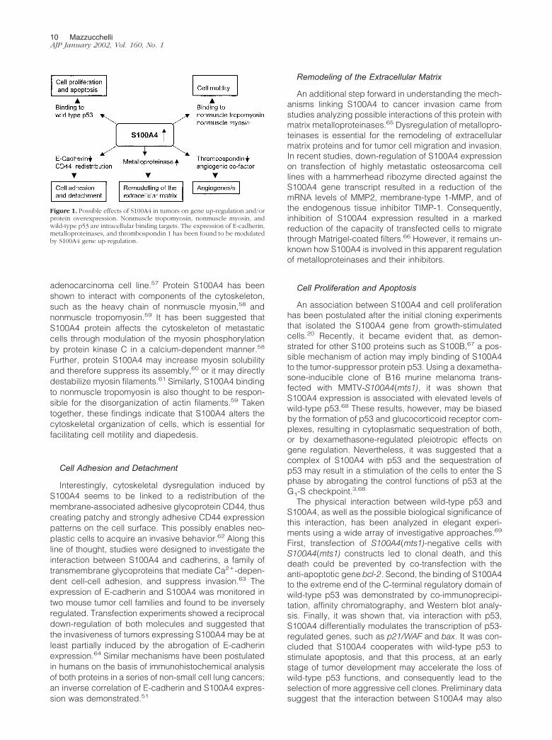
Tumor progression is characterized by complex pro-cesses such as cell motility and invasiveness, as well ascell proliferation. Evidence is accumulating for an impor-tant role of members of the protein S100 family in theseprocesses. Possible mechanisms are summarized belowand in Figure 1.
Cell Motility
A possible role of S100A4 in cell motility was sug-gested by initial studies performed with human promy-elocytic leukemia HL-60 cells27 and a mouse mammary
S100A4 in Cancer 9AJP January 2002, Vol. 160, No. 1
adenocarcinoma cell line.57 Protein S100A4 has beenshown to interact with components of the cytoskeleton,such as the heavy chain of nonmuscle myosin,58 andnonmuscle tropomyosin.59 It has been suggested thatS100A4 protein affects the cytoskeleton of metastaticcells through modulation of the myosin phosphorylationby protein kinase C in a calcium-dependent manner.58
Further, protein S100A4 may increase myosin solubilityand therefore suppress its assembly,60 or it may directlydestabilize myosin filaments.61 Similarly, S100A4 bindingto nonmuscle tropomyosin is also thought to be respon-sible for the disorganization of actin filaments.59 Takentogether, these findings indicate that S100A4 alters thecytoskeletal organization of cells, which is essential forfacilitating cell motility and diapedesis.
Cell Adhesion and Detachment
Interestingly, cytoskeletal dysregulation induced byS100A4 seems to be linked to a redistribution of themembrane-associated adhesive glycoprotein CD44, thuscreating patchy and strongly adhesive CD44 expressionpatterns on the cell surface. This possibly enables neo-plastic cells to acquire an invasive behavior.62 Along thisline of thought, studies were designed to investigate theinteraction between S100A4 and cadherins, a family oftransmembrane glycoproteins that mediate Ca2�-depen-dent cell-cell adhesion, and suppress invasion.63 Theexpression of E-cadherin and S100A4 was monitored intwo mouse tumor cell families and found to be inverselyregulated. Transfection experiments showed a reciprocaldown-regulation of both molecules and suggested thatthe invasiveness of tumors expressing S100A4 may be atleast partially induced by the abrogation of E-cadherinexpression.64 Similar mechanisms have been postulatedin humans on the basis of immunohistochemical analysisof both proteins in a series of non-small cell lung cancers;an inverse correlation of E-cadherin and S100A4 expres-sion was demonstrated.51
Remodeling of the Extracellular Matrix
An additional step forward in understanding the mech-anisms linking S100A4 to cancer invasion came fromstudies analyzing possible interactions of this protein withmatrix metalloproteinases.65 Dysregulation of metallopro-teinases is essential for the remodeling of extracellularmatrix proteins and for tumor cell migration and invasion.In recent studies, down-regulation of S100A4 expressionon transfection of highly metastatic osteosarcoma celllines with a hammerhead ribozyme directed against theS100A4 gene transcript resulted in a reduction of themRNA levels of MMP2, membrane-type 1-MMP, and ofthe endogenous tissue inhibitor TIMP-1. Consequently,inhibition of S100A4 expression resulted in a markedreduction of the capacity of transfected cells to migratethrough Matrigel-coated filters.66 However, it remains un-known how S100A4 is involved in this apparent regulationof metalloproteinases and their inhibitors.
Cell Proliferation and Apoptosis
An association between S100A4 and cell proliferationhas been postulated after the initial cloning experimentsthat isolated the S100A4 gene from growth-stimulatedcells.20 Recently, it became evident that, as demon-strated for other S100 proteins such as S100B,67 a pos-sible mechanism of action may imply binding of S100A4to the tumor-suppressor protein p53. Using a dexametha-sone-inducible clone of B16 murine melanoma trans-fected with MMTV-S100A4(mts1), it was shown thatS100A4 expression is associated with elevated levels ofwild-type p53.68 These results, however, may be biasedby the formation of p53 and glucocorticoid receptor com-plexes, resulting in cytoplasmatic sequestration of both,or by dexamethasone-regulated pleiotropic effects ongene regulation. Nevertheless, it was suggested that acomplex of S100A4 with p53 and the sequestration ofp53 may result in a stimulation of the cells to enter the Sphase by abrogating the control functions of p53 at theG1-S checkpoint.3,68
The physical interaction between wild-type p53 andS100A4, as well as the possible biological significance ofthis interaction, has been analyzed in elegant experi-ments using a wide array of investigative approaches.69
First, transfection of S100A4(mts1)-negative cells withS100A4(mts1) constructs led to clonal death, and thisdeath could be prevented by co-transfection with theanti-apoptotic gene bcl-2. Second, the binding of S100A4to the extreme end of the C-terminal regulatory domain ofwild-type p53 was demonstrated by co-immunoprecipi-tation, affinity chromatography, and Western blot analy-sis. Finally, it was shown that, via interaction with p53,S100A4 differentially modulates the transcription of p53-regulated genes, such as p21/WAF and bax. It was con-cluded that S100A4 cooperates with wild-type p53 tostimulate apoptosis, and that this process, at an earlystage of tumor development may accelerate the loss ofwild-type p53 functions, and consequently lead to theselection of more aggressive cell clones. Preliminary datasuggest that the interaction between S100A4 may also
Figure 1. Possible effects of S100A4 in tumors on gene up-regulation and/orprotein overexpression. Nonmuscle tropomyosin, nonmuscle myosin, andwild-type p53 are intracellular binding targets. The expression of E-cadherin,metalloproteinases, and thrombospondin 1 has been found to be modulatedby S100A4 gene up-regulation.
10 MazzucchelliAJP January 2002, Vol. 160, No. 1

modulate the functions of at least some p53 mutantsand therefore play important roles in advanced cancerstages.70
Angiogenesis
Angiogenesis is critical for tumor growth and cancermetastasis. Interestingly, experiments with S100A4-in-ducible cell lines grown at high density suggest thatS100A4 strongly down-regulates the thrombospondin 1(THBS1) gene,71 another p53 target, which is known torepress tumor progression by inhibition of angiogene-sis.69 Thus, it is conceivable that S100A4 also promotesangiogenesis in vivo by preventing the anti-angiogeniceffect of THBS1. Further, preliminary experiments sug-gest that S100A4 protein may act directly as an angio-genic factor.72 Tumors developing in S100A4(mts-1)transgenic mice revealed an increased vascular density.S100A4 oligomers were capable of stimulating motility,but not proliferation of endothelial cells in vitro, and in-ducing corneal neovascularization in vivo. Further stud-ies, however, will be necessary to better clarify this ap-parent co-stimulatory angiogenic effect of proteinS100A4 and to identify putative cell membrane receptorsfor extracellular forms of this protein.
Regulation of S100A4 Gene Transcription andmRNA Translation
The human S100A4 gene has been mapped on chromo-some 1. It is clustered with 12 other genes, belonging tothe S100 family, on the 1q21 region that is altered inseveral cancer types.73,74 In contrast to other gene clus-ters, however, the S100 family genes retain a specificpattern of expression, and they are most likely character-ized by independent regulation mechanisms. Interest-ingly, Northern blot analysis of normal mouse organsrevealed S100A4 mRNA in organs without protein ex-pression,1 suggesting therefore translational down-regu-lation and/or posttranslational degradation.32 Two splicevariants of the human S100A4 mRNA with some tissuespecificity of expression exist.75,76 The significance ofthese variants with respect to gene activity in differentorgans and/or cancer progression remains unclear.
S100A4 gene transcription seems to depend ongrowth-modulatory conditions of the cells. For instance, itwas found that its expression in macrophages may beaffected by molecules involved in the functional modula-tion of these cells in inflammation, such as lipopolysac-charides, tumor necrosis factor-�, concanavalin A, andby modulation of the cytosolic Ca2� concentration.1
Rosty and colleagues4 show a statistically significant as-sociation between the expression of protein S100A4 andhypomethylation in the first intron of the S100A4 gene.They also suggest that rather than an epigenetic phe-nomenon, the hypomethylation of S100A4 may reflectclonal selection during cancer progression. These find-ings are in agreement with previous studies showing thathypermethylation of the S100A4 gene is involved in tran-scription silencing.46,77–79 In this context, it is noteworthy
that the first intron of the S100A4 gene contains severalnegative and positive regulatory elements80,81 that in themouse interact with numerous factors, such as nuclearfactor-kB, or recently characterized regulatory pro-teins.82,83 These enhancer and silencer elements may bestrongly affected by the methylation status of the gene.
Conclusions and Outlooks
The information gathered throughout the past few yearsdemonstrate that protein S100A4 is involved in the regu-lation of cancer invasiveness and metastasis. Clinicalstudies are beginning to elucidate the prognostic signif-icance of this protein in human tumors. It is likely that ourknowledge on S100A4 as a prognostic, predictive, oreven diagnostic factor will dramatically increase with thedevelopment and commercial availability of antibodies.However, pathologists involved in the quest for prognos-tic markers in human neoplasia are, at the same time,also bound to focus on one of the most important objec-tives of pathology, ie, to foster a better comprehension ofthe pathogenesis of neoplasms and other diseases. Toachieve these goals, the functions of S100A4 need to befurther investigated. In the future, novel intra- and extra-cellular targets of S100A4 protein will probably be iden-tified. We still need to understand how the conformationalstatus of this protein, and in particular the formation ofprotein heterodimers with other members of the S100protein family may influence cellular functions. The studyof interactions of S100A4 with proteins involved in thecontrol of the cell cycle, either in the wild-type or in amutated form, and the interactions with adhesion mole-cules seems to be a promising research area. Hopefully,a more extensive knowledge on S100A4 will eventuallyallow the development of novel therapeutic strategies.
Acknowledgments
I thank Dr. J. A. Laissue, Dr. T. Schaffner, and Dr. J. Weisfor critical reading of the manuscript.
References
1. Lukanidin EM, Georgiev GP: Metastasis-related mts1 gene. Curr TopMicrobiol Immunol 1996, 213:171–195
2. Barraclough R: Calcium-binding protein S100A4 in health and dis-ease. Biochim Biophys Acta 1998, 1448:190–199
3. Sherbet GV, Lakshmi MS: S100A4 (MTS1) calcium binding protein incancer growth, invasion and metastasis. Anticancer Res 1998, 18:2415–2421
4. Rosty C, Ueki T, Argani P, Jansen M, Yeo CJ, Cameron JL, HrubanRH, Goggins M: Overexpression of S100A4 in pancreatic ductaladenocarcinomas is associated with poor differentiation and DNAhypomethylation. Am J Pathol 2002, 160:45–50
5. El-Rifai W, Frierson Jr HF, Harper JC, Powell SM, Knuutila S: Expres-sion profiling of gastric adenocarcinoma using cDNA array. Int JCancer 2001, 92:832–838
6. Kligman D, Hilt DC: The S100 protein family. Trends Biochem Sci1988, 13:437–443
7. Schafer BW, Heizmann CW: The S100 family of EF-hand calcium-binding proteins: functions and pathology. Trends Biochem Sci 1996,21:134–140
S100A4 in Cancer 11AJP January 2002, Vol. 160, No. 1

8. Heizmann CW, Hunziker W: Intracellular calcium-binding proteins:more sites than insights. Trends Biochem Sci 1991, 16:98–103
9. Linse S, Forsen S: Determinants that govern high-affinity calciumbinding. Adv Second Messenger Phosphoprotein Res 1995, 30:89 –151
10. Lee SW, Tomasetto C, Swisshelm K, Keyomarsi K, Sager R: Down-regulation of a member of the S100 gene family in mammary carci-noma cells and reexpression by azadeoxycytidine treatment. ProcNatl Acad Sci USA 1992, 89:2504–2508
11. Weterman MA, van Muijen GN, Bloemers HP, Ruiter DJ: Expression ofcalcyclin in human melanocytic lesions. Cancer Res 1993, 53:6061–6066
12. Baudier J, Delphin C, Grunwald D, Khochbin S, Lawrence JJ: Char-acterization of the tumor suppressor protein p53 as a protein kinaseC substrate and a S100b-binding protein. Proc Natl Acad Sci USA1992, 89:11627–11631
13. Ko LJ, Prives C: p53: puzzle and paradigm. Genes Dev 1996, 10:1054–1072
14. Barraclough R, Dawson KJ, Rudland PS: Control of protein synthesisin cuboidal rat mammary epithelial cells in culture. Changes in geneexpression accompany the formation of elongated cells. Eur J Bio-chem 1982, 129:335–341
15. Barraclough R, Savin J, Dube SK, Rudland PS: Molecular cloning andsequence of the gene for p9Ka. A cultured myoepithelial cell proteinwith strong homology to S-100, a calcium-binding protein. J Mol Biol1987, 198:13–20
16. Watanabe Y, Kobayashi R, Ishikawa T, Hidaka H: Isolation and char-acterization of a calcium-binding protein derived from mRNA termedp9Ka, pEL-98,18A2, or 42A by the newly synthesized vasorelaxantW-66 affinity chromatography. Arch Biochem Biophys 1992, 292:563–569
17. Engelkamp D, Schafer BW, Erne P, Heizmann CW: S100 alpha,CAPL, and CACY: molecular cloning and expression analysis of threecalcium-binding proteins from human heart. Biochemistry 1992, 31:10258–10264
18. Ebralidze A, Tulchinsky E, Grigorian M, Afanasyeva A, Senin V,Revazova E, Lukanidin E: Isolation and characterization of a genespecifically expressed in different metastatic cells and whose de-duced gene product has a high degree of homology to a Ca2�-binding protein family. Genes Dev 1989, 3:1086–1093
19. Goto K, Endo H, Fujiyoshi T: Cloning of the sequences expressedabundantly in established cell lines: identification of a cDNA clonehighly homologous to S-100, a calcium binding protein. J Biochem(Tokyo) 1988, 103:48–53
20. Linzer DI, Nathans D: Growth-related changes in specific mRNAs ofcultured mouse cells. Proc Natl Acad Sci USA 1983, 80:4271–4275
21. Masiakowski P, Shooter EM: Nerve growth factor induces the genesfor two proteins related to a family of calcium-binding proteins inPC12 cells. Proc Natl Acad Sci USA 1988, 85:1277–1281
22. Strutz F, Okada H, Lo CW, Danoff T, Carone RL, Tomaszewski JE,Neilson EG: Identification and characterization of a fibroblast marker:FSP1. J Cell Biol 1995, 130:393–405
23. De Vouge MW, Mukherjee BB: Transformation of normal rat kidneycells by v-K-ras enhances expression of transin 2 and an S-100-related calcium-binding protein. Oncogene 1992, 7:109–119
24. Takenaga K, Nakamura Y, Endo H, Sakiyama S: Involvement ofS100-related calcium-binding protein pEL98 (or mts1) in cell motilityand tumor cell invasion. Jpn J Cancer Res 1994, 85:831–839
25. Ambartsumian NS, Grigorian MS, Larsen IF, Karlstrom O, Sidenius N,Rygaard J, Georgiev G, Lukanidin E: Metastasis of mammary carci-nomas in GRS/A hybrid mice transgenic for the mts1 gene. Onco-gene 1996, 13:1621–1630
26. Davies MP, Rudland PS, Robertson L, Parry EW, Jolicoeur P, Barra-clough R: Expression of the calcium-binding protein S100A4 (p9Ka)in MMTV-neu transgenic mice induces metastasis of mammary tu-mours. Oncogene 1996, 13:1631–1637
27. Takenaga K, Nakamura Y, Sakiyama S: Expression of a calciumbinding protein pEL98 (mts1) during differentiation of human promy-elocytic leukemia HL-60 cells. Biochem Biophys Res Commun 1994,202:94–101
28. Gibbs FE, Barraclough R, Platt-Higgins A, Rudland PS, Wilkinson MC,Parry EW: Immunocytochemical distribution of the calcium-bindingprotein p9Ka in normal rat tissues: variation in the cellular location indifferent tissues. J Histochem Cytochem 1995, 43:169–180
29. Boni R, Burg G, Doguoglu A, Ilg EC, Schafer BW, Muller B, HeizmannCW: Immunohistochemical localization of the Ca2� binding S100proteins in normal human skin and melanocytic lesions. Br J Dermatol1997, 137:39–43
30. Ilg EC, Schafer BW, Heizmann CW: Expression pattern of S100calcium-binding proteins in human tumors. Int J Cancer 1996, 68:325–332
31. Duarte WR, Iimura T, Takenaga K, Ohya K, Ishikawa I, Kasugai S:Extracellular role of S100A4 calcium-binding protein in the periodon-tal ligament. Biochem Biophys Res Commun 1999, 255:416–420
32. Ambartsumian N, Klingelhofer J, Grigorian M, Karlstrom O, SideniusN, Georgiev G, Lukanidin E: Tissue-specific posttranscriptionaldownregulation of expression of the S100A4(mts1) gene in trans-genic animals. Invasion Metastasis 1998, 18:96–104
33. Rammes A, Roth J, Goebeler M, Klempt M, Hartmann M, Sorg C:Myeloid-related protein (MRP) 8 and MRP14, calcium-binding pro-teins of the S100 family, are secreted by activated monocytes via anovel, tubulin-dependent pathway. J Biol Chem 1997, 272:9496–9502
34. Zimmer DB, Cornwall EH, Landar A, Song W: The S100 protein family:history, function, and expression. Brain Res Bull 1995, 37:417–429
35. Tarabykina S, Scott DJ, Herzyk P, Hill TJ, Tame JR, Kriajevska M,Lafitte D, Derrick PJ, Dodson GG, Maitland NJ, Lukanidin EM, Bron-stein IB: The dimerization interface of the metastasis-associated pro-tein S100A4 (Mts1): in vivo and in vitro studies. J Biol Chem 2001,276:24212–24222
36. Tarabykina S, Kriajevska M, Scott DJ, Hill TJ, Lafitte D, Derrick PJ,Dodson GG, Lukanidin E, Bronstein I: Heterocomplex formation be-tween metastasis-related protein S100A4 (Mts1) and S100A1 as re-vealed by the yeast two-hybrid system. FEBS Lett 2000, 475:187–191
37. Novitskaya V, Grigorian M, Kriajevska M, Tarabykina S, Bronstein I,Berezin V, Bock E, Lukanidin E: Oligomeric forms of the metastasis-related Mts1 (S100A4) protein stimulate neuronal differentiation incultures of rat hippocampal neurons. J Biol Chem 2000, 275:41278–41286
38. Watanabe Y, Usada N, Minami H, Morita T, Tsugane S, Ishikawa R,Kohama K, Tomida Y, Hidaka H: Calvasculin, as a factor affecting themicrofilament assemblies in rat fibroblasts transfected by src gene.FEBS Lett 1993, 324:51–55
39. Davies BR, Davies MP, Gibbs FE, Barraclough R, Rudland PS: In-duction of the metastatic phenotype by transfection of a benign ratmammary epithelial cell line with the gene for p9Ka, a rat calcium-binding protein, but not with the oncogene EJ-ras-1. Oncogene 1993,8:999–1008
40. Lloyd BH, Platt-Higgins A, Rudland PS, Barraclough R: HumanS100A4 (p9Ka) induces the metastatic phenotype upon benign tu-mour cells. Oncogene 1998, 17:465–473
41. Parker C, Whittaker PA, Usmani BA, Lakshmi MS, Sherbet GV: Induc-tion of 18A2/mts1 gene expression and its effects on metastasis andcell cycle control. DNA Cell Biol 1994, 13:1021–1028
42. Grigorian M, Ambartsumian N, Lykkesfeldt AE, Bastholm L, Elling F,Georgiev G, Lukanidin E: Effect of mts1 (S100A4) expression on theprogression of human breast cancer cells. Int J Cancer 1996, 67:831–841
43. Maelandsmo GM, Hovig E, Skrede M, Engebraaten O, Florenes VA,Myklebost O, Grigorian M, Lukanidin E, Scanlon KJ, Fodstad O:Reversal of the in vivo metastatic phenotype of human tumor cells byan anti-CAPL (mts1) ribozyme. Cancer Res 1996, 56:5490–5498
44. Takenaga K, Nakamura Y, Sakiyama S: Expression of antisense RNAto S100A4 gene encoding an S100-related calcium-binding proteinsuppresses metastatic potential of high-metastatic Lewis lung carci-noma cells. Oncogene 1997, 14:331–337
45. Davies M, Harris S, Rudland P, Barraclough R: Expression of the rat,S-100-related, calcium-binding protein gene, p9Ka, in transgenicmice demonstrates different patterns of expression between thesetwo species. DNA Cell Biol 1995, 14:825–832
46. Pedrocchi M, Schafer BW, Mueller H, Eppenberger U, Heizmann CW:Expression of Ca(2�)-binding proteins of the S100 family in malig-nant human breast-cancer cell lines and biopsy samples. Int J Can-cer 1994, 57:684–690
47. Albertazzi E, Cajone F, Leone BE, Naguib RN, Lakshmi MS, SherbetGV: Expression of metastasis-associated genes h-mts1 (S100A4)and nm23 in carcinoma of breast is related to disease progression.DNA Cell Biol 1998, 17:335–342
12 MazzucchelliAJP January 2002, Vol. 160, No. 1

48. Platt-Higgins AM, Renshaw CA, West CR, Winstanley JH, De SilvaRudland S, Barraclough R, Rudland PS: Comparison of the metasta-sis-inducing protein S100A4 (p9ka) with other prognostic markers inhuman breast cancer. Int J Cancer 2000, 89:198–208
49. Rudland PS, Platt-Higgins A, Renshaw C, West CR, Winstanley JH,Robertson L, Barraclough R: Prognostic significance of the metasta-sis-inducing protein S100A4 (p9Ka) in human breast cancer. CancerRes 2000, 60:1595–1603
50. Ninomiya I, Ohta T, Fushida S, Endo Y, Hashimoto T, Yagi M, FujimuraT, Nishimura G, Tani T, Shimizu K, Yonemura Y, Heizmann CW,Schafer BW, Sasaki T, Miwa K: Increased expression of S100A4 andits prognostic significance in esophageal squamous cell carcinoma.Int J Oncol 2001, 18:715–720
51. Kimura K, Endo Y, Yonemura Y, Heizmann CW, Schafer BW, Wa-tanabe Y, Sasaki T: Clinical significance of S100A4 and E-cadherin-related adhesion molecules in non-small cell lung cancer. Int J Oncol2000, 16:1125–1131
52. Yonemura Y, Endou Y, Kimura K, Fushida S, Bandou E, Taniguchi K,Kinoshita K, Ninomiya I, Sugiyama K, Heizmann CW, Schafer BW,Sasaki T: Inverse expression of S100A4 and E-cadherin is associatedwith metastatic potential in gastric cancer. Clin Cancer Res 2000,6:4234–4242
53. Takenaga K, Nakanishi H, Wada K, Suzuki M, Matsuzaki O, MatsuuraA, Endo H: Increased expression of S100A4, a metastasis-associatedgene, in human colorectal adenocarcinomas. Clin Cancer Res 1997,3:2309–2316
54. Komatsu K, Kobune-Fujiwara Y, Andoh A, Ishiguro S, Hunai H, SuzukiN, Kameyama M, Murata K, Miyoshi J, Akedo H, Tatsuta M, Naka-mura H: Increased expression of S100A6 at the invading fronts of theprimary lesion and liver metastasis in patients with colorectal adeno-carcinoma. Br J Cancer 2000, 83:769–774
55. Bronckart Y, Decaestecker C, Nagy N, Harper L, Schafer BW, SalmonI, Pochet R, Kiss R, Heizman CW: Development and progression ofmalignancy in human colon tissues are correlated with expression ofspecific Ca(2�)-binding S100 proteins. Histol Histopathol 2001, 16:707–712
56. Camby I, Nagy N, Lopes MB, Schafer BW, Maurage CA, RuchouxMM, Murmann P, Pochet R, Heizmann CW, Brotchi J, Salmon I, KissR, Decaestecker C: Supratentorial pilocytic astrocytomas, astrocyto-mas, anaplastic astrocytomas and glioblastomas are characterizedby a differential expression of S100 proteins. Brain Pathol 1999,9:1–19
57. Ford HL, Salim MM, Chakravarty R, Aluiddin V, Zain SB: Expression ofMts1, a metastasis-associated gene, increases motility but not inva-sion of a nonmetastatic mouse mammary adenocarcinoma cell line.Oncogene 1995, 11:2067–2075
58. Kriajevska M, Tarabykina S, Bronstein I, Maitland N, Lomonosov M,Hansen K, Georgiev G, Lukanidin E: Metastasis-associated Mts1(S100A4) protein modulates protein kinase C phosphorylation of theheavy chain of nonmuscle myosin. J Biol Chem 1998, 273:9852–9856
59. Takenaga K, Nakamura Y, Sakiyama S, Hasegawa Y, Sato K, Endo H:Binding of pEL98 protein, an S100-related calcium-binding protein, tononmuscle tropomyosin. J Cell Biol 1994, 124:757–768
60. Kriajevska M, Bronstein IB, Scott DJ, Tarabykina S, Fischer-Larsen M,Issinger O, Lukanidin E: Metastasis-associated protein Mts1(S100A4) inhibits CK2-mediated phosphorylation and self-assemblyof the heavy chain of nonmuscle myosin. Biochim Biophys Acta 2000,1498:252–263
61. Ford HL, Silver DL, Kachar B, Sellers JR, Zain SB: Effect of Mts1 onthe structure and activity of nonmuscle myosin II. Biochemistry 1997,36:16321–16327
62. Lakshmi MS, Parker C, Sherbet GV: Expression of the transmem-brane glycoprotein CD44 and metastasis associated 18A2/MTS1gene in B16 murine melanoma cells. Anticancer Res 1997, 17:3451–3455
63. Perl AK, Wilgenbus P, Dahl U, Semb H, Christofori G: A causal role forE-cadherin in the transition from adenoma to carcinoma. Nature 1998,392:190–193
64. Keirsebilck A, Bonne S, Bruyneel E, Vermassen P, Lukanidin E,Mareel M, van Roy F: E-cadherin and metastasin (mts-1/S100A4)expression levels are inversely regulated in two tumor cell families.Cancer Res 1998, 58:4587–4591
65. Merzak A, Parker C, Koochekpour S, Sherbet GV, Pilkington GJ:Overexpression of the 18A2/mts1 gene and down-regulation of the
TIMP-2 gene in invasive human glioma cell lines in vitro. NeuropatholAppl Neurobiol 1994, 20:614–619
66. Bjornland K, Winberg JO, Odegaard OT, Hovig E, Loennechen T,Aasen AO, Fodstad O, Maelandsmo GM: S100A4 involvement inmetastasis: deregulation of matrix metalloproteinases and tissue in-hibitors of matrix metalloproteinases in osteosarcoma cells trans-fected with an anti-S100A4 ribozyme. Cancer Res 1999, 59:4702–4708
67. Lin J, Blake M, Tang C, Zimmer D, Rustandi RR, Weber DJ, Carrier F:Inhibition of p53 transcriptional activity by the S100B calcium-bindingprotein. J Biol Chem 2001, 276:35037–35041
68. Parker C, Lakshmi MS, Piura B, Sherbet GV: Metastasis-associatedmts1 gene expression correlates with increased p53 detection in theB16 murine melanoma. DNA Cell Biol 1994, 13:343–351
69. Grigorian M, Andresen S, Tulchinsky E, Kriajevska M, Carlberg C,Kruse C, Cohn M, Ambartsumian N, Christensen A, Selivanova G,Lukanidin E: Tumor suppressor p53 protein is a new target for themetastasis-associated Mts1/S100A4 protein: functional conse-quences of their interaction. J Biol Chem 2001, 276:22699–22708
70. Chen H, Fernig DG, Rudland PS, Sparks A, Wilkinson MC, Barra-clough R: Binding to intracellular targets of the metastasis-inducingprotein, s100a4 (p9ka). Biochem Biophys Res Commun 2001, 286:1212–1217
71. Roberts DD: Regulation of tumor growth and metastasis by throm-bospondin-1. FASEB J 1996, 10:1183–1191
72. Ambartsumian N, Klingelhofer J, Grigorian M, Christensen C, Kria-jevska M, Tulchinsky E, Georgiev G, Berezin V, Bock E, Rygaard J,Cao R, Cao Y, Lukanidin E: The metastasis-associated Mts1(S100A4)protein could act as an angiogenic factor. Oncogene 2001, 20:4685–4695
73. Engelkamp D, Schafer BW, Mattei MG, Erne P, Heizmann CW: SixS100 genes are clustered on human chromosome 1q21: identificationof two genes coding for the two previously unreported calcium-binding proteins S100D and S100E. Proc Natl Acad Sci USA 1993,90:6547–6551
74. Ridinger K, Ilg EC, Niggli FK, Heizmann CW, Schafer BW: Clusteredorganization of S100 genes in human and mouse. Biochim BiophysActa 1998, 1448:254–263
75. Ambartsumian N, Tarabykina S, Grigorian M, Tulchinsky E, HulgaardE, Georgiev G, Lukanidin E: Characterization of two splice variants ofmetastasis-associated human mts1 gene. Gene 1995, 159:125–130
76. Albertazzi E, Cajone F, Sherbet GV: Characterization of a splicevariant of metastasis-associated h-mts1 (S100A4) gene expressed inhuman infiltrating carcinomas of the breast. DNA Cell Biol 1998,17:1003–1008
77. Tulchinsky E, Ford HL, Kramerov D, Reshetnyak E, Grigorian M, ZainS, Lukanidin E: Transcriptional analysis of the mts1 gene with specificreference to 5� flanking sequences. Proc Natl Acad Sci USA 1992,89:9146–9150
78. Tulchinsky E, Grigorian M, Tkatch T, Georgiev G, Lukanidin E: Tran-scriptional regulation of the mts1 gene in human lymphoma cells: therole of DNA-methylation. Biochim Biophys Acta 1995, 1261:243–248
79. Chen D, Rudland PS, Chen HL, Barraclough R: Differential reactivityof the rat S100A4(p9Ka) gene to sodium bisulfite is associated withdifferential levels of the S100A4 (p9Ka) mRNA in rat mammary epi-thelial cells. J Biol Chem 1999, 274:2483–2491
80. Tulchinsky E, Prokhortchouk E, Georgiev G, Lukanidin E: A kappaB-related binding site is an integral part of the mts1 gene compositeenhancer element located in the first intron of the gene. J Biol Chem1997, 272:4828–4835
81. Chen D, Davies MP, Rudland PS, Barraclough R: Transcriptionaldown-regulation of the metastasis-inducing S100A4 (p9Ka) in benignbut not in malignant rat mammary epithelial cells by GC-factor. J BiolChem 1997, 272:20283–20290
82. Hjelmsoe I, Allen CE, Cohn MA, Tulchinsky EM, Wu LC: The kappaBand V(D)J recombination signal sequence binding protein KRC reg-ulates transcription of the mouse metastasis-associated geneS100A4/mts1. J Biol Chem 2000, 275:913–920
83. Cohn MA, Hjelmso I, Wu LC, Guldberg P, Lukanidin EM, TulchinskyEM: Characterization of Sp1, AP-1, CBF and KRC binding sites andminisatellite DNA as functional elements of the metastasis-associatedmts1/S100A4 gene intronic enhancer. Nucleic Acids Res 2001, 29:3335–3346
S100A4 in Cancer 13AJP January 2002, Vol. 160, No. 1