Embed Size (px)
Citation preview

Food Chemistry 239 (2018) 486–494
Contents lists available at ScienceDirect
Food Chemistry
journal homepage: www.elsevier .com/locate / foodchem
Protein degradation and peptide release from milk proteins in humanjejunum. Comparison with in vitro gastrointestinal simulation
http://dx.doi.org/10.1016/j.foodchem.2017.06.1340308-8146/� 2017 Elsevier Ltd. All rights reserved.
⇑ Corresponding author.E-mail address: [email protected] (I. Recio).
J. Sanchón a, S. Fernández-Tomé a, B. Miralles a, B. Hernández-Ledesma a, D. Tomé b, C. Gaudichon b,I. Recio a,⇑a Instituto de Investigación en Ciencias de la Alimentación, CIAL (CSIC-UAM, CEI UAM+CSIC), Nicolás Cabrera, 9, 28049 Madrid, SpainbAgroParisTech_UMR0914 Physiologie de la Nutrition et du Comportement Alimentaire, 16 rue Claude Bernard, 75005 Paris, France
a r t i c l e i n f o
Article history:Received 17 March 2017Received in revised form 20 June 2017Accepted 23 June 2017Available online 24 June 2017
Keywords:In vitro/in vivo digestionPeptidomicMass spectrometryMilk protein digestion
a b s t r a c t
Human jejunal digests after oral ingestion of casein and whey protein were collected by a nasogastrictube and protein degradation and peptide release was compared with that found in the digests of thesame substrates using a standardised protocol. No intact casein was detected in the jejunal nor in thein vitro samples taken during the intestinal phase, while b-lactoglobulin was found in one hour-jejunalsamples in agreement with the in vitro digestion. In vivo and in vitro digests showed comparable peptideprofiles and high number of common sequences. A selective precipitation step was used to strengthen theidentification of phosphorylated peptides. Most of the sequences found in jejunum, some of them notpreviously described, were also identified in the simulated digests. Common resistant regions to diges-tion were identified, revealing that the in vitro protocol constitutes a good approximation to the physio-logical gastrointestinal digestion of milk proteins.
� 2017 Elsevier Ltd. All rights reserved.
1. Introduction
Digestion is an intricate process that food undergoes upon oralingestion. Proteins are known to be extensively hydrolysedthroughout the digestive tract by gastrointestinal enzymes andbrush border membrane peptidases, leading to the release ofnumerous peptides and free amino acids. However, some specificregions of proteins have demonstrated resistance against thedigestive phenomena. Food protein-derived peptides, present inthe gut lumen, might be biologically and metabolically relevantfor different physiological functions, exerting not only beneficialproperties (Rutherfurd-Markwick, 2012) but also deleteriouseffects, such as those produced by epitopes involved in food aller-gies (Jiménez-Saiz, Benedé, Molina, & López-Expósito, 2015).
Because of the potential health implications of dietary peptides,several studies aimed at the identification of the peptides releasedin the gastrointestinal tract during protein digestion have previ-ously been performed (Picariello, Mamone, Nitride, Addeo, &Ferranti, 2013; Sánchez-Rivera, Martínez-Maqueda, Cruz-Huerta,Miralles, & Recio, 2014). Among food proteins, milk proteins area main source of exogenous biologically active peptides(Hernández-Ledesma, García-Nebot, Fernández-Tomé, Amigo, &
Recio, 2014; Nongonierma & FitzGerald, 2015) and represent astable complex food matrix commonly used as a suitable modelin digestion experiments. Some in vivo studies have been per-formed on the presence of milk-derived peptides in the gastroin-testinal lumen of humans (Boutrou et al., 2013; Chabance et al.,1998; Svedberg, de Haas, Leimenstoll, Paul, & Teschemacher,1985) and animals (Barbé et al., 2014; Bouzerzour et al., 2012) afteringestion of dairy products, but the reported data although essen-tial is still scarce and a notable diversity has been recently stated(Boutrou, Henry, & Sánchez-Rivera, 2015).
Knowledge on the factors that influence protein degradationand lead to peptide release and stability, is also provided by sev-eral in vitro methods, simulating the digestion of food and phar-maceuticals (Dupont et al., 2010; Kaukonen, Boyd, Charman, &Porter, 2004; Kopf-Bolanz et al., 2012; Martos, Contreras,Molina, & López-Fandiño, 2010; Versantvoort, Oomen, Van deKamp, Rompelberg, & Sips, 2005). In this context, a harmonizedstatic in vitro digestion method was recently developed by theCOST Action INFOGEST FA1005 network (Minekus et al., 2014),but it remains difficult to overcome the wide variation betweenthe individual parameters used on these methodologies, and tofully mimic the kinetic behaviour of the complex in vivo digestionprocess by a static model, prompting further experiments todirectly compare the in vitro results towards in vivo data (Eggeret al., 2016).
J. Sanchón et al. / Food Chemistry 239 (2018) 486–494 487
The aim of the present study was to directly compare thein vitro results with in vivo data to provide additional informationon the interaction between milk proteins and the digestive tract,and to shed light on their physiological-derived consequences.Accordingly, this work compared the peptidome of samplesobtained after gastrointestinal digestion of milk casein and wheyprotein by the harmonized INFOGEST in vitro digestion method,with in vivo digests collected from human jejunum. Nine differenttime combinations of the in vitro gastric and intestinal phases werecarried out and were compared with in vivo jejunal samplesobtained 1, 2, 3 and 4 h after protein ingestion. Protein degradationwas determined by SDS-PAGE, and the characterisation at a pep-tide level of the digestion products was evaluated by HPLC-tandem mass spectrometry (HPLC-MS/MS). Moreover, enrichmentby selective precipitation was performed on casein samples tostudy the caseinphosphopeptide (CPP) fraction.
2. Materials and methods
2.1. Human jejunal effluents
Human jejunal effluents were obtained at the Human NutritionResearch Centre of Bobigny, as previously described (Boutrou et al.,2013; Mahé et al., 1996; Marsset-Baglieri et al., 2014). All proce-dures were approved by the Ethics Committee for Saint-Germain-en-Laye Hospital. Written informed consent wasobtained from all participants. The study was registered underwww.ClinicalTrials.gov (NCT00862329). Briefly, individualsingested in fasting conditions 30 g of casein or whey, dissolved in500 mL of water. Samples were obtained by a nasogastric tube thatmigrated to the proximal jejunum by aspiration of jejunal effluentsat 1, 2, 3 and 4 h after oral administration of the protein solution.Jejunal effluents were collected on ice, freeze-dried and kept at�80 �C until analysis. Nitrogen content in freeze-dried sampleswas determined by elemental analysis and further analyses wereperformed on protein basis. In this study, jejunal samples collectedfrom a total of 9 volunteers were analysed: 5 who had ingestedcasein and 4 who had ingested whey protein.
2.2. In vitro simulated gastrointestinal digestion
Casein and whey milk protein powders were digested accordingto the in vitro harmonized protocol (Minekus et al., 2014). Briefly,freeze-dried casein or whey protein samples were dissolved insimulated saliva fluid at 30 mg of protein/mL without amylasebecause of the absence of starch in the samples. Then, the mixwas diluted at a ratio of 50:50 (v:v) in simulated gastric fluid con-taining pepsin from porcine gastric mucosa (2000 U/mL of digest,EC 3.4.23.1, Sigma-Aldrich, St. Louis, MO, USA). Samples were with-drawn at 10, 20, 30, 60 and 120 min during gastric digestion andthe reaction was stopped by adjusting the pH at 7.0 with NaOH1 M and snap freezing in liquid nitrogen. Intestinal phase was car-ried out by mixing the gastric phase with the same volume of sim-ulated intestinal fluid containing pancreatin from porcine pancreas(100 U trypsin activity/mL of final mixture, Sigma-Aldrich) andporcine bile extract (10 mM in the final mixture, Sigma-Aldrich).All simulated fluids were tempered at 37 �C before use. Digestionsof each protein powder were performed in duplicate by incubatingat 37 �C in an orbital shaker at 150 rpm. Intestinal phases werestarted from 20 and 120 min of gastric phase digests, performedduring 60 and 120 min, the digestion was stopped with Pefabloc�
SC (5 mM, Sigma-Aldrich) and snap freezing. Samples werefreeze-dried and kept at �20 �C until analysis. A digestion blankconsisting of the mix of enzymes used in digestions at the sameconcentration without substrate protein was prepared. Enzyme
activities and bile concentration were measured according to theassay described in the protocol (Minekus et al., 2014).
2.3. SDS-PAGE and Western blotting
Samples were dissolved at 1 mg of protein/mL in sample bufferthat contained Tris-HCl (0.05 M, pH 6.8, Sigma-Aldrich), SDS (1.6%,w:v, Merck, Darmstadt, Germany), glycerol (8%, v:v, PanreacQuímica SAU, Castellar del Vallés, Barcelona, Spain), b-mercaptoethanol (2%, v:v, Sigma-Aldrich) and bromophenol blueindicator (0.002%, w:v, Merck), heated at 95 �C for 5 min andloaded on 12% Bis-Trispolyacrilamide gels (Criterion_XT, Bio-Rad,Hercules, CA, USA). Electrophoretic separations were run at 100 Vfor 5 min and then at 150 V, using the XT MES running buffer(Bio-Rad) in the criterion cell (Bio-Rad). A molecular weight mar-ker (Precision Plus ProteinTM Unstained standard, Bio-Rad) was usedon each gel. Gels were stained with Coomasie Blue (Instant blue,Expedeon, Swavesey, UK) and images were taken with a MolecularImager� VersaDocTM MP 5000 system (Bio-Rad) and processed withQuantity One�1-D analysis software (Bio-Rad).
Western blotting detection of intact casein was performed bySDS-PAGE analysis of the human jejunal digests after casein inges-tion. Gels were run in duplicate and after separation one gel wasstained using Coomasie Blue as described above and the otherwas soaked in transfer buffer (48 mM Tris, 39 mM glycine, 20%methanol, pH 9.2) for 30 min. Proteins were electroblotted ontonitrocellulose membranes using a trans-Blot semi-dry transfer cell(BioRad) at 18 V for 30 min. Membranes were blocked by incuba-tion in pH 7.6 Tris buffered saline containing 0.05% Tween 20(TBST) and 1% bovine serum albumin (BSA) for 3 h at room temper-ature. After three washing steps with TBST, the membrane wasincubated with casein polyclonal antibody developed in rabbit(Biorbyt Ltd., Cambridge, UK) diluted 1/1000 (v:v) in TBST contain-ing 0.1% BSA overnight at 4 �C. The incubation was followed by fivewashing steps with TBST of 5 min each. Finally, the membrane wasincubated overnight at 4 �C with monoclonal peroxidase-conjugated swine anti-rabbit IgG antibody (Dako, Glostrup, Den-mark) 1/5000 diluted in TBST containing 0.1% BSA followed bysix washing steps with TBST of 5 min each. The membrane wasdeveloped by chemiluminescence with Amersham TM ECL Prime(GE Healthcare, Chalfont St Giles, UK). Image acquisition was per-formed using the VersaDoc Imaging System. The positive controlcontained 20 mg of casein for the SDS-PAGE and western blotanalyses.
2.4. Analysis by HPLC–tandem mass spectrometry (HPLC–MS/MS)
Freeze-dried samples were reconstituted in solvent A (water:-formic acid, 100:0.1, v:v) and centrifuged at 13000g, 10 min, beforeinjection of the supernatant. From casein samples, an enrichmentselective precipitation of CPPs was performed by adding calciumchloride (1%, w:v) and ethanol (50%, v:v) as described bySánchez-Rivera et al. (2014). Whey samples were treated for60 min at 37 �C with 1,4-dithiothreitol 70 mM (Sigma-Aldrich) toreduce samples in order to improve the identification ofdisulfide-linked fragments.
Samples were analysed by HPLC-MS/MS in duplicate using anAgilent 1100 HPLC system (Agilent Technologies, Waldbron, Ger-many), equipped with a Mediterranea Sea18 column(150 � 2.1 mm, Teknokroma, Barcelona, Spain). HPLC system wasconnected to an Esquire 3000 linear ion trap mass spectrometer(Bruker Daltonics GmbH, Bremen, Germany) equipped with anelectrospray ionization source. The injection volume was 50 mLand the flow was set at 0.2 mL/min. Peptide elution was performedwith a linear gradient from 0 to 45% of solvent B (acetonitrile:-formic acid, 100:0.1, v:v) in 115 min. The spectra were recorded

488 J. Sanchón et al. / Food Chemistry 239 (2018) 486–494
over the mass/charge (m/z) range 100–700, 100–1700 and 100–2000, selecting 500, 1200, and 750 as target mass, respectively.For CPPs, same solvents and gradient, although in 60 min, wereused, and spectra were recorded over the m/z range 100–2100,with a target mass of 1200. The results were processed by usingData Analysis (version 4.0, Bruker Daltonics). Homemade databaseof most abundant bovine casein and whey protein, including majorgenetic variants, was used for the peptide sequencing in MASCOTv2.4 software (Matrix Science). No specific enzyme cleavage wasused. Peptide mass tolerance was set to 0.1% and 0.5 Da for MSand MS/MS analysis, respectively. Moreover, Biotools version 3.2was used for the interpretation of the matched MS/MS spectra.Each identification peptide spectrum was revised manually,regardless of its P value.
2.5. Statistical analysis
Spearman correlation matrices were built on the basis of fre-quency of appearance of amino acids data. This frequency ofappearance of each amino acid in the protein sequences was alsoused to perform Principal Component Analysis (PCA). Statisticasoftware for Windows version 7.0 (StatSoft Inc., Tulsa, OK, USA)was used for the calculations.
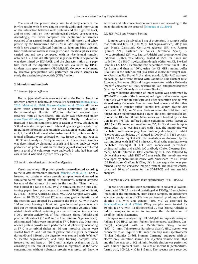
Fig. 1. SDS-PAGE comparison of jejunal casein digests with in vitro simulated gastroinsimulated gastrointestinal whey protein digestion (C and D). Each lane in A and C corresjejunal sampling (1, 2, 3 and 4 h). Each lane in B and D correspond to different times (min)WP, whey protein; Blank, gastrointestinal digestion enzymes without milk protein; HPL,a-lactalbumin.
3. Results and discussion
3.1. Protein degradation during human and in vitro gastrointestinaldigestion
Protein patterns obtained by SDS-PAGE of the jejunal effluentscollected at 1, 2, 3 and 4 h after oral administration of casein andwhey protein are shown in Fig. 1 (A and C, respectively). In allthe jejunal digests from casein (Fig. 1A), there was a clear bandwith molecular weight (MW) 50 kDa, which likely corresponds tohuman pancreatic lipase, as previously described by other authors(Kopf-Bolanz et al., 2012). In addition, there were several lightbands with mobility between 25 and 37 kDa, close to those ofintact casein. However, no differences were observed at differentsampling times or between volunteers and they did not react withantibodies against casein by immunoblotting (Supporting Informa-tion Fig 1), thus, these bands could probably correspond to endoge-nous proteins. In a previous report, the nitrogen composition,endogenous and exogenous, in the upper jejunum of human volun-teers after ingestion of casein or whey protein was investigated(Mahé et al., 1996). It was found that casein was slowly recoveredin the jejunum and mainly in the form of degraded peptides whichis consistent with our results where no intact protein is detected.Similarly, other in vivo studies in minipigs cannulated at
testinal casein digestion (A and B) and jejunal whey protein digests with in vitroponds to different subjects (referred to with capital letters from A to I) and times ofof gastric (G) and intestinal (I) digestion. MM, molecular weight marker; CN, casein;human pancreatic lipase; PPL, porcine pancreatic lipase; b-Lg, b-lactoglobulin; a-La,

J. Sanchón et al. / Food Chemistry 239 (2018) 486–494 489
duodenum have shown that after ingestion of non-heated liquidmilk, intact casein can be detected during the first minutes ofdigestion, but they are rapidly digested in the next 20 min (Barbéet al., 2014).
When applying the standardised in vitro digestion conditions onthe same casein substrate (Fig. 1B), a gradual degradation of thecasein fraction with the digestion time was observed in the gastricphase. Electrophoretic bands corresponding to casein wereobserved up to 30 min peptic digestion but they were absent inthe samples obtained in the gastric phase at 60 and 120 min. Inthe samples withdrawn during the intestinal phase an intenseband corresponding to porcine pancreatic lipase could be observedca. 50 kDa, as well as, other minor bands corresponding to otherpancreatic enzymes between 20 and 35 kDa, which is compatiblewith the MW of the porcine pancreatic enzymes trypsin(23.3 kDa), chymotrypsin (25.5–29.10 kDa) or elastase (25.9 kDa),as previously shown (Kopf-Bolanz et al., 2012). The rapid degrada-tion of the casein fraction of milk during in vitro gastric digestionagrees with previous reports using this standardised method in dif-ferent laboratories (Egger et al., 2016), where the casein bandswere not detected at the end of the gastric phase (120 min) andby other authors using slightly different in vitro conditions (Kopf-Bolanz et al., 2012; Ménard et al., 2014; Picariello et al., 2010).
Fig. 1C shows the SDS-PAGE analyses of the jejunal contents ofdifferent volunteers at different times after whey protein intake.The electrophoretic band corresponding to b-lactoglobulin was vis-ible in the samples taken at 1 h in all volunteers with variations inintensity. In some volunteers, this protein was also detected in thesample taken at 2 h after oral administration of whey protein (vol-unteers H and I). Again, this agrees with previous results on nitro-gen content in jejunum where b-lactoglobulin could be recovered,
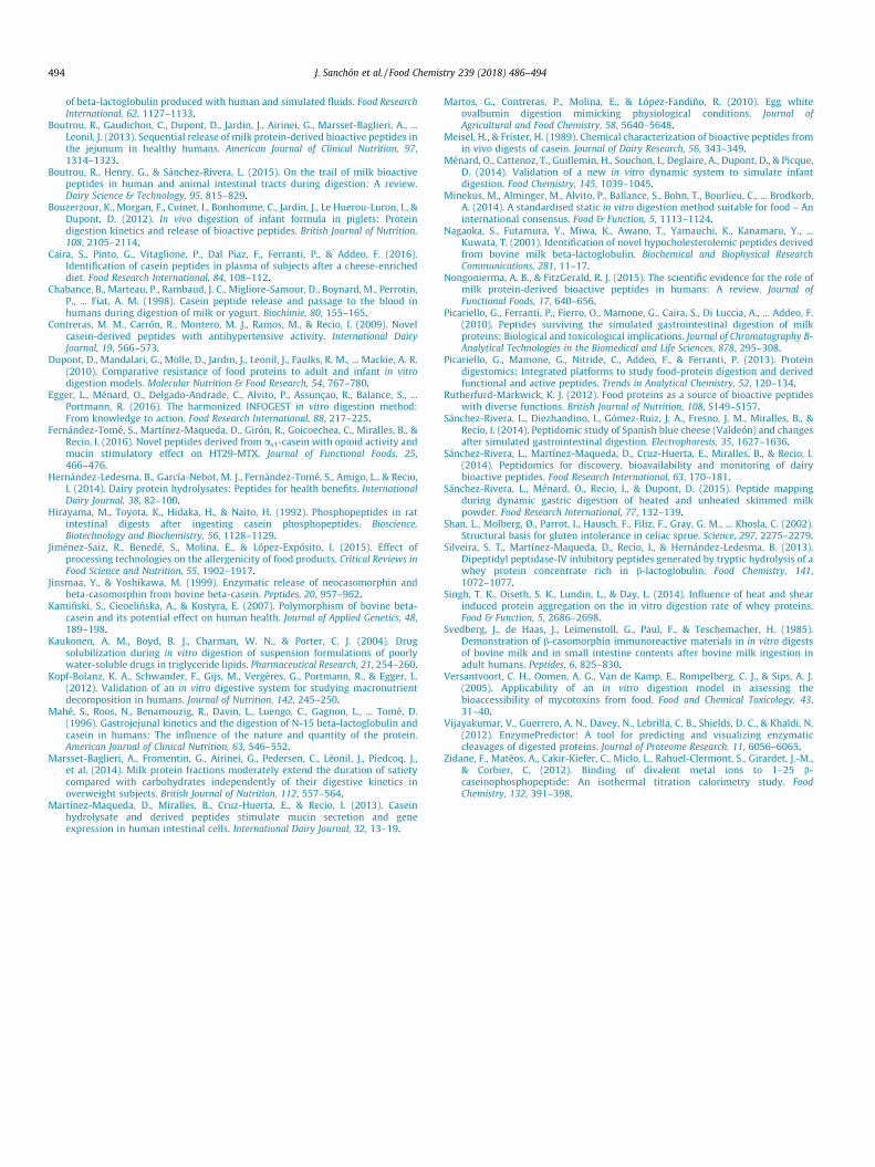
Fig. 2. Heat map built with the frequency of appearance of each amino acid identified aobtained from human jejunal digests obtained at 1, 2, 3 and 4 h after oral administratdigestion of the same substrate. The green colour represents low frequency and red higgastric; I, intestinal. (For interpretation of the references to colour in this figure legend,
mostly in the form of intact protein (Mahé et al., 1996). Fig. 1Cshows that in volunteer I, an important amount of a-lactalbumincould be found 1 h after administration. As expected, duringin vitro digestion of whey protein (Fig. 1D) b-lactoglobulin wasresistant to digestion by pepsin but it disappeared completely after60 min of intestinal digestion regardless the duration of the gastricphase (20 or 120 min). The resistance of this protein to pepticdigestion has been largely documented in vivo (Bouzerzour et al.,2012) and in vitro (Dupont et al., 2010; Kopf-Bolanz et al., 2012),as well as, the effect of heat treatment on the susceptibility of thisprotein to gastric digestion (Barbé et al., 2014; Sánchez-Rivera,Ménard, Recio, & Dupont, 2015).
3.2. Peptidomic characterisation of in vivo and in vitro digests
Jejunal digests obtained at four different time points and in vitrodigests withdrawn under gastric and intestinal phases were anal-ysed under equal conditions by HPLC-MS/MS. The peptides identi-fied in the jejunal digests from b-casein, as1-casein, b-lactoglobulin, and a-lactalbumin are shown in SupplementaryTables S1–S4. To compare the peptide profile obtained from theanalysis of the jejunal digests and in vitro digests, heat maps werebuilt (Fig. 2). These graphs represent the appearance frequency ofeach amino acid identified as part of a peptide sequence for a givenprotein. This representation gives qualitative information aboutthe protein coverage and about those protein regions where pep-tides were identified. Each line in the heat map corresponds to ahuman volunteer (Fig. 2A and C) or a different duplicate in thein vitro assays (Fig. 2B and D). In the in vivo digests, there is a com-mon pattern at the different time points and for the different vol-unteers, although certain inter-individual variability can be
s part of a peptide sequence from b- and as1-casein. A and C correspond to profilesion of casein and B and D correspond to profiles obtained from simulated in vitroh frequency, being the maximum 24, 9, 18 and 8 in A, B, C, and D, respectively. G,the reader is referred to the web version of this article.)

490 J. Sanchón et al. / Food Chemistry 239 (2018) 486–494
inferred. In addition, the peptide profiles of jejunal digests arecomparable to the intestinal phase of the in vitro simulated gas-trointestinal digestion (Fig. 2A and B). The b-casein N-terminalregion and the region 57–99 are highly represented in both cases,but the peptides in the central and final region of the protein (resi-due 115–175) were more abundant in the jejunal digests. For as1-casein (Fig. 2C and D), the peptide profile was much more complexin vivo than in vitro. While in the simulated in vitro digestions pep-tides corresponded to the N- and C-terminal regions, in the jejunaldigests, peptides were released from the whole protein chain. Thus,many peptides belonging to the central domain of the as1-caseinprotein chain could be identified in the jejunal digests which wereabsent in the in vitro digests.
The frequency of appearance of each amino acid for b-caseinand as1-casein were used to compare the in vitro and the in vivodigestion. Interindividual and in vivo/in vitro Spearman correla-tions were calculated. The correlation coefficient for the in vivo/in vitro intestinal digests comparison reached 0.71 ± 0.08 whichis within the range found when comparing different individuals0.58–0.73. To compare these results, a PCA was applied to the fre-quency of appearance of each amino acid for b-casein and as1-casein. Fig. 3 shows the clustering of the in vivo and in vitro diges-tion samples in the PC1-PC2 score plot. As expected, the samplesfrom the in vitro gastric phase were plotted separately from jejunaldigests and from in vitro simulated gastrointestinal digestion, andthese two latter types of digests could be grouped in the samplecluster. There were two jejunal samples separated from the rest(C 2 h and B 4 h) that reflects the high in vivo variability. This anal-ysis confirmed the similarity between the jejunal and the in vitrointestinal digests at peptide level for the digestion of the caseinfraction.
Fig. 4 shows the peptide pattern of the two major proteins, b-lactoglobulin (Fig. 4A and B) and a-lactalbumin (Fig. 4C and D),obtained when whey protein was digested. In this case, althoughsome b-lactoglobulin-derived peptides were found in the 30 min-gastric phase, most of peptides were identified in the intestinalphase during in vitro gastrointestinal simulation. This agrees withthe low protein degradation pattern obtained by SDS-PAGE andthe previously mentioned resistance of b-lactoglobulin to theaction of pepsin. Similarly, other in vitro digestion studies have
Fig. 3. Principal component analysis score plot calculated with the frequency of appearaDifferent human subjects (blue triangles) are referred to with capital letters from A to Edigests are represented with red squares. G, gastric; I, intestinal, followed by the time expin this figure legend, the reader is referred to the web version of this article.)
reported the release of certain peptide sequences from b-lactoglobulin during gastric digestion (Benedé et al., 2014; Eggeret al., 2016). Discrepancies between different studies are explainedbecause the susceptibility of this protein to peptic hydrolysis mayvary with the denaturation degree of b-lactoglobulin by heating orother treatments (Singh, Oiseth, Lundin, & Day, 2014; Sánchez-Rivera et al., 2015). Likewise, only few a-lactalbumin peptideswere identified at the end of the gastric phase, consistent withthe late degradation of this protein observed by SDS-PAGE. Duringthe in vitro intestinal phase, several b-lactoglobulin sequenceswere released mainly from regions comprised between the 42–68 and 107–135, with few peptides from the N- and C-terminalregions of the protein (Fig. 4B). The peptidic profile obtained fromjejunal digests was more complex than the in vitro one, with ahigher number of different peptides along the whole protein chain.A considerable number of b-lactoglobulin peptides found in jeju-num belonged to regions which were also overrepresented in theintestinal in vitro profile (40–60; 120–140) (Fig. 4B). Previous stud-ies have also shown a higher abundancy of b-lactoglobulin pep-tides from region 40 to 60 in human jejunal contents afteringestion of whey protein (Boutrou et al., 2013), in piglet jejunumafter administration of infant formula (Bouzerzour et al., 2012),and after the incubation of b-lactoglobulin with human gastroin-testinal fluids (Benedé et al., 2014). In the case of the a-lactalbumin protein, both peptidic profiles, in vitro and in vivowerecoincident in several regions: 40–52, 63–68, and 80–90(Fig. 4C and D). Interestingly, peptides were found in jejunal sam-ples withdrawn during the first two hours, but only few a-lactalbumin peptides were identified at 3 and 4 h. In this sense,casein-derived peptides maintained their presence along the diges-tion times evaluated (Fig. 2), while the number of whey-derivedpeptides started to decrease after 3 h of ingestion. Similarly,Boutrou et al. (2013) also found these differences between caseinand whey protein, regarding the kinetics of dietary nitrogen fluxand the detection and size distribution of peptides in humanjejunum.
A correlation matrix for the in vitro/in vivo comparison of b-lactoglobulin and a-lactalbumin-derived peptides was also built.In this case, the first two samples from the in vitro gastric phasewere not included neither jejunal samples at 4 h due to the low
nce of each amino acid identified as part of a peptide from b-casein and as1-casein.followed by the time of jejunal sampling (1, 2, 3 and 4 h). In vitro gastrointestinalressed in minutes of in vitro digestion. (For interpretation of the references to colour

Fig. 4. Heat map built with the frequency of appearance of each amino acid identified as part of a peptide sequence from b-lactoglobulin and a-lactalbumin protein chain. Aand C correspond to profiles obtained from human jejunal digests obtained at 1, 2, 3 and 4 h after oral administration of whey proteins and B and D correspond to profilesobtained from simulated in vitro digestion of the same substrate. The green colour represents low frequency and red high frequency, being the maximum 32, 12, 8 and 9 in A,B, C, and D, respectively. G, gastric; I, intestinal. (For interpretation of the references to colour in this figure legend, the reader is referred to the web version of this article.)
J. Sanchón et al. / Food Chemistry 239 (2018) 486–494 491
number of identified peptides. The average of Spearman correla-tion coefficient within jejunum digests from different individualsranged from 0.43 to 0.76, similar to the average Spearman correla-tion coefficient found for the in vivo/in vitro comparison,0.74 ± 0.16. Moreover, these results were examined by PCA andtwo groups were distinguished. One group was composed by theintestinal digests (in vitro and in vivo) while the other cluster com-prised the in vitro gastric digests (data not shown). These resultswere similar to those found with casein as substrate.
3.3. Identification of phosphorylated peptides
All casein digests were subjected to a selective precipitationprocedure with CaCl2 to identify phosphorylated regions in thein vivo and in vitro digests. A total of 19 different phosphorylatedsequences from b-casein were found in the jejunum digests(Fig. 5). They contained from one to four phosphorylated Ser butonly b-casein f15–24 included the cluster sequence SerP-SerP-SerP-Glu-Glu, typically recognised as the binding region of miner-als (Zidane et al., 2012). In addition, other CPPs with residues of Gluclose to phosphorylated Ser residues were identified. Various phos-phorylated fragments belonging to the N-terminal region, b-casein
f6-16, f7-16, f8-16 and f9-16, were identified in all jejunal digests,but especially, in the samples taken at 1 and 2 h where these phos-phorylated peptides were found in all volunteers (Fig. 5). One ofthe sequences, b-casein f7-16, had been previously identified byChabance et al. (1998) in the human duodenum after milk inges-tion. In addition, these authors had identified the sequence 155–165, that comprises b-casein f157-165, one of the fragments foundin the jejunum. Most of the phosphorylated peptides from b-caseinfound in jejunum were also identified in the simulated intestinaldigests (Supplementary Fig. 2), although in some cases, longersequences were shown. This was the case of the peptide f7-35 thatcomprised some of the previously mentioned jejunal sequences.Regarding as1-casein, 15 different phosphopeptides were identi-fied in the jejunum digests (Fig. 6). The most consistent regionwas as1-casein f42-52 that appeared at all jejunal time pointsand was also persistently found in vitro (Supplementary Fig. 3).Peptides belonging to the as1-casein domain 43–52 have also beenidentified in human plasma after cheese consumption (Caira et al.,2016). The region as1-casein f66-70 merits being mentionedbecause it includes the sequence SerP-SerP-SerP-Glu-Glu and,although it had been found in digests from rats (Hirayama,Toyota, Hidaka, & Naito, 1992) and mini pigs (Barbé et al., 2014;

Fig. 5. Phosphorylated peptides derived from b-casein identified in jejunal digests at different times from ingestion. Each grey colour denotes a different subject. In peptideswith multiple phosphorylation sites the position of the phosphorylated residues may vary. Phosphorylated serine residues are represented by a red coloured X. (Forinterpretation of the references to colour in this figure legend, the reader is referred to the web version of this article.)
Fig. 6. Phosphorylated peptides derived from as1-casein identified in jejunal digests, at different times from ingestion. Each grey colour denotes a different subject. Inpeptides with multiple phosphorylation sites the position of the phosphorylated residues may vary. Phosphorylated serine residues are represented by a red coloured X. (Forinterpretation of the references to colour in this figure legend, the reader is referred to the web version of this article.)
492 J. Sanchón et al. / Food Chemistry 239 (2018) 486–494
Meisel & Frister, 1989), no peptides including this domain hadbeen previously reported in humans. By contrast, other identifiedsequences, namely the as1-casein f110-119, had been previouslyidentified in human intestinal digests (Boutrou et al., 2013).
3.4. Evaluation of the peptide sequences identified in human jejunum
After casein intake, a total of 415 different peptides were recov-ered from human jejunal effluents, and 155 were found followingthe in vitro protocol. In the case of the whey proteins, 230 peptideswere obtained in vivo and 80 from in vitro digestion. The total num-ber of peptides identified in vivo was higher than in vitro, probablydue to the inter-individual variability found. With the aim of iden-tifying the enzymes responsible for the enzymatic cleavages, ananalysis by using the online software application Enzyme Predictor
was performed (Vijayakumar et al., 2012). From the comparisonbetween in vivo and in vitro results, it was observed that severalcleavages appeared exclusively in the jejunal digests. Some of themcorresponded to enzymes included in the simulated juices, such as,pepsin, contained in the gastric fluids, trypsin, chymotrypsin, andelastase, contained in the pancreatic extract. The missing cleavagesin the in vitro digests could indicate that the digestion conditions,in terms of concentration, presence of enzyme coadjuvants, or agi-tation, were not optimal for their occurrence.
Many studies have been focused on the resistance of certainpeptide fragments during gastrointestinal digestion, and especiallypeptides with an attributed physiological effect. From the identi-fied casein sequences, the percentage of peptides containing pro-line was 85% for b-casein, 73% for as1-casein, 45% for as2-casein,and 77% for j-casein. Therefore, regions rich in proline residues

J. Sanchón et al. / Food Chemistry 239 (2018) 486–494 493
were resistant to gastrointestinal digestion and reached jejunum.Interestingly, the peptides whose sequences did not contain pro-line showed in most cases (75% on average) included aspartic orglutamic acid residues. Similarly, from the identified whey proteinpeptides, only a 5% did not contain proline or negatively chargedresidues. In vitro, comparable results were found, i.e., proline-containing and peptides with negatively charged residues survivedto gastrointestinal digestion, pointing to a special resistance ofthese regions to the action of gastrointestinal enzymes.
Regarding the formation of bioactive peptides during gastroin-testinal digestion, the opioid peptide b-casomorphin-7 (b-casein60YPFPGPI66) and various peptides containing the same sequencewere found in jejunum after consumption of casein (Supplemen-tary Table S5), as well as in the intestinal phase of in vitro caseindigestion. These peptides had been previously quantified in jejunalcontents and it was estimated that the amount considering b-casomorphin-7 and its precursors could reach a concentration ca.17 lmol/L (Boutrou et al., 2013). It has been described that pepsincleaves the 58L-V59 peptide bond in b-casein, and leucine-aminopeptidase removes the valine at position 59, rendering thetyrosine residue at the N-terminus, characteristic of opioid pep-tides (Jinsmaa & Yoshikawa, 1999). At the C-terminal end of theb-casomorphin-7, the cleavage 66I-X67 is favoured in the geneticvariant A1 of b-casein, where 67X corresponds to histidine, withrespect the A2 variant, where 67X corresponds to proline. The mix-ture of peptides released from this region is due to the presence ofthese two major genetic variants in the precursor casein. Most ofthe longer peptides containing b-casomorphin corresponded toA2 b-casein (i.e. with proline at position 67), while free b-casomorphin-7 could likely be generated from the A1 b-casein vari-ant. Previous reports have shown that in hydrolysed milk with A1
b-casein, the level of b-casomorphin-7 was 4-fold than in A2 milk(Kaminski, Cieoelinska, & Kostyra, 2007). Similarly, neocasomor-phin and various longer forms could be also identified in the jeju-nal digests after administration of casein. In addition to opioidpeptides, these results also prove the resistance of peptidesequences with reported antihypertensive activity (SupplementaryTable S5). For some of the volunteers, the reported activesequences were found in human jejunum, suggesting resistanceof these regions to gastrointestinal digestion, and the possibilityof these peptides being released during casein digestion. This isthe case of the antihypertensive peptides b-CN 134HLPLP138, andas1-CN 143AYFYPEL149, for which the reported active forms werefound in the casein jejunal digests at different sampling times. Thislatter peptide had been previously found in gastric human digestsafter milk ingestion (Chabance et al., 1998), and in addition to itsantihypertensive effects in spontaneously hypertensive rats(Contreras, Carrón, Montero, Ramos, & Recio, 2009), it is alsoknown for exerting intestinal mucin stimulatory properties inhuman goblet HT29-MTX cells (Martínez-Maqueda, Miralles,Cruz-Huerta, & Recio, 2013), and opioid activity in the guinea pigileum assay (Fernández-Tomé et al., 2016). On the contrary, forother peptides, such as, the antihypertensive tripeptides, IPP andVPP, only longer forms were found in vivo, which reflects the resis-tance of certain peptide bounds to be hydrolysed by the action ofthe gastrointestinal enzymes. In the region comprising the peptide74IPP76, the cleavage 73N-I74 was observed in some cases but thecleavage 76P-L77 was not found. The difficulty of the gastrointesti-nal enzymes to hydrolyse the proline C-terminal peptide bonds isalso illustrated in Supplementary Table S5, where different pep-tides from the C-terminal region the b-casein molecule, peptidescomprising neocasomorphin and other antihypertensive peptideswere protected from hydrolysis. It has been proposed that thepost-proline cleaving protease activity is absent in human gastricand pancreatic juices which is related with the resistance ofgluten-derived peptides and the inflammatory response in celiac
patients (Shan et al., 2002). However, several peptide fragmentsfound in jejunum ended by proline, resulting from the post-proline cleavage, while this cleavage seemed to be protected inother cases, which suggests the influence of the residues close tothe bond to be hydrolysed.
In the case of the whey peptides identified in jejunal digests,several peptides with a proven biological activity were found. Thisis the case of the b-lactoglobulin 71IIAEK75 and 9GLDIQK14 withhypocholesterolemic activity (Nagaoka et al., 2001), and b-lactoglobulin 78IPAVF82 with dipeptidyl peptidase-inhibitory activ-ity (Silveira, Martínez-Maqueda, Recio, & Hernández-Ledesma,2013).
4. Conclusions
In the present work, a comparison of the standardised in vitrodigestion model (Infogest) with in vivo digestion data from humanjejunum is shown. Although some differences were found, thein vitro protocol resembles the in vivo intestinal digestion concern-ing protein degradation and peptide release. Regarding proteindegradation, casein reached the jejunum in the form of degradedpeptides while intact b-lactoglobulin was visible in the samplestaken at 1 h in all volunteers. At the end of the in vitro gastroin-testinal simulation a peptide pattern was comparable to that foundin jejunum. It is important to highlight that the common regionswhich resist gastrointestinal digestion correspond to proline richpeptides or peptides containing negatively charged residues afterboth, casein and whey protein digestion. It was shown that the cor-relation found for the in vivo/in vitro comparison by using the fre-quency of appearance of each amino acid was similar to the inter-individual variability. Most of the phosphorylated peptides from b-casein found in jejunum were also identified in the in vitro intesti-nal digests, although some longer sequences were also found in thein vitro digests. Therefore, these results illustrate that the proposedin vitro digestion protocol constitutes a good approximation to thephysiological gastrointestinal digestion of milk proteins.
Conflict of interest statement
The authors declare no competing financial interest.
Acknowledgements
This work has received financial support from project AGL2015-66886-R from the Spanish Ministry of Economy and Competitive-ness (MINECO). S. F.-T. acknowledges MINECO for his FPI fellow-ship. J. S. is the recipient of a ‘‘CSIC-CM-FSE, Iniciativa de EmpleoJuvenil” contract. Irene Pastor Cardeña is acknowledged for herexcellent technical assistance and Phillip John Bentley for languageediting.
Appendix A. Supplementary data
Supplementary data associated with this article can be found, inthe online version, at http://dx.doi.org/10.1016/j.foodchem.2017.06.134.
References
Barbé, F., Le Feunteun, S., Remond, D., Ménard, O., Jardin, J., Henry, G., ... Dupont, D.(2014). Tracking the in vivo release of bioactive peptides in the gut duringdigestion: Mass spectrometry peptidomic characterization of effluents collectedin the gut of dairy matrix fed mini-pigs. Food Research International, 63,147–156.
Benedé, S., Lopez-Exposito, I., Gimenez, G., Grishina, G., Bardina, L., Sampson, H. A.,... Molina, E. (2014). Mapping of IgE epitopes in in vitro gastroduodenal digests
494 J. Sanchón et al. / Food Chemistry 239 (2018) 486–494
of beta-lactoglobulin produced with human and simulated fluids. Food ResearchInternational, 62, 1127–1133.
Boutrou, R., Gaudichon, C., Dupont, D., Jardin, J., Airinei, G., Marsset-Baglieri, A., ...Leonil, J. (2013). Sequential release of milk protein-derived bioactive peptides inthe jejunum in healthy humans. American Journal of Clinical Nutrition, 97,1314–1323.
Boutrou, R., Henry, G., & Sánchez-Rivera, L. (2015). On the trail of milk bioactivepeptides in human and animal intestinal tracts during digestion: A review.Dairy Science & Technology, 95, 815–829.
Bouzerzour, K., Morgan, F., Cuinet, I., Bonhomme, C., Jardin, J., Le Huerou-Luron, I., &Dupont, D. (2012). In vivo digestion of infant formula in piglets: Proteindigestion kinetics and release of bioactive peptides. British Journal of Nutrition,108, 2105–2114.
Caira, S., Pinto, G., Vitaglione, P., Dal Piaz, F., Ferranti, P., & Addeo, F. (2016).Identification of casein peptides in plasma of subjects after a cheese-enricheddiet. Food Research International, 84, 108–112.
Chabance, B., Marteau, P., Rambaud, J. C., Migliore-Samour, D., Boynard, M., Perrotin,P., ... Fiat, A. M. (1998). Casein peptide release and passage to the blood inhumans during digestion of milk or yogurt. Biochimie, 80, 155–165.
Contreras, M. M., Carrón, R., Montero, M. J., Ramos, M., & Recio, I. (2009). Novelcasein-derived peptides with antihypertensive activity. International DairyJournal, 19, 566–573.
Dupont, D., Mandalari, G., Molle, D., Jardin, J., Leonil, J., Faulks, R. M., ... Mackie, A. R.(2010). Comparative resistance of food proteins to adult and infant in vitrodigestion models. Molecular Nutrition & Food Research, 54, 767–780.
Egger, L., Ménard, O., Delgado-Andrade, C., Alvito, P., Assunçao, R., Balance, S., ...Portmann, R. (2016). The harmonized INFOGEST in vitro digestion method:From knowledge to action. Food Research International, 88, 217–225.
Fernández-Tomé, S., Martínez-Maqueda, D., Girón, R., Goicoechea, C., Miralles, B., &Recio, I. (2016). Novel peptides derived from as1-casein with opioid activity andmucin stimulatory effect on HT29-MTX. Journal of Functional Foods, 25,466–476.
Hernández-Ledesma, B., García-Nebot, M. J., Fernández-Tomé, S., Amigo, L., & Recio,I. (2014). Dairy protein hydrolysates: Peptides for health benefits. InternationalDairy Journal, 38, 82–100.
Hirayama, M., Toyota, K., Hidaka, H., & Naito, H. (1992). Phosphopeptides in ratintestinal digests after ingesting casein phosphopeptides. Bioscience,Biotechnology and Biochemistry, 56, 1128–1129.
Jiménez-Saiz, R., Benedé, S., Molina, E., & López-Expósito, I. (2015). Effect ofprocessing technologies on the allergenicity of food products. Critical Reviews inFood Science and Nutrition, 55, 1902–1917.
Jinsmaa, Y., & Yoshikawa, M. (1999). Enzymatic release of neocasomorphin andbeta-casomorphin from bovine beta-casein. Peptides, 20, 957–962.
Kaminski, S., Cieoelinska, A., & Kostyra, E. (2007). Polymorphism of bovine beta-casein and its potential effect on human health. Journal of Applied Genetics, 48,189–198.
Kaukonen, A. M., Boyd, B. J., Charman, W. N., & Porter, C. J. (2004). Drugsolubilization during in vitro digestion of suspension formulations of poorlywater-soluble drugs in triglyceride lipids. Pharmaceutical Research, 21, 254–260.
Kopf-Bolanz, K. A., Schwander, F., Gijs, M., Vergères, G., Portmann, R., & Egger, L.(2012). Validation of an in vitro digestive system for studying macronutrientdecomposition in humans. Journal of Nutrition, 142, 245–250.
Mahé, S., Roos, N., Benamouzig, R., Davin, L., Luengo, C., Gagnon, L., ... Tomé, D.(1996). Gastrojejunal kinetics and the digestion of N-15 beta-lactoglobulin andcasein in humans: The influence of the nature and quantity of the protein.American Journal of Clinical Nutrition, 63, 546–552.
Marsset-Baglieri, A., Fromentin, G., Airinei, G., Pedersen, C., Léonil, J., Piedcoq, J.,et al. (2014). Milk protein fractions moderately extend the duration of satietycompared with carbohydrates independently of their digestive kinetics inoverweight subjects. British Journal of Nutrition, 112, 557–564.
Martínez-Maqueda, D., Miralles, B., Cruz-Huerta, E., & Recio, I. (2013). Caseinhydrolysate and derived peptides stimulate mucin secretion and geneexpression in human intestinal cells. International Dairy Journal, 32, 13–19.
Martos, G., Contreras, P., Molina, E., & López-Fandiño, R. (2010). Egg whiteovalbumin digestion mimicking physiological conditions. Journal ofAgricultural and Food Chemistry, 58, 5640–5648.
Meisel, H., & Frister, H. (1989). Chemical characterization of bioactive peptides fromin vivo digests of casein. Journal of Dairy Research, 56, 343–349.
Ménard, O., Cattenoz, T., Guillemin, H., Souchon, I., Deglaire, A., Dupont, D., & Picque,D. (2014). Validation of a new in vitro dynamic system to simulate infantdigestion. Food Chemistry, 145, 1039–1045.
Minekus, M., Alminger, M., Alvito, P., Ballance, S., Bohn, T., Bourlieu, C., ... Brodkorb,A. (2014). A standardised static in vitro digestion method suitable for food – Aninternational consensus. Food & Function, 5, 1113–1124.
Nagaoka, S., Futamura, Y., Miwa, K., Awano, T., Yamauchi, K., Kanamaru, Y., ...Kuwata, T. (2001). Identification of novel hypocholesterolemic peptides derivedfrom bovine milk beta-lactoglobulin. Biochemical and Biophysical ResearchCommunications, 281, 11–17.
Nongonierma, A. B., & FitzGerald, R. J. (2015). The scientific evidence for the role ofmilk protein-derived bioactive peptides in humans: A review. Journal ofFunctional Foods, 17, 640–656.
Picariello, G., Ferranti, P., Fierro, O., Mamone, G., Caira, S., Di Luccia, A., ... Addeo, F.(2010). Peptides surviving the simulated gastrointestinal digestion of milkproteins: Biological and toxicological implications. Journal of Chromatography B-Analytical Technologies in the Biomedical and Life Sciences, 878, 295–308.
Picariello, G., Mamone, G., Nitride, C., Addeo, F., & Ferranti, P. (2013). Proteindigestomics: Integrated platforms to study food-protein digestion and derivedfunctional and active peptides. Trends in Analytical Chemistry, 52, 120–134.
Rutherfurd-Markwick, K. J. (2012). Food proteins as a source of bioactive peptideswith diverse functions. British Journal of Nutrition, 108, S149–S157.
Sánchez-Rivera, L., Diezhandino, I., Gómez-Ruiz, J. A., Fresno, J. M., Miralles, B., &Recio, I. (2014). Peptidomic study of Spanish blue cheese (Valdeón) and changesafter simulated gastrointestinal digestion. Electrophoresis, 35, 1627–1636.
Sánchez-Rivera, L., Martínez-Maqueda, D., Cruz-Huerta, E., Miralles, B., & Recio, I.(2014). Peptidomics for discovery, bioavailability and monitoring of dairybioactive peptides. Food Research International, 63, 170–181.
Sánchez-Rivera, L., Ménard, O., Recio, I., & Dupont, D. (2015). Peptide mappingduring dynamic gastric digestion of heated and unheated skimmed milkpowder. Food Research International, 77, 132–139.
Shan, L., Molberg, Ø., Parrot, I., Hausch, F., Filiz, F., Gray, G. M., ... Khosla, C. (2002).Structural basis for gluten intolerance in celiac sprue. Science, 297, 2275–2279.
Silveira, S. T., Martínez-Maqueda, D., Recio, I., & Hernández-Ledesma, B. (2013).Dipeptidyl peptidase-IV inhibitory peptides generated by tryptic hydrolysis of awhey protein concentrate rich in b-lactoglobulin. Food Chemistry, 141,1072–1077.
Singh, T. K., Oiseth, S. K., Lundin, L., & Day, L. (2014). Influence of heat and shearinduced protein aggregation on the in vitro digestion rate of whey proteins.Food & Function, 5, 2686–2698.
Svedberg, J., de Haas, J., Leimenstoll, G., Paul, F., & Teschemacher, H. (1985).Demonstration of b-casomorphin immunoreactive materials in in vitro digestsof bovine milk and in small intestine contents after bovine milk ingestion inadult humans. Peptides, 6, 825–830.
Versantvoort, C. H., Oomen, A. G., Van de Kamp, E., Rompelberg, C. J., & Sips, A. J.(2005). Applicability of an in vitro digestion model in assessing thebioaccessibility of mycotoxins from food. Food and Chemical Toxicology, 43,31–40.
Vijayakumar, V., Guerrero, A. N., Davey, N., Lebrilla, C. B., Shields, D. C., & Khaldi, N.(2012). EnzymePredictor: A tool for predicting and visualizing enzymaticcleavages of digested proteins. Journal of Proteome Research, 11, 6056–6065.
Zidane, F., Matéos, A., Cakir-Kiefer, C., Miclo, L., Rahuel-Clermont, S., Girardet, J.-M.,& Corbier, C. (2012). Binding of divalent metal ions to 1–25 b-caseinophosphopeptide: An isothermal titration calorimetry study. FoodChemistry, 132, 391–398.