Embed Size (px)
Citation preview

The FASEB Journal • Research Communication
Lysosomal degradation of endocytosed proteinsdepends on the chloride transport protein ClC-7
Lena Wartosch,*,† Jens C. Fuhrmann,*,1 Michaela Schweizer,‡ Tobias Stauber,* andThomas J. Jentsch*,2
*Leibniz-Institut fur Molekulare Pharmakologie (FMP) and Max-Delbruck-Centrum fur MolekulareMedizin (MDC), Berlin, Germany; †Freie Universitat, Berlin, Germany; and ‡Zentrum fur MolekulareNeurobiologie (ZMNH), Universitat Hamburg, Hamburg, Germany
ABSTRACT Mutations in either ClC-7, a late endoso-mal/lysosomal member of the CLC family of chloridechannels and transporters, or in its �-subunit Ostm1cause osteopetrosis and lysosomal storage disease inmice and humans. The severe phenotype of miceglobally deleted for ClC-7 or Ostm1 and the absence ofstorage material in cultured cells hampered investiga-tions of the mechanism leading to lysosomal pathologyin the absence of functional ClC-7/Ostm1 transporters.Tissue-specific ClC-7-knockout mice now reveal thataccumulation of storage material occurs cell-autono-mously in neurons or renal proximal tubular cellslacking ClC-7. Almost all ClC-7-deficient neurons die.The activation of glia is restricted to brain regionswhere ClC-7 has been inactivated. The effect of ClC-7disruption on lysosomal function was investigated inrenal proximal tubular cells, which display high endo-cytotic activity. Pulse-chase endocytosis experiments invivo with mice carrying chimeric deletion of ClC-7 inproximal tubules allowed a direct comparison of thehandling of endocytosed protein between cells express-ing or lacking ClC-7. Whereas protein was endocytosedsimilarly in cells of either genotype, its half-life in-creased significantly in ClC-7-deficient cells. These ex-periments demonstrate that lysosomal pathology is acell-autonomous consequence of ClC-7 disruption andthat ClC-7 is important for lysosomal protein degrada-tion.—Wartosch, L., Fuhrmann, J. C., Schweizer, M.,Stauber, T., Jentsch, T. J. Lysosomal degradation ofendocytosed proteins depends on the chloride trans-port protein ClC-7. FASEB J. 23, 000–000 (2009).www.fasebj.org
Key Words: neurodegeneration � neuronal ceroid lipofuscinosis� NCL � lysosomal storage disease
Clc-7 is a ubiquitously expressed member of theCLC gene family of Cl� channels and Cl�/H� ex-changers (1). Of the 9 mammalian CLC proteins, 4members are plasma membrane Cl� channels, whereasothers, including ClC-7, are sited predominantly inmembranes of the endolysosomal pathway (2). Thepresence of a certain “proton glutamate” in ClC-3through ClC-7 suggested that ClC-7 functions as a
Cl�/H� exchanger, in analogy to the bacterial EcClC-1(3) and mammalian ClC-4 and ClC-5 proteins (4, 5).Indeed lysosomes, which prominently express ClC-7and its �-subunit Ostm1 (6–8), show Cl�/H�-exchangeactivity (9). ClC-7/Ostm1 is also found on late endo-somes and a specialized acid-secreting plasma mem-brane domain of osteoclasts (6–8). The latter mem-brane (“ruffled border”) is believed to be formed bylysosomal exocytosis.
Compartments of the endocytic pathway are progres-sively acidified along their way from the plasma mem-brane to the lysosome. Their low luminal pH is impor-tant for such processes as receptor-ligand interaction,endolysosomal trafficking, and luminal enzymatic activ-ity (10), rendering it a key parameter for processingand degradation of endocytosed material. The activeaccumulation of protons is achieved mainly throughV-type H�-ATPases that act as electrogenic protonpumps. Intracellular CLC proteins are thought to facil-itate endosomal/lysosomal acidification by providingan electrical shunt for proton pumping (2).
Disruption of specific vesicular CLC proteins hasdiverse effects, such as impaired renal endocytosis andkidney stone formation with a loss of ClC-5 (11–13) orneurodegeneration with the loss of ClC-3 (14). The lossof either ClC-7 (7) or its �-subunit Ostm1 (8, 15) leadsto excessive bone mineralization (osteopetrosis) and toa severe degeneration of the brain and retina (6–8).The neurodegeneration observed in mice lacking ei-ther ClC-7 or Ostm1 displays typical features of humanneuronal ceroid lipofuscinosis (NCL), a neurodegen-erative lysosomal storage disease (6, 8). Neurologicalabnormalities and blindness have also been describedin osteopetrotic patients carrying mutations in CLCN7(16) or OSTM1 on both alleles (17). In Clcn7�/� mice,the accumulation of lysosomal storage material in neu-ronal cell bodies precedes neuronal cell loss that is
1 Present address: Metanomics GmbH, Tegeler Weg 33,D-10589 Berlin, Germany.
2 Correspondence: Leibniz-Institut fur Molekulare Pharma-kologie and Max-Delbruck-Centrum fur Molekulare Medizin,Robert-Rossle-Str. 10, D-13125 Berlin, Germany. E-mail: [email protected]
doi: 10.1096/fj.09-130880
10892-6638/09/0023-0001 © FASEB
The FASEB Journal article fj.09-130880. Published online August 6, 2009.

evident as early as 30 d after birth (P30) (6). The exactmechanism for lysosomal deposition with a loss ofClC-7/Ostm1 activity is unknown. Neither lysosomalpH nor the activity of the lysosomal enzyme tripepti-dylpeptidase I (TPP I) were altered in cultured ClC-7-deficient neurons or fibroblasts (6). The short life spanand complex phenotype of constitutive ClC-7-knockout(KO) mice (6, 7) was a severe obstacle in the analysis oflysosomal function in vivo and prevented us fromfollowing the progression of neurodegeneration.
Newly generated conditional KO mice now revealthat accumulation of lysosomal storage material isintrinsic to cells lacking ClC-7. The massive activation ofmicroglia and astrocytes is limited to brain regionswhere ClC-7 has been deleted. Chimeric deletion ofClC-7 in proximal tubular cells (PTCs) enabled us tocompare directly the uptake and degradation of pro-teins between cells of either genotype. We show thatClC-7 is important for the lysosomal degradation ofproteins but not for their endocytotic uptake.
MATERIALS AND METHODS
Mice
To generate “floxed” ClC-7 mice, a part of the murine ClC-7gene (Clcn7), extending from the intron preceding exon 6 tothe intron after exon 16, was cloned into a pKO Scramblerplasmid (Lexicon Genetics Incorporated, Woodland, TX,USA). A loxP site with a 5� EcoRI site was introduced in theintron upstream of exon 12, a neomycin resistance (neo)cassette flanked by loxP sites behind exon 13, and a diphthe-ria toxin A cassette at the 5� end of the targeting construct(Supplemental Fig. 1A). The vector was electroporated intoR1 embryonal stem cells, and clones were tested for homol-ogous recombination by Southern blot analysis (Supplemen-tal Fig. 1B). After transfection with a plasmid expressing Crerecombinase, clones were tested for removal of the neocassette by Southern blotting (Supplemental Fig. 1C). C57BL/6blastocysts injected with correctly targeted cells were im-planted into foster mothers. Male chimeras were bred withC57BL/6 females to yield heterozygous floxed ClC-7 mice(Clcn7�/lox).
Forebrain-specific ClC-7-KO mice were obtained by cross-ing Clcn7 lox/lox mice with mice (in a mixed C57BL/6-129/SVJ background) expressing Cre recombinase under controlof the EMX1 promotor (18), mice with partial ClC-7 deletionin proximal tubules (PTs) by crossing Clcn7 lox/lox mice withApoE-cre mice (19) and backcrossing into C57BL/6 for �5generations. In all experiments, wild-type (WT; Clcn7�/�),Clcn7�/lox, Clcn7 lox/lox, Clcn7�/lox;cre, or Clcn7�/�;cre litter-mates served as controls.
Clcn7�/� and Clcn5�/� mice were described previously (7,11). Deleter-Cre mice (20) were provided by Ralf Kuhn (Helm-holtz Zentrum Munchen, Munich, Germany), EMX1-Cremice (18) by Takuji Iwasato and Shigeyoshi Itohara (BrainScience Institute, RIKEN, Saitama, Japan), and ApoE-Cremice (19) by Thomas Willnow (MDC, Berlin, Germany). Allexperiments conformed to the animal protection laws ofGermany.
Histology and immunohistochemistry
Primary antibodies used were own KO-controlled rabbit anti-ClC-7 (7), guinea-pig anti-ClC-7 (8), rabbit anti-Ostm1 (8),
rabbit anti-ClC-5 (21), rabbit anti-ClC-3 (22), mouse anti-lysobisphosphatidic acid (LBPA; ref. 23), rat anti-lysosome-associated membrane protein-1 (lamp-1; clone 1D4B; BDPharmingen, Heidelberg, Germany), mouse anti-villin (Acris,Hiddenhausen, Germany), mouse anti-parvalbumin (Swant,Bellinzona, Switzerland), mouse anti-NeuN (Chemicon, Te-meculla, CA, USA), rabbit anti-IBA1 (Wako Chemicals, Rich-mond, VA, USA), and mouse anti-glial fibrillary acidic protein(GFAP; Sigma, Deisenhofen, Germany). Secondary antibod-ies conjugated to Alexa Fluor 488, 546, or 633 (MolecularProbes, Karlsruhe, Germany) or horseradish peroxidase(HRP; Dianova, Hamburg, Germany) were used.
HRP was detected by incubation in solution containing0.05% (w/v) 3,3�-diaminobenzidine (DAB), 0.03% (w/v)H2O2, and 0.04% nickel ammonium sulfate in 0.01 M phos-phate buffer (PB). Endogenous peroxidase was exhausted bypreincubation for 30 min in 0.3% H2O2 in 0.1 M PB.GSA-biotin (Griffonia simplicifolia agglutinin; Vector Laborato-ries, Burlingame, CA, USA) was detected using Streptavidin-Cy3 (Molecular Probes, Karlsruhe, Germany).
Perfusion of deeply anesthetized mice, fixation, and prep-aration of tissue and staining procedures were performed asdescribed previously (6). For alkaline phosphatase-stainingthe Sigma Fast™ Fast Red TR/Naphtol AS-MX substrate kit(Sigma) was used according to the manufacturer’s instruc-tions.
Electron microscopy procedures were described previously(6). For semithin sections, tissues were sliced to 0.7 �m andstained with 0.1% (w/v) toluidine blue.
In vivo endocytosis of protein
Bovine �-lactoglobulin (L3908; Sigma) was labeled with ei-ther Alexa Fluor 546 or 700 succinimidyl esters (MolecularProbes). Dye solution in DMSO (50 �l of 10 mg/ml) wasmixed with 1 ml �-lactoglobulin in PBS (10 mg/ml) andincubated under stirring for 1.5 h at RT in the dark. Uncou-pled dye was removed by passing through Sephadex™ G-25PD10 (GE Healthcare, Freiburg, Germany).
Mice were injected with labeled �-lactoglobulin (5 �g/gbody weight for kidney-specific ClC-7-KO mice, 20 �g/g forClC-7/ClC-5 double-KO mice) or with HRP type VI (300�g/g) (Sigma) in PBS into the tail vein. Mice were anesthe-tized and perfused transcardially for 3 min using either PBSwith 0.01% (w/v) heparin alone (for tissue lysates) or fol-lowed by 4% (w/v) PFA in PBS (immunohistochemistry).
Quantification of protein degradation
Mice were injected with �-lactoglobulin Alexa Fluor 546 andfixed as described above. After immunostaining kidney sec-tions for ClC-7 and villin, images were acquired with an SP2confocal microscope equipped with an �40 1.25-NA oil-immersion lens (Leica, Mannheim, Germany). Imaging set-tings for the �-lactoglobulin Alexa Fluor 546 recording wereidentical for all tubules of the same time point. Fluorescencewas quantified using the program NIH ImageJ 1.34 (http://rsbweb.nih.gov/ij/). In a chimeric PT, a region of interest(ROI) was drawn around ClC-7-expressing and -deficient PTareas, respectively, including the villin-stained brush border.For each tubule, the fluorescence intensity of �-lactoglobulinAlexa Fluor 546 was measured for the same number of WTand ClC-7-KO ROIs and the mean intensity was calculated.Background fluorescence was determined by measuring ROIsin the basal region of PTs fixed 10 min after injection, whenthose regions are clear of endocytosed protein. The averagedbackground fluorescence of 5 PTs was subtracted from the�-lactoglobulin Alexa Fluor 546 fluorescence in WT and
2 Vol. 23 December 2009 WARTOSCH ET AL.The FASEB Journal � www.fasebj.org

ClC-7-KO cells, respectively. For each PT, the ratio of thecorrected WT and ClC-7-KO �-lactoglobulin fluorescence wascalculated. Three independent experiments were performed,and more than 20 chimeric PTs per time point and animalwere analyzed. Statistical differences were calculated with aStudent’s t test for paired data.
SDS-PAGE and immunoblot
Tissue homogenate preparation and immunoblots were per-formed as described previously (6). Equal amounts of proteinwere separated by SDS-PAGE using 4–12% NuPage® gradientbis-Tris gels in NuPage MES buffer (Invitrogen, Karlsruhe,Germany). �-lactoglobulin Alexa Fluor 700 was detectedusing a LI-COR® Odyssey fluorescent reader (LI-COR Bio-sciences, Bad Homburg, Germany) at 680-nm excitation and780-nm emission wavelengths. For immunoblots, anti-�-tubu-lin, anti-actin (both Sigma), anti-LC3 (AP1802a; Abgent, SanDiego, CA, USA), anti-ClC-7 (7), anti-ClC-3 (22), anti-ClC-5(11), anti-Lamp-1 (BD Pharmingen) primary antibodies, andHRP-conjugated secondary antibodies (Dianova) were used.
RESULTS
Neurodegeneration in mice with forebrain-specificClC-7 deletion
To inactivate ClC-7 in a tissue-specific manner, wegenerated mice in which exons 12 and 13 of the Clcn7gene are flanked by loxP sites (Supplemental Fig. 1).Mice homozygous for the floxed Clcn7 allele exhibitednormal levels of the ClC-7 protein (Supplemental Fig.2A) and did not display any obvious phenotype regard-ing size, weight, or bone density (Supplemental Fig.2B). Cre-mediated excision of Clcn7 exons 12 and 13leads to a premature stop codon in exon 14, withinhelix I of the trans-membrane domain. A truncatedamino-terminal portion of ClC-7 could not be detected(data not shown), owed to either nonsense-mediatedRNA decay or protein instability. Ubiquitous deletionof the floxed Clcn7 allele by crossing Clcn7lox/lox micewith the deleter strain (20) produced the same pheno-type as the constitutive ClC-7 KO (7), including severeosteopetrosis (Supplemental Fig. 2C).
We first crossed floxed ClC-7 mice with the EMX1-crestrain (18). This strain expresses Cre recombinasemainly in the hippocampus and cortical structures ofthe forebrain (18), as confirmed by reporter geneanalysis (Fig. 1A). The forebrain-restricted ClC-7 dele-tion was visualized by the reduction of ClC-7 immuno-reactivity in the cortex and hippocampus (Fig. 1B, Cand Supplemental Fig. 3A). In other brain regions, e.g.,the cerebellum, ClC-7 levels appeared unchanged (Fig.1C and Supplemental Fig. 3C). ClC-7 deletion in cortexand hippocampus was incomplete because parvalbu-min-positive, inhibitory neurons of the cortex andhippocampus still expressed ClC-7 (Supplemental Fig.3B). This finding agrees with the predominant expres-sion of Cre recombinase in excitatory neurons of theforebrain in EMX1-cre mice (18).
Forebrain-specific ClC-7-KO mice lived to the same
age as WT littermates. They neither differed in size norshowed any sign of osteopetrosis (data not shown).Neurological abnormalities such as hind-limb claspingbecame apparent at the age of 4–5 mo (data notshown). At the same time, KO mice displayed a drasticreduction of the cortex (Fig. 1D–I). Nissl-stained sagit-tal brain sections revealed a massive loss of corticalneurons (Fig. 1E, H). In contrast, regions in whichClC-7 was not deleted (such as the cerebellum) werespared from neurodegeneration. Additionally, an enor-mous expansion of the lateral ventricle became obvious(Fig. 1H, I). An incipient hippocampal degenerationhad been observed in constitutive ClC-7-KO micearound P30 but could not be followed much furtherbecause of the early death of those animals (6). At 1.5yr of age, the cortex and hippocampus of forebrain-specific ClC-7-KO mice had almost disappeared (Fig.1F, I), and semithin sections revealed a nearly completeloss of all cortical neurons in 1-yr-old forebrain-specificClC-7-KO mice (Supplemental Fig. 4). The progressivedegeneration of the hippocampus was followed byhematoxylin-eosin staining (Fig. 1J–O). Consistent withour previous results (6), neurodegeneration started inthe CA3 region of the hippocampus, which showedsevere neuronal cell loss as early as 30 d after birth (Fig.1J, M). In 20-wk-old KO mice, the dentate gyrus showedconspicuous degeneration (Fig. 1K, N), and at 1.5 yr ofage, hippocampal structures were no longer detectable(Fig. 1L, O).
Lysosomal storage disease entails region-restrictedglia activation
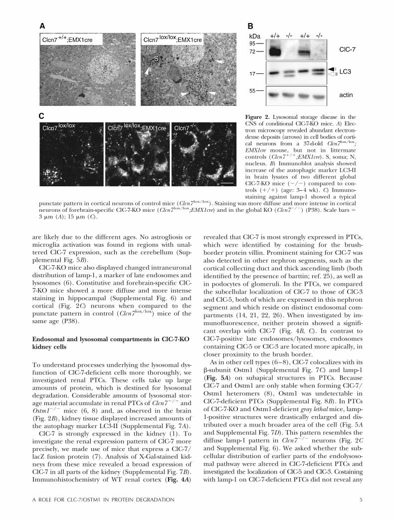
Constitutive ClC-7-KO mice show an accumulation ofelectron-dense, osmiophilic lysosomal storage materialin hippocampal and cortical neurons (6). These depos-its, which display typical features of NCL, becameapparent at 11 d after birth and increased with age (6).Electron microscopy on forebrain-specific ClC-7-KOmice revealed that prominent storage material wasdistributed throughout the somata of cortical neuronsat P37 (Fig. 2A) and at the age of 8 wk (data notshown). Immunoblotting brain lysates for LC3 showeda strong increase of the LC3-II form in constitutiveClC-7-KO mice (Fig. 2B), indicating an increase inautophagy (24).
A hallmark of NCL and of other neurodegenerativecentral nervous system (CNS) pathologies is astrogliosisand activation of microglia. Both features were ob-served in constitutive Clcn7�/� mice (6). We now showthat these pathological changes are restricted to re-gions displaying lysosomal storage and neurodegenera-tion (Fig. 3). In forebrain-specific ClC-7-KO mice,astrogliosis and microglia activation appeared at first inthe region of the hippocampus (Fig. 3A) from where itthen spread over the cortex (Fig. 3B and SupplementalFig. 5A, B). Immunofluorescence staining shows thatactivated, enlarged microglia express ClC-7 (Fig. 3B).Differences in the depicted microglia morphology be-tween forebrain-specific and complete ClC-7-KO mice
3A ROLE FOR CLC-7/OSTM1 IN PROTEIN DEGRADATION

Figure 1. Neurodegeneration in forebrain-specific ClC-7-KO mice. A) Cre expression in the EMX1-cre mice crossed with theZ/AP reporter strain. Cre-expressing cells were stained red due to the expression of alkaline phosphatase. Cre-negative cells werestained blue by the activity of �-galactosidase. B, C) Antibody detection of ClC-7 using diaminobenzidine on brain slices of 2-wk-oldcontrol mice (Clcn7�/�;EMX1cre) (B) and Clcn7 lox/lox;EMX1cre mice (C) showed deletion of ClC-7 in the cortex and hippocampusof forebrain-specific KO mice. D–I) Conspicuous loss of cortical and hippocampal neurons in whole-brain (D, G) or Nissl-stainedsagittal brain sections of 20-wk-old (E, H) and 1.5-yr-old Clcn7 lox/lox;EMX1cre mice (F, I). J–O) Hematoxylin-eosin staining on sagittalbrain sections in the hippocampal region in WT (J–L) and Clcn7 lox/lox;EMX1cre (M–O) mice. Neuronal loss in forebrain-specificClC-7-KO mice started in the hippocampal CA3 region (M, arrow) and later extended to the dentate gyrus (N, arrow). Nohippocampal structures could be detected in Clcn7 lox/lox;EMX1cre mice at 1.5 yr of age (O, asterisk). Scale bars � 2 mm (A–C, E, F,H, I); 200 �m (J–O).
4 Vol. 23 December 2009 WARTOSCH ET AL.The FASEB Journal � www.fasebj.org
are likely due to the different ages. No astrogliosis ormicroglia activation was found in regions with unal-tered ClC-7 expression, such as the cerebellum (Sup-plemental Fig. 5B).
ClC-7-KO mice also displayed changed intraneuronaldistribution of lamp-1, a marker of late endosomes andlysosomes (6). Constitutive and forebrain-specific ClC-7-KO mice showed a more diffuse and more intensestaining in hippocampal (Supplemental Fig. 6) andcortical (Fig. 2C) neurons when compared to thepunctate pattern in control (Clcn7 lox/lox) mice of thesame age (P38).
Endosomal and lysosomal compartments in ClC-7-KOkidney cells
To understand processes underlying the lysosomal dys-function of ClC-7-deficient cells more thoroughly, weinvestigated renal PTCs. These cells take up largeamounts of protein, which is destined for lysosomaldegradation. Considerable amounts of lysosomal stor-age material accumulate in renal PTCs of Clcn7�/� andOstm1�/� mice (6, 8) and, as observed in the brain(Fig. 2B), kidney tissue displayed increased amounts ofthe autophagy marker LC3-II (Supplemental Fig. 7A).
ClC-7 is strongly expressed in the kidney (1). Toinvestigate the renal expression pattern of ClC-7 moreprecisely, we made use of mice that express a ClC-7/lacZ fusion protein (7). Analysis of X-Gal-stained kid-neys from these mice revealed a broad expression ofClC-7 in all parts of the kidney (Supplemental Fig. 7B).Immunohistochemistry of WT renal cortex (Fig. 4A)
revealed that ClC-7 is most strongly expressed in PTCs,which were identified by costaining for the brush-border protein villin. Prominent staining for ClC-7 wasalso detected in other nephron segments, such as thecortical collecting duct and thick ascending limb (bothidentified by the presence of barttin; ref. 25), as well asin podocytes of glomeruli. In the PTCs, we comparedthe subcellular localization of ClC-7 to those of ClC-3and ClC-5, both of which are expressed in this nephronsegment and which reside on distinct endosomal com-partments (14, 21, 22, 26). When investigated by im-munofluorescence, neither protein showed a signifi-cant overlap with ClC-7 (Fig. 4B, C). In contrast toClC-7-positive late endosomes/lysosomes, endosomescontaining ClC-5 or ClC-3 are located more apically, incloser proximity to the brush border.
As in other cell types (6–8), ClC-7 colocalizes with its�-subunit Ostm1 (Supplemental Fig. 7C) and lamp-1(Fig. 5A) on subapical structures in PTCs. BecauseClC-7 and Ostm1 are only stable when forming ClC-7/Ostm1 heteromers (8), Ostm1 was undetectable inClC-7-deficient PTCs (Supplemental Fig. 8B). In PTCsof ClC-7-KO and Ostm1-deficient gray lethal mice, lamp-1-positive structures were drastically enlarged and dis-tributed over a much broader area of the cell (Fig. 5Aand Supplemental Fig. 7D). This pattern resembles thediffuse lamp-1 pattern in Clcn7�/� neurons (Fig. 2Cand Supplemental Fig. 6). We asked whether the sub-cellular distribution of earlier parts of the endolysoso-mal pathway were altered in ClC-7-deficient PTCs andinvestigated the localization of ClC-5 and ClC-3. Costainingwith lamp-1 on ClC-7-deficient PTCs did not reveal any
Figure 2. Lysosomal storage disease in theCNS of conditional ClC-7-KO mice. A) Elec-tron microscopy revealed abundant electron-dense deposits (arrows) in cell bodies of corti-cal neurons from a 37-d-old Clcn7 lox/lox;EMX1cre mouse, but not in littermatecontrols (Clcn7�/�;EMX1cre). S, soma; N,nucleus. B) Immunoblot analysis showedincrease of the autophagic marker LC3-IIin brain lysates of two different globalClC-7-KO mice (�/�) compared to con-trols (�/�) (age: 3–4 wk). C) Immuno-staining against lamp-1 showed a typical
punctate pattern in cortical neurons of control mice (Clcn7 lox/lox). Staining was more diffuse and more intense in corticalneurons of forebrain-specific ClC-7-KO mice (Clcn7 lox/lox;EMX1cre) and in the global KO (Clcn7�/�) (P38). Scale bars �3 �m (A); 15 �m (C).
5A ROLE FOR CLC-7/OSTM1 IN PROTEIN DEGRADATION

Figure 3. Activation of astrocytes and microgliosis in forebrain-specific ClC-7-KO mice. Brain sections stained with amicroglia-binding lectin (GSA) and with an antibody against GFAP (staining astrocytes) (A) or with antibodies against ClC-7 andthe microglia marker protein IBA1 (B). A) Massive activation of microglia and astrocytes in the hippocampus of 31-d-oldClcn7 lox/lox;EMX1cre mice. B) ClC-7 is expressed in cortical neurons (asterisks) of control (Clcn7�/�;EMX1cre) mice. Microgliaare activated in the cortex of 46-d-old Clcn7 lox/lox;EMX1cre mice and strongly express ClC-7. No ClC-7 signal was detected inactivated microglia of globally-deleted Clcn7�/� mice (age P31). Scale bars � 300 �m (A); 40 �m (B).
6 Vol. 23 December 2009 WARTOSCH ET AL.The FASEB Journal � www.fasebj.org

major changes in morphology and distribution of ei-ther ClC-5-positive (early) or ClC-3-positive (late) endo-somes (Fig. 5B, C). Neither did we find altered expres-sion levels of ClC-3, ClC-5, or lamp-1 in immunoblotanalysis of kidney tissue (Supplemental Fig. 7E).
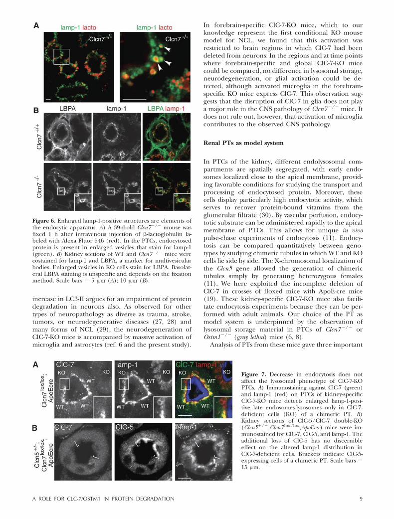
We next asked whether the enlarged lamp-1-positivestructures in ClC-7-KO PTCs are integral parts of theendocytic system. We performed in vivo pulse-chase ex-periments in which we injected intravenously the low-molecular-weight (18 kDa) protein �-lactoglobulin that isendocytosed from the primary urine by PTCs (11). At 1 hafter injection, fluorescently labeled �-lactoglobulinwas found in lumina of enlarged lamp-1-positive com-partments of ClC-7-deficient PTCs (Fig. 6A), demon-strating that these structures are connected to theendocytic system. The presence of LBPA on thesestructures (Fig. 6B) strongly suggests that these vesiclesshare features with multivesicular endosomes (23) ratherthan being solely of lysosomal nature.
Slower protein degradation in ClC-7-KO mice
To study the uptake and degradation of endocytosedproteins in WT and ClC-7-KO PTCs under identicalconditions, we investigated PTs that contain cells ex-pressing or lacking ClC-7 side by side. To this end, wecrossed our floxed ClC-7 mouse with mice that expressCre recombinase under a renal cortex-specific ApoEpromotor element, which, however, is not active inevery PTC (19).
We first examined lamp-1 expression in those chi-meric tubules. Only cells lacking ClC-7, but not neigh-boring WT cells, displayed enlarged lamp-1-positivestructures (Fig. 7A). Hence, the enlargement of lamp-1-positive structures is a cell-autonomous effect of ClC-7deletion. We next attempted to test whether this phe-notype might be rescued by a reduction in endocyticuptake. To this end, we crossed the kidney-specificClC-7-KO mice to ClC-5-KO mice (11), which exhibitdrastically reduced endocytosis in PTCs (11, 13). Lamp-1-positive structures appeared unaltered in PTCs lack-ing only ClC-5 (Supplemental Fig. 8C). No discernibleeffect of ClC-5 absence on the ClC-7-KO-induced al-tered distribution and size of lamp-1-positive structureswas observed in PTs that were chimeric for ClC-5 due torandom X-chromosomal inactivation (Fig. 7B).
After chasing endocytosed protein for 1 h into PTlysosomes of kidney-specific ClC-7-KO mice, KO cellsdisplayed much stronger fluorescence of labeled �-lac-toglobulin than neighboring WT cells of the sametubule (Fig. 8A and Supplemental Fig. 8A, B). Thestronger accumulation of �-lactoglobulin in ClC-7-KOcells might be explained either by increased endocyto-sis or by impaired protein degradation. To distinguishbetween these possibilities, we performed pulse-chaseendocytosis experiments. Fluorescence of labeled �-lac-toglobulin was quantified for PT sections expressing orlacking ClC-7 (Fig. 8A, B). At 10 min after injection,�-lactoglobulin fluorescence did not differ between WTand KO cells. Hence, ClC-7 disruption has no appre-
Figure 4. Expression of ClC-7 in the kidney.A) Staining with antibodies against ClC-7 (green),barttin (red), and villin (blue) revealed ClC-7expression in different cortical nephron seg-ments, such as proximal tubules (PT), corticalcollecting duct (CCD), and thick ascending limb(TAL), and in podocytes of the glomerulus(GL). B, C) A limited overlap exists betweenClC-7 and ClC-3 signals (B). The majority ofClC-7-positive late endosomes and lysosomes aredistinct from the more apical ClC-3-positive (B)and ClC-5-positive (C) compartments (endo-somes). Scale bars � 40 �m (A); 15 �m (B, C).
7A ROLE FOR CLC-7/OSTM1 IN PROTEIN DEGRADATION

ciable effect on apical endocytosis. At 20 min afterinjection, more �-lactoglobulin fluorescence was de-tected in Clcn7�/� than WT cells of the same tubule.The same was true for 30, 40, and 60 min and 3 h (Fig.8A, B), strongly suggesting defective degradation ofendocytosed protein in ClC-7-deficient cells. Althoughslowed down, lysosomal degradation was not abolishedcompletely by the loss of ClC-7, as is evident from thereduced fluorescence after 1 h of chase (Fig. 8A). At24 h after injection, labeled �-lactoglobulin was absentfrom both WT and ClC-7-KO cells (data not shown).
To exclude that the longer half-life of �-lactoglobulinfluorescence in cells lacking ClC-7 is only due to adifferent handling of the fluorescent moiety, e.g., owingto slowed-down exit of the dye over the lysosomalmembrane, we analyzed renal degradation of labeled�-lactoglobulin by SDS-PAGE (Fig. 8C). Because of theearly death of the global ClC-7-KO mice, we had tocompare control with kidney-specific ClC-7-KO mice,even though this comparison would underestimate anyeffect because ClC-7 is only partially deleted. Lysates ofchimeric ClC-7-KO and WT kidneys displayed a singlefluorescent band with the size of uninjected labeled�-lactoglobulin at 10 min after injection (Fig. 8C;arrowhead), indicating that no significant lysosomaldegradation of �-lactoglobulin had occurred yet. After30 min, an additional band (Fig. 8C; open arrowhead),most likely a tagged �-lactoglobulin degradation prod-uct, appeared in both KO and control lysates. Thisband was less intense in lysates from (partial) KOkidneys. At 3 h, signals of both the undegraded protein
and the degradation product were stronger in kidney-specific ClC-7-KO than in control mice, likely becausemost of the �-lactoglobulin had been degraded incontrol mice by this time. A similar protein degradationdefect was observed with injected albumin (data notshown) or with the fluid-phase endocytosis marker HRP(Supplemental Fig. 8D).
We next performed endocytosis/degradation experi-ments in chimeric ClC-5/ClC-7 double-deficient mice. At60 min after �-lactoglobulin injection, fluorescence inPTCs lacking only ClC-7 was much stronger than inClC-7/ClC-5 double-KO cells of the same tubule (Fig. 8D).
DISCUSSION
Progression of neurodegeneration and microgliaactivation
Like in the global ClC-7-KO, neurodegeneration inforebrain-specific ClC-7-KO mice began in the CA3region of the hippocampus. It proceeded much fur-ther, as these mice have a normal life span. By the ageof 1.5 yr, we found a nearly complete loss of neurons inthe areas where ClC-7 was disrupted, whereas regionsexpressing ClC-7 were unaffected. This finding suggeststhat lysosomal storage and neurodegeneration dependon the absence of ClC-7 in a cell-autonomous manner,as we have shown stringently for lysosomal storage inthe kidney. Although we could not study protein deg-radation in the brain as we did in the kidney, the
Figure 5. Massive enlargement of lamp-1- but not of ClC-3-and ClC-5-positive compartments in PTs of Clcn7�/� mice.Kidney sections of WT and Clcn7�/� mice were costainedfor the late endosomal/lysosomal marker lamp-1 withClC-7 (A), ClC-5 (B), or ClC-3 (C) as well as for the brushborder protein villin (A, B). A) In WT cells, ClC-7 colocal-ized with lamp-1. In Clcn7� /� PTCs, lamp-1-positive struc-tures were enlarged drastically and distributed broadly overthe cells. B) ClC-5 does not colocalize with lamp-1, neitherin WT nor in Clcn7 � /� PTCs. C) Only very little colocal-ization of ClC-3 and lamp-1 was observed in WT cells andon enlarged vesicles of KO PTCs. Different appearance ofenlarged lamp-1-positive structures is due to variationsbetween animals. Scales bars � 15 �m.
8 Vol. 23 December 2009 WARTOSCH ET AL.The FASEB Journal � www.fasebj.org
increase in LC3-II argues for an impairment of proteindegradation in neurons also. As observed for othertypes of neuropathology as diverse as trauma, stroke,tumors, or neurodegenerative diseases (27, 28) andmany forms of NCL (29), the neurodegeneration ofClC-7-KO mice is accompanied by massive activation ofmicroglia and astrocytes (ref. 6 and the present study).
In forebrain-specific ClC-7-KO mice, which to ourknowledge represent the first conditional KO mousemodel for NCL, we found that this activation wasrestricted to brain regions in which ClC-7 had beendeleted from neurons. In the regions and at time pointswhere forebrain-specific and global ClC-7-KO micecould be compared, no difference in lysosomal storage,neurodegeneration, or glial activation could be de-tected, although activated microglia in the forebrain-specific KO mice express ClC-7. This observation sug-gests that the disruption of ClC-7 in glia does not playa major role in the CNS pathology of Clcn7�/� mice. Itdoes not rule out, however, that activation of microgliacontributes to the observed CNS pathology.
Renal PTs as model system
In PTCs of the kidney, different endolysosomal com-partments are spatially segregated, with early endo-somes localized close to the apical membrane, provid-ing favorable conditions for studying the transport andprocessing of endocytosed protein. Moreover, thesecells display particularly high endocytotic activity, whichserves to recover protein-bound vitamins from theglomerular filtrate (30). By vascular perfusion, endocy-totic substrate can be administered rapidly to the apicalmembrane of PTCs. This allows for unique in vivopulse-chase experiments of endocytosis (11). Endocy-tosis can be compared quantitatively between geno-types by studying chimeric tubules in which WT and KOcells lie side by side. The X-chromosomal localization ofthe Clcn5 gene allowed the generation of chimerictubules simply by generating heterozygous females(11). We here exploited the incomplete deletion ofClC-7 in crosses of floxed mice with ApoE-cre mice(19). These kidney-specific ClC-7-KO mice also facili-tate endocytosis experiments because they can be per-formed with adult animals. Our choice of the PT asmodel system is underpinned by the observation oflysosomal storage material in PTCs of Clcn7�/� orOstm1�/� (gray lethal) mice (6, 8).
Analysis of PTs from these mice gave three important
Figure 6. Enlarged lamp-1-positive structures are elements ofthe endocytic apparatus. A) A 39-d-old Clcn7�/� mouse wasfixed 1 h after intravenous injection of �-lactoglobulin la-beled with Alexa Fluor 546 (red). In the PTCs, endocytosedprotein is present in enlarged vesicles that stain for lamp-1(green). B) Kidney sections of WT and Clcn7�/� mice werecostained for lamp-1 and LBPA, a marker for multivesicularbodies. Enlarged vesicles in KO cells stain for LBPA. Basolat-eral LBPA staining is unspecific and depends on the fixationmethod. Scale bars � 5 �m (A); 10 �m (B).
Figure 7. Decrease in endocytosis does notaffect the lysosomal phenotype of ClC-7-KOPTCs. A) Immunostaining against ClC-7 (green)and lamp-1 (red) on PTCs of kidney-specificClC-7-KO mice detects enlarged lamp-1-posi-tive late endosomes-lysosomes only in ClC-7-deficient cells (KO) of a chimeric PT. B)Kidney sections of ClC-5/ClC-7 double-KO(Clcn5�/�;Clcn7 lox/lox;ApoEcre) mice were im-munostained for ClC-7, ClC-5, and lamp-1. Theadditional loss of ClC-5 has no discernibleeffect on the altered lamp-1 distribution inClC-7-deficient cells. Brackets indicate ClC-5-expressing cells of a chimeric PT. Scale bars �15 �m.
9A ROLE FOR CLC-7/OSTM1 IN PROTEIN DEGRADATION
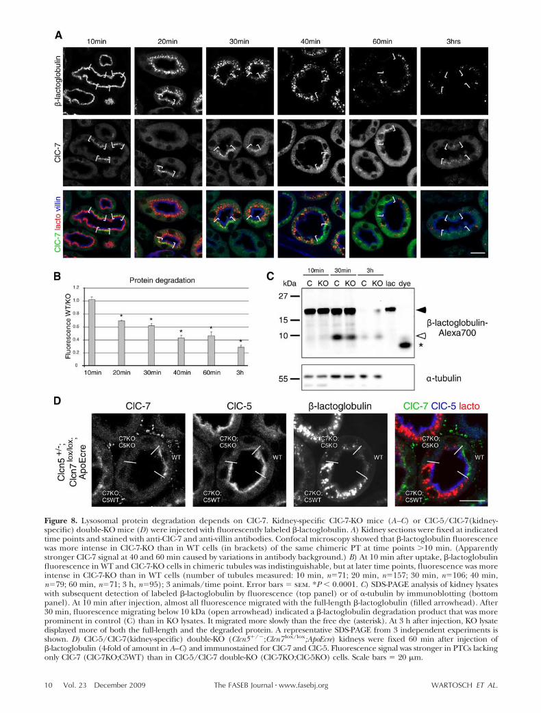
Figure 8. Lysosomal protein degradation depends on ClC-7. Kidney-specific ClC-7-KO mice (A–C) or ClC-5/ClC-7(kidney-specific) double-KO mice (D) were injected with fluorescently labeled �-lactoglobulin. A) Kidney sections were fixed at indicatedtime points and stained with anti-ClC-7 and anti-villin antibodies. Confocal microscopy showed that �-lactoglobulin fluorescencewas more intense in ClC-7-KO than in WT cells (in brackets) of the same chimeric PT at time points 10 min. (Apparentlystronger ClC-7 signal at 40 and 60 min caused by variations in antibody background.) B) At 10 min after uptake, �-lactoglobulinfluorescence in WT and ClC-7-KO cells in chimeric tubules was indistinguishable, but at later time points, fluorescence was moreintense in ClC-7-KO than in WT cells (number of tubules measured: 10 min, n�71; 20 min, n�157; 30 min, n�106; 40 min,n�79; 60 min, n�71; 3 h, n�95); 3 animals/time point. Error bars � sem. *P 0.0001. C) SDS-PAGE analysis of kidney lysateswith subsequent detection of labeled �-lactoglobulin by fluorescence (top panel) or of �-tubulin by immunoblotting (bottompanel). At 10 min after injection, almost all fluorescence migrated with the full-length �-lactoglobulin (filled arrowhead). After30 min, fluorescence migrating below 10 kDa (open arrowhead) indicated a �-lactoglobulin degradation product that was moreprominent in control (C) than in KO lysates. It migrated more slowly than the free dye (asterisk). At 3 h after injection, KO lysatedisplayed more of both the full-length and the degraded protein. A representative SDS-PAGE from 3 independent experiments isshown. D) ClC-5/ClC-7(kidney-specific) double-KO (Clcn5�/�;Clcn7 lox/lox;ApoEcre) kidneys were fixed 60 min after injection of�-lactoglobulin (4-fold of amount in A–C) and immunostained for ClC-7 and ClC-5. Fluorescence signal was stronger in PTCs lackingonly ClC-7 (ClC-7KO;C5WT) than in ClC-5/ClC-7 double-KO (ClC-7KO;ClC-5KO) cells. Scale bars � 20 �m.
10 Vol. 23 December 2009 WARTOSCH ET AL.The FASEB Journal � www.fasebj.org

results: 1) The accumulation of storage material andthe generation of abnormally large vesicular structuresare cell-intrinsic, occurring only in cells lacking ClC-7;2) apical endocytotic uptake of protein does not de-pend appreciably on ClC-7; and 3) ClC-7 disruptiondrastically slows, but does not abolish, proteolysis ofendocytosed protein.
A possibly direct effect of ion composition on proteindegradation
Lysosomal protein degradation depends on the activityof acidic hydrolases. The loss of certain lysosomalenzyme activities underlies several different forms oflysosomal storage disease (31). In analogy to the role ofClC-3 and ClC-5 in acidifying endosomes (21, 32) andof ClC-7 in acidifying the resorption lacuna of oste-oclasts (7), ClC-7 is believed to help in late endosomal/lysosomal acidification by shunting electric currents ofthe H�-ATPase (2, 7). Thus, slower protein degrada-tion in ClC-7-KO cells might reflect a lowered activity ofacidic hydrolases caused by an elevated luminal pH.During the transport to lysosomes, prelysosomal com-partments might be acidified at a slower rate. Steady-state lysosomal pH of cultured neurons or fibroblastsderived from WT, Clcn7�/�, or Ostm1�/� mice, how-ever, was undistinguishable by ratiometric fluorescencemeasurement (6, 8). By contrast, a recent report as-serted that lysosomal pH of HeLa cells became morealkaline when ClC-7 was partially knocked down (9).However, that study used only single antisense andcontrol siRNAs to manipulate ClC-7 levels. Disconcert-ingly, lysosomal pH reportedly was more acidic with thenegative control than in untreated cells. Moreover, thatstudy used nonratiometric measurements of total lyso-tracker fluorescence, which is not only a function ofvesicular pH, but also of the number and volumes ofvesicles. These latter parameters were not determined (9).
In addition to luminal pH, lysosomal chloride con-centration might influence lysosomal protein degrada-tion (2, 33, 34). For instance, there might be (so farunknown) chloride-coupled transporters for degrada-tion products in lysosomal membranes. Chloride mayalso influence the activity of degradation enzymes, asshown for the lysosomal protease cathepsin C (33).Like other vesicular CLCs (4, 5), ClC-7 has beensuggested to function as a Cl�/H� exchanger (2, 9),with a likely stoichiometry of 2Cl�/1H�. The largeinside-out H� gradient across lysosomal membranespredicts a steady-state lysosomal Cl� accumulation thatwould be reduced in ClC-7-KO cells even in the pres-ence of unchanged lysosomal pH. Likewise, AtClC-aaccumulates nitrate, an important plant nutrient, invacuoles of Arabidopsis thaliana (35). Impaired proteindegradation, whether owed to changed chloride or pHin (or on the way to) lysosomes, might lead to theobserved enlarged lamp1-positive structures.
Trafficking defect upon ClC-7 deficiency
To investigate whether the generation of large lamp-1-positive vesicles in Clcn7�/� cells depends on theaccumulation of undegraded endocytic cargo, we stud-ied PTCs lacking both ClC-7 and ClC-5. Despite thestrong impairment of endocytosis caused by ClC-5disruption (11, 13, 36), similar enlarged vesicles wereobserved in double-KO cells. ClC-5, however, is not onlycrucial for the lysosomal delivery of substrate for deg-radation, but also of lysosomal degradative enzymes(37). Therefore, the additional loss of ClC-5 may fur-ther impair lysosomal degradation in Clcn7�/� cells,rendering predictions on cargo accumulation difficult.Our in vivo endocytosis/degradation assay shows thatafter a 1 h chase, ClC-5/ClC-7 double-KO cells con-tained much less tagged protein than Clcn7�/� cells,with fluorescence levels being similar to WT. We con-clude that the accumulation of undegraded cargo isunlikely to be a major factor in the generation of thoseabnormal vesicles.
Although endocytosed protein reached those lamp-1-positive structures after a 1-h chase, those enlargedvesicles do not necessarily represent lysosomes, sincelamp-1 is also present on late endosomes. Indeed, thosevesicles were also positive for LBPA, a lipid enriched inmultivesicular endosomes (23). This observation sug-gests a vesicular trafficking defect, which could alsoaffect the delivery of degradative enzymes. PTCs may beparticularly vulnerable in this respect, because of aprominent role of apical endocytosis in deliveringcertain proteolytic enzymes to lysosomes (37). Thisdifference in lysosomal enzyme trafficking might ex-plain the apparent discrepancy between the presentobservation of impaired proteolysis and our previousfinding that the activity of the lysosomal enzyme TPP Iwas normal in cultured Clcn7�/� neurons and fibro-blasts (6). Alternatively, TPP I plays no important rolein the degradative pathways observed here.
A vesicular trafficking defect might also resolve theconundrum that the KO of ClC-7 abolished the acidi-fication of the osteoclast resorption lacuna (7) but didnot change lysosomal steady-state pH (6, 8). Indeed,the acid-secreting ruffled border membrane of oste-oclasts, which is formed by the exocytotic insertion oflysosomal membranes, was underdeveloped in Clcn7�/�
mice (7). Similar to the role of ClC-5 in renalendocytosis (11, 36), the loss of ClC-7 might affectintracellular trafficking by a reduced acidificationrate of late endosomes. The recruitment of somecomponents of the vesicular transport machinery ispH-sensitive (38 – 40), and transport between variouscompartments of the endocytic pathway depends onvesicular H�-ATPase activity (41, 42). So far, no roleof chloride in membrane trafficking is known apartfrom its indirect role in supporting vesicular acidifi-cation. However, novel roles of vesicular chloride areemerging. For instance, a recent report described theregulation of an early endosomal Ca2� channel byluminal [Cl�] (43). Some evidence indicates that the
11A ROLE FOR CLC-7/OSTM1 IN PROTEIN DEGRADATION

release of vesicular Ca2� affects vesicle fusion andtrafficking (44).
Relationship to other CLCs
The existence of 5 distinct vesicular CLC proteins in theendosomal-lysosomal pathway (ClC-3 through ClC-7) raisesthe possibility of a partial compensation of the loss ofClC-7 by other CLCs. Although the localization ofdifferent vesicular CLC transporters has been studiedby immunocytochemistry in transfected cells (26) andby subcellular fractionation of native tissues (6), nocostaining of different endogenous intracellular CLCproteins has been available. Our results show that themajority of ClC-5- or ClC-3-positive endosomes is dis-tinct from ClC-7-positive late endosomes/lysosomes,agreeing with their localization to earlier endocytoticcompartments (2). We have observed previously that aproportion of ClC-3 and ClC-6 moved toward denser,lysosome-containing fractions in brain lysates of ClC-7-KO mice (45). We have found in this work that theexpression level and distribution of ClC-5 was un-changed by the loss of ClC-7 and that enlarged lamp-1-positive late endosomes/lysosomes of ClC-7-KO PTCsdid not stain for ClC-5. Both in WT and in ClC-7-KOmice, we found a minor overlap of ClC-3 staining withlamp-1, as shown previously on “normal” lamp-1-posi-tive structures in transfected cells (14). The localizationof ClC-3, which showed unaltered expression levels,seemed to be largely unchanged by ClC-7 disruption.The only other late endosomal CLC protein, ClC-6, isnot expressed in the kidney (45). Therefore, a majorcompensatory effect of ClC-5 or ClC-3 on lysosomalprotein degradation in ClC-7-KO PTCs can be ex-cluded.
CONCLUSIONS
Using a novel floxed ClC-7 mouse model, we havedemonstrated for the first time that the lysosomalchloride transporter ClC-7/Ostm1 is important for thedegradation of endocytosed protein, at least in thekidney. The failure to degrade endocytosed proteinsadequately may lead to the lysosomal storage diseaseobserved in mice and humans lacking this chloridetransport activity.
We thank the following colleagues for generously makingmaterial available: Takuji Iwasato and Shigeyoshi Itohara (BrainScience Institute, RIKEN, Saitama, Japan) for EMX1-cre mice,Thomas Willnow (MDC, Berlin, Germany) for ApoE-cre mice,and Jean Gruenberg (University of Geneva, Geneva, Switzer-land) for the LBPA antibody. We thank Sebastian Bachmann forhelpful discussions; Irm Hermans-Borgmeyer and Tina Koppel-mann for help in generating Clcn7 lox/lox mice; Nicole Kronke,Patrick Seidler, and Stephanie Wernick for technical assistance;York Rudhard for EMX1-Z/AP reporter staining; and GunterDelling for help with X-ray analysis. This work was supported bygrants of the Deutsche Forschungsgemeinschaft and the Euro-pean Union (Euregene) to T.J.J. and a Boehringer Ingelheimfellowship to L.W.
REFERENCES
1. Brandt, S., and Jentsch, T. J. (1995) ClC-6 and ClC-7 are twonovel broadly expressed members of the CLC chloride channelfamily. FEBS Lett. 377, 15–20
2. Jentsch, T. J. (2007) Chloride and the endosomal-lysosomalpathway: emerging roles of CLC chloride transporters. J. Physiol.578, 633–640
3. Accardi, A., and Miller, C. (2004) Secondary active transportmediated by a prokaryotic homologue of ClC Cl- channels.Nature 427, 803–807
4. Picollo, A., and Pusch, M. (2005) Chloride/proton antiporteractivity of mammalian CLC proteins ClC-4 and ClC-5. Nature436, 420–423
5. Scheel, O., Zdebik, A. A., Lourdel, S., and Jentsch, T. J. (2005)Voltage-dependent electrogenic chloride/proton exchange byendosomal CLC proteins. Nature 436, 424–427
6. Kasper, D., Planells-Cases, R., Fuhrmann, J. C., Scheel, O., Zeitz,O., Ruether, K., Schmitt, A., Poet, M., Steinfeld, R., Schweizer,M., Kornak, U., and Jentsch, T. J. (2005) Loss of the chloridechannel ClC-7 leads to lysosomal storage disease and neurode-generation. EMBO J. 24, 1079–1091
7. Kornak, U., Kasper, D., Bosl, M. R., Kaiser, E., Schweizer, M.,Schulz, A., Friedrich, W., Delling, G., and Jentsch, T. J. (2001)Loss of the ClC-7 chloride channel leads to osteopetrosis in miceand man. Cell 104, 205–215
8. Lange, P. F., Wartosch, L., Jentsch, T. J., and Fuhrmann, J. C.(2006) ClC-7 requires Ostm1 as a beta-subunit to support boneresorption and lysosomal function. Nature 440, 220–223
9. Graves, A. R., Curran, P. K., Smith, C. L., and Mindell, J. A.(2008) The Cl-/H� antiporter ClC-7 is the primary chloridepermeation pathway in lysosomes. Nature 453, 788–792
10. Mellman, I., Fuchs, R., and Helenius, A. (1986) Acidification ofthe endocytic and exocytic pathways. Annu. Rev. Biochem. 55,663–700
11. Piwon, N., Gunther, W., Schwake, M., Bosl, M. R., and Jentsch,T. J. (2000) ClC-5 Cl- -channel disruption impairs endocytosis ina mouse model for Dent’s disease. Nature 408, 369–373
12. Lloyd, S. E., Pearce, S. H., Fisher, S. E., Steinmeyer, K., Schwap-pach, B., Scheinman, S. J., Harding, B., Bolino, A., Devoto, M.,Goodyer, P., Rigden, S. P., Wrong, O., Jentsch, T. J., Craig, I. W.,and Thakker, R. V. (1996) A common molecular basis for threeinherited kidney stone diseases. Nature 379, 445–449
13. Wang, S. S., Devuyst, O., Courtoy, P. J., Wang, X. T., Wang, H.,Wang, Y., Thakker, R. V., Guggino, S., and Guggino, W. B.(2000) Mice lacking renal chloride channel, CLC-5, are a modelfor Dent’s disease, a nephrolithiasis disorder associated withdefective receptor-mediated endocytosis. Hum. Mol. Genet. 9,2937–2945
14. Stobrawa, S. M., Breiderhoff, T., Takamori, S., Engel, D.,Schweizer, M., Zdebik, A. A., Bosl, M. R., Ruether, K., Jahn, H.,Draguhn, A., Jahn, R., and Jentsch, T. J. (2001) Disruption ofClC-3, a chloride channel expressed on synaptic vesicles, leadsto a loss of the hippocampus. Neuron 29, 185–196
15. Chalhoub, N., Benachenhou, N., Rajapurohitam, V., Pata, M.,Ferron, M., Frattini, A., Villa, A., and Vacher, J. (2003)Grey-lethal mutation induces severe malignant autosomalrecessive osteopetrosis in mouse and human. Nat. Med. 9,399 – 406
16. Frattini, A., Pangrazio, A., Susani, L., Sobacchi, C., Mirolo, M.,Abinun, M., Andolina, M., Flanagan, A., Horwitz, E. M., Mihci,E., Notarangelo, L. D., Ramenghi, U., Teti, A., Van Hove, J.,Vujic, D., Young, T., Albertini, A., Orchard, P. J., Vezzoni, P.,and Villa, A. (2003) Chloride channel ClCN7 mutations areresponsible for severe recessive, dominant, and intermediateosteopetrosis. J. Bone Miner. Res. 18, 1740–1747
17. Pangrazio, A., Poliani, P. L., Megarbane, A., Lefranc, G., Lanino,E., Di Rocco, M., Rucci, F., Lucchini, F., Ravanini, M., Facchetti,F., Abinun, M., Vezzoni, P., Villa, A., and Frattini, A. (2006)Mutations in OSTM1 (grey lethal) define a particularly severeform of autosomal recessive osteopetrosis with neural involve-ment. J. Bone Miner. Res. 21, 1098–1105
18. Iwasato, T., Datwani, A., Wolf, A. M., Nishiyama, H., Taguchi, Y.,Tonegawa, S., Knopfel, T., Erzurumlu, R. S., and Itohara, S.(2000) Cortex-restricted disruption of NMDAR1 impairs neuro-nal patterns in the barrel cortex. Nature 406, 726–731
12 Vol. 23 December 2009 WARTOSCH ET AL.The FASEB Journal � www.fasebj.org

19. Leheste, J. R., Melsen, F., Wellner, M., Jansen, P., Schlichting,U., Renner-Muller, I., Andreassen, T. T., Wolf, E., Bachmann,S., Nykjaer, A., and Willnow, T. E. (2003) Hypocalcemia andosteopathy in mice with kidney-specific megalin gene defect.FASEB J. 17, 247–249
20. Schwenk, F., Baron, U., and Rajewsky, K. (1995) A Cre-trans-genic mouse strain for the ubiquitous deletion of loxP-flankedgene segments including deletion in germ cells. Nucl. Acids Res.23, 5080–5081
21. Gunther, W., Luchow, A., Cluzeaud, F., Vandewalle, A., andJentsch, T. J. (1998) ClC-5, the chloride channel mutated inDent’s disease, colocalizes with the proton pump in endocytoti-cally active kidney cells. Proc. Natl. Acad. Sci. U. S. A. 95, 8075–8080
22. Maritzen, T., Keating, D. J., Neagoe, I., Zdebik, A. A., andJentsch, T. J. (2008) Role of the vesicular chloride transporterClC-3 in neuroendocrine tissue. J. Neurosci. 28, 10587–10598
23. Kobayashi, T., Stang, E., Fang, K. S., de Moerloose, P., Parton,R. G., and Gruenberg, J. (1998) A lipid associated with theantiphospholipid syndrome regulates endosome structure andfunction. Nature 392, 193–197
24. Rubinsztein, D. C., Cuervo, A. M., Ravikumar, B., Sarkar, S.,Korolchuk, V., Kaushik, S., and Klionsky, D. J. (2009) In searchof an “autophagomometer.” Autophagy 5, 585–589
25. Estevez, R., Boettger, T., Stein, V., Birkenhager, R., Otto, E.,Hildebrandt, F., and Jentsch, T. J. (2001) Barttin is a Cl- channelbeta-subunit crucial for renal Cl- reabsorption and inner ear K�secretion. Nature 414, 558–561
26. Suzuki, T., Rai, T., Hayama, A., Sohara, E., Suda, S., Itoh, T.,Sasaki, S., and Uchida, S. (2006) Intracellular localization of ClCchloride channels and their ability to form hetero-oligomers.J. Cell. Physiol. 206, 792–798
27. Kreutzberg, G. W. (1996) Microglia: a sensor for pathologicalevents in the CNS. Trends Neurosci. 19, 312–318
28. Pekny, M., and Nilsson, M. (2005) Astrocyte activation andreactive gliosis. Glia 50, 427–434
29. Cooper, J. D., Russell, C., and Mitchison, H. M. (2006) Progresstowards understanding disease mechanisms in small vertebratemodels of neuronal ceroid lipofuscinosis. Biochim. Biophys. Acta.1762, 873–889
30. Christensen, E. I., and Birn, H. (2002) Megalin and cubilin:multifunctional endocytic receptors. Nat. Rev. Mol. Cell. Biol. 3,256–266
31. Gieselmann, V. (1995) Lysosomal storage diseases. Biochim.Biophys. Acta. 1270, 103–136
32. Hara-Chikuma, M., Yang, B., Sonawane, N. D., Sasaki, S.,Uchida, S., and Verkman, A. S. (2005) ClC-3 chloride channelsfacilitate endosomal acidification and chloride accumulation.J. Biol. Chem. 280, 1241–1247
33. Cigic, B., and Pain, R. H. (1999) Location of the binding site forchloride ion activation of cathepsin C. Eur. J. Biochem. 264,944–951
34. Faundez, V., and Hartzell, H. C. (2004) Intracellular chloridechannels: determinants of function in the endosomal pathway.Sci. STKE 2004, re8
35. De Angeli, A., Monachello, D., Ephritikhine, G., Frachisse, J. M.,Thomine, S., Gambale, F., and Barbier-Brygoo, H. (2006) Thenitrate/proton antiporter AtCLCa mediates nitrate accumula-tion in plant vacuoles. Nature 442, 939–942
36. Gunther, W., Piwon, N., and Jentsch, T. J. (2003) The ClC-5chloride channel knock-out mouse—an animal model forDent’s disease. Pflugers Arch. 445, 456–462
37. Nielsen, R., Courtoy, P. J., Jacobsen, C., Dom, G., Lima, W. R.,Jadot, M., Willnow, T. E., Devuyst, O., and Christensen, E. I.(2007) Endocytosis provides a major alternative pathway forlysosomal biogenesis in kidney proximal tubular cells. Proc. Natl.Acad. Sci. U. S. A. 104, 5407–5412
38. Aniento, F., Gu, F., Parton, R. G., and Gruenberg, J. (1996) Anendosomal beta COP is involved in the pH-dependent forma-tion of transport vesicles destined for late endosomes. J. Cell Biol.133, 29–41
39. Zeuzem, S., Feick, P., Zimmermann, P., Haase, W., Kahn, R. A.,and Schulz, I. (1992) Intravesicular acidification correlates withbinding of ADP-ribosylation factor to microsomal membranes.Proc. Natl. Acad. Sci. U. S. A. 89, 6619–6623
40. Maranda, B., Brown, D., Bourgoin, S., Casanova, J. E., Vinay, P.,Ausiello, D. A., and Marshansky, V. (2001) Intra-endosomalpH-sensitive recruitment of the Arf-nucleotide exchange factorARNO and Arf6 from cytoplasm to proximal tubule endosomes.J. Biol. Chem. 276, 18540–18550
41. Clague, M. J., Urbe, S., Aniento, F., and Gruenberg, J. (1994)Vacuolar ATPase activity is required for endosomal carriervesicle formation. J. Biol. Chem. 269, 21–24
42. Van Weert, A. W., Dunn, K. W., Gueze, H. J., Maxfield, F. R., andStoorvogel, W. (1995) Transport from late endosomes to lyso-somes, but not sorting of integral membrane proteins in endo-somes, depends on the vacuolar proton pump. J. Cell Biol. 130,821–834
43. Saito, M., Hanson, P. I., and Schlesinger, P. (2007) Luminalchloride-dependent activation of endosome calcium channels:patch clamp study of enlarged endosomes. J. Biol. Chem. 282,27327–27333
44. Luzio, J. P., Bright, N. A., and Pryor, P. R. (2007) The role ofcalcium and other ions in sorting and delivery in the lateendocytic pathway. Biochem. Soc. Trans. 35, 1088–1091
45. Poet, M., Kornak, U., Schweizer, M., Zdebik, A. A., Scheel, O.,Hoelter, S., Wurst, W., Schmitt, A., Fuhrmann, J. C., Planells-Cases, R., Mole, S. E., Hubner, C. A., and Jentsch, T. J. (2006)Lysosomal storage disease upon disruption of the neuronalchloride transport protein ClC-6. Proc. Natl. Acad. Sci. U. S. A.103, 13854–13859
Received for publication January 30, 2009.Accepted for publication July 16, 2009.
13A ROLE FOR CLC-7/OSTM1 IN PROTEIN DEGRADATION