Embed Size (px)
Citation preview

R E S E A R CH A R T I C L E
Protective role of cortistatin in pulmonary inflammation andfibrosis
Margarita Barriga1 | Raquel Benitez1 | Viviane Ferraz-de-Paula1,2 |
Marina Garcia-Frutos1 | Marta Caro1 | Gema Robledo1 | Francisco O'Valle3 |
Jenny Campos-Salinas1 | Mario Delgado1
1Department of Immunology and Cell Biology,
Institute of Parasitology and Biomedicine
Lopez-Neyra IPBLN-CSIC, Granada, Spain
2Department of Clinical and Toxicological
Analysis, School of Pharmaceutical Sciences,
University of S~ao Paulo, S~ao Paulo, Brazil
3Pathology Department, School of Medicine,
IBIMER, CIBM, University of Granada and
Biosanitary Research Institute IBS-Granada,
Granada, Spain
Correspondence
Mario Delgado, Department of Immunology
and Cell Biology, Institute of Parasitology and
Biomedicine Lopez-Neyra IPBLN-CSIC, PT
Salud, Granada, Spain.
Email: [email protected]
Funding information
Fundaç~ao de Amparo à Pesquisa do Estado de
S~ao Paulo, Grant/Award Number:
12/21767-5; Ministerio de Ciencia e
Innovaci�on, Grant/Award Number:
SAF2015-67787-R
Background and Purpose: Acute lung injury (ALI), acute respiratory distress syn-
drome (ARDS) and pulmonary fibrosis remain major causes of morbidity, mortality
and a healthcare burden in critically ill patient. There is an urgent need to identify
factors causing susceptibility and for the design of new therapeutic agents. Here, we
evaluate the effectiveness of the immunomodulatory neuropeptide cortistatin to
regulate pulmonary inflammation and fibrosis in vivo.
Experimental Approach: ALI/ARDS and pulmonary fibrosis were induced experimen-
tally in wild-type and cortistatin-deficient mice by pulmonary infusion of the bacterial
endotoxin LPS or the chemotherapeutic drug bleomycin, and the histopathological
signs, pulmonary leukocyte infiltration and cytokines, and fibrotic markers were
evaluated.
Key Results: Partially deficient mice in cortistatin showed exacerbated pulmonary
damage, pulmonary inflammation, alveolar oedema and fibrosis, and subsequent
increased respiratory failure and mortality when challenged to LPS or bleomycin,
even at low doses. Treatment with cortistatin reversed these aggravated phenotypes
and protected from progression to severe ARDS and fibrosis, after high exposure to
both injury agents. Moreover, cortistatin-deficient pulmonary macrophages and
fibroblasts showed exaggerated ex vivo inflammatory and fibrotic responses, and
treatment with cortistatin impaired their activation. Finally, the protective effects of
cortistatin in ALI and pulmonary fibrosis were partially inhibited by specific antago-
nists for somatostatin and ghrelin receptors.
Conclusion and Implications: We identified cortistatin as an endogenous inhibitor of
pulmonary inflammation and fibrosis. Deficiency in cortistatin could be a marker of
poor prognosis in inflammatory/fibrotic pulmonary disorders. Cortistatin-based ther-
apies could emerge as attractive candidates to treat severe ALI/ARDS, including
SARS-CoV-2-associated ARDS.
Abbreviations: ALI, acute lung injury; ARDS, acute respiratory distress syndrome; BALF, bronchoalveolar lavage fluid; CST, cortistatin; Cort, cortistatin gene; CTGF, connective tissue growth
factor; [D-Lys3]-GHRP-6, [D-Lys3]-growth hormone-releasing peptide 6; i.n., intranasally; IPF, idiopathic pulmonary fibrosis; CXCL2/MIP-2, macrophage inflammatory protein-2; SARS-CoV-2,
severe acute respiratory syndrome coronavirus 2; SST, somatostatin receptor; αSMA, α-smooth muscle actin.
Received: 17 February 2021 Revised: 3 June 2021 Accepted: 24 June 2021
DOI: 10.1111/bph.15615
This is an open access article under the terms of the Creative Commons Attribution License, which permits use, distribution and reproduction in any medium,
provided the original work is properly cited.
© 2021 The Authors. British Journal of Pharmacology published by John Wiley & Sons Ltd on behalf of British Pharmacological Society.
Br J Pharmacol. 2021;1–21. wileyonlinelibrary.com/journal/bph 1
K E YWORD S
acute lung injury, fibroblasts, macrophages, neuropeptide, pulmonary inflammation
1 | INTRODUCTION
Despite major treatment efforts made over the past decades, acute
lung injury (ALI) and its most severe form, acute respiratory distress
syndrome (ARDS), characterized by refractory hypoxia, severe inflam-
mation, increased vascular permeability and diffuse alveolar damage,
remains a major cause of morbidity and mortality in critically ill
patients (Matthay et al., 2017). ARDS can occur as a result of different
clinical conditions, such as infections, pulmonary contusion and inhala-
tion injury, that directly damage the pulmonary epithelial and endo-
thelial cells and compromise alveolar–capillary barrier. ARDS is caused
and sustained by an uncontrolled inflammatory activation character-
ized by massive release of cytokines and chemokines, diffuse lung
oedema, inflammatory cell infiltration and disseminated coagulation.
In this sense, evidence indicates that the massive pulmonary infiltra-
tion (neutrophils and macrophages) and the subsequent inflammatory
cytokine storm are closely related to secondary complications such as
lung injury/ARDS, multiorgan failure and ultimately poor prognosis
in the new severe acute respiratory syndrome coronavirus
2 (SARS-CoV-2) pandemic (Mehta et al., 2020; Zhou et al., 2020).
Moreover, in patients who develop ARDS, the progression of ALI to
pulmonary fibrosis portends a fatal outcome, with severe disruption
of lung function and elevated mortality (George et al., 2020). As in
other cases of pulmonary fibrosis which are caused by persistent
infection, oxidative stress and inflammatory insults, the injury to alve-
olar epithelial cells activates pulmonary fibroblasts, promoting their
transformation to extracellular matrix-producing myofibroblasts
(Wynn, 2011). These findings highlight the urgent need to develop
safe and effective therapeutic agents with capacity to limit both
inflammatory and fibrotic responses in injured lung. Moreover, due to
the heterogeneous progression and severity of disease in patients
with ALI/ARDS, it is critical to identify factors and genes that predis-
pose/protect from the development of the most severe forms of lung
injury and progressive lung fibrosis.
Cortistatin-14 (CST-14) is a cyclic neuropeptide belonging to the
somatostatin family that has emerged as a potent immunomodulatory
agent (Gonzalez-Rey & Delgado, 2008) with capacity to protect
against exacerbated inflammatory and autoimmune responses in
various experimental models of sepsis, rheumatoid arthritis, colitis,
myocarditis and multiple sclerosis (Delgado-Maroto et al., 2017;
Gonzalez-Rey, Chorny, Del Moral, et al., 2007; Gonzalez-Rey, Chorny,
Robledo, & Delgado, 2006; Gonzalez-Rey, Varela, Sheibanie,
et al., 2006; Souza-Moreira et al., 2013). These effects are exerted
through the regulation of a plethora of inflammatory cytokines and
chemokines, and by deactivating macrophages and lymphocytes, indi-
cating that it is a multitargeted and safe modulator of the cytokine
storm in various tissues. However, the endogenous role of cortistatin
in the modulation of immune response has been scarcely investigated,
with some paradoxical effects found in cortistatin-deficient animals
(Qiu et al., 2020; Souza-Moreira et al., 2013). Moreover, its role in ALI
and fibrotic disorders is completely unknown, although some data
point out to a potential antifibrotic action of this neuropeptide. Thus,
cortistatin receptors (somatostatin (SST)5 receptors and ghrelin
receptor GHSR) are expressed in fibroblasts, and other SST receptor
agonists have been described that exert antifibrotic responses in vari-
ous tissues, including lung (Borie et al., 2008; Egger et al., 2014; Tug
et al., 2013). Therefore, cortistatin could converge immunomodulatory
and antifibrotic properties that synergistically might contribute to
ameliorate inflammatory and fibroproliferative disorders in the lung. In
this study, we will evaluate the therapeutic potential of cortistatin
in two well-established experimental models of ALI and pulmonary
fibrosis, as well as the immune and antifibrotic mechanisms involved.
We will also investigate the role of cortistatin as a potential endoge-
nous protective factor in the progression to severe ALI and pulmonary
fibrosis in mice that are partially or totally deficient in this
neuropeptide.
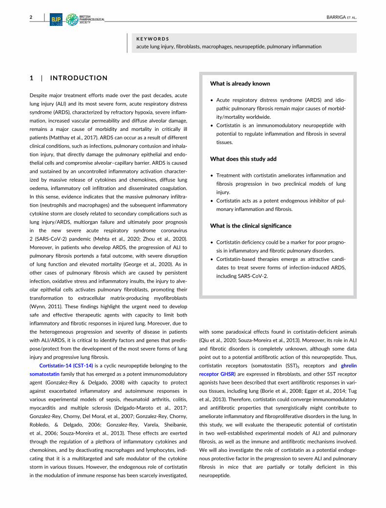
What is already known
• Acute respiratory distress syndrome (ARDS) and idio-
pathic pulmonary fibrosis remain major causes of morbid-
ity/mortality worldwide.
• Cortistatin is an immunomodulatory neuropeptide with
potential to regulate inflammation and fibrosis in several
tissues.
What does this study add
• Treatment with cortistatin ameliorates inflammation and
fibrosis progression in two preclinical models of lung
injury.
• Cortistatin acts as a potent endogenous inhibitor of pul-
monary inflammation and fibrosis.
What is the clinical significance
• Cortistatin deficiency could be a marker for poor progno-
sis in inflammatory and fibrotic pulmonary disorders.
• Cortistatin-based therapies emerge as attractive candi-
dates to treat severe forms of infection-induced ARDS,
including SARS-CoV-2.
2 BARRIGA ET AL.

2 | METHODS
2.1 | Animals and ethic statement
The experiments reported in this study followed the ethical guidelines
for investigations of experimental animals approved by the Animal
Care and Use Board and the Ethical Committee of Spanish Council of
Scientific Research (Number 388/2015) and performed in accordance
with the guidelines from Directive 2010/63/EU of the European Par-
liament on the protection of animals used for scientific purposes. Ani-
mal studies are reported in compliance with the ARRIVE guidelines
(Percie du Sert et al., 2020) and with the recommendations made by
the British Journal of Pharmacology (Lilley et al., 2020). Mice lacking
the gene for cortistatin (Cort�/�) were a generous gift of Dr Luis de
Lecea (Stanford University, La Jolla, CA, USA) and were generated in a
C57BL/6 background and backcrossed with C57BL/6 mice for 10 gen-
erations as previously described (Cordoba-Chacon et al., 2011). Mice
heterozygous (Cort+/�) for cortistatin were generated by crossing
female Cort�/� and male Cort+/+ mice. Cort+/� breeding pairs
were used to generate a littermate colony of wild-type (Cort+/+), het-
erozygous (Cort+/�) and knockout (Cort�/�) mice for cortistatin.
Both male and female mice (20- to 24-g body weight, 8–10 weeks
old) were used in all experiments described in this study, and no dif-
ferences were found between sexes. All animals were housed in a
controlled-temperature/humidity environment (22 ± 1�C, 60–70%
relative humidity) in individual cages (10 mice per cage, with wood
shaving bedding and nesting material), with a 12-h light/dark cycle
(lights on at 7:00 AM) and fed with rodent chow (Global Diet 2018,
Harlan) and tap water ad libitum. Mice were allowed to acclimatize to
the experimental room for 1 h before experiments. Mice were ran-
domly assigned to the different experimental groups. Experiments
were designed to make sample sizes relatively equal. However, this
was not possible in some experiments due to the differential mortality
rates occurring between genotypes and response to bleomycin. None
of the animals were excluded from the study. Power calculations were
performed using the software G*Power (www.gpower.hhu.de, RRID:
SCR_013726) to ensure that adequate group sizes were used for the
studies detailed below. For in vivo animal models, we calculated a min-
imum size of five to eight mice per group in order to have a power
>0.95 of detecting approximately a 30% change, assuming an SD of
30% at a significance level of P < 0.05, expecting an effect size of
1.82 for ANOVA tests. In primary cell cultures, for effect sizes
between 3.1 and 4, experiments were repeated at least four times to
obtain P < 0.05 and a power >0.95.
2.2 | Materials
Unless otherwise indicated, all purchased reagents used in this study
were from Sigma-Aldrich (St. Louis, MO, USA). Bleomycin sulphate
(with specific activity of 1.6–2.0 U�mg�1, from Sigma-Aldrich, cat#B-
8616) was dissolved in saline solution (0.9% NaCl) at 1 mg�ml�1
(1.8 U�ml�1) and stored at �20�C and was diluted in saline at the
indicated doses immediately before its injection in animals. Mouse
cortistatin-29 (from Bachem, Bubendorf, Switzerland, cat#H-6444)
was dissolved in ddH2O and stored at �80�C at a dose of 0.1 mM
and was diluted in ddH2O (used as vehicle) at the indicated dose and
volume immediately before its use in the experimental models. LPS
(from Escherichia coli serotype 055:B5, Sigma-Aldrich, cat#L-2880)
was dissolved in saline at 2 mg�ml�1 and stored at �20�C until its use.
The antagonist for SST1–5 receptors, cyclosomatostatin (Sigma-
Aldrich, cat#C4801), and the antagonist for ghrelin receptor, [D-Lys3]-
growth hormone-releasing peptide 6 ([D-Lys3]-GHRP-6, Sigma-
Aldrich, cat#G4535), were dissolved in ddH2O at 0.1 mM and stored
at �80�C until their use and diluted in saline solution at the indicated
dose and volume immediately before their use in the experimental
models. Doses of LPS, bleomycin, cortistatin, cyclosomatostatin and
[D-Lys3]-GHRP-6 used in this study were chosen on the basis of our
previous experience and that of other laboratories with these pep-
tides and substances (Delgado-Maroto et al., 2017; Gonzalez-Rey,
Chorny, Varela, et al., 2006; Gonzalez-Rey, Varela, Sheibanie,
et al., 2006; Kolb et al., 2020; Morell et al., 2014; Tsushima
et al., 2009).
2.3 | Antibodies
Antibodies used for flow cytometry were as follows:- anti-mouse
CD16/CD32 (clone 2.4G2, BD Pharmingen, San Jose, CA, USA, cat
#553130, RRID:AB_394655), phycoerythrin (PE)-labelled anti-mouse
CD45 monoclonal antibody (clone 16A, BD Pharmingen,
cat#553099, RRID:AB_394625), allophycocyanin (APC)-Cy7-labelled
anti-mouse CD3 (clone 17A2, BD Pharmingen, cat#560590, RRID:
AB_1727461), APC-labelled anti-mouse CD64 (clone X54-5/7.1,
eBioscience/Thermo Fisher, cat#17-0641-82, RRID:AB_2735010) or
FITC-labelled anti-mouse Ly6G antibodies (clone RB6-8C5, BD Bio-
science, cat#553126, RRID:AB_394642). Capture and biotinylated
antibodies used for sandwich ELISA were as follows:- for TNF-αdetection (BD Pharmingen): capture antibody (clone G281-2626,
cat#551225, RRID:AB_394102) and biotin antibody (clone MP6-XT3,
cat#554415, RRID:AB_395378); for IL-6 detection (BD Pharmingen):
capture antibody (clone MP5-20F3, cat#554398, RRID:AB_2127480)
and biotin antibody (clone MP5-32C11, cat#554402, RRID:
AB_395368); for TGF-β1 detection (BD Pharmingen): capture anti-
body (clone A75-2, cat#555052, RRID:AB_395673) and biotin anti-
body (clone A75-3, cat#555053, RRID:AB_395674); for macrophage
inflammatory protein-2 (CXCL2/MIP-2) detection (PeproTech,
London, UK): capture antibody (cat#500-P130, RRID:AB_148106)
and biotin antibody (cat#500-P130Bt, RRID:AB_148107); and for
IL-1β detection (PeproTech): capture antibody (cat#500-P51, RRID:
AB_147963) and biotin antibody (cat#500-P51Bt, RRID:AB_147630).
Antibodies used for immunofluorescence analysis of tissues were as
follows: mouse anti-mouse α-smooth muscle actin (αSMA) antibody
(clone 1A4, Sigma-Aldrich, cat#A5228, RRID:AB_262054) and Alexa
Fluor 568-conjugated goat anti-mouse antibody (Life Biotechnol-
ogies/Thermo Fisher, cat#A11004, RRID:AB_2534072). Primary
BARRIGA ET AL. 3
antibodies used for western blot analysis of pulmonary fibroblasts, all
from Cell Signaling (Danvers, MA, USA), were as follows: rabbit
anti-mouse phospho-Akt (cat#2968, RRID:AB_1264114), mouse
anti-mouse Akt (cat#2920, RRID:AB_1147620), mouse anti-mouse
phospho-p38 MAPK (cat#9216, RRID:AB_331296), rabbit anti-
mouse p38 MAPK (cat#9212, RRID:AB_330713), mouse anti-mouse
phospho-p42/p44 MAPK (ERK1/2, cat#9106, RRID:AB_331768) and
rabbit anti-mouse ERK1/2 (cat#4695, RRID:AB_390779). Secondary
antibodies used for western blot analysis of pulmonary fibroblasts, all
from LI-COR Biosciences (Lincoln, NE, USA), were as follows:
IRDye 800CW-conjugated goat anti-mouse (cat#926-32210,
RRID:AB_621842), IRDye 680RD-conjugated goat anti-rabbit
(cat#926-68071, RRID:AB_10956166), IRDye 680RD-conjugated
goat anti-mouse (cat#926-68070, RRID:AB_10956588) and
IRDye 800CW-conjugated goat anti-rabbit (cat#926-32211, RRID:
AB_621843). Antibodies for immunofluorescence analysis of pulmo-
nary fibroblasts were as follows: rabbit anti-mouse Smad2/3
antibody (Cell Signaling, cat#5678, RRID:AB_10693547) and Alexa
Fluor 488-conjugated donkey anti-rabbit antibody (Thermo Fisher,
ca#A21206, RRID:AB_2535792).
2.4 | Induction of acute lung injury (ALI)
To investigate the effect of cortistatin deficiency in the severity of
ALI, Cort+/+, Cort+/� and Cort�/� mice were infused intranasally
(i.n.) with the bacterial endotoxin LPS (1-mg�kg�1 mouse, in 20-μl
volume onto the nares, approximately 20-μg LPS per mouse), a
dose that causes mild–moderate forms of ALI. To determine the
therapeutic effect of cortistatin in the progression of ALI, Cort+/+
mice were infused i.n. with LPS (2-mg�kg�1 mouse, in 40-μl vol-
ume, approximately 40-μg LPS per mouse), an endotoxin dose that
causes severe forms of ALI, and then treated intraperitoneally
120 min, 24 h and 48 h later with vehicle or cortistatin (1 nmol per
mouse, in 200-μl volume, approximately 140-μg cortistatin kg�1
mouse). Mice infused i.n. with saline, instead of LPS, were used as
basal controls of reference. When indicated, cyclosomatostatin and
[D-Lys3]-GHRP-6 were infused i.n. (10 μg per mouse, in 20-μl vol-
ume onto the nares, approximately 500-μg�kg�1 mouse) 30 min
before every cortistatin injection. On different times after LPS or
saline inhalation, animals were killed by carbon dioxide (in all cases,
death was ensured by further exsanguination) and bronchoalveolar
lavage fluid (BALF) and lungs were isolated and processed for anal-
ysis of leukocyte infiltration, cytokine contents, vascular permeabil-
ity, histopathology and myeloperoxidase activity as described
below.
2.5 | Induction of experimental lung fibrosis
To investigate the effect of cortistatin deficiency in severity of
lung fibrosis, bleomycin was administered intratracheally
(at 1.8-U�kg�1 mouse, in 50-μl volume) to anaesthetized
(i.p., ketamine 80 mg, xylazine 10-mg�kg�1 mouse) Cort+/+, Cort
+/� and Cort�/� mice. When indicated, other doses of bleomycin
(from 1.2- to 5-U�kg�1 mouse) were assayed (see Figure 5). To
investigate the therapeutic effect of cortistatin in lung fibrosis, Cort
+/+ mice were injected intratracheally with bleomycin (3.2-U�kg�1
mouse, in 50-μl volume) and treated three times per week with
vehicle or cortistatin via a local intranasal pathway (at 50-pmol cor-
tistatin per mouse, in 20-μl volume) or a systemic intraperitoneally
pathway (at 1 nmol cortistatin per mouse, in 200-μl volume),
starting immediately (protective acute regime) or 5 days (therapeu-
tic regime) after bleomycin injection. Mice injected intratracheally
with saline, instead of bleomycin, were used as basal controls of
reference. When indicated, cyclosomatostatin and [D-Lys3]-GHRP-6
were infused i.n. (10 μg per mouse, in 20-μl volume onto the nares,
approximately 500-μg�kg�1 mouse) 30 min before every cortistatin
injection. Survival and body weight were daily monitored for
3 weeks. On different times after bleomycin injection, animals were
killed by carbon dioxide and BALFs and lungs were isolated and
analysed for leukocyte infiltration, cytokine contents, vascular
permeability, histopathological signs and fibrotic markers (collagen
content and gene expression) as described below.
2.6 | Histopathological analysis
For histopathological evaluation, freshly collected lungs were fixed in
10% buffered formalin, embedded in paraffin and sectioned. Cross-
sections (5-μm) were stained with haematoxylin/eosin (H&E), with
Masson's trichrome or with Picrosirius Red using standard techniques.
Images were acquired in an Axio Scope.A1 microscope (Carl Zeiss,
Germany) using 5� and 10� objectives and 10� ocular and analysed
with Zen 2011 Light Edition software (Carl Zeiss). All histopathological
analysis and determinations were performed in a blinded manner by
at least two independent researchers in whole-lung sections.
ALI-induced histopathology was scored in H&E-stained lung sections
determining the extent of inflammatory cell infiltration on alveolar
walls, alveolar haemorrhage and alveolar septum congestion, using a
semiquantitative scale from 0 (normal and no focal inflammatory infil-
trates) to 4 (severe infiltration and damage in lung structure).
Bleomycin-induced pulmonary fibrosis was scored in Masson's
trichrome-stained lung sections according to a semiquantitative scale
(0 to 4) evaluating alveolar thickness, damage of lung structure and
fibrosis extension (Ashcroft et al., 1988):- 0, normal lung or minimal
fibrous thickening of alveolar or bronchial walls, 1, moderate
thickening of the wall, with less than 25% of fibrotic area, but without
obvious damage to lung architecture, 2, formation of fibrous bands,
fibrous masses in 25–50% of lung area, and definitive damage of lung
structure, 3, severe distortion of the structure and large fibrous areas
(>50% of a cross-section involved) and 4, total fibrous obliteration
of the field. Results show the mean value of at least three
nonoverlapping randomly selected areas per lung section (with 5�objective) and three representative sections per mouse (discarding at
least 200 μm between sections).
4 BARRIGA ET AL.

2.7 | Bronchoalveolar lavage fluid (BALF)collection and analysis
A cannula (21G) was inserted into the trachea, and ice-cold
PBS/100-mM EDTA (0.8 ml) was instilled twice into the lung. BALF
was harvested and centrifuged (400 g, 8 min, 4�C). Cell pellets
were resuspended in PBS and used to determine total cell numbers
using a standard haemocytometer and to analyse the percentage
of neutrophils, macrophages and T lymphocytes in BALF by flow
cytometry as described below (Tager et al., 2008). Alternatively,
cell populations were examined by counting at least 200 cells on
Wright–Giemsa-stained BALF CytoSpin preparations. The superna-
tants collected from BALFs were used to determine the levels of
cytokines and chemokines using sandwich ELISAs (see above for
specific capture and biotinylated antibodies) following the manufac-
turer's recommendations, to measure total protein content using a
BCA Protein Assay Kit (Pierce/Thermo Fisher) and to determine
the levels of mouse albumin using an ELISA kit (Abcam, Cambridge,
UK, cat#ab207620).
2.8 | Measurement of pulmonary myeloperoxidaseactivity
Oxidative stress and neutrophil infiltration in the lung was also moni-
tored by measuring myeloperoxidase activity by using a method
reported previously (Gonzalez-Rey, Chorny, Varela, et al., 2006). In
brief, left lung lobules were homogenized at 50 mg�ml�1 in phosphate
buffer (50 mM, pH 6.0) with 0.5% hexadecyltrimethylammonium bro-
mide. Samples were frozen, thawed three times and centrifuged
(30,000 g, 20 min). The supernatants were diluted at 1:30 with assay
buffer consisting in 50-mM phosphate buffer pH 6.0 with
167-μg�ml�1 o-dianisidine and 0.0005% H2O2, and the colorimetric
reaction was measured at 450 nm between 1 and 3 min (ΔA450) in a
spectrophotometer (VersaMax Microplate Reader, from Molecular
Devices, San Jose, CA, USA). Myeloperoxidase activity per gram of
wet lung was calculated as follows: 13.5 � ΔA450 ∕ lung weight. The
coefficient 13.5 was empirically determined such that 1-U
myeloperoxidase activity is the amount of enzyme that will reduce
1 μmol H2O2 min�1.
2.9 | Pulmonary macrophage cultures
Pulmonary macrophages were enriched by plastic adherence (120 min
at 37�C, in 24-well plates) of BALFs collected from Cort+/+, Cort+/�or Cort�/� mice 24 h after LPS-induced ALI. After washing and
removal of nonadherent cells, the adherent macrophages (5 � 104
cells) were cultured in RPMI complete medium (RPMI-1640 sup-
plemented with 10% FBS, 100-U�ml�1 penicillin/streptomycin, 2-mM
L-glutamine and 50-μM 2-mercaptoethanol, all from Gibco/Thermo
Fisher) in the absence (unstimulated) or presence (stimulated) of LPS
(0.5 μg�ml�1) and treated with or without cortistatin (10 nM). After
24 h, the levels of TNF-α in culture supernatants were determined by
ELISA.
2.10 | Measurement of pulmonary vascularpermeability
Pulmonary vascular permeability was quantified by measuring albumin
and protein contents in BALFs (see above) and using the Evans blue
dye extravasation assay (Tager et al., 2008). Briefly, Evans blue dye
(20 mg�kg�1) was injected through mouse tail vain 3 h before kill. At
the time of killing, blood was collected into a heparinized syringe by
cardiac puncture and mice were then perfused with PBS through the
right ventricle to remove intravascular dye from the lungs. Lungs were
dissected and homogenized, and Evans blue dye was extracted by the
addition of 2 vol of formamide followed by incubation overnight at
60�C. After centrifugation (5000 g, 30 min), the absorption of Evans
blue in lung supernatants and plasma was measured at 620 nm and
corrected for the presence of haem pigments as follows:
A620 � (1.426 � A740 + 0.030). We calculated an Evans blue index as
the ratio of the amount of dye in the lungs to the plasma dye concen-
tration. Pulmonary vascular permeability was also monitored through
the ratio between lung weight measured immediately after its excision
(wet weight) and lung weight after 5 days in an oven at 60�C (dry
weight).
2.11 | Flow cytometric analysis
BALF cell pellets (105 cells) were incubated with anti-mouse CD16/
CD32 antibody (1:100, 4�C, 10 min) to avoid non-specific binding to
Fc receptors and with 7-aminoactinomycin D (1:100, Calbiochem/
Sigma-Aldrich) to exclude dead cells. After washing in PBS/0.1% BSA,
cells were surface stained with fluorophore-conjugated antibodies for
CD45 and for CD3, CD64 or Ly6G (each at 4–5 μg�ml�1, 30 min, 4�C)
and were analysed in a FACSCalibur flow cytometer (BD Biosciences).
Data were acquired until at least 20,000 events were collected from a
live gate using forward/side scatter plots and 7-aminoactinomycin D
staining. Percentages of CD64+ macrophages, CD3+ T lymphocytes
and Ly6G+ neutrophils were analysed in a gated CD45+ cell
population using FlowJo v9 software (RRID:SCR_008520) and the
differential number of each cell subpopulation in BALF was calculated
by multiplying this percentage by the total number of collected
BALF cells.
2.12 | Immunofluorescence analysis of pulmonarymyofibroblasts
Formalin-fixed lung sections were incubated in 10-nM sodium cit-
rate/0.05% Tween 20 (20 min, 100�C) for antigen retrieval, cooled
in water and then incubated twice during 5 min in PBS/0.025% Tri-
ton X-100. Sections were blocked with 10% goat serum/1% BSA
BARRIGA ET AL. 5

(120 min, 20�C) and incubated with primary anti-αSMA antibody
(diluted at 1:1000 in PBS/1% BSA, overnight, 4�C). After extensive
washing with PBS/0.025% Triton X-100, sections were incubated
with secondary Alexa Fluor 568-conjugated antibody (diluted at
1:1000 in PBS/1% BSA, 60 min, 20�C). Nuclei were DAPI counter-
stained (diluted at 1:1000 in PBS, 5 min, 20�C) and sections were
mounted in Mowiol. Sections in which we omitted primary anti-
body were used as negative controls, showing in all cases lack of
fluorescence signal. Sections were examined in an Olympus IX81
fluorescence microscope (Olympus Life Science, Hamburg,
Germany) and the images were acquired at 100� magnification
(Olympus CellSens Imaging software) using the same parameters
and region of interest (ROI) between samples and were quantified
for the mean of fluorescence intensity using the Fiji ImageJ soft-
ware. The Immuno-related procedures used comply with the
recommendations made by the British Journal of Pharmacology
(Alexander et al., 2018).
2.13 | Measurement of collagen content in tissues
The collagen content in lungs of mice was measured using
the hydroxyproline assay (Reddy & Enwemeka, 1996). Briefly,
right lung lobes were hydrolysed in 6-N HCl (approximately
100 mg tissue ml�1) at 95�C for 20 h and shaking. After centrifuga-
tion (13,000 g, 15 min, 20�C), supernatants were diluted to reach a
final concentration of 4-N HCl, transferred to 96-well plates and
oxidized with 1.2% chloramine-T/10% propanol in citrate acetate
buffer pH 6.5 (720-mM sodium acetate, 1% acetic acid, 200-mM
citric acid and 680-mM sodium hydroxide) for 25 min at 20�C and
shacking. Ehrlich's reagent (1 vol, 15% p-dimethylaminobenzaldehyde
in propanol/perchloric acid 2:1, vol:vol) was added to wells and
incubated at 60�C during 1 h. Absorbance at 550 nm was measured
in a spectrophotometer and extrapolated to a standard hydroxypro-
line curve. Collagen content was calculated by multiplying the
hydroxyproline measurements by 7.40 (a coefficient according to
the fact that hydroxyproline represents 13.5% of amino acids in col-
lagen sequence) and then expressed in μg relative to the weight of
tissue.
2.14 | Isolation and culture of primary fibroblasts
Lung lobules were collected from Cort+/+ and Cort+/� mice
(10 weeks old) and mechanically dissected in small pieces using ster-
ile scalpels. Tissue fragments were digested in DMEM/F12 medium
(Gibco) supplemented with 100-U�ml�1 penicillin/streptomycin,
2-mM L-glutamine and 140-U�L�1 Liberase (Thermolysin Low, from
Roche, Basel, Switzerland) at 37�C with shaking. After 60 min,
digested tissues were centrifuged (525 g, 5 min, 20�C) and cell pel-
lets were washed three times with complete DMEM/F12 medium
(supplemented with 15% FBS, 100-U�ml�1 penicillin/streptomycin
and 2-mM L-glutamine) and then cultured in complete DMEM/F12
medium in 75-cm2 Nunc flasks (Nunc/Thermo Fisher), at 37�C, 5%
CO2. After 3–7 days of culture, medium was replaced by MEMα
complete medium (MEMα supplemented with 15% FBS, 100-U�ml�1
penicillin/streptomycin and 2-mM L-glutamine, all from Gibco) and
adhered fibroblasts were cultured until 80% confluence, harvested
by adding trypsin–EDTA solution (Sigma-Aldrich, cat#T4049) and
maintained at 5 � 105 cells per flask (in 175-cm2 Nunc flasks) at
37�C, 5% CO2 until their use. To evaluate gene expression by real-
time qPCR, 4 � 104 fibroblasts were cultured in six-well Nunc
plates, cultured until 80% confluence, synchronized to G0 phase by
incubation in free-FBS MEMα (overnight at 37�C, 5% CO2) and then
cultured in complete MEMα in the absence or presence of TGF-β1
(10 ng�ml�1, PeproTech) for 24 h. When indicated, cortistatin was
added at 100 nM to cultures simultaneously with TGF-β1 stimula-
tion. To evaluate Smad2/3 nuclear translocation by immunofluores-
cence analysis, 103 fibroblasts were cultured until 80% confluence
in glass coverslips, which were inserted in 24-well Nunc plates,
synchronized and cultured in complete MEMα in the absence or
presence of TGF-β1 (10 ng�ml�1) for 60 min. To evaluate protein
expression by western blot, 5 � 105 fibroblasts were cultured in
75-cm2 Nunc flasks until 80% confluence, synchronized and cul-
tured in complete MEMα in the absence or presence of TGF-β1
(10 ng�ml�1) for 24 h.
Cell viability of fibroblast cultures was evaluated using Alamar
Blue assay and ATP determinations. In brief, 1500 fibroblasts were
seeded in 96-well plates, synchronized and cultured for different
times and Alamar Blue reagent (10% vol/vol, Sigma-Aldrich) was
added during the last 4 h of the culture and measured its reduction by
fluorescence (excitation 550 nm/emission 590 nm) in a fluorescence
plate reader (Tecan, Männedorf, Switzerland). Moreover, levels of
ATP were determined using CellTiter-Glo Luminescent Cell Viability
Assay kit (Promega, Madison, WI, USA) following manufacturer's
instructions.
Migration of fibroblasts was determined using an in vitro
wound healing assay. In brief, 103 fibroblasts were seeded in
Culture-Inserts 2 Well in μ-Dish 35 mm (Ibidi, Gräfelfing, Germany)
and then cultured to confluence. After cell synchronization, the
inserts were removed and complete MEMα medium was added. At
different time points, wells were observed in an Olympus
microscope and images were acquired at 100� magnification under
phase-contrast mode. The percentage of unhealed wound area was
quantified using Fiji ImageJ software and the MRI Wound
Healing Tool (http://dev.mri.cnrs.fr/projects/imagej-macros/wiki/
Wound_Healing_Tool).
2.15 | Immunofluorescence analysis of primaryfibroblasts
Lung fibroblasts were cultured in coverslips as described above
and then fixed with 4% paraformaldehyde/2% glucose during
15 min at 20�C. After extensive washing with PBS, cells were incu-
bated with 30-mM glycine for 5 min and permeabilized with 0.1%
6 BARRIGA ET AL.

Triton X-100 (15 min, 20�C). Coverslips were blocked with PBS/5%
FBS/0.3% Triton X-100 (60 min, 20�C) and incubated with primary
anti-Smad2/3 antibody (diluted at 1:200 in PBS/1% BSA/0.3%
Triton X-100, overnight, 4�C). After extensive washing with
PBS/0.025% Triton X-100, samples were incubated with secondary
Alexa Fluor 488-conjugated antibody (60 min, 20�C, diluted at
1:1000 in PBS/1% BSA/0.3% Triton X-100). Nuclei were DAPI
counterstained (1:500 in PBS, 5 min, 20�C) and were mounted in
Mowiol. Samples in which we omitted the primary antibodies were
used as negative controls, showing in all cases lack of fluorescence
signal. Samples were examined in an Olympus IX81 fluorescence
microscope and the images were acquired at 400� magnification
(Olympus CellSens Imaging software) using the same parameters
and ROI for five independent experiments (in duplicates), and
fluorescence intensity (integrated density) located specifically in
nuclei was determined using the Fiji ImageJ software. At least a
mean of 200 nuclei per experiment was quantified in a blinded
fashion in each experimental group. Experimental details of
immunofluorescence analysis conform to BJP guidelines
(Alexander et al., 2018).
2.16 | Determination of gene expression by real-time PCR
Total RNA was isolated from lung lobes by tissue homogenization
(Ultra-Turrax T-25, from IKA, Staufen, Germany) at 13,500 rpm for
40 s in TriPure reagent (Roche), following the manufacturer' protocol.
Fibroblasts were cultured and activated as described above and then
directly collected from culture plates by adding TriPure solution. Pre-
cipitated RNA (1 μg) was treated with DNase I (1 U) and then reversed
transcribed using RevertAid First Strand cDNA Synthesis Kit (200 U,
Thermo Fisher) and random hexamer primers (5 μM) at 42�C for
60 min in a Mastercycler EP Gradient Thermocycler (Eppendorf,
Madrid, Spain). SYBER green quantitative PCR (SensiFast Sybr No-
Rox mix, from Bioline, Germany) was performed on the CFX96 Real-
Time PCR system (Bio-Rad, Hercules, CA, USA) using the following
conditions: 94�C for 5 min followed by 40 cycles at 94�C for 30 s,
annealing at 60�C for 30 s and extension at 72�C for 30 s. Primer
sequences for cortistatin, αSMA, connective tissue growth factor
(CTGF), collagen 1α2 (Col1a2), fibronectin (FN) and Ribosomal Protein
Lateral Stalk Subunit P0 (RPLP0) were (50 to 30): Cort forward, GCCTT
CTGACTTTCCTTGCC; Cort reverse, GAAAGCTCCCCGCTGATTGA;
αSMA forward, CAGGGAGTAATGGTTGGAAT; αSMA reverse, TCTC
AAACATAATCTGGGT; CTGF forward, AGAACTGTGTACGGAGCGT
G; CTGF reverse, GTGCACCATCTTTGGCAGTG; Col1a2 forward, TC
TCCTGGAAATGTTGGCCCATCT; Col1a2 reverse, AATCCGATGTTG
CCAGCTTCACCT; FN forward, GACCCTTACACGGTTTCCCA; FN
reverse, TCATCCGCTGGCCATTTTCT; RPLP0 forward, TGCACTCTC
GCTTTCTGGAG; and RPLP0 reverse, CTGACTTGGTTGCTTTGGCG.
The expression of each gene was normalized against the expression
of the housekeeping gene RPLP0 in every PCR reaction and estimat-
ing fold-change expression with delta–delta Ct method.
2.17 | Western blot analysis of fibroblast cultures
Mouse pulmonary fibroblasts were cultured and activated as
described above and then lysed by incubation with lysis buffer con-
taining 50-mM Tris–HCl pH 7.4, 150-mM NaCl, 1-mM EDTA, 1% Tri-
ton X-100, 1% sodium deoxycholic acid, 0.1% SDS, 10-μg�ml�1
protease inhibitor cocktail (cat#P8465) and phosphatase inhibitor
(PhosSTOP, from Roche) for 2 h at 4�C and shaking. Lysates were
centrifuged (21,000 g, 15 min, 4�C) and supernatants containing pro-
tein extracts (10 μg) were separated on 12% SDS-PAGE and blotted
onto PVDF membranes (ImmMobilon-FL PVDF, Millipore/Thermo
Fisher) using a semidry system (transfer buffer: 25 mM pH 8.3,
192-mM glycine and 20% methanol). Membranes were blocked with
TBS-T buffer (10-mM Tris, 150-mM NaCl, pH 7.5 and 0.1% Tween
20) and 5% BSA for 1 h at 20�C and subsequently probed overnight
at 4�C with pairs of primary antibodies directed against Akt and
phospho-Akt, p38 MAPK and phospho-p38 MAPK, or phospho-
ERK1/2 and ERK1/2 (diluted at 1:1000 in TBS-T/2% BSA). Immu-
nodetection of primary antibodies was performed by incubation with
secondary antibodies labelled to the near-IR fluorophores IRDye
800CW (green dye, for phosphorylated kinases) or IRDye 680RD (red
dye, for nonphosphorylated kinases) diluted at 1:20,000 in TBS-T/2%
BSA/0.02% SDS for 1 h at 20�C. Images of blots were acquired in an
Odyssey CLx (LI-COR Biosciences) and fluorescence intensities of
specific bands corresponding to phosphorylated and non-
phosphorylated (used to normalize protein expression) forms of each
kinase were quantified using Fiji ImageJ software. Experimental
details of western blot analysis conform to BJP guidelines (Alexander
et al., 2018).
2.18 | Data and statistical analysis
All experiments are randomized and blinded. All data are expressed as
mean ± SD, unless when specified (i.e. Figure 6). To control for
unwanted sources of variation between individual experiments, data
obtained from qPCR and western blot analysis of fibroblast cultures
(Figure 6b,c,e) were normalized to the mean of unstimulated Cort+/+
fibroblasts. No data were excluded and outliers were included in data
analysis and presentation. Group size is the number of independent
animals or cell cultures, and statistical analysis was performed using
these independent values. The data and statistical analysis comply
with the recommendations of the British Journal of Pharmacology on
experimental design and analysis in pharmacology (Curtis et al., 2018).
In accordance with journal policy, statistical analysis was performed
only when a minimum of n = 5 independent samples was acquired.
We analysed data for statistical differences between groups using the
unpaired Student's t-test or the non-parametric Mann–Whitney U-
test and. Post hoc tests were conducted only if F in ANOVA achieved
P < 0.05. Survival curves were analysed by the Kaplan–Meier log-rank
test. All analyses were performed using GraphPad Prism v5.0 software
(La Jolla, CA, USA, RRID:SCR_002798). We considered P values <0.05
(two tailed) as significant.
BARRIGA ET AL. 7

2.19 | Nomenclature of targets and ligands
Key protein targets and ligands in this article are hyperlinked
to corresponding entries in the IUPHAR/BPS Guide to
PHARMACOLOGY http://www.guidetopharmacology.org and are
permanently archived in the Concise Guide to PHARMACOLOGY
2019/20 (Alexander et al., 2019).
3 | RESULTS
3.1 | Protective role of cortistatin in bacterialendotoxin-induced ALI
We first investigated the role played by cortistatin in pulmonary
inflammation by using a well-characterized experimental model of ALI
that is induced in mice by intranasal injection of the bacterial endo-
toxin LPS, which mirrors important aspects of human ARDS and is
widely used to assay novel therapeutic agents (Tsushima et al., 2009).
Pulmonary administration of LPS (2 mg�kg�1) caused a rapid influx of
circulating inflammatory cells, mainly of neutrophils, into the alveolar
spaces (Figure 1a), which was accompanied by excessive levels of
inflammatory cytokines and chemokines (Figure 1b) and resulted in
enhanced permeability of pulmonary capillaries, alveolar protein leak-
age and interstitial oedema (Figure 1c). Histopathological analysis of
H&E-stained lung sections confirmed that pulmonary infusion of LPS
resulted in marked damage in lung structure, including substantial
inflammatory cell infiltration, abundant alveolar exudation and dissem-
inated haemorrhages, indicating the occurrence of ALI (Figure 1d). We
observed that the systemic administration of cortistatin ameliorated
the severity of ALI, as denoted by a significant reduction in leukocyte
infiltration, lung inflammation, interstitial and alveolar oedema, and
the histopathological signs (Figure 1a–d). Moreover, macrophages iso-
lated from BALFs of mice with LPS-induced ALI that were treated
with cortistatin produced less inflammatory factors than those iso-
lated from untreated mice (Figure 1e). In this sense, ex vivo
treatment with cortistatin significantly reduced the production of
inflammatory cytokines by macrophages isolated from BALF of mice
with ALI (Figure 1f). These data suggest that cortistatin impairs the
acute inflammatory cascade induced by exposition to bacterial endo-
toxins in the lung and avoid subsequent disruption of epithelial
integrity.
In order to investigate the ability of endogenous cortistatin to
regulate pulmonary inflammation, we induced ALI in mice that par-
tially (Cort+/�) or fully (Cort�/�) lack the cortistatin gene. Infusion of
LPS at low dose (1 mg�kg�1) resulted in mild signs of ALI in wild-type
mice, whereas caused exacerbated development of ALI in mice with a
total, and even with a partial, deficiency in cortistatin (Figure 2). In
comparison with wild-type mice (Cort+/+), we observed excessive
leukocyte infiltration (Figure 2a), severe histopathological signs of pul-
monary inflammation, oedema and damage (Figure 2b), and enhanced
presence of inflammatory mediators (Figure 2c) and protein leakage
(Figure 2d) in BALFs of Cort+/� and Cort�/� mice. Moreover,
pulmonary macrophages isolated from Cort+/� and Cort�/� mice
with ALI significantly produced more inflammatory TNF-α than mac-
rophages isolated from BALF of Cort+/+ mice (Figure 2e). Impor-
tantly, exogenous administration of cortistatin to Cort+/� mice
reversed the severe ALI phenotype that we observed in these animals
(Figure 3). Moreover, in vitro treatment with cortistatin impaired the
enhanced TNF-α production by BALF macrophages isolated from Cort
+/� and Cort�/� mice with ALI (Figure 2e). These findings indicate
that a partial deficiency of cortistatin predisposes to develop exacer-
bated acute pulmonary inflammatory responses and severe lung
damage after exposition to bacterial endotoxins.
3.2 | Deficiency in cortistatin exacerbatespulmonary fibrosis in bleomycin-challenged mice
We further investigated the role played by cortistatin in a well-
characterized model of pulmonary fibrosis induced by intratracheal
injection of bleomycin, which shares significant similarities with
human idiopathic pulmonary fibrosis (IPF) and has been widely used
for studying pulmonary fibrogenesis and evaluating the effect of ther-
apeutic antifibrotic strategies (Kolb et al., 2020). In this model, as
occurred with exposition to infection, toxins or radiation, the antineo-
plastic drug bleomycin causes alveolar epithelial injury that induces
the release of profibrotic cytokines/growth factors (i.e. TGF-β1, TNF-
α and CTGF), which activate lung fibroblasts and their subsequent
transformation into αSMA-expressing myofibroblasts that are respon-
sible of the excessive extracellular matrix protein deposition that char-
acterizes the fibrotic lung. We first found that mice lacking cortistatin
showed significantly earlier and higher mortality after bleomycin chal-
lenge, relative to wild-type animals (Figure 4). Significant was the fact
that bleomycin at doses as low as 1.2–1.8 U�kg�1, which did not com-
promise survival of Cort+/+ mice, increased the mortality rate above
50% in Cort+/� and Cort�/� mice. In order to investigate the clinical
markers and mechanisms involved in this susceptibility to bleomycin,
we further used a dose of 1.8 U�kg�1. Exposure of Cort+/� and
Cort�/� mice to this dose of bleomycin resulted in a significant loss
of body weight (up to 20%) and death rates ranging from 75% to 80%
(Figure 5a). Histopathological examination of Masson's trichrome-
stained pulmonary sections showed that, whereas lungs of bleomycin-
challenged Cort+/+ mice had moderate thickening of the alveolar
walls, less than 25% of fibrotic area and no obvious damage in lung
architecture, mice that are partially or fully deficient in cortistatin had
lungs with large fibrous areas (above 60%), in many cases with total
fibrous obliteration of the field, showing severe distortion of pulmo-
nary structure (Figure 5b). We observed this increased fibrosis score
in cortistatin-deficient mice as early as 7 days after bleomycin instilla-
tion, coinciding with the drop in survival (Figure 5a,b). Moreover, the
exacerbated bleomycin-induced lung fibrosis observed in Cort+/�and Cort�/� mice was accompanied by an excessive early leukocyte
infiltration (mainly composed by neutrophils and macrophages) and
increased levels of inflammatory mediators (Figure 5c). Moreover,
deficiency in cortistatin increased pulmonary vascular leak after
8 BARRIGA ET AL.

bleomycin-induced injury, as indicated by elevated total protein con-
tent in BALFs and Evans blue extravasation in lungs of Cort+/� and
Cort�/� mice in comparison with Cort+/+ mice (Figure 5d). Finally,
we observed that bleomycin challenge resulted in marked enhance-
ment of fibrotic markers and fibrogenic mediators in mice lacking
cortistatin. Thus, collagen contents, TGF-β1 levels and gene expres-
sion of CTGF were significantly elevated in lungs and BALFs collected
from Cort+/� and Cort�/� mice 7–10 days after bleomycin instilla-
tion (Figure 5c,e). Moreover, immunofluorescence analysis of lung
sections showed that deficiency in cortistatin significantly increased
F IGURE 1 Treatment with cortistatin (CST) ameliorates acute lung injury (ALI). (a) Mice with LPS-induced ALI were treated intraperitoneallywith vehicle or cortistatin (LPS + CST) as described in the scheme. Animals treated intranasally (i.n.) with saline were used as basal controls. Thenumber of total cells and leukocyte subpopulations in bronchoalveolar lavage fluid (BALF) was determined at the indicated times.
Myeloperoxidase (MPO) activity in lung extracts was determined to confirm the presence of neutrophils. n = 8 mice per group. (b) Cytokinecontents in BALF isolated 12 h after LPS-induced ALI (n = 6 mice per group). (c) Lung oedema was determined by measuring protein and albumincontents in BALFs and wet/dry weight ratios of lungs isolated 48 h after LPS-induced ALI (n = 7 mice per group). (d) Histopathological scoreswere determined in H&E-stained sections of lungs collected 48 h after LPS-induced ALI (n = 8 mice per group, scale bar: 100 μm).(e) Spontaneous production of inflammatory cytokines by macrophages isolated from BALFs recovered 48 h after LPS-induced ALI (n = 5 miceper group, each by duplicated cell cultures). (f) Cytokine production by BALF macrophages collected 2 days after LPS-induced ALI andrestimulated ex vivo with medium or LPS in the absence or presence of CST (n = 5 mice per group, each by duplicated cell cultures). Results arethe mean ± SD with dots representing individual values of biologically independent animals. *P < 0.05 versus LPS + vehicle
BARRIGA ET AL. 9

the presence of αSMA-expressing myofibroblasts in peribronchiolar
and parenchymal areas of dense fibrotic remodelling at 3 weeks after
bleomycin challenge (Figure 5e). All together, these findings indicate
that cortistatin is a key regulator of pathological pulmonary fibrosis
induced by tissue damage agents and that these effects are probably
exerted both by indirectly limiting pulmonary inflammation and by
directly impairing fibrogenic responses.
3.3 | Cortistatin-deficient fibroblasts showincreased fibrogenic responses
To investigate whether endogenous cortistatin directly regulates
fibrosis, independently of its immunoregulatory effects, we evaluated
the fibrogenic responses of primary pulmonary fibroblasts isolated
from wild-type and cortistatin-deficient mice. Because in vivo experi-
ments demonstrated that partially deficient and totally deficient mice
for cortistatin showed similar exacerbated pulmonary fibrosis in
response to bleomycin, and as individuals with partial deficiency
in this neuropeptide will be more frequent within the human popula-
tion than those fully deficient, we focused in vitro experiments on
Cort+/� fibroblasts. First, we confirmed that mouse lungs and pulmo-
nary fibroblasts expressed cortistatin (Figure 6a). Interestingly, activa-
tion of pulmonary fibroblasts with TGF-β1 almost completely
inhibited cortistatin gene expression and bleomycin challenge moder-
ately reduced its expression in lungs (Figure 6a). Cort+/� pulmonary
fibroblasts expressed significantly higher levels of the profibrogenic
factor CTGF and the myofibroflastic marker αSMA than Cort+/+
F IGURE 2 Deficiency in cortistatin exacerbates severity of acute lung injury (ALI) and pulmonary inflammation. ALI was induced in wild-type(Cort+/+), partially deficient (Cort+/�) or totally deficient (Cort�/�) mice for cortistatin by intranasal instillation of LPS (1 mg�kg�1). (a) Thenumber of total cells in bronchoalveolar lavage fluids (BALFs) and myeloperoxidase (MPO) activity in lung extracts was determined in samplesisolated 24 h after LPS inhalation. n = 10 mice per group. (b) Histopathological scores were determined in H&E-stained lung sections isolated24 h after LPS-induced ALI (n = 7 mice per group, scale bar: 200 μm). (c) Cytokine contents in BALF collected 12 h after LPS-induced ALI (n = 5mice per group). (d) Lung oedema was assayed by measuring the protein contents in BALFs collected 48 h after LPS-induced ALI (n = 7–10 miceper group). (e) Production of inflammatory TNF-α by macrophages isolated from BALFs collected from Cort+/+, Cort+/� or Cort�/� mice 24 hafter LPS-induced ALI and ex vivo restimulated with medium or LPS in the absence (none, open circles) or presence (closed circles) of cortistatin(n = 5 mice per group, each by duplicated cell cultures). Results are the mean ± SD with dots representing individual values of biologicallyindependent animals. *P < 0.05 versus Cort+/+ mice. #P < 0.05 versus untreated macrophages in (e)
10 BARRIGA ET AL.
fibroblasts (Figure 6b). Interestingly, deficiency in cortistatin
generated a fibroblast that, in basal conditions, expressed CTGF levels
comparable with those expressed by TGF-β1-activated Cort+/+
fibroblasts (Figure 6b), suggesting the existence of an overactivated
state in cortistatin-deficient fibroblasts. The fibrogenic phenotype
found in Cort+/� pulmonary fibroblasts correlated with the hyper-
activation of various intracellular factors that are critically involved in
profibrogenic signalling (Wynn, 2011; Wynn & Ramalingam, 2013).
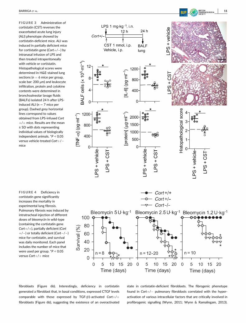
F IGURE 3 Administration ofcortistatin (CST) reverses theexacerbated acute lung injury(ALI) phenotype showed bycortistatin-deficient mice. ALI wasinduced in partially deficient micefor cortistatin gene (Cort+/�) byintranasal infusion of LPS andthen treated intraperitoneally
with vehicle or cortistatin.Histopathological scores weredetermined in H&E-stained lungsections (n = 6 mice per group,scale bar: 200 μm) and leukocyteinfiltration, protein and cytokinecontents were determined inbronchoalveolar lavage fluids(BALFs) isolated 24 h after LPS-induced ALI (n = 7 mice pergroup). Dashed grey horizontallines correspond to valuesobtained from LPS-infused Cort+/+ mice. Results are the mean± SD with dots representingindividual values of biologicallyindependent animals. *P < 0.05versus vehicle-treated Cort+/�mice
F IGURE 4 Deficiency incortistatin gene significantlyincreases the mortality inexperimental lung fibrosis.Pulmonary fibrosis was induced byintratracheal injection of differentdoses of bleomycin in wild-type(containing the cortistatin geneCort+/+), partially deficient (Cort+/�) or totally deficient (Cort�/�)mice for cortistatin, and survivalwas daily monitored. Each panelincludes the number of mice thatwere used per group. *P < 0.05versus Cort+/+ mice
BARRIGA ET AL. 11
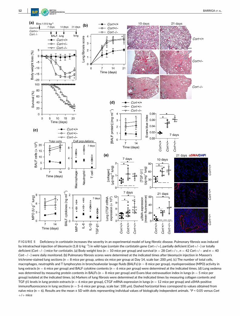
F IGURE 5 Deficiency in cortistatin increases the severity in an experimental model of lung fibrotic disease. Pulmonary fibrosis was inducedby intratracheal injection of bleomycin (1.8 U�kg�1) in wild-type (contain the cortistatin gene Cort+/+), partially deficient (Cort+/�) or totallydeficient (Cort�/�) mice for cortistatin. (a) Body weight loss (n = 10 mice per group) and survival (n = 28 Cort+/+, n = 42 Cort+/� and n = 40Cort�/�) were daily monitored. (b) Pulmonary fibrosis scores were determined at the indicated times after bleomycin injection in Masson'strichrome-stained lung sections (n = 8 mice per group, unless six mice per group at Day 14, scale bar: 200 μm). (c) The number of total cells,macrophages, neutrophils and T lymphocytes in bronchoalveolar lavage fluids (BALFs) (n = 8 mice per group), myeloperoxidase (MPO) activity inlung extracts (n = 6 mice per group) and BALF cytokine contents (n = 6 mice per group) were determined at the indicated times. (d) Lung oedemawas determined by measuring protein contents in BALFs (n = 8 mice per group) and Evans blue extravasation index in lungs (n = 5 mice pergroup) isolated at the indicated times. (e) Markers of lung fibrosis were determined at the indicated times by measuring collagen contents andTGF-β1 levels in lung protein extracts (n = 6 mice per group), CTGF mRNA expression in lungs (n = 12 mice per group) and αSMA-positiveimmunofluorescence in lung sections (n = 5–6 mice per group, scale bar: 100 μm). Dashed horizontal lines correspond to values obtained fromnaïve mice (n = 6). Results are the mean ± SD with dots representing individual values of biologically independent animals. *P < 0.05 versus Cort+/+ mice
12 BARRIGA ET AL.

Thus, the activation and subsequent nuclear translocation of Smad2/3
was significantly increased in Cort+/� fibroblasts (Figures 6c and
S1A). Moreover, the basal levels of activated phosphorylated forms of
various protein kinases, including Akt, p38 MAPK and ERK1/2, were
markedly increased in cortistatin-deficient fibroblasts, showing similar
or even higher activation levels than those showed by TGF-β1-acti-
vated Cort+/+ fibroblasts (Figures 6c and S1B). Furthermore, in com-
parison with wild-type fibroblasts, cortistatin-deficient pulmonary
fibroblasts showed accelerated and increased migratory responses in
a wound healing assay (Figure 6d). However, lack of cortistatin did
affect neither fibroblast growth nor viability (Figure S2). We finally
assessed its antifibrotic effect by adding cortistatin to cultures of
TGF-β1-activated Cort+/+ and Cort+/� fibroblasts. As expected,
treatment with cortistatin significantly reduced the expression of
profibrotic markers and extracellular matrix components, including
αSMA, CTGF, Col1a2 and fibronectin, in activated lung fibroblasts
(Figure 6e). Therefore, these findings indicate that cortistatin could
act as an endogenous break of activation, migration and differentia-
tion of fibroblasts.
3.4 | Treatment with cortistatin amelioratesbleomycin-induced pulmonary inflammation andfibrosis
Our previous results indicate that cortistatin has a critical role in the
regulation of pulmonary inflammation and fibrosis and that adminis-
tration of cortistatin is a potential strategy for the prevention and
F IGURE 6 Cortistatin (CST)-deficient fibroblasts show exacerbated profibrotic responses. (a) Gene expression of cortistatin by unstimulatedand TGF-β1-activated mouse primary lung fibroblasts (n = 3 cultures) and by lungs isolated from naïve or bleomycin (Bleo)-treated mice(n = 5 mice per group). (b–d) Primary lung fibroblasts were isolated from wild-type (Cort+/+) and partially deficient (Cort+/�) mice for cortistatin
were cultured in the absence (�) or presence (+) of TGF-β1 (10 ng�ml�1) stimulation. (b) αSMA and CTGF gene expression was determined after24-h culture (13 unstimulated cultures and nine stimulated cultures). (c) Nuclear translocation of activated Smad2/3 was determined byimmunofluorescence analysis and the levels of activated phosphorylated Akt, p38 MAPK and ERK1/2 were analysed by western blot after 1 h ofculture (n = 4–5 independent experiments, in triplicates). See Figure S1 for representative immunofluorescence images and western blots.(d) Fibroblast migration activity was measured at different time points using an in vitro wound healing assay (n = 6 independent cultures). (e)Fibroblasts isolated from Cort+/+ and Cort+/� lungs were stimulated with TGF-β1 in the absence (�) or presence (+) of cortistatin, and the geneexpression of αSMA, CTGF, collagen Iα2 (Col1a2) and fibronectin was determined after 24-h culture (n = 5–6 independent cultures). Results arethe mean ± SEM with dots representing individual values of independent experiments. Data in (b), (c) and (e) are expressed as fold change relativeto the mean of unstimulated Cort+/+ fibroblasts. *P < 0.05
BARRIGA ET AL. 13
treatment of lung injury-induced fibrosis. The systemic injection of
cortistatin at the early stage significantly prevented the profound
body weight loss, high mortality and severe pulmonary fibrosis
that were induced by the administration of high-dose bleomycin
(Figure 7a). These protective effects correlated with inhibition of pul-
monary inflammation, injury and vascular leak (Figure 7a). Importantly,
cortistatin also therapeutically attenuated bleomycin-induced mortal-
ity and the severity of pulmonary fibrosis at the later stages following
bleomycin instillation (Figure 7b). Indeed, treatment of animals with
cortistatin beginning 5 days after bleomycin challenge, once that pul-
monary inflammation was fully established, significantly improved
fibrosis score, reduced collagen deposition and decreased the pres-
ence of αSMA-expressing myofibroblasts in lung (Figure 7b). We
found similar therapeutic efficiencies using both systemic (i.p.) and
local (i.n., at 20-fold lower doses) routes of administration (Figure 7b).
Noteworthy from a therapeutic point of view is the fact that cor-
tistatin treatment was able to reverse the susceptibility to suffer
severe pulmonary fibrosis in mice that had a partial deficiency in cor-
tistatin after their exposition to low doses of bleomycin (Figure 8).
These results suggest that cortistatin-based therapies could impair
pulmonary inflammation and attenuate the established pulmonary
fibrosis.
3.5 | Involvement of somatostatin and ghrelinreceptors in the therapeutic effect of cortistatin inpulmonary inflammation and fibrosis
We finally investigated the receptors through which cortistatin could
exert its anti-inflammatory and antifibrotic activities in vivo. Previous
evidence demonstrates the capacity of cortistatin to bind, between
others, to SST receptors and ghrelin receptor (Gonzalez-Rey &
Delgado, 2007), supports that both receptors play major roles in the
immunomodulatory effect of cortistatin in several organs (Gonzalez-
Rey & Delgado, 2008) and indicates that signalling through SST recep-
tors and ghrelin receptor exerts antifibrotic actions other tissues
(Borie et al., 2008; Egger et al., 2014; Tug et al., 2013). Local adminis-
tration by intranasal infusion of either a potent SST1–5 antagonist or a
specific ghrelin receptor antagonist partially blocked the protective
effects observed for cortistatin in Cort+/� mice subjected to
LPS-induced ALI (Figure 9a) and bleomycin-induced pulmonary
fibrosis (Figure 9b). These results suggest that cortistatin exerts its
anti-inflammatory and antifibrotic actions in the lungs by signalling
through both SST receptors and ghrelin receptor.
4 | DISCUSSION
Inflammation and wound healing are two physiological processes
aimed at restoring normal tissue structure and function after an insult
or injury. However, they can be more damaging than the insult itself if
uncontrolled, excessive or prolonged. In the lung, a dysregulated
inflammation causes excessive leukocyte accumulation and increased
permeability of endothelial and alveolar epithelial barriers. A wound
healing response that has gone out of control after lung injury causes
pulmonary fibrosis, which is characterized by progressive loss of
alveolar structure, disruption of the epithelial–endothelial barrier,
activation of fibroblasts and their differentiation to myofibroblasts,
excessive deposition of extracellular matrix and tissue remodelling.
Far of restoring host pulmonary homeostasis, aberrant inflammatory
and fibrotic responses, in some cases being part of the same cascade,
contribute to the pathogenesis of severe lung disorders, such as
ALI/ARDS and IPF. A precise balance of inflammatory/fibrogenic
versus immunomodulatory/antifibrotic factors must exist to tune ade-
quately these responses. The identification of the factors that limit or
reverse both processes is critical for understanding the pathophysiol-
ogy and identifying new therapeutic targets for these disorders. In this
study, by using two well-characterized experimental models of ARDS
and pulmonary fibrosis, we point out to cortistatin as an endogenous
protective factor. We found that a deficiency in cortistatin predis-
poses for developing exacerbated inflammatory and fibrotic responses
in injured lungs after exposition to bacterial endotoxins or chemother-
apeutic drugs, even at low doses, and to subsequently suffer more
severe disease progression and increased mortality. Moreover, our
data show that a treatment with cortistatin is able to mitigate these
pathological processes.
We envision the involvement of various non-excluding and
complementary cellular and molecular mechanisms that could explain
the protective effect of cortistatin in pulmonary inflammation and
fibrosis (Figure 10). First, previous reports demonstrated the anti-
inflammatory activity of cortistatin on macrophages and described its
protective effect in murine models of sepsis and endotoxemia,
acting mainly by regulating a wide range of inflammatory mediators
(Gonzalez-Rey, Chorny, Robledo, & Delgado, 2006; Gonzalez-Rey,
Varela, Sheibanie, et al., 2006). Here, we have confirmed that in the
lung cortistatin down-regulates the production of various inflamma-
tory cytokines and chemokines by activated BALF macrophages. Fur-
ther, and importantly, we found that infiltrating pulmonary
macrophages, that are deficient in cortistatin produced excessive
levels of cytokines that are responsible for the pathophysiology of
ALI/ARDS. This effect could be initiated in an autocrine/paracrine
manner, as macrophages express both cortistatin (Dalm et al., 2003;
Gonzalez-Rey, Chorny, Robledo, & Delgado, 2006; Gonzalez-Rey,
Varela, Sheibanie, et al., 2006; Markovics et al., 2012), as well as both
somatostatin and ghrelin receptors, which are involved in the anti-
inflammatory activity of cortistatin (Gonzalez-Rey & Delgado, 2007).
Indeed, we found that blockade of any of these receptors, mainly the
ghrelin receptor, impaired the protective action of cortistatin in ALI.
Moreover, its action on macrophages could orchestrate the infiltration
of other leukocytes (i.e. neutrophils) involved in pulmonary inflamma-
tion and fibrosis. Furthermore, the immunomodulatory activity of cor-
tistatin on lymphocytes is widely recognized besides having a potent
suppressive effect on Th1 cell-mediated inflammatory responses.
Further, we have previously found that a major mechanism involved
in the generation of immune tolerance by cortistatin is the induction
of regulatory T cells (Gonzalez-Rey, Chorny, & Delgado, 2007),
14 BARRIGA ET AL.

F IGURE 7 Treatment with cortistatin (CST) reduces mortality and disease severity of experimental lung fibrosis. Mice with bleomycin (Bleo)-induced severe pulmonary fibrosis were treated with vehicle or with cortistatin following an early protective regime (a) or a delayed therapeuticregime (b) as indicated in the two schemes. Mice injected intratracheally with saline instead of bleomycin were used as basal controls (n = 6).
Mortality, body weight loss (eight mice per group) and histopathological signs of lung fibrosis (eight mice per group in a and 7–13 mice per groupin b) were determined at the indicated time points. Pulmonary leukocyte infiltration and the levels of inflammatory cytokines and oedema (proteinlevels) were assayed in bronchoalveolar lavage fluids (BALFs; six mice per group). Fibrogenic markers including the content of collagen (10 miceper group) and the presence of αSMA-positive myofibroblasts (expressed as fluorescence mean, seven mice per group) were determined in lungsisolated at the indicated time points. Scale bars: 150 μm. Results are the mean ± SD with dots representing individual values of biologicallyindependent animals. *P < 0.05 versus Bleo + vehicle-treated mice
BARRIGA ET AL. 15

a T-cell subpopulation that is involved in amelioration of ALI/ARDS
and pulmonary fibrosis (D'Alessio et al., 2009). These findings open
the possibility that, by regulating the inflammatory/immune response
in the lung, cortistatin could impair an initial step in the profibrotic
cascade and thus mitigate subsequent progression to pulmonary
fibrosis. Although the immunomodulatory action of cortistatin could
contribute to its antifibrotic role, evidence suggests a direct and
additional action of cortistatin on the fibrogenic effectors in the lung.
We found that pulmonary fibroblasts isolated from cortistatin-
deficient mice showed overactivated TGF-β1-signalling pathways,
including Smad2/3, Akt and MAPKs (p38 and ERK1/2), that drive the
expression of genes (for example, collagen, CTGF and αSMA) that are
involved in pathological fibrosis (Wynn, 2011). Again, this effect
could be mediated in a paracrine fashion, because fibroblasts express
both cortistatin and its receptors (Borie et al., 2008; Egger
et al., 2014; this study; Tug et al., 2013). In this respect, we observed
that exogenously added cortistatin inhibited directly the expression
of several mediators and markers of fibrosis by TGF-β1-activated
fibroblasts. In agreement, various studies have reported the effect of
cortistatin in MAPKs and Akt in other cell types (Duran-Prado
et al., 2013; Morell et al., 2014) and other somatostatin receptor ago-
nists regulate the activation of fibroblasts (Borie et al., 2008; Wang
et al., 2013). Moreover, by using selective antagonists, we provide
evidence that somatostatin and ghrelin receptors play major roles in
the antifibrotic effects of cortistatin in the lung. The potential capac-
ity of cortistatin to synergistically signal through both classes of
receptors could suppose an advantage versus somatostatin and
ghrelin in regulating fibrotic responses. Interestingly, we observed a
negative correlation between expression of cortistatin and the activa-
tion of pulmonary fibroblasts, confirming that cortistatin acts as an
endogenous break that needs to be released for allowing fibroblast
activation. Finally, the fact that treatment with cortistatin efficiently
reduced fibrotic responses in bleomycin-challenged mice, when initi-
ated once the inflammatory response was fully established in the
lung, also supports the capacity of cortistatin to directly limit the
fibrogenic response.
Our findings have multiple clinical implications from both thera-
peutic and from a diagnostic point of view. First, we demonstrated
that cortistatin-based therapies emerge as attractive alternatives for
treatment of pulmonary disorders which involve hyperactivated
inflammatory and fibrotic responses, such as in ALI/ARDS and IPF.
Despite the enormous progression made in the identification of the
pathogenic mechanisms involved in the initiation and progression of
these diseases, they are an enduring problem in respiratory and critical
medicine that remains therapeutically unresolved, as they remain a
major cause of morbidity and mortality worldwide (Matthay
et al., 2017). This urgent need has acquired a global dimension lately
during the pandemic COVID-19, with the association of severe ARDS
F IGURE 8 Exogenous administration of cortistatin (CST) reversed the exacerbated fibrogenic phenotype observed in cortistatin-deficientmice. Lung fibrosis was induced in partially deficient mice (Cort+/�) for cortistatin and treated with vehicle or cortistatin as indicated in thescheme. Bleomycin (Bleo)-challenged Cort+/+ mice were used as controls of reference. Mortality, histopathological signs of fibrosis (5–11 mice
per group), pulmonary collagen content (7–8 mice per group) and the presence of αSMA-positive myofibroblasts (five mice per group) weredetermined in lungs isolated at the indicated time points. Scale bars: 100 μm. Results are the mean ± SD with dots representing individual valuesof biologically independent animals. *P < 0.05
16 BARRIGA ET AL.

and pulmonary fibrosis which has a poor prognosis in SARS-
CoV-2-infected patients (Mehta et al., 2020). Due to the redundancy
and complexity of the cytokine and fibrogenic network, the
multitargeted action of cortistatin as an immunomodulatory agent on
a plethora of mediators of the cytokine storm offers obvious
advantages versus other therapies based on neutralization of a single
F IGURE 9 Involvement of somatostatin receptors and ghrelin receptor in the therapeutic effect of cortistatin (CST) in acute lung injury (ALI)and pulmonary fibrosis. (a) ALI was induced in partially deficient mice for cortistatin (Cort+/�) by intranasal injection of LPS and then treatedintraperitoneally with vehicle or cortistatin. Saline, a SST1–5 antagonist (cyclosomatostatin, cycloSOM), or a ghrelin receptor (GHSR) antagonist
([D-Lys3]-GHRP-6) was injected intranasally (i.n.) 30 min before cortistatin injection as indicated in the scheme. Leukocyte infiltration, and proteinand cytokine contents were determined in bronchoalveolar lavage fluids (BALFs) isolated 24 h after LPS-induced ALI (n = 7 mice per group). (b)Lung fibrosis was induced in Cort+/� mice with bleomycin and then treated with vehicle or cortistatin as indicated in the scheme. Saline,cycloSOM or [D-Lys3]-GHRP-6 was injected i.n. 30 min before cortistatin injection. Histopathological signs of fibrosis and the contents ofcollagen were determined in lungs collected 10 days after bleomycin challenge (5–8 mice per group). Results are the mean ± SD with dotsrepresenting individual values of biologically independent animals. *P < 0.05
BARRIGA ET AL. 17
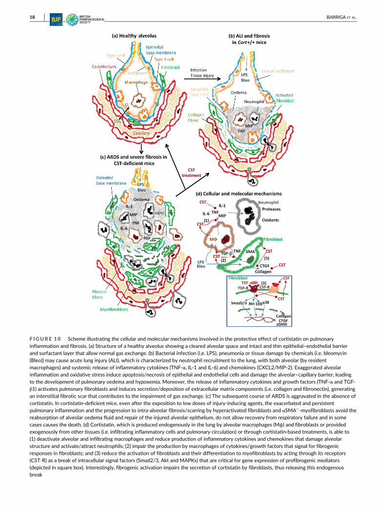
F IGURE 10 Scheme illustrating the cellular and molecular mechanisms involved in the protective effect of cortistatin on pulmonaryinflammation and fibrosis. (a) Structure of a healthy alveolus showing a cleared alveolar space and intact and thin epithelial–endothelial barrierand surfactant layer that allow normal gas exchange. (b) Bacterial infection (i.e. LPS), pneumonia or tissue damage by chemicals (i.e. bleomycin
(Bleo)) may cause acute lung injury (ALI), which is characterized by neutrophil recruitment to the lung, with both alveolar (by residentmacrophages) and systemic release of inflammatory cytokines (TNF-α, IL-1 and IL-6) and chemokines (CXCL2/MIP-2). Exaggerated alveolarinflammation and oxidative stress induce apoptosis/necrosis of epithelial and endothelial cells and damage the alveolar–capillary barrier, leadingto the development of pulmonary oedema and hypoxemia. Moreover, the release of inflammatory cytokines and growth factors (TNF-α and TGF-β1) activates pulmonary fibroblasts and induces secretion/deposition of extracellular matrix components (i.e. collagen and fibronectin), generatingan interstitial fibrotic scar that contributes to the impairment of gas exchange. (c) The subsequent course of ARDS is aggravated in the absence ofcortistatin. In cortistatin-deficient mice, even after the exposition to low doses of injury-inducing agents, the exacerbated and persistentpulmonary inflammation and the progression to intra-alveolar fibrosis/scarring by hyperactivated fibroblasts and αSMA+-myofibroblasts avoid thereabsorption of alveolar oedema fluid and repair of the injured alveolar epithelium, do not allow recovery from respiratory failure and in somecases causes the death. (d) Cortistatin, which is produced endogenously in the lung by alveolar macrophages (Mɸ) and fibroblasts or providedexogenously from other tissues (i.e. infiltrating inflammatory cells and pulmonary circulation) or through cortistatin-based treatments, is able to(1) deactivate alveolar and infiltrating macrophages and reduce production of inflammatory cytokines and chemokines that damage alveolarstructure and activate/attract neutrophils; (2) impair the production by macrophages of cytokines/growth factors that signal for fibrogenicresponses in fibroblasts; and (3) reduce the activation of fibroblasts and their differentiation to myofibroblasts by acting through its receptors(CST-R) as a break of intracellular signal factors (Smad2/3, Akt and MAPKs) that are critical for gene expression of profibrogenic mediators(depicted in square box). Interestingly, fibrogenic activation impairs the secretion of cortistatin by fibroblasts, thus releasing this endogenousbreak
18 BARRIGA ET AL.

molecule (i.e. monoclonal antibodies). Moreover, cortistatin-based
therapies could limit the start of late-onset pulmonary fibrosis in
patients with ARDS or COVID-19 in post-infection stages. It is impor-
tant to mention that cortistatin has a favourable safety profile in
humans and has demonstrated clinical efficacy in patients with Cus-
hing's disease (Giordano et al., 2007). Furthermore, the interest of the
pharmaceutical companies in developing cortistatin-based analogues
with improved half-life in serum has increased lately and a recent
report demonstrated their efficiency in inflammatory conditions (Rol
et al., 2021). We have recently witnessed an example of repositioning
of a therapy based on another immunomodulatory and antifibrotic
neuropeptide, vasoactive intestinal peptide (VIP) or aviptadil (Chorny
& Delgado, 2008; Prasse et al., 2010), for the treatment of ARDS in
patients with severe COVID-19 (Scavone et al., 2020; clinical trial:
NCT04311697).
Second, the fact that a simple partial deficiency in cortistatin
could predispose patients for developing exacerbated inflammatory
and fibrotic responses and could be used to anticipate the diagnosti-
cally the developmen of more severe forms of pulmonary disorders.
In this sense, this study opens the possibility of initiating further
clinical research that could corroborate plasma cortistatin level as a
biomarker of protection or prognosis in patients with ARDS or IPF.
If this is the case, our results also suggest that a treatment based
on cortistatin injection would easily correct this deficiency and
improve disease progression. Although the endogenous and environ-
mental factors that could influence the body levels of cortistatin are
mostly unknown, one could anticipate that these levels will change
during our life under different circumstances and scenarios, and thus
our susceptibility to suffer certain disorders. In any case, a percent-
age of the human population is heterozygous for cortistatin from
birth, because cortistatin gene is located at chromosome 1p36.22
and monosomy of 1p36 is the most common subtelomeric terminal
deletion syndrome (Jordan et al., 2015). Besides a spectrum of neu-
rological and sensorial defects, these individuals develop cardiac
fibrosis and cardiomyopathy, however no studies have described
increased incidence of pulmonary disorders, despite their short life
expectancy.
Finally, bleomycin is a chemotherapeutic drug used for treating
many types of cancer. Unfortunately, a proportion of bleomycin-
treated patients develop severe side effects that are associated to
appearance of pulmonary and skin fibrosis that mostly cause treat-
ment to be withdrawn. Understanding that a deficiency in a factor like
cortistatin could predispose patients to such adverse effects could
help selection of the correct chemotherapy or try to correct the
adverse effects by cortistatin-based treatments.
In summary, this study provides new insights into the function of
cortistatin in pulmonary inflammation and demonstrates a novel role
of this neuropeptide in fibrosis, which could be extrapolated beyond
lung tissue. Overall, we demonstrate that deficiency in cortistatin
could be considered a potential biomarker of susceptibility to suffer
severe forms of pulmonary disorders including ARDS, IPF or pneumo-
nia and provide a powerful rationale for the assessment of the effi-
cacy of cortistatin or its stable analogues as therapeutic approaches
to the treatment of diseases that are still unsolved from a clinical point
of view. However, because the effects observed are based on preclini-
cal mouse models of ALI/ARDS in the absence of viral or bacterial
infection, and many pulmonary fibrotic disorders are of idiopathic
nature, extrapolations to clinical practice have to be made with
caution.
ACKNOWLEDGEMENTS
We thank to Dr Luis de Lecea (Stanford University, CA, USA) for pro-
viding the Cort�/� mice to establish initially the colony of animals
used in this study. This study was mainly supported by the Spanish
Ministry of Science and Innovation (Ministerio de Ciencia e Innova-
ci�on [MICINN], Grant SAF2015-67787-R). R.B. was a recipient of FPI
fellowship from the Spanish Ministry of Science and Innovation, and
M.G.-F. was a recipient of FPU fellowship from the Spanish Ministry
of Universities. V.F.-P. was a recipient of postdoctoral fellowship from
Fundaç~ao de Amparo à Pesquisa do Estado de S~ao Paulo—FAPESP
(12/21767-5).
AUTHOR CONTRIBUTIONS
M.B. and J.C.-S. conducted most of the experiments and analysed the
data. M.C. and G.R. carried out the histological processing and ELISA
determinations. V.F.-P. conducted the part of in vivo experimental
models. R.B., M.G.-F. and F.O. performed the histopathological
studies. M.D. conceived and designed the study, conducted the in vivo
experimental models, analysed the data and wrote the manuscript. All
the authors edited the manuscript.
CONFLICT OF INTEREST
The authors declare no conflicts of interest.
DECLARATION OF TRANSPARENCY AND SCIENTIFIC
RIGOUR
This Declaration acknowledges that this paper adheres to the princi-
ples for transparent reporting and scientific rigour of preclinical
research as stated in the British Journal of Pharmacology guidelines for
Design & Analysis, Immunoblotting and Immunochemistry and Animal
Experimentation and as recommended by funding agencies, publishers
and other organizations engaged with supporting research.
DATA AVAILABILITY STATEMENT
The data that support the findings of this study are available from the
corresponding author upon reasonable request.
ORCID
Mario Delgado https://orcid.org/0000-0003-1893-5982
REFERENCES
Alexander, S. P. H., Christopoulos, A., Davenport, A. P., Kelly, E.,
Mathie, A., Peters, J. A., Veale, E. L., Armstrong, J. F., Faccenda, E.,
Harding, S. D., Pawson, A. J., Sharman, J. L., Southan, C., Davies, J. A.,
& CGTP Collaborators. (2019). The Concise Guide to PHARMACOL-
OGY 2019/20: G protein-coupled receptors. British Journal of Pharma-
cology, 176, S21–S141.
BARRIGA ET AL. 19

Alexander, S. P. H., Roberts, R. E., Broughton, B. R. S., Sobey, C. G.,
George, C. H., Stanford, S. C., Cirino, G., Docherty, J. R.,
Giembycz, M. A., Hoyer, D., Insel, P. A., Izzo, A. A., Ji, Y.,
MacEwan, D. J., Mangum, J., Wonnacott, S., & Ahluwalia, A. (2018).
Goals and practicalities of immunoblotting and immunohistochemistry:
A guide for submission to the British Journal of Pharmacology. British
Journal of Pharmacology, 175, 407–411. https://doi.org/10.1111/bph.14112
Ashcroft, T., Simpson, J. M., & Timbrell, V. (1988). Simple method of esti-
mating severity of pulmonary fibrosis on a numerical scale. Journal of
Clinical Pathology, 41, 467–470. https://doi.org/10.1136/jcp.41.4.467Borie, R., Fabre, A., Prost, F., Marchal-Somme, J., Lebtahi, R., Marchand-
Adam, S., Aubier, M., Soler, P., & Crestani, B. (2008). Activation of
somatostatin receptors attenuates pulmonary fibrosis. Thorax, 63,
251–258. https://doi.org/10.1136/thx.2007.078006Chorny, A., & Delgado, M. (2008). Neuropeptides rescue mice from lethal
sepsis by downregulating secretion of the late-acting inflammatory
mediator high mobility group box 1. The American Journal of Pathology,
172, 1297–1307. https://doi.org/10.2353/ajpath.2008.070969Cordoba-Chacon, J., Gahete, M. D., Pozo-Salas, A. I., Martínez-
Fuentes, A. J., de Lecea, L., Gracia-Navarro, F., Kineman, R. D.,
Castaño, J. P., & Luque, R. M. (2011). Cortistatin is not a somatostatin
analogue but stimulates prolactin release and inhibits GH and ACTH in
a gender-dependent fashion: Potential role of ghrelin. Endocrinology,
152, 4800–4812. https://doi.org/10.1210/en.2011-1542Curtis, M. J., Alexander, S., Cirino, G., Docherty, J. R., George, C. H.,
Giembycz, M. A., Hoyer, D., Insel, P. A., Izzo, A. A., Ji, Y.,
MacEwan, D. J., Sobey, C. G., Stanford, S. C., Teixeira, M. M.,
Wonnacott, S., & Ahluwalia, A. (2018). Experimental design and analy-
sis and their reporting II: Updated and simplified guidance for authors
and peer reviewers. British Journal of Pharmacology, 175, 987–993.https://doi.org/10.1111/bph.14153
D'Alessio, F. R., Tsushima, K., Aggarwal, N. R., West, E. E., Willett, M. H.,
Britos, M. F., Pipeling, M. R., Brower, R. G., Tuder, R. M., McDyer, J. F.,
& King, L. S. (2009). CD4+ CD25+ Foxp3+ Tregs resolve experimental
lung injury in mice and are present in humans with acute lung injury.
The Journal of Clinical Investigation, 119, 2898–2913. https://doi.org/10.1172/JCI36498
Dalm, V. A., van Hagen, P. M., van Koetsveld, P. M., Achilefu, S.,
Houtsmuller, A. B., Pols, D. H., van der Lely, A. J., Lamberts, S. W., &
Hofland, L. J. (2003). Expression of somatostatin, cortistatin, and
somatostatin receptors in human monocytes, macrophages, and
dendritic cells. American Journal of Physiology. Endocrinology and
Metabolism, 285, E344–E353. https://doi.org/10.1152/ajpendo.
00048.2003
Delgado-Maroto, V., Falo, C. P., Forte-Lago, I., Adan, N., Morell, M.,
Maganto-Garcia, E., Robledo, G., O'Valle, F., Lichtman, A. H.,
Gonzalez-Rey, E., & Delgado, M. (2017). The neuropeptide
cortistatin attenuates experimental autoimmune myocarditis via
inhibition of cardiomyogenic T cell-driven inflammatory responses.
British Journal of Pharmacology, 174, 267–280. https://doi.org/10.
1111/bph.13682
Duran-Prado, M., Morell, M., Delgado-Maroto, V., Castaño, J. P., Aneiros-
Fernandez, J., de Lecea, L., Culler, M. D., Hernandez-Cortes, P.,
O'Valle, F., & Delgado, M. (2013). Cortistatin inhibits migration and
proliferation of human vascular smooth muscle cells and decreases
neointimal formation on carotid artery ligation. Circulation Research,
112, 1444–1455. https://doi.org/10.1161/CIRCRESAHA.112.300695
Egger, C., Gérard, C., Vidotto, N., Accart, N., Cannet, C., Dunbar, A.,
Tigani, B., Piaia, A., Jarai, G., Jarman, E., Schmid, H. A., & Beckmann, N.
(2014). Lung volume quantified by MRI reflects extracellular-matrix
deposition and altered pulmonary function in bleomycin models of
fibrosis: Effects of SOM230. American Journal of Physiology. Lung Cellu-
lar and Molecular Physiology, 306, L1064–L1077. https://doi.org/10.1152/ajplung.00027.2014
George, P. M., Wells, A. U., & Jenkins, R. G. (2020). Pulmonary fibrosis and
COVID-19: The potential role for antifibrotic therapy. The Lancet
Respiratory Medicine, 8, 807–815. https://doi.org/10.1016/S2213-
2600(20)30225-3
Giordano, R., Picu, A., Bonelli, L., Broglio, F., Prodam, F., Grottoli, S.,
Muccioli, G., Ghigo, E., & Arvat, E. (2007). The activation of
somatostatinergic receptors by either somatostatin-14 or cortistatin-
17 often inhibits ACTH hypersecretion in patients with Cushing's dis-
ease. European Journal of Endocrinology, 157, 393–398. https://doi.org/10.1530/EJE-07-0147
Gonzalez-Rey, E., Chorny, A., Del Moral, R. G., Varela, N., & Delgado, M.
(2007). Therapeutic effect of cortistatin on experimental arthritis by
downregulating inflammatory and Th1 responses. Annals of the Rheu-
matic Diseases, 66, 582–588. https://doi.org/10.1136/ard.2006.
062703
Gonzalez-Rey, E., Chorny, A., & Delgado, M. (2007). Regulation of immune
tolerance by anti-inflammatory neuropeptides. Nature Reviews.
Immunology, 7, 52–63. https://doi.org/10.1038/nri1984Gonzalez-Rey, E., Chorny, A., Robledo, G., & Delgado, M. (2006).
Cortistatin, a new antiinflammatory peptide with therapeutic effect on
lethal endotoxemia. The Journal of Experimental Medicine, 203,
563–571. https://doi.org/10.1084/jem.20052017
Gonzalez-Rey, E., Chorny, A., Varela, N., Robledo, G., & Delgado, M.
(2006). Urocortin and adrenomedullin prevent lethal endotoxemia by
down-regulating the inflammatory response. The American Journal of
Pathology, 168, 1921–1930. https://doi.org/10.2353/ajpath.2006.
051104
Gonzalez-Rey, E., & Delgado, M. (2007). Anti-inflammatory neuropeptide
receptors: New therapeutic targets for immune disorders? Trends in
Pharmacological Sciences, 28, 482–491. https://doi.org/10.1016/j.tips.2007.07.001
Gonzalez-Rey, E., & Delgado, M. (2008). Emergence of cortistatin as a new
immunomodulatory factor with therapeutic potential in immune disor-
ders. Molecular and Cellular Endocrinology, 286, 135–140. https://doi.org/10.1016/j.mce.2007.08.001
Gonzalez-Rey, E., Varela, N., Sheibanie, A. F., Chorny, A., Ganea, D., &
Delgado, M. (2006). Cortistatin, an antiinflammatory peptide with
therapeutic action in inflammatory bowel disease. Proceedings of the
National Academy of Sciences of the United States of America, 103,
4228–4233. https://doi.org/10.1073/pnas.0508997103Jordan, V. K., Zaveri, H. P., & Scott, D. A. (2015). 1p36 deletion syndrome:
An update. The Application of Clinical Genetics, 8, 189–200. https://doi.org/10.2147/TACG.S65698
Kolb, P., Upagupta, C., Vierhout, M., Ayaub, E., Bellaye, P. S., Gauldie, J.,
Shimbori, C., Inman, M., Ask, K., & Kolb, M. R. J. (2020). The impor-
tance of interventional timing in the bleomycin model of pulmonary
fibrosis. The European Respiratory Journal, 55, 1901105. https://doi.
org/10.1183/13993003.01105-2019
Lilley, E., Stanford, S. C., Kendall, D. E., Alexander, S. P.,
Cirino, G., Docherty, J. R., George, C. H., Insel, P. A.,
Izzo, A. A., Ji, Y., Panettieri, R. A., Sobey, C. G., Stefanska, B.,
Stephens, G., Teixeira, M., & Ahluwalia, A. (2020). ARRIVE 2.0 and the
British Journal of Pharmacology: Updated guidance for 2020. British
Journal of Pharmacology, 177, 3611–3616. https://doi.org/10.1111/bph.15178
Markovics, A., Szoke, �E., Sándor, K., Börzsei, R., Bagoly, T., Kemény, �A.,
Elekes, K., Pintér, E., Szolcsányi, J., & Helyes, Z. (2012). Comparison of
the anti-inflammatory and anti-nociceptive effects of cortistatin-14
and somatostatin-14 in distinct in vitro and in vivo model systems.
Journal of Molecular Neuroscience, 46, 40–50. https://doi.org/10.1007/s12031-011-9577-4
Matthay, M. A., McAuley, D. F., & Ware, L. B. (2017). Clinical trials in acute
respiratory distress syndrome: Challenges and opportunities. The Lan-
cet Respiratory Medicine, 5, 524–534. https://doi.org/10.1016/S2213-2600(17)30188-1
20 BARRIGA ET AL.

Mehta, P., McAuley, D. F., Brown, M., Sanchez, E., Tattersall, R. S.,
Manson, J. J., & HLH Across Speciality Collaboration, UK. (2020).
COVID-19: Consider cytokine storm syndromes and immunosuppres-
sion. Lancet, 395, 1033–1034. https://doi.org/10.1016/S0140-6736(20)30628-0
Morell, M., Camprubí-Robles, M., Culler, M. D., de Lecea, L., & Delgado, M.
(2014). Cortistatin attenuates inflammatory pain via spinal and periph-
eral actions. Neurobiology of Disease, 63, 141–154. https://doi.org/10.1016/j.nbd.2013.11.022
Percie du Sert, N., Hurst, V., Ahluwalia, A., Alam, S., Avey, M. T., Baker, M.,
Browne, W. J., Clark, A., Cuthill, I. C., Dirnagl, U., Emerson, M.,
Garner, P., Holgate, S. T., Howells, D. W., Karp, N. A., Lazic, S. E.,
Lidster, K., MacCallum, C. J., Macleod, M., … Würbel, H. (2020). The
ARRIVE guidelines 2.0: Updated guidelines for reporting animal
research. British Journal of Pharmacology, 177, 3617–3624. https://doi.org/10.1111/bph.15193
Prasse, A., Zissel, G., Lützen, N., Schupp, J., Schmiedlin, R., Gonzalez-
Rey, E., Rensing-Ehl, A., Bacher, G., Cavalli, V., Bevec, D., Delgado, M.,
& Müller-Quernheim, J. (2010). Inhaled vasoactive intestinal peptide
exerts immunoregulatory effects in sarcoidosis. American Journal of
Respiratory and Critical Care Medicine, 182, 540–548. https://doi.org/10.1164/rccm.200909-1451OC
Qiu, C., Li, J., Luo, D., Chen, X., Qu, R., Liu, T., Li, F., & Liu, Y. (2020).
Cortistatin protects against inflammatory airway diseases through
curbing CCL2 and antagonizing NF-κB signaling pathway. Biochemical
and Biophysical Research Communications, 531, 595–601. https://doi.org/10.1016/j.bbrc.2020.07.088
Reddy, G. K., & Enwemeka, C. S. (1996). A simplified method for the analy-
sis of hydroxyproline in biological tissues. Clinical Biochemistry, 29,
225–229. https://doi.org/10.1016/0009-9120(96)00003-6Rol, A., Todorovski, T., Martin-Malpartida, P., Escola, A., González-Rey, E.,
Arag�on, E., Verdaguer, X., Vallès-Miret, M., Farrera-Sinfreu, J., Puig, E.,
& Fernández-Carneado, J. (2021). Structure-based design of a cor-
tistatin analogue with immunoregulatory activity in models of inflam-
matory bowel disease. Nature Communications, 12, 1869. https://doi.
org/10.1038/s41467-021-22076-5
Scavone, C., Brusco, S., Bertini, M., Sportiello, L., Rafaniello, C., Zoccoli, A.,
Berrino, L., Racagni, G., Rossi, F., & Capuano, A. (2020). Current phar-
macological treatments for COVID-19: What's next? British Journal of
Pharmacology, 177, 4813–4824. https://doi.org/10.1111/bph.15072Souza-Moreira, L., Morell, M., Delgado-Maroto, V., Pedreño, M., Martinez-
Escudero, L., Caro, M., O'Valle, F., Luque, R., Gallo, M., de Lecea, L.,
Castaño, J. P., & Gonzalez-Rey, E. (2013). Paradoxical effect of cor-
tistatin treatment and its deficiency on experimental autoimmune
encephalomyelitis. Journal of Immunology, 191, 2144–2154. https://doi.org/10.4049/jimmunol.1300384
Tager, A. M., LaCamera, P., Shea, B. S., Campanella, G. S.,
Selman, M., Zhao, Z., Polosukhin, V., Wain, J., Karimi-Shah, B. A.,
Kim, N. D., Hart, W. K., Pardo, A., Blackwell, T. S., Xu, Y., Chun, J., &
Luster, A. D. (2008). The lysophosphatidic acid receptor LPA1 links
pulmonary fibrosis to lung injury by mediating fibroblast recruitment
and vascular leak. Nature Medicine, 14, 45–54. https://doi.org/10.
1038/nm1685
Tsushima, K., King, L. S., Aggarwal, N. R., De Gorordo, A., D'Alessio, F. R.,
& Kubo, K. (2009). Acute lung injury review. Internal Medicine, 48,
621–630. https://doi.org/10.2169/internalmedicine.48.1741
Tug, T., Kara, H., Karaoglu, A., Karatas, F., Turgut, N. H., Ayan, E., Boran, C.,
& Tug, E. (2013). The effect of octreotide, an analog of somatostatin,
on bleomycin-induced interstitial pulmonary fibrosis in rats. Drug and
Chemical Toxicology, 36, 181–186. https://doi.org/10.3109/
01480545.2012.710618
Wang, J., Wang, L., Song, G., & Han, B. (2013). The mechanism through
which octreotide inhibits hepatic stellate cell activity. Molecular Medi-
cine Reports, 7, 1559–1564. https://doi.org/10.3892/mmr.2013.1385
Wynn, T. A. (2011). Integrating mechanisms of pulmonary fibrosis. The
Journal of Experimental Medicine, 208, 1339–1350. https://doi.org/10.1084/jem.20110551
Wynn, T. A., & Ramalingam, T. R. (2013). Mechanisms of fibrosis: Thera-
peutic translation for fibrotic disease. Nature Medicine, 18, 1028–1040.
Zhou, F., Yu, T., Du, R., Fan, G., Liu, Y., Liu, Z., Xiang, J., Wang, Y., Song, B.,
Gu, X., & Guan, L. (2020). Clinical course and risk factors for mortality
of adult inpatients with COVID-19 in Wuhan, China: A retrospective
cohort study. Lancet, 395, 1054–1062. https://doi.org/10.1016/
S0140-6736(20)30566-3
SUPPORTING INFORMATION
Additional supporting information may be found online in the
Supporting Information section at the end of this article.
How to cite this article: Barriga, M., Benitez, R., Ferraz-de-
Paula, V., Garcia-Frutos, M., Caro, M., Robledo, G., O'Valle, F.,
Campos-Salinas, J., & Delgado, M. (2021). Protective role of
cortistatin in pulmonary inflammation and fibrosis. British
Journal of Pharmacology, 1–21. https://doi.org/10.1111/bph.
15615
BARRIGA ET AL. 21