Embed Size (px)
Citation preview

NATURE MEDICINE • VOLUME 8 • NUMBER 8 • AUGUST 2002 787
NEWS & VIEWS
Protect the killer: CTLs need defenses against the tumorEvasion of the immune system is an all too common feature of cancer. A new study suggests one
evasion mechanism: induction of T-cell apoptosis through B7-H1, a molecule expressed on the surface of many tumor cells (pages 793–800).
Suppression of immunological reactiv-ity is one of the major mechanisms by
which neoplastic cells are thought to escape host defenses. Tumor-reactiveCD4+ and CD8+ T cells can be found inthe peripheral blood, lymph nodes andtumors of some tumor-bearing patients,and in some cases, elevated numbers oftumor-infiltrating T lymphocytes (TILs)correlate with better prognosis1. But un-fortunately, in most cases tumors con-tinue to grow, suppressing anti-tumorimmune responses through mechanismsthat are, as yet, not wholly clear.However, it is known that tumors canevade immune attack by downregulatingMHC expression on their surface or byproducing inhibitory cytokines such astransforming growth factor-β (TGF-β) orinterleukin-10 (IL-10).
In this issue, Dong et al. show that tu-mors can actively inhibit immune re-sponses by expression of B7 homolog 1(B7-H1), also known as programmed
death ligand 1 (PD-L1)2. B7-H1 is a B7family member and ligand for pro-grammed death-1 (PD-1), a member ofthe CD28 family3–6. Receptor–ligandpairs of the B7-CD28 superfamily havekey roles in regulating T-cell activationand tolerance7. Dong et al. built on re-cent findings that B7-H1 is expressed onmany tumors5 and examine the conse-quences of this expression. They reportthat B7-H1 on tumors can interact withreceptors on cytotoxic T lymphocytes(CTLs) and promote CTL death. Theythen go on to provide a potential avenuefor therapy by showing that antibodiesagainst B7-H1 enhance killing of tumorcells by protecting CTLs from B7-H1-in-duced death.
Some pathways within the B7-CD28
superfamily provide a critical, positivecostimulatory signal that augments andsustains T-cell responses. This costimula-tory signal works in conjunction with anantigen-specific signal provided by theinteraction of the peptide antigen–majorhistocompatibility complex (MHC) withthe T-cell receptor (TCR). Other B7-CD28superfamily pathways contribute keynegative signals that limit, terminateand/or attenuate T-cell responses.
The B7-1/B7-2–CD28/CTLA-4 (cyto-toxic T-lymphocyte antigen-4) costimu-latory pathway is the best characterized.However, this pathway is complex inthat the B7 molecules B7-1 (CD80) andB7-2 (CD86) can each engage two recep-tors, the stimulatory CD28 receptor andthe inhibitory CTLA-4 (CD152) receptor.The components of the pathway de-scribed by Dong et al.—the PD-1 receptorand its two ligands, B7-H1 (PD-L1) andPD-L2 (B7-DC)—belong to the most re-cently defined pathway within the B7-
GORDON J. FREEMAN1, ARLENE H. SHARPE2 & VIJAY K. KUCHROO3
tect resident neurons, but also providea permissive environment for the mobi-lization of endogenous progenitorcells14. If EPO can stimulate vascularneurotrophin production, then itmight serve as a critical activator of anendothelial-based paracrine pathwayleading to local neural protection.
In a field littered with past reportsthat have raised expectations only toultimately falter, skepticism is war-ranted in assessing new claims.Nonetheless, the data presented byGorio et al. are clear and provocative,and exciting in their potential for earlyapplication. Past efforts at developing avariety of neuroprotective agents havefoundered in early clinical trials, typi-cally because of their poor tolerance orfrank toxicity in ill patients. In sharpand reassuring contrast, EPO has beenused for over 15 years in the treatmentof dialysis and chemotherapy-associ-ated anemias, as well as other disordersof erythropoiesis (as well as illicitly byathletes, seeking to improve their oxy-gen-carrying capacity). It has provensafe and effective, with virtually notoxicity in the setting of a wide range ofdiseases and drug treatments. In the
face of this latest strong report by Gorioet al., it does not seem premature tobegin assessing EPO’s therapeutic effi-cacy in patients. Indeed, its clinicalevaluation may be reasonably enter-tained not only for SCI patients, butalso for victims of other acute cerebralischemic processes, including trau-matic brain injury, subarachnoid hem-orrhage and stroke.
1. Sekhon, L. & Fehlings, M. Epidemiology, demo-graphics, and pathophysiology of acute spinalcord injury. Spine 26, S2–S12 (2001).
2. Gorio, A. et al. Recombinant human erythropoi-etin counteracts secondary injury and markedlyenhances neurological recovery from experimen-tal spinal cord trauma. Proc. Natl. Acad. Sci. USA99, 9450–9455 (2002).
3. Basso, D.M., Beattie, M. & Bresnahan, J. A sensi-tive and reliable locomotor rating scale for openfield testing in rats. J. Neurotrauma 12, 1–21(1995).
4. Juul, S., Anderson, D., Li, Y. & Christensen, R.Erythropoietin and erythropoietin receptor in thedeveloping human central nervous system.Pediatric Res. 43, 40–49 (1998).
5. Bernaudin, M. et al. A potential role for erythro-poietin in focal permanent cerebral ischemia inmice. J. Cereb. Blood Flow Metab. 19, 643–651(1999).
6. Brines, M. et al. Erythropoietin crosses the blood-brain barrier to protect against experimentalbrain injury. Proc. Natl. Acad. Sci. 97,10526–10531 (2000).
7. Lin, J. et al. Gap junction-mediated propagationand amplification of cell injury. Nature Neurosci.
1, 494–500 (1998).8. Celik, M. et al. Erythropoietin prevents motor
neuron apoptosis and neurologic disability in ex-perimental spinal cord ischemic injury. Proc. Natl.Acad. Sci. 99, 2258–2263 (2002).
9. Springborg, J. et al. A single subcutaneous bolusof erythropoietin normalizes cerebral blood flowautoregulation after subarachnoid hemorrhage inrats. British J. Pharmacol. 135, 823–829 (2002).
10. Digicaylioglu, M. & Lipton, S.A. Erythropoietin-mediated neuroprotection involves cross-talk be-tween Jak2 and NF-κB signaling cascades. Nature412, 641–647 (2001).
11. O’Neill, L. & Kaltscmidt, C. NF-κB: a crucial tran-scription factor for glial and neuronal cell func-tion. Trends in Neurosci. 20, 252–258 (1997).
12. Mattson, M., Goodman, Y., Luo, H., Fu, W. &Furukawa, K. Activation of NF-κB protects hip-pocampal neurons against oxidative stress-in-duced apoptosis. J. Neurosci. Res. 49, 681–697(1997).
13. Leventhal, C., Rafii, S., Rafii, D., Shahar, A. &Goldman, S.A. Endothelial trophic support ofneuronal production and recruitment by theadult mammalian subependyma. Molec. Cell.Neurosci. 13, 450–464 (1999).
14. Palmer, T. Adult neurogenesis and the vascularNietzsche. Neuron 34, 856–858 (2002).
1Department of Neurology and NeuroscienceWeill Medical College of Cornell UniversityNew York, New York, USA2Department of Cell Biology and AnatomyNew York Medical CollegeValhalla, New York, USAEmail: [email protected][email protected]
©20
02 N
atu
re P
ub
lish
ing
Gro
up
h
ttp
://w
ww
.nat
ure
.co
m/n
atu
rem
edic
ine

788 NATURE MEDICINE • VOLUME 8 • NUMBER 8 • AUGUST 2002
NEWS & VIEWS
bition, probably because of higher pro-duction of IL-2 by CD4+ T cells5,8. In lym-phoid tissue, B7-1-CD28 and B7-2-CD28interactions may overcome the im-munoinhibitory effects of B7-H1 on CD4+
T cells, but in the absence of B7-1 and B7-2 on peripheral target tissues and tumors,the immunoinhibitory effects of B7-H1may predominate. Further in vivo work isneeded to determine the contribution ofB7-H1 to tumor progression, but its in situexpression on a broad range of solid tu-mors is striking.
The current and previous studies byDong et al. using resting T cells and B7-H1–immunoglobulin (Ig) fusion protein
suggest that B7-H1 could provide a posi-tive costimulatory signal, stimulating T-cell proliferation and inducing IL-10production3,6. In contrast, other studieshave suggested that the interaction of B7-H1 with PD-1 results in inhibition of T-cellproliferation, reduced cytokine produc-tion and cell-cycle arrest4,5,8. The currentstudy begins to reconcile these seeminglyopposite results by providing evidence fora second receptor for B7-H1. A CTL clonethat does not express PD-1 still bound B7-H1–Ig, indicating the presence of a secondreceptor for B7-H1. Incubation of the CTLclone with a B7-H1+ breast-tumor cell lineresulted in increased apoptosis of CTL.
CD28 superfamily. The functions of thisnew pathway are just beginning to bestudied3–6,8. In contrast to other CD28family members, PD-1 expression is notrestricted to T cells. PD-1 is expressed onactivated, but not resting, CD4+ andCD8+ T cells, B cells and myeloid cells5,8,9.PD-1-deficient mice have an autoim-mune phenotype, providing direct invivo evidence that PD-1 acts as a negativeregulator of immune responses10,11.
Dong et al. and others5 have shownthat many human tumors, including car-cinomas of the breast, lung, ovary andcolon, express B7-H1, whereas the corre-sponding normal tissues do not. Dong etal. show that this expression has func-tional significance. They find that thepresence of B7-H1 on tumor cells can ac-tively inhibit immune responses by pro-moting the apoptosis of effector CTLs viainduction of Fas ligand (FasL) and IL-10,thereby providing a novel means forevading immune destruction by CTLs(Fig. 1a). Monoclonal antibody againstB7-H1 reduced this apoptosis and led togreater tumor-cell killing. Notably, IFN-γ,a cytokine produced by CTLs, was foundto induce expression of B7-H1 on manytumor cell lines. Thus, effector CTLs mayhasten their own demise by productionof IFN-γ and subsequent upregulation ofB7-H1 expression on the tumor3–5.
Given that other pathways within theB7-C28 superfamily also regulate anti-tumor immunity, Dong et al. investigatedthe functional interactions between B7-H1 and another family member, B7-1. Inthis experiment, they used the P815 mas-tocytoma tumor cell line, which cangrow progressively in mice and kill them.B7-1-transfected cells stimulate a potentantitumor immune response that leads totumor rejection. However, B7-H1- andB7-1-cotransfected cells grew and killedthe host, indicating that B7-H1-mediatedinhibition of the anti-tumor immune re-sponse is strong enough to overcome po-tent costimulation mediated by the B7-1receptor, CD28. These results are consis-tent with previous studies8, showing thatB7-H1-mediated inhibition of CD8+ T-cellactivation cannot be overcome by CD28-mediated costimulation but can be over-come by exogenous IL-2. Together, thesestudies suggest that the level of IL-2 willdetermine whether B7-H1 can lead toCD8+ T-cell apoptosis. B7-H1 seems to bea particularly potent inhibitor of CD8+ T-cell responses5,8. In contrast, in CD4+ Tcells, strong CD28-mediated costimula-tion can overcome B7-H1-mediated inhi-
Activation Differentiation
Tumor
Target organLymphoid tissue
Effector T cellPrimed T cellNaive T cell
B7-H1/PD-L1
B7-H1/PD-L1
IL-10/Fas-L
(suppression/anergy?)
CD8 T cell apoptosis by:Fas/Fas-L
IL-10Growth factor deprivation
SHP-2
P
Second receptor
Decreased TCR signalapoptosis (?)
Fas-ligandIL-10Apoptosis
TCR/CD3MHC
Solid tumor Cytotoxic T Lymphocyte
Y
Secondreceptor
PD-1
a
b
B7-H1/PD-L1
Second receptor
PD-1
Antigen
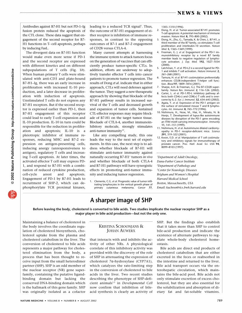
Fig. 1 Consequences of B7-H1 engagement. a, B7-H1 and T-cell apoptosis. Tumors express B7-H1/PD-L1, which can potentially interact with at least two different receptors on activated cyto-toxic T lymphocytes (CTLs). When the T-cell receptor (TCR) is activated by binding toMHC–peptide, the interaction of B7-H1 with its receptors provides second signals that downregu-late the effector functions and survival of CTLs. Ligation of the second receptor may upregulateFas-Ligand and IL-10 production, resulting in T-cell apoptosis. Interaction of B7-H1 with PD-1 re-cruits SHP-2, resulting in dephosphorylation of TCR proximal kinases, reduced TCR signal and de-creased cytokine production, and ultimately cell-cycle arrest or death by growth-factordeprivation. b, Hypothetical model for temporal expression of B7-H1 receptors on T cells. Naive Tcells do not express B7-H1 receptors. Activated T cells may rapidly express the second receptor forB7-H1, whose ligation upregulates IL-10 production and FasL expression. PD-1, a known receptorfor B7-H1, is expressed later after activation on effector T cells. Tumors may engage either recep-tor, resulting in inhibition of the CTL function by IL-10 production and/or induce cell death byFasL upregulation or growth-factor deprivation.
Rene
e Lu
cas
©20
02 N
atu
re P
ub
lish
ing
Gro
up
h
ttp
://w
ww
.nat
ure
.co
m/n
atu
rem
edic
ine
NATURE MEDICINE • VOLUME 8 • NUMBER 8 • AUGUST 2002 789
NEWS & VIEWS
Antibodies against B7-H1 but not PD-1–Igfusion protein reduced the apoptosis ofthe CTL clone. These data suggest that en-gagement of the second receptor for B7-H1 functions in T- cell apoptosis, perhapsby inducing FasL.
The divergent data on B7-H1 functionwould make even more sense if PD-1and the second receptor are expressedwith different kinetics and on differentsubpopulations of T cells (Fig. 1b).When human primary T cells were stim-ulated with anti-CD3 and plate-boundB7-H1–Ig, there was an early increase inproliferation with increased IL-10 pro-duction, and a later decrease in prolifer-ation with induction of apoptosis.Unstimulated T cells do not express anyB7-H1 receptors. But if the second recep-tor is expressed earlier than PD-1, thenB7-H1 engagement of this receptorcould lead to early T-cell expansion andIL-10 production. IL-10 in turn could beresponsible for the reduction in prolifer-ation and apoptosis; IL-10 is apleiotropic inhibitor of immune re-sponses, reducing MHC and B7-2 ex-pression on antigen-presenting cells,inducing anergy (unresponsiveness toantigen), regulatory T cells and increas-ing T-cell apoptosis. At later times, theactivated effector T cell may express PD-1, and respond to B7-H1 with a combi-nation of reduced cytokine production,cell-cycle arrest and apoptosis.Engagement of PD-1 by B7-H1 leads torecruitment of SHP-2, which can de-phosphorylate TCR proximal kinases,
leading to a reduced TCR signal5. Thus,the outcome of B7-H1 engagement of ei-ther receptor is inhibition of immune re-sponses, in contrast to the opposingoutcomes of B7-1 and B7-2 engagementof CD28 versus CTLA-4.
Many current attempts at harnessingthe immune system to attack tumors focuson the generation of vaccines that can effi-ciently produce tumor-specific CTLs. Inaddition, efforts are underway to adop-tively transfer effector T cells into cancerpatients to promote tumor regression. Theresults of Dong et al. indicate that in eitherapproach, CTLs will need defenses againstthe tumor. They suggest a new therapeuticapproach by showing that blockade of theB7-H1 pathway results in increased sur-vival of the T cells and decreased growthand expansion of tumor cells. SustainedCTL-effector responses may require block-ade of B7-H1 on the target tumor tissue.Blockade of CTLA-4, another immunoin-hibitory molecule, strongly stimulatesanti-tumor immunity12.
Like any compelling study, this oneleads the way to the next set of experi-ments. In this case, the next step is to ad-dress whether blockade of B7-H1 willstimulate anti-tumor immunity againstnaturally occurring B7-H1+ tumors in vivoand whether blockade of both CTLA-4and B7-H1 pathways will have synergisticeffects in promoting anti-tumor immu-nity and inducing tumor regression.
1. Clemente, C.G. et al. Prognostic value of tumor infil-trating lymphocytes in the vertical growth phase ofprimary cutaneous melanoma. Cancer 77,
1303–1310 (1996).2. Dong, H. et al. Tumor-associated B7-H1 promotes
T-cell apoptosis: A potential mechanism of immuneevasion. Nature Med. 8, 793–800 (2002).
3. Dong, H., Zhu, G., Tamada, K. & Chen, L. B7-H1, athird member of the B7 family, co-stimulates T-cellproliferation and interleukin-10 secretion. NatureMed. 5, 1365–1369 (1999).
4. Freeman, G. J. et al. Engagement of the PD-1 im-munoinhibitory receptor by a novel B7 familymember leads to negative regulation of lympho-cyte activation. J. Exp. Med. 192, 1027–1034(2000).
5. Latchman, Y. et al. PD-L2 is a second ligand for PD-1 and inhibits T cell activation. Nature Immunol. 2,261–268 (2001).
6. Tamura, H. et al. B7-H1 costimulation preferentiallyenhances CD28-independent T-helper cell func-tion. Blood 97, 1809–1816 (2001).
7. Sharpe, A.H. & Freeman, G.J. The B7-CD28 super-family. Nature Rev. Immunol. 2, 116–126. (2002).
8. Carter, L. et al. PD-1:PD-L inhibitory pathway af-fects both CD4(+) and CD8(+) T cells and is over-come by IL-2. Eur. J. Immunol. 32, 634–643 (2002).
9. Agata, Y. et al. Expression of the PD-1 antigen onthe surface of stimulated mouse T and B lympho-cytes. Int. Immunol. 8, 765–772 (1996).
10. Nishimura, H., Nose, M., Hiai, H., Minato, N. &Honjo, T. Development of lupus-like autoimmunediseases by disruption of the PD-1 gene encodingan ITIM motif-carrying immunoreceptor. Immunity11, 141–151 (1999).
11. Nishimura, H. et al. Autoimmune dilated cardiomy-opathy in PD-1 receptor-deficient mice. Science291, 319–322 (2001).
12. Kwon, E.D. et al. Manipulation of T cell costimula-tory and inhibitory signals for immunotherapy ofprostate cancer. Proc. Natl. Acad. Sci. USA 94,8099–8103 (1997).
1Department of Adult Oncology,Dana-Farber Cancer Institute2Department of Pathology and3Center for Neurologic DiseasesBrigham and Women’s HospitalHarvard Medical SchoolBoston, Massachusetts, USAEmail: [email protected]
Maintaining a balance of cholesterol inthe body involves the coordinate regu-lation of cholesterol biosynthesis, cho-lesterol uptake from the plasma andcholesterol catabolism in the liver. Theconversion of cholesterol to bile acidsrepresents a major pathway for choles-terol elimination from the body, aprocess that has been thought to re-ceive input from the small heterodimerpartner (SHP). SHP is an odd member ofthe nuclear receptor (NR) gene super-family, containing the putative ligand-binding domain but lacking theconserved DNA-binding domain whichis the hallmark of this gene family. SHPwas originally isolated as a cofactor
that interacts with and inhibits the ac-tivity of other NRs. A physiologicalcorrelate of this inhibitory activity wasprovided with the discovery of the roleof SHP in attenuating the expression ofcholesterol 7α-hydroxylase (CYP7A1),which catalyzes the rate-limiting stepin the conversion of cholesterol to bileacids in the liver. Two recent studiesdescribing the phenotype of SHP-defi-cient animals1,2 in Developmental Cellnow confirm that inhibition of bile-acid synthesis is clearly an activity of
SHP. But the findings also establishthat it takes more than SHP to controlbile-acid production and indicate theexistence of alternate pathways to reg-ulate whole-body cholesterol home-ostasis.
Bile acids are direct end products ofcholesterol catabolism that are eitherexcreted in the feces or reabsorbed inthe intestine and returned to the liver.Bile acid transport occurs via the en-terohepatic circulation, which main-tains the bile-acid pool. Bile acids notonly stimulate excretion of excess cho-lesterol, but they are also essential forthe solubilization and absorption of di-etary fat and fat-soluble vitamins.
A sharper image of SHPBefore leaving the body, cholesterol is converted to bile acids. Two studies implicate the nuclear receptor SHP as a
major player in bile-acid production—but not the only one.
KRISTINA SCHOONJANS & JOHAN AUWERX
©20
02 N
atu
re P
ub
lish
ing
Gro
up
h
ttp
://w
ww
.nat
ure
.co
m/n
atu
rem
edic
ine