Embed Size (px)
Citation preview

Prostate Imaging Basics
Resident Academic Half-DayOctober 2, 2012
Rebecca Hibbert, MD, FRCPC

Outline
• Anatomy
• Relevant concepts
• Biopsy
• Prostate Ca – imaging with US and MRI
• Other disease processes of the prostate and seminal vesicles

Anatomy
Central gland = Central zone + Transition zone
Peripheral Zone where most cancers occur

Anatomy
• Radiologically relevant:– Central and peripheral gland– Base, mid, and apex (sextants)– Right and left

Relevant Concepts
• Serum PSA > 4 ng/mL is abnormal.
• 95% of prostate cancers are adenocarcinomas• 70% arise in peripheral zone• 30% arise in central gland (20% in transition zone, 10% in
central zone)
• Nearly one third of biopsy-proven prostate cancers present with normal PSA levels
• 70–80% of patients with elevated PSA levels do not have prostate carcinoma

Relevant Concepts
• Local or regional disease • 5-year survival of 100%
• With distant metastases • 5-year survival drops to 34%
• Variety of treatment options – watchful waiting– hormonal treatment– radical prostatectomy (open, laparoscopic, or robotic) – various forms of radiation therapy (including external
beam and brachytherapy)– combined approaches

Gleason Score
• Score < 6• Well-differentiated cancers• Good prognosis
• Score 8-10• Worst prognosis• Highest risk of recurrence
• Score 7• Variable prognosis• Indeterminate risk of recurrence
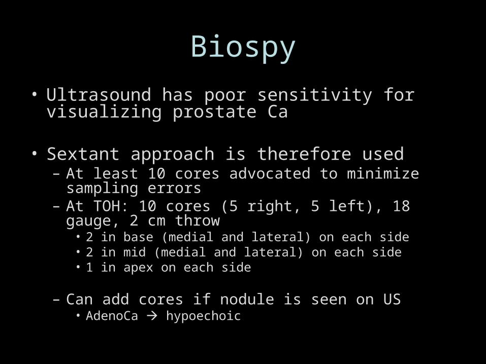
Biospy
• Ultrasound has poor sensitivity for visualizing prostate Ca
• Sextant approach is therefore used– At least 10 cores advocated to minimize sampling errors– At TOH: 10 cores (5 right, 5 left), 18 gauge, 2 cm throw
• 2 in base (medial and lateral) on each side• 2 in mid (medial and lateral) on each side• 1 in apex on each side
– Can add cores if nodule is seen on US• AdenoCa hypoechoic
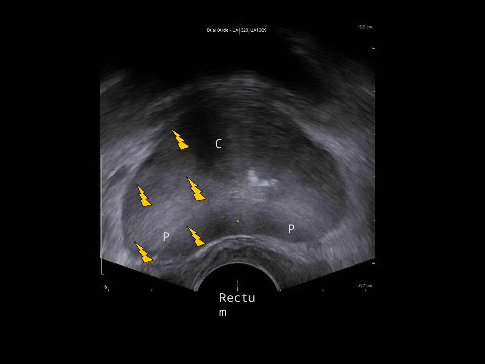
Rectum
C
PP

Prostate Ca on US


Other structures visible on US
SV = Seminal vesiclesArrows = Vas deferens

MRI – Normal appearance
• Peripheral zone high T2 signal intensity• Capsule rim of low T2 signal• Central gland intermediate T2 signal intensity
(more compact smooth muscle and sparser glandular elements)
• Neurovascular bundles course posterolateral to prostate capsule bilaterally at 5- and 7-o’clock

Sag T2 with endorectal coil
Urethra
Symphysis pubis
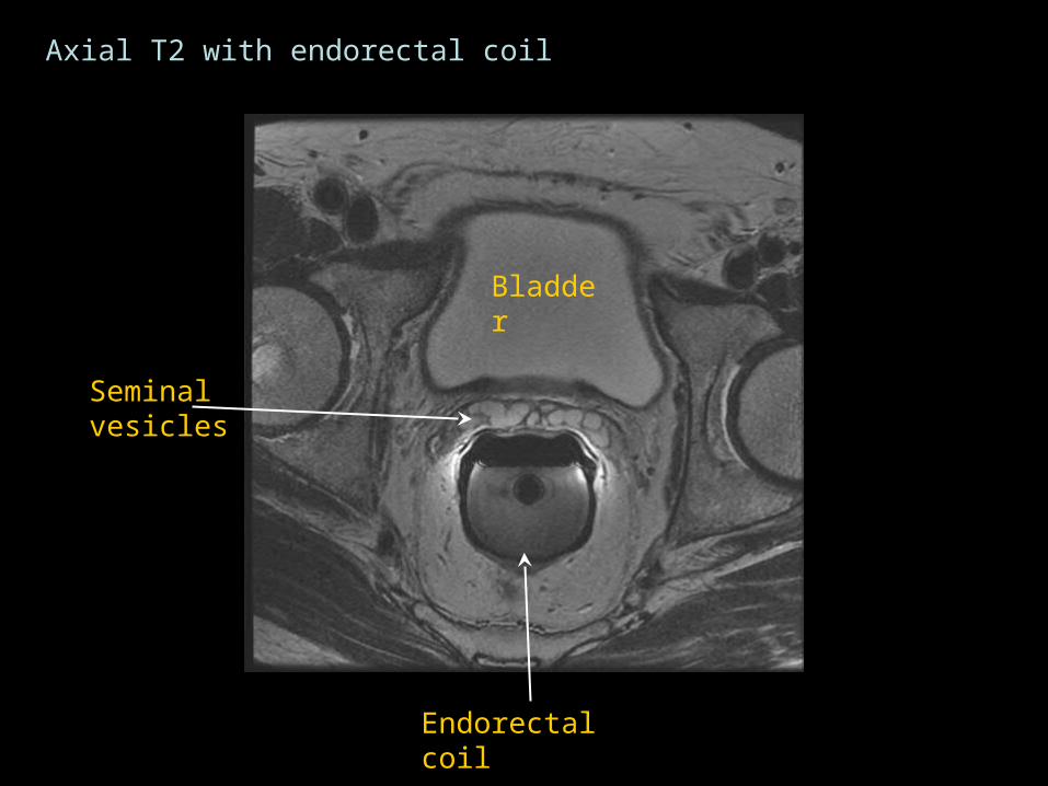
Endorectal coilBladder
Prostate
Endorectal coil
Bladder
Seminal vesicles
Axial T2 with endorectal coil

Prostate
(central gland)
Axial T2 with endorectal coil

Prostate
(Peripheral zone)
Axial T2 with endorectal coil
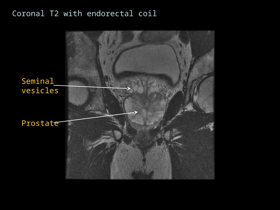
Coronal T2 with endorectal coil
Seminal vesicles
Prostate

Vas Deferens
Role of MRI
• Staging high risk patients
• Evaluating rising PSA in post-prostatectomy patients in the absence of disease elsewhere
• Pre-ablation planning (cryotherapy)
MRI Technique
• Current clinical standard is to perform prostate MRI using endorectal and pelvic phased array coils on a magnet that is at least 1.5 T.
• Endorectal coils and high-resolution images are necessary for accurate localization and staging of prostate cancer
• 8-10 week wait between biopsy and MRI is recommended• Postbiopsy hemorrhage can distort image quality and
mask tumor

MRI Technique
• T1W images from aortic bifurcation to pelvis • to check for postbiopsy hemorrhage • to check for metastases to bone and lymph nodes
• Multiplanar high-resolution fast spin-echo (FSE) T2-weighted images
• Enables detection and localization of tumor
• Diffusion weighted imaging (DWI)• improves detection and localization• higher b values (1000–2000 s/mm2) are better
• 3D gradient echo unenhanced and multiphase contrast-enhanced images
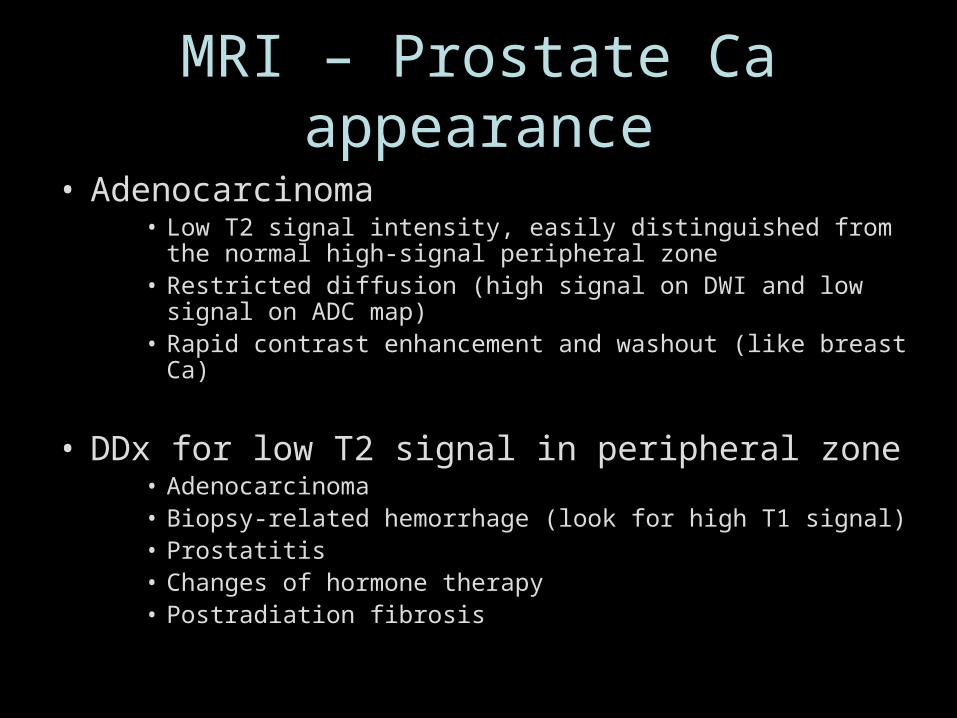
MRI – Prostate Ca appearance
• Adenocarcinoma• Low T2 signal intensity, easily distinguished from the normal
high-signal peripheral zone• Restricted diffusion (high signal on DWI and low signal on ADC
map)• Rapid contrast enhancement and washout (like breast Ca)
• DDx for low T2 signal in peripheral zone• Adenocarcinoma• Biopsy-related hemorrhage (look for high T1 signal)• Prostatitis• Changes of hormone therapy• Postradiation fibrosis

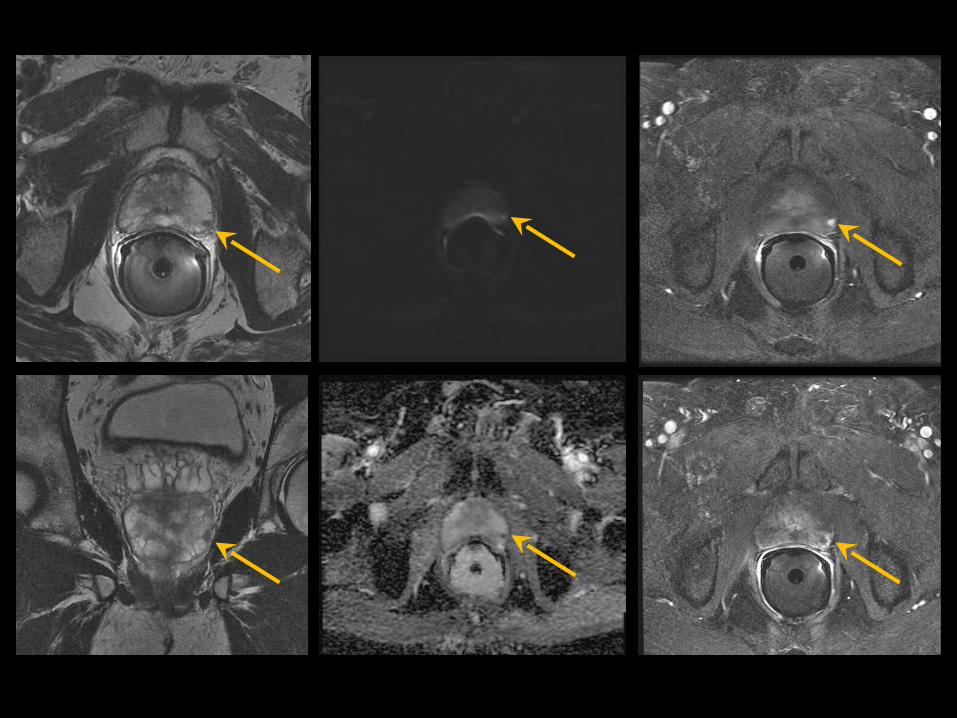
High-resolution T2-weighted images with endorectal coil
Axial Coronal
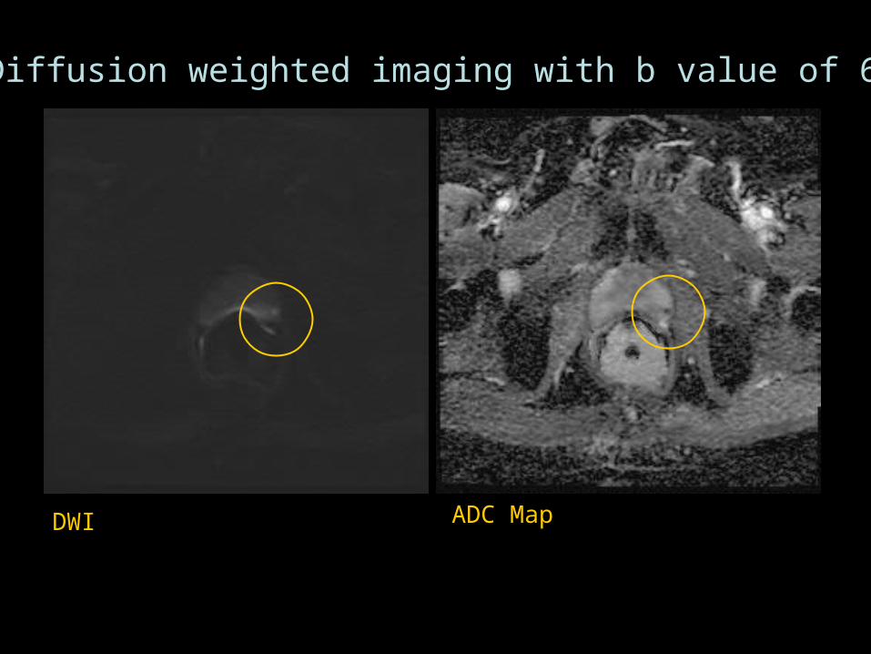
Diffusion weighted imaging with b value of 600
DWI ADC Map
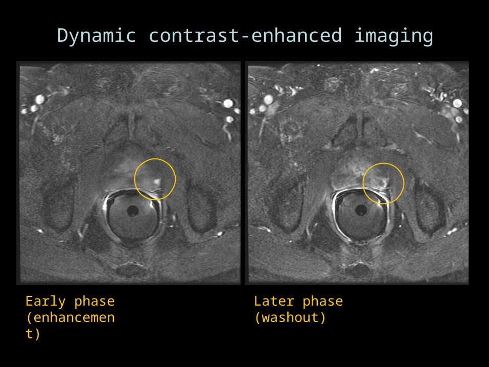
Dynamic contrast-enhanced imaging
Early phase (enhancement)
Later phase (washout)
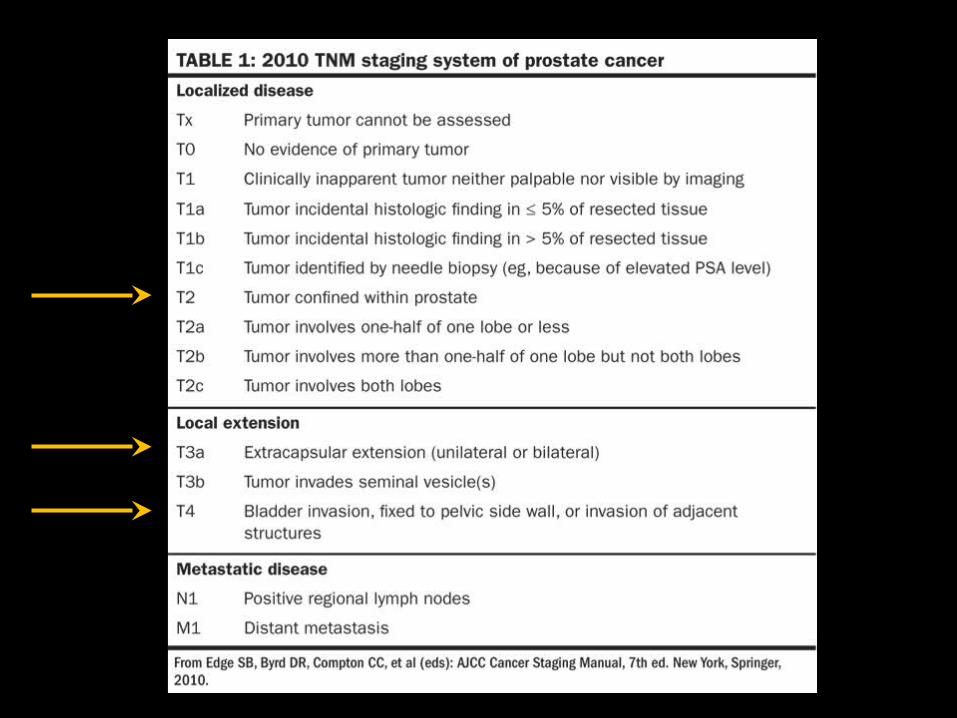

Signs of Extracapsular Extension
• Asymmetric prostate capsular bulge with irregular margins
• Obliteration of the rectoprostatic angle• Asymmetry of neurovascular bundle• Tumor encasement of the neurovascular bundle• Seminal vesicle invasion
Seminal Vesicle (SV) Invasion
• Diagnostic Criteria:– Loss of normal SV architecture – SV enlargement with a low-signal-intensity mass on
T2-weighted images
• Caveat– After radiation, chemo or hormonal therapy, the SVs
often demonstrate decreased size, diffuse wall thickening, or diffuse low signal intensity on T2-weighted images. Can mimic tumor invasion.


Other disease processes
• Infection/inflammation– Prostatitis or seminal vesiculitis– Abscess (prostate or SVs)

54 year-old male with leukemia and severe graft versus host disease of bowel. Elevated PSA.


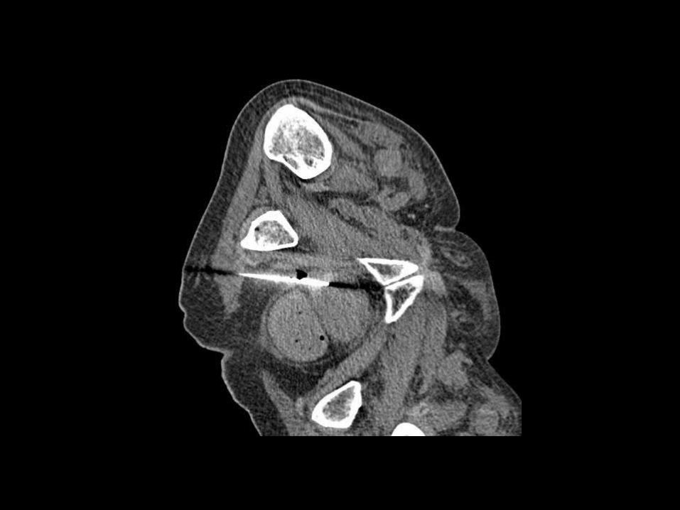


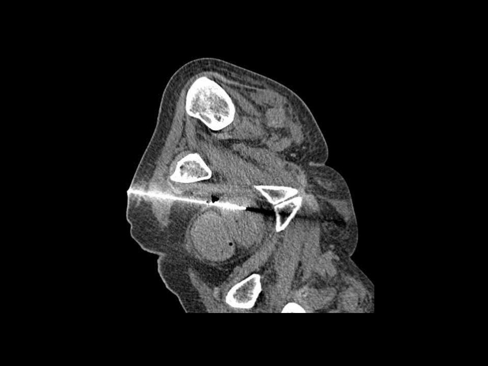


PATH: areas of necrosis and presence of fungal forms (yeast, hyphae, and pseudohyphae) consistent with Candida species.

Cystic prostate masses
• Utricular cyst• Communicates with prostatic urethra • May contain spermatozoa• Confined within prostate at the midline• Associated with GU abnormalities (hypospadias,
cryptorchidism, unilateral renal agenesis)
• Müllerian duct cyst • Does not communicate with urethra • May extend above the prostate• Not associated with other abnormalities

From: Kim B, Kawashima A, Ryu J, Takahashi N, Hartman RP, and King BF. Imaging of the Seminal Vesicle and Vas Deferens. Radiographics. July 2009, 29, 1105-1121.

Seminal vesicle cysts
• If bilateral, think ADPCKD
• If unilateral, think renal agenesis
• Other: acquired from inflammation and obstruction of the ejaculatory ducts and seminal vesicles secondary to urinary infection and calculi.