Embed Size (px)
Citation preview

FEBS Letters 582 (2008) 537–542
Proneural bHLH neurogenin 2 differentially regulatesNurr1-induced dopamine neuron differentiation in rat and mouse
neural precursor cells in vitro
Chang-Hwan Parkb,c,d, Jin Sun Kangb,c,d, Eun-Hye Yoonb,c,d, Jae-Won Shima,c,d,Haeyoung Suh-Kime, Sang-Hun Leea,c,d,*
a Department of Biochemistry and Molecular Biology, College of Medicine, Hanyang University, No. 17 Haengdang-dong, Sungdong-gu,Seoul 133-791, Republic of Korea
b Department of Microbiology, College of Medicine, Hanyang University, No. 17 Haengdang-dong, Sungdong-gu, Seoul 133-791, Republic of Koreac Institute of Mental Health, Hanyang University, No. 17 Haengdang-dong, Sungdong-gu, Seoul 133-791, Republic of Korea
d Cell Therapy Research Center, Hanyang University, No. 17 Haengdang-dong, Sungdong-gu, Seoul 133-791, Republic of Koreae Department of Anatomy and Brain Disease Research Center, College of Medicine, Ajou University, Suwon 442-749, South Korea
Received 27 October 2007; revised 13 December 2007; accepted 8 January 2008
Available online 31 January 2008
Edited by Ned Mantei
Abstract Roles of Nurr1 and neurogenin 2 (Ngn2) have beenshown in midbrain dopamine (DA) neuron development. Wepresent here rat and mouse species-dependent differences ofNurr1 and Ngn2 actions in DA neuron differentiation. Nurr1exogene expression caused an efficient generation of tyrosinehydroxylase (TH)-positive DA cells from rat neural precursorcells (NPCs). Nurr1-induced TH+ cell yields were low andhighly variable depending on the origins of NPCs in mousecultures. Coexpression of Ngn2 repressed Nurr1-induced gener-ation of TH+ cells in rat cultures. In clear contrast, a robustenhancement in Nurr1-induced DA cell yields was observed inmouse NPCs by Ngn2. These findings imply that DA neuronsmay develop differently in the midbrains of these two species.� 2008 Federation of European Biochemical Societies. Publishedby Elsevier B.V. All rights reserved.
Keywords: Dopaminergic differentiation; Neural precursorcells; Neurogenin 2; Species-dependent differences; Nurr1
1. Introduction
Midbrain dopamine (DA) neurons play principal roles in the
regulation of motor behaviors, emotion, and cognition. Dys-
function of these cells is related to Parkinson�s disease, schizo-
phrenia, and drug addiction. Development of the midbrain
dopaminergic system has been extensively studied because of
its physiological and clinical significance. In vivo functional
analyses in mice mainly contribute to identifying the critical
factors of midbrain DA neuron development. However, the
mechanisms of generating midbrain DA neurons are still
largely unknown. Neural stem/precursor cells cultured
in vitro are regarded as an appropriate tool for studies that
aim to gain a deeper understanding of the mechanisms and
signals that regulate the differentiation of stem/precursor cells
into specific neuronal populations. In addition, research has
*Corresponding author. Address: Department of Biochemistry andMolecular Biology, College of Medicine, Hanyang University, No. 17Haengdang-dong, Sungdong-gu, Seoul 133-791, Republic of Korea.Fax: +82 2 2294 6270.E-mail address: [email protected] (S.-H. Lee).
0014-5793/$34.00 � 2008 Federation of European Biochemical Societies. Pu
doi:10.1016/j.febslet.2008.01.018
been focused on the promise of stem cell-based replacement
therapies for neuro-degenerative disorders.
Nurr1 is a transcription factor of the orphan nuclear recep-
tor superfamily that is highly expressed in the developing mid-
brain. Mice lacking Nurr1 exhibit a specific loss of midbrain
DA neurons [1–3], suggesting that Nurr1 is essential for mid-
brain DA neuron development. With respect to the production
of midbrain DA neurons for experimental and transplantable
purposes in regenerative medicine, interest in Nurr1 has been
renewed because engineered Nurr1expression in vitro suffi-
ciently induces the acquisition of DA neuron phenotypes in
naıve non-dopaminergic neural precursor cells (NPCs) [4].
However, Nurr1-induced DA cells were either undifferentiated
non-neuronal cells [5,6] or morphologically and functionally
immature DA neurons [4]. In the search for factors regulating
neuronal differentiation of Nurr1-DA cells, neurogenin (Ngn)
2 and Mash1 proneural basic helix-loop-helix (bHLH) factors
are the first candidates because of their well-acknowledged
pan-neurogenic nature for review, see [7] and proposed roles
in midbrain DA neuron development [8–10].
We have recently demonstrated that coexpression of Mash1,
a proneural transcription factor homologous with drosophila
achaete-scute bHLH, resulted in the morphological and func-
tional differentiation of Nurr1-expressing NPC toward mature
DA neurons exhibiting biochemical and electrophysiological
functionalities as presynaptic DA neurons [10,11]. Interest-
ingly and surprisingly, however, another class of the atonal-re-
lated bHLHs including Ngn1, Ngn2, and neuroD remarkably
repressed Nurr1-induced expression of DA neuronal markers
in the cultures for NPCs derived from rat embryonic brains
[10,11]. These findings are in contrast to the loss-of-function
studies in mice that demonstrate a reduced number of DA neu-
rons in developing midbrains by Ngn2 deletion, which suggests
that Ngn2 is required in mouse midbrain DA neuron develop-
ment [8,9]. The inconsistency of the in vivo and in vitro find-
ings may be attributable to the differences between the two
experimental systems with a possibility that the in vitro NPC
cultures may not reflect physiologic developmental events in
certain cases. Alternatively, the contrasting regulatory roles
of Ngn2 could represent a real species-dependent difference
in midbrain DA neuron development. To help answer
this question, we examined Ngn2 roles in Nurr1-induced DA
blished by Elsevier B.V. All rights reserved.

538 C.-H. Park et al. / FEBS Letters 582 (2008) 537–542
neuron differentiation in the cultures of NPCs derived from rat
and mouse brains.
2. Materials and methods
2.1. Cultures for NPCsCultures of NPCs derived from rodent embryonic brains were per-
formed as described previously [4] with some modifications. Briefly,embryonic brain tissues were dissected from the cortex (Ctx), lateralganglionic eminence (LGE, anlage of striatum), and ventral midbrain(VM) of rats (Sprague–Dawley (SD), KOATECK, Seoul, Korea) atembryonic day 12 (E12)–E14 or from the identical brain regions ofmouse embryos (ICR, KOATECK, E10.5–E12.5). After mechanicaltrituration, cells were plated at 20,000 cells/cm2 on culture dishespre-coated with poly-LL-ornithine (PO)/fibronectin (FN) in N2B med-ium (DMEM/F12, 4.4 lM insulin, 100 mg/l transferrin, 30 nM sele-nite, 0.6 lM putrescine, 20 nM progesterone, 0.2 mM ascorbic acid,2 mM LL-glutamine, 8.6 mM DD(+) glucose, 20 mM NaHCO3, B27(Invitrogen, Carlsbad, CA)) supplemented with basic fibroblast growthfactor (bFGF; 20 ng/ml, R&D Systems, Minneapolis, MN), and epi-dermal growth factor (EGF, 20 ng/ml, R&D) and were cultured for4–6 days as a monolayer on the adherent surface. To obtain a uniformpopulation of NPCs, clusters of cells, which were formed by the prolif-eration of NPCs with the mitogens (bFGF and EGF), were passagedby dissociating them into single cells and plating them onto freshlyPO/FN-coated coverslips (12-mm diameter; Marienfeld GmbH &Co. KG, Lauda-Knlgshofen, Germany) [12]. After subsequent precur-sor expansion up to 50–60% cell confluence, cells were retrovirallytransduced as described below. On the day following infection, cell dif-ferentiation was induced in N2B medium in the absence of the mito-gens for 3–10 days. All the data in this study were obtained in thepassaged cultures grown on the adherent surface, unless otherwisenoted. Cultures were maintained at 37 �C in a 5% CO2 incubator.Medium was changed every other day and mitogens were added daily.
‘‘Neurosphere’’ culture, another widely used culture method forNPCs, was carried out under identical experimental conditions tothose described above, except cells were seeded in low adhesive culturedishes (No. 3262 Ultra Low Attachment; Corning, NY) without thePO/FN-coating procedure to prevent cells from attaching to the cul-ture dish surface. Four to six days after cell expansion, floating cellaggregates (neurospheres) were mechanically dissociated into smallpieces and subjected to retroviral infection in a cell suspension. Celldifferentiation was induced by plating the fragmented neurosphereson PO/FN-coated surfaces and culturing for 3–10 days in N2B.
NPCs were also isolated and cultured from the subventricular zone(SVZ) of adult rat or mouse brains [13]. For details, see the supplemen-tary methods.
2.2. Retroviral construction and transductionMouse Nurr1 (mNurr1, Genebank number: GI 7305324), mNgn2
(GI 34328159), rat Mash1 (rMash1, GI 11693147), and human Nurr1(hNurr1, GI 27894347) and hNgn2 (GI 22477416) cDNAs were engi-neered into the retroviral vector pCL as previously described [11].Details for retroviral construction and transduction are given in thesupplementary methods.
2.3. Cell counting and statistical analysisCell counting was performed in uniform microscopic fields that were
randomly chosen across the culture area, using an eyepiece grid at afinal magnification of 200· or 400·. Statistical comparisons were per-formed using SPSS software (version 11.0; SPSS Inc., Chicago, IL).One-way or two-way ANOVA, followed by Tukey�s post hoc compar-ison was applied where appropriate.
Methods for immunochemical, immunoblot, RT-PCR and DArelease analyses are described in the supplementary methods.
3. Results and discussion
In order to compare the roles of Ngn2 in Nurr1-induced
dopaminergic differentiation in rats and mice, NPCs were iso-
lated and cultured from rat and mouse embryonic brains. To
exclude any potential experimental bias, the comparative stud-
ies were carried out under various experimental conditions,
which included the gathering of precursor cells from various
brain regions at variable developmental stages and different
culture methods, such as adherent monolayer and floating
neurosphere cultures. In particular, we paid special attention
to creating identical experimental settings in the rat and mouse
cultures.
As was described previously [4], exogenous expression of
Nurr1 in rat NPCs efficiently yielded cells expressing TH, a
key enzyme of DA biosynthesis, regardless of embryonic age
(E12, E14) or the brain region (Ctx, LGE, VM) from which
the precursors were isolated (Fig. 1A–F). Of the cells trans-
duced with Nurr1 (Nurr1-IRES-eGFP), as visualized by
GFP expression, 49–66% were positive for TH at 2 days after
transduction (Fig. 1M). Comparisons of the TH+ cell yields
induced by Nurr1 transduction were made between the rat
and mouse NPC cultures derived from identical regions at
corresponding embryonic days (rat E12 versus mouse E10.5
and rat E14 versus mouse E12.5). Viral transductions were
efficient (>90% GFP+/total DAPI+ cells) and indistinguish-
able in both rat and mouse cultures. Nurr1-induced TH+ cell
yields from the mouse NPCs were extremely low, compared
with those of the corresponding rat cultures. For instance,
the percentages of TH+ cells out of the GFP+ infected cells
in rat E14 versus mouse E12.5 cultures were 55.3 ± 4.1% versus
2.5 ± 0.2% (cortex), 59.1 ± 8.2% versus 3.4 ± 0.1% (LGE), and
49.1 ± 3.6% versus 7.9 ± 0.2% (VM) (n = 300 microscopic
fields for each value from three independent experiments).
Unlike the invariably high embryonic age- and region-indepen-
dent TH+ cell yields in the rat cultures, Nurr1-induced TH+
cell yields were highly variable (2–15% of GFP+ cells), depend-
ing on the mouse brain regions and embryonic ages of the
precursors. The higher TH+ cell yields were attained in the
mouse NPCs derived from younger embryos, and the VM
showed the greatest Nurr1 effect in yielding TH+ cells among
the brain regions tested. Thus, the most efficient TH+ cell yield
among the mouse cultures tested was observed in the NPCs de-
rived from the VM at E10.5. These findings are consistent with
previous studies showing that Nurr1-induced TH expression in
mouse precursor cells requires factors derived from astrocytes
of embryonic midbrain-origin [14,15] and that cells from early
embryonic midbrains secrete autocrine/paracrine factors for
in vitro precursor differentiation of DA neurons [16].
As previously reported [10,11], coexpression of Ngn2 in rat
NPCs resulted in a striking repression of Nurr1-induced deriva-
tion of TH+ cells. The inhibitory role of Ngn2 was observed in
all rat cultures derived from different developmental ages and
brain regions (Fig. 2A, B, E and data not shown). To our sur-
prise, however, the opposite effect of Ngn2 was observed in the
mouse cultures; forced expression of Ngn2 resulted in a
remarkable (5.3–8.6-fold) enhancement of Nurr1-induced
TH+ cell yields in all the mouse cultures tested (Fig. 2C, D
and F). For instance, the percentages of TH+ cells among all
cells were 38.1 ± 1.7% and 7.3 ± 0.5% (n = 300 from three inde-
pendent experiments) in E12.5 VM cultures transduced with
Nurr1 + Ngn2 and Nurr1 alone, respectively. Immunoblots
for TH protein levels further confirmed the contrasting roles
of Ngn2 in mouse and rat NPC cultures (Fig. 2G).
Neuron numbers were also greatly increased in the Ngn2
co-transduced VM cultures (TuJ1+ cells out of all cells were
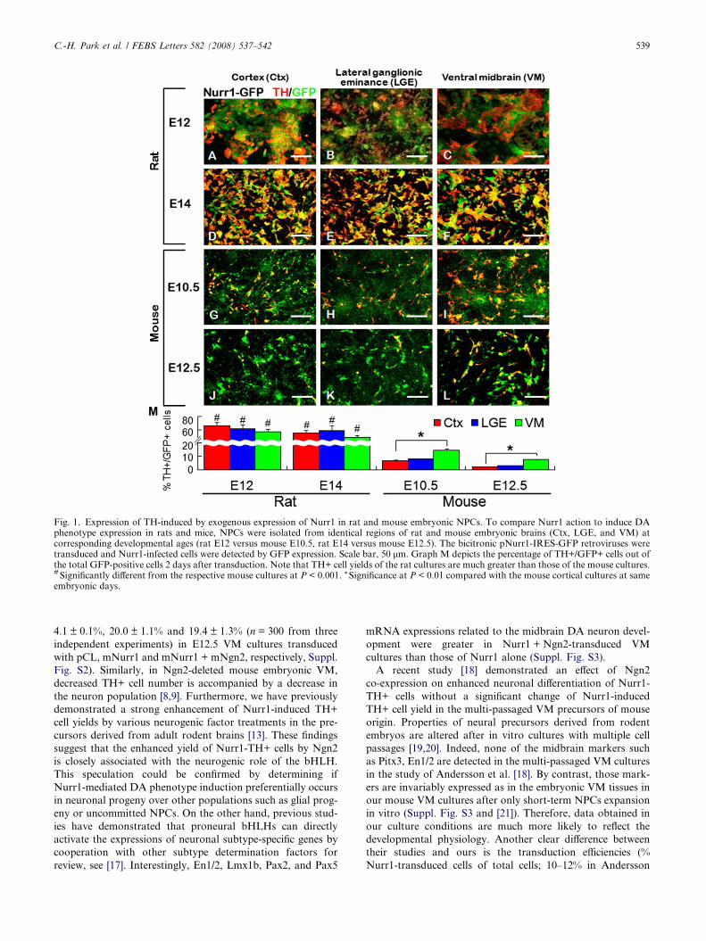
Fig. 1. Expression of TH-induced by exogenous expression of Nurr1 in rat and mouse embryonic NPCs. To compare Nurr1 action to induce DAphenotype expression in rats and mice, NPCs were isolated from identical regions of rat and mouse embryonic brains (Ctx, LGE, and VM) atcorresponding developmental ages (rat E12 versus mouse E10.5, rat E14 versus mouse E12.5). The bicitronic pNurr1-IRES-GFP retroviruses weretransduced and Nurr1-infected cells were detected by GFP expression. Scale bar, 50 lm. Graph M depicts the percentage of TH+/GFP+ cells out ofthe total GFP-positive cells 2 days after transduction. Note that TH+ cell yields of the rat cultures are much greater than those of the mouse cultures.# Significantly different from the respective mouse cultures at P < 0.001. * Significance at P < 0.01 compared with the mouse cortical cultures at sameembryonic days.
C.-H. Park et al. / FEBS Letters 582 (2008) 537–542 539
4.1 ± 0.1%, 20.0 ± 1.1% and 19.4 ± 1.3% (n = 300 from three
independent experiments) in E12.5 VM cultures transduced
with pCL, mNurr1 and mNurr1 + mNgn2, respectively, Suppl.
Fig. S2). Similarly, in Ngn2-deleted mouse embryonic VM,
decreased TH+ cell number is accompanied by a decrease in
the neuron population [8,9]. Furthermore, we have previously
demonstrated a strong enhancement of Nurr1-induced TH+
cell yields by various neurogenic factor treatments in the pre-
cursors derived from adult rodent brains [13]. These findings
suggest that the enhanced yield of Nurr1-TH+ cells by Ngn2
is closely associated with the neurogenic role of the bHLH.
This speculation could be confirmed by determining if
Nurr1-mediated DA phenotype induction preferentially occurs
in neuronal progeny over other populations such as glial prog-
eny or uncommitted NPCs. On the other hand, previous stud-
ies have demonstrated that proneural bHLHs can directly
activate the expressions of neuronal subtype-specific genes by
cooperation with other subtype determination factors for
review, see [17]. Interestingly, En1/2, Lmx1b, Pax2, and Pax5
mRNA expressions related to the midbrain DA neuron devel-
opment were greater in Nurr1 + Ngn2-transduced VM
cultures than those of Nurr1 alone (Suppl. Fig. S3).
A recent study [18] demonstrated an effect of Ngn2
co-expression on enhanced neuronal differentiation of Nurr1-
TH+ cells without a significant change of Nurr1-induced
TH+ cell yield in the multi-passaged VM precursors of mouse
origin. Properties of neural precursors derived from rodent
embryos are altered after in vitro cultures with multiple cell
passages [19,20]. Indeed, none of the midbrain markers such
as Pitx3, En1/2 are detected in the multi-passaged VM cultures
in the study of Andersson et al. [18]. By contrast, those mark-
ers are invariably expressed as in the embryonic VM tissues in
our mouse VM cultures after only short-term NPCs expansion
in vitro (Suppl. Fig. S3 and [21]). Therefore, data obtained in
our culture conditions are much more likely to reflect the
developmental physiology. Another clear difference between
their studies and ours is the transduction efficiencies (%
Nurr1-transduced cells of total cells; 10–12% in Andersson
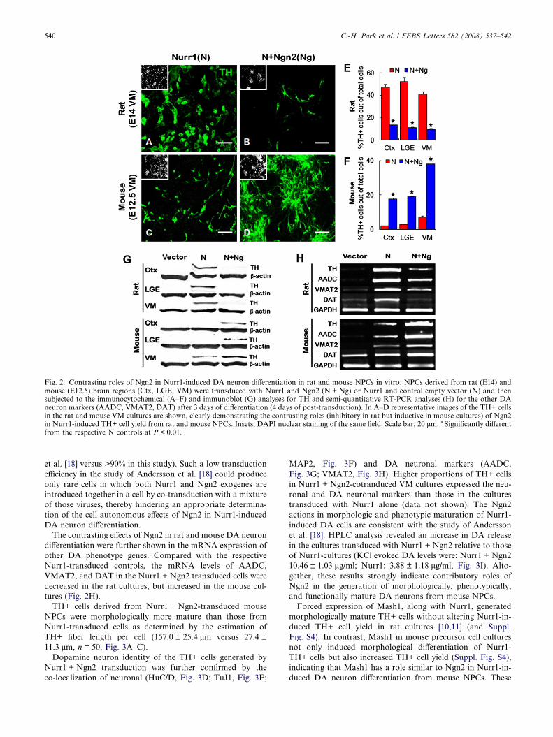
Fig. 2. Contrasting roles of Ngn2 in Nurr1-induced DA neuron differentiation in rat and mouse NPCs in vitro. NPCs derived from rat (E14) andmouse (E12.5) brain regions (Ctx, LGE, VM) were transduced with Nurr1 and Ngn2 (N + Ng) or Nurr1 and control empty vector (N) and thensubjected to the immunocytochemical (A–F) and immunoblot (G) analyses for TH and semi-quantitative RT-PCR analyses (H) for the other DAneuron markers (AADC, VMAT2, DAT) after 3 days of differentiation (4 days of post-transduction). In A–D representative images of the TH+ cellsin the rat and mouse VM cultures are shown, clearly demonstrating the contrasting roles (inhibitory in rat but inductive in mouse cultures) of Ngn2in Nurr1-induced TH+ cell yield from rat and mouse NPCs. Insets, DAPI nuclear staining of the same field. Scale bar, 20 lm. * Significantly differentfrom the respective N controls at P < 0.01.
540 C.-H. Park et al. / FEBS Letters 582 (2008) 537–542
et al. [18] versus >90% in this study). Such a low transduction
efficiency in the study of Andersson et al. [18] could produce
only rare cells in which both Nurr1 and Ngn2 exogenes are
introduced together in a cell by co-transduction with a mixture
of those viruses, thereby hindering an appropriate determina-
tion of the cell autonomous effects of Ngn2 in Nurr1-induced
DA neuron differentiation.
The contrasting effects of Ngn2 in rat and mouse DA neuron
differentiation were further shown in the mRNA expression of
other DA phenotype genes. Compared with the respective
Nurr1-transduced controls, the mRNA levels of AADC,
VMAT2, and DAT in the Nurr1 + Ngn2 transduced cells were
decreased in the rat cultures, but increased in the mouse cul-
tures (Fig. 2H).
TH+ cells derived from Nurr1 + Ngn2-transduced mouse
NPCs were morphologically more mature than those from
Nurr1-transduced cells as determined by the estimation of
TH+ fiber length per cell (157.0 ± 25.4 lm versus 27.4 ±
11.3 lm, n = 50, Fig. 3A–C).
Dopamine neuron identity of the TH+ cells generated by
Nurr1 + Ngn2 transduction was further confirmed by the
co-localization of neuronal (HuC/D, Fig. 3D; TuJ1, Fig. 3E;
MAP2, Fig. 3F) and DA neuronal markers (AADC,
Fig. 3G; VMAT2, Fig. 3H). Higher proportions of TH+ cells
in Nurr1 + Ngn2-cotranduced VM cultures expressed the neu-
ronal and DA neuronal markers than those in the cultures
transduced with Nurr1 alone (data not shown). The Ngn2
actions in morphologic and phenotypic maturation of Nurr1-
induced DA cells are consistent with the study of Andersson
et al. [18]. HPLC analysis revealed an increase in DA release
in the cultures transduced with Nurr1 + Ngn2 relative to those
of Nurr1-cultures (KCl evoked DA levels were: Nurr1 + Ngn2
10.46 ± 1.03 lg/ml; Nurr1: 3.88 ± 1.18 lg/ml, Fig. 3I). Alto-
gether, these results strongly indicate contributory roles of
Ngn2 in the generation of morphologically, phenotypically,
and functionally mature DA neurons from mouse NPCs.
Forced expression of Mash1, along with Nurr1, generated
morphologically mature TH+ cells without altering Nurr1-in-
duced TH+ cell yield in rat cultures [10,11] (and Suppl.
Fig. S4). In contrast, Mash1 in mouse precursor cell cultures
not only induced morphological differentiation of Nurr1-
TH+ cells but also increased TH+ cell yield (Suppl. Fig. S4),
indicating that Mash1 has a role similar to Ngn2 in Nurr1-in-
duced DA neuron differentiation from mouse NPCs. These
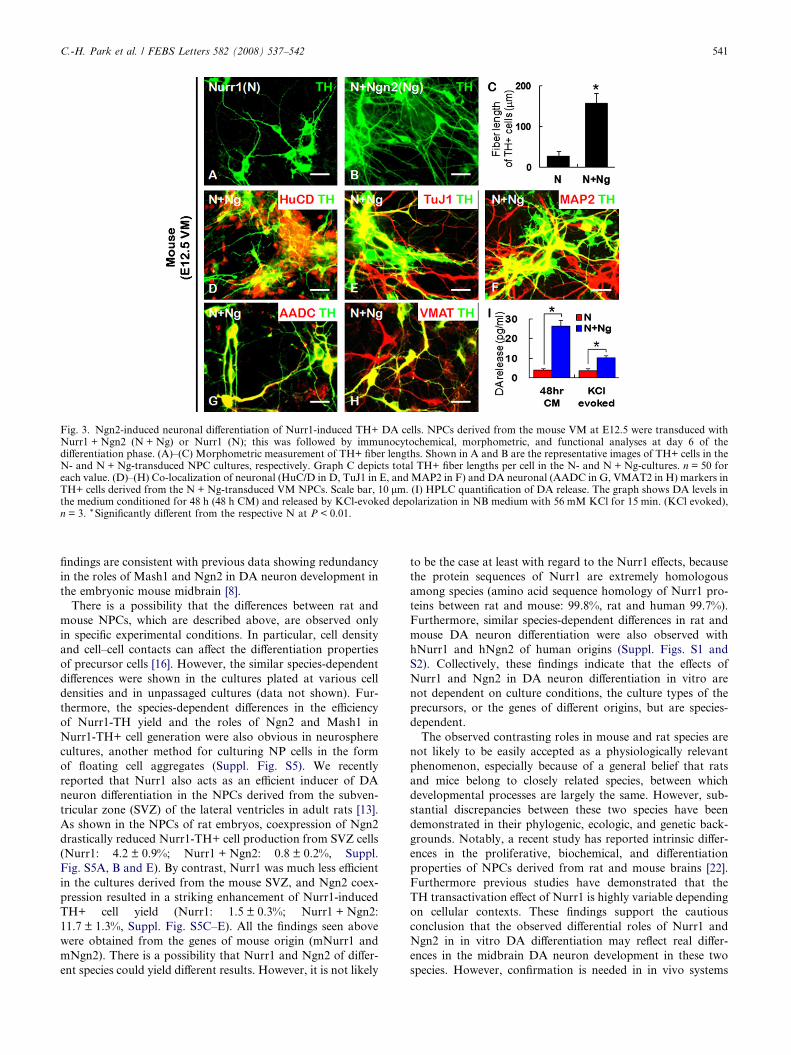
Fig. 3. Ngn2-induced neuronal differentiation of Nurr1-induced TH+ DA cells. NPCs derived from the mouse VM at E12.5 were transduced withNurr1 + Ngn2 (N + Ng) or Nurr1 (N); this was followed by immunocytochemical, morphometric, and functional analyses at day 6 of thedifferentiation phase. (A)–(C) Morphometric measurement of TH+ fiber lengths. Shown in A and B are the representative images of TH+ cells in theN- and N + Ng-transduced NPC cultures, respectively. Graph C depicts total TH+ fiber lengths per cell in the N- and N + Ng-cultures. n = 50 foreach value. (D)–(H) Co-localization of neuronal (HuC/D in D, TuJ1 in E, and MAP2 in F) and DA neuronal (AADC in G, VMAT2 in H) markers inTH+ cells derived from the N + Ng-transduced VM NPCs. Scale bar, 10 lm. (I) HPLC quantification of DA release. The graph shows DA levels inthe medium conditioned for 48 h (48 h CM) and released by KCl-evoked depolarization in NB medium with 56 mM KCl for 15 min. (KCl evoked),n = 3. * Significantly different from the respective N at P < 0.01.
C.-H. Park et al. / FEBS Letters 582 (2008) 537–542 541
findings are consistent with previous data showing redundancy
in the roles of Mash1 and Ngn2 in DA neuron development in
the embryonic mouse midbrain [8].
There is a possibility that the differences between rat and
mouse NPCs, which are described above, are observed only
in specific experimental conditions. In particular, cell density
and cell–cell contacts can affect the differentiation properties
of precursor cells [16]. However, the similar species-dependent
differences were shown in the cultures plated at various cell
densities and in unpassaged cultures (data not shown). Fur-
thermore, the species-dependent differences in the efficiency
of Nurr1-TH yield and the roles of Ngn2 and Mash1 in
Nurr1-TH+ cell generation were also obvious in neurosphere
cultures, another method for culturing NP cells in the form
of floating cell aggregates (Suppl. Fig. S5). We recently
reported that Nurr1 also acts as an efficient inducer of DA
neuron differentiation in the NPCs derived from the subven-
tricular zone (SVZ) of the lateral ventricles in adult rats [13].
As shown in the NPCs of rat embryos, coexpression of Ngn2
drastically reduced Nurr1-TH+ cell production from SVZ cells
(Nurr1: 4.2 ± 0.9%; Nurr1 + Ngn2: 0.8 ± 0.2%, Suppl.
Fig. S5A, B and E). By contrast, Nurr1 was much less efficient
in the cultures derived from the mouse SVZ, and Ngn2 coex-
pression resulted in a striking enhancement of Nurr1-induced
TH+ cell yield (Nurr1: 1.5 ± 0.3%; Nurr1 + Ngn2:
11.7 ± 1.3%, Suppl. Fig. S5C–E). All the findings seen above
were obtained from the genes of mouse origin (mNurr1 and
mNgn2). There is a possibility that Nurr1 and Ngn2 of differ-
ent species could yield different results. However, it is not likely
to be the case at least with regard to the Nurr1 effects, because
the protein sequences of Nurr1 are extremely homologous
among species (amino acid sequence homology of Nurr1 pro-
teins between rat and mouse: 99.8%, rat and human 99.7%).
Furthermore, similar species-dependent differences in rat and
mouse DA neuron differentiation were also observed with
hNurr1 and hNgn2 of human origins (Suppl. Figs. S1 and
S2). Collectively, these findings indicate that the effects of
Nurr1 and Ngn2 in DA neuron differentiation in vitro are
not dependent on culture conditions, the culture types of the
precursors, or the genes of different origins, but are species-
dependent.
The observed contrasting roles in mouse and rat species are
not likely to be easily accepted as a physiologically relevant
phenomenon, especially because of a general belief that rats
and mice belong to closely related species, between which
developmental processes are largely the same. However, sub-
stantial discrepancies between these two species have been
demonstrated in their phylogenic, ecologic, and genetic back-
grounds. Notably, a recent study has reported intrinsic differ-
ences in the proliferative, biochemical, and differentiation
properties of NPCs derived from rat and mouse brains [22].
Furthermore previous studies have demonstrated that the
TH transactivation effect of Nurr1 is highly variable depending
on cellular contexts. These findings support the cautious
conclusion that the observed differential roles of Nurr1 and
Ngn2 in in vitro DA differentiation may reflect real differ-
ences in the midbrain DA neuron development in these two
species. However, confirmation is needed in in vivo systems
542 C.-H. Park et al. / FEBS Letters 582 (2008) 537–542
to assure that the species difference is a biologically relevant
one.
Nurr1 gene manipulation in precursor or stem cells is partic-
ularly important in generating transplantable DA neurons in
the cell replacement approaches for Parkinson�s patients.
However, it was recently reported that Nurr1, dissimilar to
its action of DA cell differentiation in rodent cells, is unable
to yield TH+ DA cells from human NPCs [23]. This is another
example of the species-dependent differences of Nurr1 action,
indicating that simple and direct application of Nurr1 exoge-
nous expression is infeasible for generating human DA neu-
rons. In this regard, further studies are needed to elucidate
the mechanisms for the species-dependent roles of Nurr1 and
Ngn2 that were observed in this study; these studies could pro-
vide possible clues for successful Nurr1-utilized generation of
transplantable human DA neurons.
Acknowledgements: We are grateful to Dr. Toshiyuki Ohtsuka (KyotoUniversity, Japan) for his critical advice on this study. This work wassupported by SC2130 (Stem Cell Research Center of the 21st CenturyFrontier Research Program) funded by the Ministry of Science andTechnology, Republic of Korea.
Appendix A. Supplementary material
Supplementary data associated with this article can be
found, in the online version, at doi:10.1016/j.febslet.2008.
01.018.
References
[1] Zetterstrom, R.H., Solomin, L., Jansson, L., Hoffer, B.J., Olson,L. and Perlmann, T. (1997) Dopamine neuron agenesis in Nurr1-deficient mice. Science 276, 248–250.
[2] Baffi, J.S., Palkovits, M., Castillo, S.O., Mezey, E. and Nikodem,V.M. (1999) Differential expression of tyrosine hydroxylase incatecholaminergic neurons of neonatal wild-type and Nurr1-deficient mice. Neuroscience 93, 631–642.
[3] Le, W., Conneely, O.M., Zou, L., He, Y., Saucedo-Cardenas, O.,Jankovic, J., Mosier, D.R. and Appel, S.H. (1999) Selectiveagenesis of mesencephalic dopaminergic neurons in Nurr1-deficient mice. Exp. Neurol. 159, 451–458.
[4] Kim, J.Y., Koh, H.C., Lee, J.Y., Chang, M.Y., Kim, Y.C.,Chung, H.Y., Son, H., Lee, Y.S., Studer, L., McKay, R. and Lee,S.H. (2003) Dopaminergic neuronal differentiation from ratembryonic neural precursors by Nurr1 overexpression. J. Neuro-chem. 85 (6), 1443–1454.
[5] Sonntag, K.C., Simantov, R., Kim, K.S. and Isacson, O. (2004)Temporally induced Nurr1 can induce a non-neuronal dopami-nergic cell type in embryonic stem cell differentiation. Eur. J.Neurosci. 19 (5), 1141–1152.
[6] Sakurada, K., Ohshima-Sakurada, M., Palmer, T.D. and Gage,F.H. (1999) Nurr1, an orphan nuclear receptor, is a transcrip-tional activator of endogenous tyrosine hydroxylase in neuralprogenitor cells derived from the adult brain. Development 126(18), 4017–4026.
[7] Kageyama, R. and Nakanishi, S. (1997) Helix-loop-helix factorsin growth and differentiation of the vertebrate nervous system.Curr. Opin. Genet. Dev. 7, 659–665.
[8] Kele, J., Simplicio, N., Ferri, A.L., Mira, H., Guillemot, F.,Arenas, E. and Ang, S.L. (2006) Neurogenin 2 is required for thedevelopment of ventral midbrain dopaminergic neurons. Devel-opment 133, 495–505.
[9] Andersson, E., Jensen, J.B., Parmar, M., Guillemot, F. andBjorklund, A. (2006) Development of the mesencephalic dopa-minergic neuron system is compromised in the absence ofneurogenin 2. Development 133, 507–516.
[10] Park, C.H., Kang, J.S., Kim, J.S., Chung, S., Koh, J.Y., Yoon,E.H., Jo, A.Y., Chang, M.Y., Koh, H.C., Hwang, H., Suh-Kim, S., Lee, Y.S., Kim, K.S. and Lee, S.H. (2006) Differentialactions of the proneural genes Mash1 and Neurogenins inNurr1-induced dopamine neuron differentiation. J. Cell Sci. 119,2310–2320.
[11] Park, C.H., Kang, J.S., Shin, Y.H., Chang, M.Y., Chung, S.,Koh, H.C., Zhu, M.H., Oh, S.B., Lee, Y.S., Panagiotakos, G.,Tabar, V., Studer, L. and Lee, S.H. (2006) Acquisition of in vitroand in vivo functionality of Nurr1-induced dopamine neurons.FASEB J. 20 (14), 2553–2555.
[12] Johe, K.K., Hazel, T.G., Muller, T., Dugich-Djordjevic, M.M.and McKay, R.D. (1996) Single factors direct the differentiationof stem cells from the fetal and adult central nervous system. GeneDev. 15, 3129–3140.
[13] Shim, J.W., Park, C.H., Bae, Y.C., Bae, J.Y., Chung, S., Chang,M.Y., Koh, H.C., Lee, H.S., Hwang, S.J., Lee, K.H., Lee, Y.S.,Choi, C.Y. and Lee, S.H. (2007) Generation of functionaldopamine neurons from neural precursor cells isolated from thesubventricular zone and white matter of the adult rat brain usingNurr1 overexpression. Stem Cell 25, 1252–1262.
[14] Wagner, J., Akerud, P., Castro, D.S., Holm, P.C., Canals, J.M.,Snyder, E.Y., Perlmann, T. and Arenas, E. (1999) Induction of amidbrain dopaminergic phenotype in Nurr1-overexpressing neu-ral stem cells by type 1 astrocytes. Nat. Biotech. 17, 653–659.
[15] Castelo-Branco, G., Sousa, K.M., Bryja, V., Pinto, L., Wagner, J.and Arenas, E. (2006) Ventral midbrain glia express region-specific transcription factors and regulate dopaminergic neuro-genesis through Wnt-5a secretion. Mol. Cell. Neurosci. 31, 251–262.
[16] Ko, J.Y., Lee, J.Y., Park, C.H. and Lee, S.H. (2005) Effect ofCell-density on in vitro dopaminergic differentiation of mesence-phalic precursor cells. Neuroreport 16, 499–503.
[17] Bertrand, N., Castro, D.S. and Guillemot, F. (2002) Proneuralgenes and the specification of neural cell types. Nat. Rev.Neurosci. 3, 517–530.
[18] Andersson, E.K.I., Irvin, D.K., Ahlsio, J. and Parmar, M. (2007)Ngn2 and Nurr1 act in synergy to induce midbrain dopaminergicneurons from expanded neural stem and progenitor cells. Exp.Cell. Res. 313, 1172–1180.
[19] Yan, J., Studer, L. and McKay, R.D.G. (2001) Ascorbic acidincreases the yield of dopaminergic neurons derived from basicfibroblast growth factor expanded mesencephalic precursors. J.Neurochem. 76, 307–311.
[20] Chang, M.Y., Park, C.H., Lee, S.Y. and Lee, S.H. (2004)Properties of cortical precursor cells cultured long term aresimilar to those of precursors at later developmental stages. Dev.Brain Res. 153, 89–96.
[21] Jo, A.Y., Park, C.H., Aizawa, S. and Lee, S.H. (2007) Contrastingand brain region-specific roles of neurogenin 2 and Mash1 inGABAergic neuron differentiation in vitro. Exp. Cell. Res. 313,4066–4081.
[22] Ray, J. and Gage, F.H. (2006) Differential properties of adult ratand mouse brain-derived neural stem/progenitor cells. Mol. Cell.Neurosci. 31, 560–573.
[23] Jin, H., Romano, G., Marshall, C., Donaldson, A.E., Suon, S.and Iacovitti, L. (2006) Tyrosine hydroxylase gene regulation inhuman neuronal progenitor cells does not depend on Nurr1 as inthe murine and rat systems. J. Cell. Physiol. 207, 49–57.