Embed Size (px)
Citation preview

have reviewed 20 patients with partial 11q trisomy whereCC aplasia was confirmed in 2/4 patients, and with growthand mental retardation in almost all patients.14
Magnetic resonance imaging is the best modality forevaluation of the CC.15 Agenesis has been described in1.6% of consecutive MRI undertaken at a pediatric refer-ence centre,16 while in other nonpediatric institutions theprevalence of CC aplasia in MRIs done for various nonur-gent indications, has been 0.07%.12 Another series by Mar-zal et al. found evidence of CC aplasia in seven out of 135children who had been referred with structural defects inthe central nervous system.10
In some cases a partial agenesis of the CC is accompa-nied by enlargement of the anterior commissure or the hip-pocampal commissure that may mimic an isolated callosalsplenium.17 Whether isolated callosal hypogenesis is ofclinical importance, and to what degree, is debatable asthere are asymptomatic patients where the anomaly isdetected by cerebral imaging, and neither developmentaldisabilities nor interhemispheric transfer dysfunction areobserved in these patients.18
In conclusion, a very young patient with parathyroidadenoma and an incidental CC aplasia has been presented.To our knowledge a relationship between CC aplasia andparathyroid adenoma has not been established. However,from our discussion above there is evidence that a propor-tion of parathyroid adenomas and corpus callosal aplasiasare caused by genetic abnormalities of the same chromo-some, that is chromosome 11. Our patient and his family re-fused genetic analysis. Whether 11q trisomy or monosomyor other types of chromosomal defects related to asymp-tomatic CC aplasia could lead to gene alterations on chro-mosome 11 that might trigger parathyroid cell replication,is to be further evaluated by appropriate epidemiologicalstudies. We advise that patients with CC aplasia shouldbe screened for primary hyperparathyroidism, until the pos-sible association of the two abnormalities is clarified.
References
1. Carling T. Molecular pathology of parathyroid tumors. Trends
Endocrinol Metab 2001;12 (2):53–8.2. Miedlich S, Krohn K, Paschke R. Update on genetic and clinical aspects
of primary hyperparathyroidism. Clin Endocrinol 2003;59:539–54.3. Arnold A, Staunton CE, Kim HG, et al. Monoclonality and
abnormal parathyroid hormone genes in parathyroid adenomas.New Engl J Med 1988;318:658–62.
4. Motokura T, Bloom T, Kim HG, et al. Novel cyclin encoded by aBcl1-linked candidate oncogene. Nature 1991;350:512–5.
5. Arnold A, Shattuck TM, Mallya SM, et al. Molecular pathogenesisof primary hyperparathyroidism. J Bone Miner Res 2002;17 (Suppl.2):N30–6.
6. Teh BT, Farnebo F, Twigg S, et al. Familial isolated hyperparathy-roidism maps to the hyperparathyroidism-jaw tumor locus in 1q21-Q32in a subset of families. Clin Endocrinol Metab 1998;83:2114–20.
7. Arnold A, Kim HG. Clonal loss of one chromosome 11 in aparathyroid adenoma. J Clin Endocrinol Metab 1989;69:496–9.
8. Kendall B. Dysgenesis of the corpus callosum. Neuroradiology
1983;25:239.9. Wang Z, Osawa M, Fukuyama Y. Morphometric study of the corpus
callosum in Fukuyama type congenital muscular dystrophy bymagnetic resonance imaging. No To Hattatsu 1995;17:104–10.
10. Marzal E, Jamroz E, Pilch J, et al. Agenesis of corpus callosum:clinical description and etiology. J Child Neurol 2000;15:401–5.
11. Serur D, Jeret J, Wisniewski K. Agenesis of the corpus callosum: clinicalneuroradiologicalandcytogeneticstudies.Neuropediatrics1988;19:87–91.
12. Shevell M. Clinical and diagnostic profile of agenesis of the corpuscallosum. J Child Neurol 2002;17:896–900.
13. Hustinx R, Verloes A, Grattagliano B, et al. Monosomy 11q: reportof two familial cases and review of the literature. Am J Med Genet
1993;47:312–7.14. Pihko H, Therman E, Uchida I. Partial 11q trisomy syndrome. Hum
Genet 1981;58:129–34.15. Curnes J, Laster D, Koubek T, et al. MRI of corpus callosal
syndromes. Am J Neuroradiol 1986;7:617–22.16. Bodensteiner J, Schaefer G, Breeding L, et al. Hypoplasia of the corpus
callosum: a study of 445 consecutive MRI scans. J Clin Neurol
1994;9:47–9.17. Barkovich AJ. Magnetic resonance imaging: role in the understanding
of cerebral malformations. No To Hattatsu 2002;24:2–12.18. Lemesle M, Gidour M, Madinier G, et al. Agenesis of the corpus
callosum: modes of manifestation in adults. Rev Neurol 1997;153:256–61.
doi:10.1016/j.jocn.2006.01.020
Prolonged interval between sentinel pseudotumoral demyelinationand development of primary CNS lymphoma
Steven Ng a,*, Helmut Butzkueven b, Renate Kalnins c, Christopher Rowe a
a Department of Nuclear Medicine and Centre for PET, Austin Health, 145 Studley Road, Heidelberg, Melbourne, Victoria 3084, Australiab Department of Neurology, Royal Melbourne Hospital, Parkville, Melbourne, Victoria 3050, Australia
c Department of Anatomical Pathology, Austin Health, Heidelberg, Melbourne, Victoria 3084, Australia
Received 31 January 2006; accepted 8 May 2006
* Corresponding author. Tel.: +61 3 94965534; fax: +61 3 94585023.E-mail address: [email protected] (S. Ng).
1126 Case Reports / Journal of Clinical Neuroscience 14 (2007) 1126–1129
Abstract
Primary central nervous system lymphoma (PCNSL) can be associated with preceding demyelinating pseudotumoral brain lesions.The ‘sentinel’ demyelinating lesions recede spontaneously or with corticosteroid, and are followed by development of PCNSL typicallywithin 12 months. This report describes a 29 year-old post-partum woman who developed PCNSL 4 years after a biopsy-proven pseu-dotumoral demyelinating episode. She presented with focal seizures in February 2005. She subsequently developed hemiparesis andraised intracranial pressure. MRI showed two contrast enhancing lesions in the right frontal lobe, which were hypermetabolic on18F-FDG PET. A provisional diagnosis of tumefactive multiple sclerosis was made. Symptoms recurred despite multiple courses of highdose corticosteroid. Brain biopsy confirmed large B-cell non-Hodgkin’s lymphoma. This patient illustrates the importance of consideringPCNSL in patients presenting with a space-occupying lesion, even with previously confirmed demyelination, and that the intervalbetween the two events may be several years.� 2006 Elsevier Ltd. All rights reserved.
Keywords: PCNSL; Lymphoma; Demyelination; Pseudotumoral; Sentinel; Post-partum
1. Case report
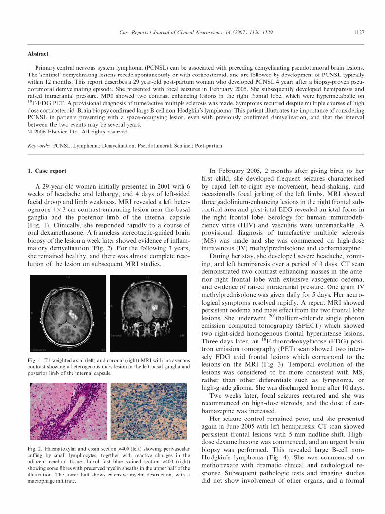
A 29-year-old woman initially presented in 2001 with 6weeks of headache and lethargy, and 4 days of left-sidedfacial droop and limb weakness. MRI revealed a left heter-ogenous 4 · 3 cm contrast-enhancing lesion near the basalganglia and the posterior limb of the internal capsule(Fig. 1). Clinically, she responded rapidly to a course oforal dexamethasone. A frameless stereotactic-guided brainbiopsy of the lesion a week later showed evidence of inflam-matory demyelination (Fig. 2). For the following 3 years,she remained healthy, and there was almost complete reso-lution of the lesion on subsequent MRI studies.
In February 2005, 2 months after giving birth to herfirst child, she developed frequent seizures characterisedby rapid left-to-right eye movement, head-shaking, andoccasionally focal jerking of the left limbs. MRI showedthree gadolinium-enhancing lesions in the right frontal sub-cortical area and post-ictal EEG revealed an ictal focus inthe right frontal lobe. Serology for human immunodefi-ciency virus (HIV) and vasculitis were unremarkable. Aprovisional diagnosis of tumefactive multiple sclerosis(MS) was made and she was commenced on high-doseintravenous (IV) methylprednisolone and carbamazepine.
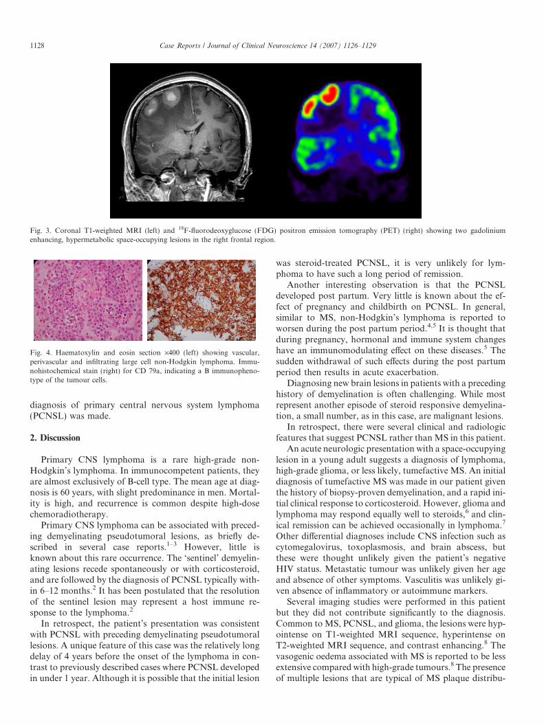
During her stay, she developed severe headache, vomit-ing, and left hemiparesis over a period of 3 days. CT scandemonstrated two contrast-enhancing masses in the ante-rior right frontal lobe with extensive vasogenic oedema,and evidence of raised intracranial pressure. One gram IVmethylprednisolone was given daily for 5 days. Her neuro-logical symptoms resolved rapidly. A repeat MRI showedpersistent oedema and mass effect from the two frontal lobelesions. She underwent 201thallium-chloride single photonemission computed tomography (SPECT) which showedtwo right-sided homogenous frontal hyperintense lesions.Three days later, an 18F-fluorodeoxyglucose (FDG) posi-tron emission tomography (PET) scan showed two inten-sely FDG avid frontal lesions which correspond to thelesions on the MRI (Fig. 3). Temporal evolution of thelesions was considered to be more consistent with MS,rather than other differentials such as lymphoma, orhigh-grade glioma. She was discharged home after 10 days.
Two weeks later, focal seizures recurred and she wasrecommenced on high-dose steroids, and the dose of car-bamazepine was increased.
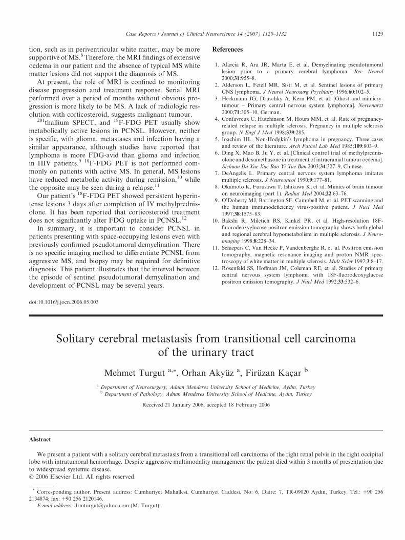
Her seizure control remained poor, and she presentedagain in June 2005 with left hemiparesis. CT scan showedpersistent frontal lesions with 5 mm midline shift. High-dose dexamethasone was commenced, and an urgent brainbiopsy was performed. This revealed large B-cell non-Hodgkin’s lymphoma (Fig. 4). She was commenced onmethotrexate with dramatic clinical and radiological re-sponse. Subsequent pathologic tests and imaging studiesdid not show involvement of other organs, and a formal
Fig. 1. T1-weighted axial (left) and coronal (right) MRI with intravenouscontrast showing a heterogenous mass lesion in the left basal ganglia andposterior limb of the internal capsule.
Fig. 2. Haematoxylin and eosin section ·400 (left) showing perivascularcuffing by small lymphocytes, together with reactive changes in theadjacent cerebral tissue. Luxol fast blue stained section ·400 (right)showing some fibres with preserved myelin sheaths in the upper half of theillustration. The lower half shows extensive myelin destruction, with amacrophage infiltrate.
Case Reports / Journal of Clinical Neuroscience 14 (2007) 1126–1129 1127
diagnosis of primary central nervous system lymphoma(PCNSL) was made.
2. Discussion
Primary CNS lymphoma is a rare high-grade non-Hodgkin’s lymphoma. In immunocompetent patients, theyare almost exclusively of B-cell type. The mean age at diag-nosis is 60 years, with slight predominance in men. Mortal-ity is high, and recurrence is common despite high-dosechemoradiotherapy.
Primary CNS lymphoma can be associated with preced-ing demyelinating pseudotumoral lesions, as briefly de-scribed in several case reports.1–3 However, little isknown about this rare occurrence. The ‘sentinel’ demyelin-ating lesions recede spontaneously or with corticosteroid,and are followed by the diagnosis of PCNSL typically with-in 6–12 months.2 It has been postulated that the resolutionof the sentinel lesion may represent a host immune re-sponse to the lymphoma.2
In retrospect, the patient’s presentation was consistentwith PCNSL with preceding demyelinating pseudotumorallesions. A unique feature of this case was the relatively longdelay of 4 years before the onset of the lymphoma in con-trast to previously described cases where PCNSL developedin under 1 year. Although it is possible that the initial lesion
was steroid-treated PCNSL, it is very unlikely for lym-phoma to have such a long period of remission.
Another interesting observation is that the PCNSLdeveloped post partum. Very little is known about the ef-fect of pregnancy and childbirth on PCNSL. In general,similar to MS, non-Hodgkin’s lymphoma is reported toworsen during the post partum period.4,5 It is thought thatduring pregnancy, hormonal and immune system changeshave an immunomodulating effect on these diseases.5 Thesudden withdrawal of such effects during the post partumperiod then results in acute exacerbation.
Diagnosing new brain lesions in patients with a precedinghistory of demyelination is often challenging. While mostrepresent another episode of steroid responsive demyelina-tion, a small number, as in this case, are malignant lesions.
In retrospect, there were several clinical and radiologicfeatures that suggest PCNSL rather than MS in this patient.
An acute neurologic presentation with a space-occupyinglesion in a young adult suggests a diagnosis of lymphoma,high-grade glioma, or less likely, tumefactive MS. An initialdiagnosis of tumefactive MS was made in our patient giventhe history of biopsy-proven demyelination, and a rapid ini-tial clinical response to corticosteroid. However, glioma andlymphoma may respond equally well to steroids,6 and clin-ical remission can be achieved occasionally in lymphoma.7
Other differential diagnoses include CNS infection such ascytomegalovirus, toxoplasmosis, and brain abscess, butthese were thought unlikely given the patient’s negativeHIV status. Metastatic tumour was unlikely given her ageand absence of other symptoms. Vasculitis was unlikely gi-ven absence of inflammatory or autoimmune markers.
Several imaging studies were performed in this patientbut they did not contribute significantly to the diagnosis.Common to MS, PCNSL, and glioma, the lesions were hyp-ointense on T1-weighted MRI sequence, hyperintense onT2-weighted MRI sequence, and contrast enhancing.8 Thevasogenic oedema associated with MS is reported to be lessextensive compared with high-grade tumours.8 The presenceof multiple lesions that are typical of MS plaque distribu-
Fig. 3. Coronal T1-weighted MRI (left) and 18F-fluorodeoxyglucose (FDG) positron emission tomography (PET) (right) showing two gadoliniumenhancing, hypermetabolic space-occupying lesions in the right frontal region.
Fig. 4. Haematoxylin and eosin section ·400 (left) showing vascular,perivascular and infiltrating large cell non-Hodgkin lymphoma. Immu-nohistochemical stain (right) for CD 79a, indicating a B immunopheno-type of the tumour cells.
1128 Case Reports / Journal of Clinical Neuroscience 14 (2007) 1126–1129
tion, such as in periventricular white matter, may be moresupportive of MS.8 Therefore, the MRI findings of extensiveoedema in our patient and the absence of typical MS whitematter lesions did not support the diagnosis of MS.
At present, the role of MRI is confined to monitoringdisease progression and treatment response. Serial MRIperformed over a period of months without obvious pro-gression is more likely to be MS. A lack of radiologic res-olution with corticosteroid, suggests malignant tumour.
201thallium SPECT, and 18F-FDG PET usually showmetabolically active lesions in PCNSL. However, neitheris specific, with glioma, metastases and infection having asimilar appearance, although studies have reported thatlymphoma is more FDG-avid than glioma and infectionin HIV patients.9 18F-FDG PET is not performed com-monly on patients with active MS. In general, MS lesionshave reduced metabolic activity during remission,10 whilethe opposite may be seen during a relapse.11
Our patient’s 18F-FDG PET showed persistent hyperin-tense lesions 3 days after completion of IV methylprednis-olone. It has been reported that corticosteroid treatmentdoes not significantly alter FDG uptake in PCNSL.12
In summary, it is important to consider PCNSL inpatients presenting with space-occupying lesions even withpreviously confirmed pseudotumoral demyelination. Thereis no specific imaging method to differentiate PCNSL fromaggressive MS, and biopsy may be required for definitivediagnosis. This patient illustrates that the interval betweenthe episode of sentinel pseudotumoral demyelination anddevelopment of PCNSL may be several years.
References
1. Alarcia R, Ara JR, Marta E, et al. Demyelinating pseudotumorallesion prior to a primary cerebral lymphoma. Rev Neurol
2000;31:955–8.2. Alderson L, Fetell MR, Sisti M, et al. Sentinel lesions of primary
CNS lymphoma. J Neurol Neurosurg Psychiatry 1996;60:102–5.3. Heckmann JG, Druschky A, Kern PM, et al. [Ghost and mimicry-
tumour – Primary central nervous system lymphoma]. Nervenarzt
2000;71:305–10, German.4. Confavreux C, Hutchinson M, Hours MM, et al. Rate of pregnancy-
related relapse in multiple sclerosis. Pregnancy in multiple sclerosisgroup. N Engl J Med 1998;339:285.
5. Ioachim HL. Non-Hodgkin’s lymphoma in pregnancy. Three casesand review of the literature. Arch Pathol Lab Med 1985;109:803–9.
6. Ding X, Mao B, Ju Y, et al. [Clinical control trial of methylprednis-olone and dexamethasone in treatment of intracranial tumour oedema].Sichuan Da Xue Xue Bao Yi Xue Ban 2003;34:327–9, Chinese.
7. DeAngelis L. Primary central nervous system lymphoma imitatesmultiple sclerosis. J Neurooncol 1990;9:177–81.
8. Okamoto K, Furusawa T, Ishikawa K, et al. Mimics of brain tumouron neuroimaging (part 1). Radiat Med 2004;22:63–76.
9. O’Doherty MJ, Barrington SF, Campbell M, et al. PET scanning andthe human immunodeficiency virus-positive patient. J Nucl Med
1997;38:1575–83.10. Bakshi R, Miletich RS, Kinkel PR, et al. High-resolution 18F-
fluorodeoxyglucose positron emission tomography shows both globaland regional cerebral hypometabolism in multiple sclerosis. J Neuro-
imaging 1998;8:228–34.11. Schiepers C, Van Hecke P, Vandenberghe R, et al. Positron emission
tomography, magnetic resonance imaging and proton NMR spec-troscopy of white matter in multiple sclerosis. Mult Scler 1997;3:8–17.
12. Rosenfeld SS, Hoffman JM, Coleman RE, et al. Studies of primarycentral nervous system lymphoma with 18F-fluorodeoxyglucosepositron emission tomography. J Nucl Med 1992;33:532–6.
doi:10.1016/j.jocn.2006.05.003
Solitary cerebral metastasis from transitional cell carcinomaof the urinary tract
Mehmet Turgut a,*, Orhan Akyuz a, Firuzan Kacar b
a Department of Neurosurgery, Adnan Menderes University School of Medicine, Aydın, Turkeyb Department of Pathology, Adnan Menderes University School of Medicine, Aydın, Turkey
Received 21 January 2006; accepted 18 February 2006
Abstract
We present a patient with a solitary cerebral metastasis from a transitional cell carcinoma of the right renal pelvis in the right occipitallobe with intratumoral hemorrhage. Despite aggressive multimodality management the patient died within 3 months of presentation dueto widespread systemic disease.� 2006 Elsevier Ltd. All rights reserved.
* Corresponding author. Present address: Cumhuriyet Mahallesi, Cumhuriyet Caddesi, No: 6, Daire: 7, TR-09020 Aydın, Turkey. Tel.: +90 2562134874; fax: +90 256 2120146.
E-mail address: [email protected] (M. Turgut).
Case Reports / Journal of Clinical Neuroscience 14 (2007) 1129–1132 1129