Embed Size (px)
Citation preview
AUSSOIS, FRANCE 35th IMMS 2017
1
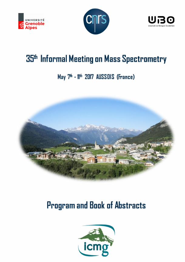
35th INFORMAL MEETING ON MASS SPECTROMETRY
7th-11th May 2017, AUSSOIS, FRANCE
organized by :
Institut de Chimie Moléculaire de Grenoble de l’Université de Grenoble Alpes (UGA)
and
Université de Bretagne Occidentale (UBO)
Chairmen: Yves Gimbert, Antony Memboeuf
Co-chairmen: Pietro Traldi, Karoly Vékey
ORGANIZING COMMITTEE
Jamila Burlet (CNRS-UGA) Amélie Durand (CNRS-UGA)
Valérie Feraux (CNRS-azur-colloque) Laure Fort (UGA)
Yves Gimbert (CNRS, UGA) Rodolphe Guéret (UGA) Antony Memboeuf (UBO)
Emilie Morales (CNRS-azur-colloque)
SCIENTIFIC COMMITTEE
Sandrine Bourgoin-Voillard, UGA Yves Gimbert, CNRS- UGA
Denis Lesage, Sorbonne University-University Paris 6 Antony Memboeuf, UBO
David Rondeau, University Rennes Jean-Claude Tabet, Sorbonne University-University Paris 6-CEA Saclay
Pietro Traldi, CNR Padova Italy Karoly Vékey, Hungarian Academy of Science
AUSSOIS, FRANCE 35th IMMS 2017
2
THE 35th INFORMAL MEETING HAS BEEN SUPPORTED AND SPONSORED BY:
CNRS, DR11-ALPES
Institut de Chimie Moléculaire de Grenoble
--
AGILENT TECHNOLOGIES (SILVER SPONSOR)
WATERS
(SILVER SPONSOR)
VEOLIA
Société Française de Spectromètrie deMasse
Laboratoire de Chimie Electrochimie Moléculaire et Chimie Analytique (Université de Bretagne Occidentale)
Université de Grenoble Alpes
AUSSOIS, FRANCE 35th IMMS 2017
3
Program
Sunday, 7th May 2017
17.00-‐20.00 Registration at the Center CNRS, Aussois 20.30 Informal get together party
Monday, 8th May 2017
8.15-‐8.45 Registration at the Center CNRS, Aussois 8.45-‐9.00 Opening
Chairman: Peter Burgers 9.00-‐9.20 Glenn Carroy
COLLISION-INDUCED GUEST RELEASE FROM GAS PHASE INCLUSION COMPLEXES : A MECHANISTIC STUDY (MO1)
9.20-‐9.40 Parisa Bayat INVESTIGATING BINDING ENERGIES OF HOST-GUEST COMPLEXES IN THE GAS-PHASE (MO2)
9.40-‐10.00 Jean-‐Claude Tabet HB VERSUS SB FORMS OF CATIONIZED NON-COVALENT COMPLEXES BETWEEN PHOSPHO-HEXOSE AND ARG-ARG: H/D EXCHANGE STUDY AND CALCULATIONS (MO3)
10.00-‐11.00 Coffee Break Poster Session Chairman: Jean-‐Claude Tabet 11.00-‐11.20 Peter C. Burgers
INTERACTION OF METAL CATIONS WITH FUNCTIONALISED HYDROCARBONS: SOLVATION OF METAL IONS BY THE HYDROCARBON CHAIN (MO4)
11.20-‐11.40 Gilles Frison METAL COMPLEXES IN THE GAS PHASE: REDUCTION AND CHARACTERIZATION BY IRMPD (MO5)
AUSSOIS, FRANCE 35th IMMS 2017
4
11.40-‐12.00 Héloïse Dossmann VUV SPECTROSCOPY OF MOLYBDENUM COMPLEXES (MO6)
12.00-‐14.00 Lunch Time Poster Session Chairman: Günter Allmaier 14.00-‐14.20 Andras Acs IN SEARCH OF TUMOR MARKERS: GLYCOMIC CHARACTERIZATION
OF PSA (Prostate Specific Antigen) (MO7) 14.20-‐14.40 Samantha Reale
MODELING FUNGAL MELANIN BUILDUP: BIOMIMETIC POLYMERIZATION OF DIHYDROXYNAPHTHALENES MAPPED BY ESI-MASS SPECTROMETRY (MO8)
14.40-‐15.00 Annunziata Lapolla IN VIVO GLYCATED HUMAN SERUM ALBUMIN IMPAIRS CHOLESTEROL EFFLUX FROM MACROPHAGES (MO9)
15.00-‐16.00 Coffee Break Poster Session Chairman: Pietro Traldi 16.00-‐16.20 Emmanuelle Mebold
MALDI-TOF MASS SPECTROMETRY: A POWERFUL TOOL FOR STRUCTURAL CHARACTERIZATION OF SYNTHETIC POLYMERS (MO10)
16.20-‐16.40 Marek Sebela MALDI-TOF MASS SPECTROMETRY OF CYANOBACTERIA (MO11)
16.40-‐17.00 Giuseppe Grasso THE USE OF MASS SPECTROMETRY TO STUDY THE COVALENT MODIFICATION OF AMYLOID BETA PROTEIN DUE TO OXYDATIVE STRESS: INSIGHTS INTO THE PATHOPHYSIOLOGY OF AD (MO12)
19.30 Social dinner
AUSSOIS, FRANCE 35th IMMS 2017
5
Tuesday, 9th May 2017
Chairman: Antony Memboeuf 8.40-‐9.00 Pascal Gerbaux
COLLISION-INDUCED DISSOCIATION OF PEPTOID IONS: DECIPHERING THE KINETIC ENERGY DEPENDENCE OF THE COMPETITIVE FRAGMENTATION MECHANISM (TU13)
9.00-‐9.20 Claudio Iacobucci
A NOVEL CID-CLEAVABLE AZO CROSS-LINKERS FOR PEPTIDE STRUCTURE ANALYSIS BY FREE RADICAL INITIATED PEPTIDE SEQUENCING (FRIPS) (TU14)
9.20-‐9.40 Karoly Vékey SEQUENCING THE OLIGOSACCHARIDE PART OF GLYCOPEPTIDE USING LOW ENERGY CID (TU15)
9.40-‐10.00 Günter Allmaier
LOW VERSUS TRUE HIGH ENERGY COLLISION INDUCED DISSOCIATION OF SODIATED DI- AND OLIGOSACCHARIDES PRECUSORS (TU16)
10.00-‐11.00 Coffee Break Poster Session Chairman: Karoly Vékey 11.00-‐11.20 Dany Jeanne Dit Fouque
TOWARDS THE QUANTIFICATION OF TOPOLOGICAL PEPTIDE ISOMERS USING MULTISTAGE MASS SPECTROMETRY (TU17)
11.20-‐11.40 Julie Cautereels
QUANTUM CHEMICAL MASS SPECTROMETRY: THE INFLUENCE OF INTER-SIDE-CHAIN INTERACTION ON THE FRAGMENTATION OF PEPTIDES (TU18)
11.40-‐12.10 AGILENT (TU19) 12.10-‐14.00 Lunch Time Poster Session
AUSSOIS, FRANCE 35th IMMS 2017
6
Chairman: Philippe Dugourd 14.00-‐14.20 Corentin Decroo
ION MOBILITY MASS SPECTROMETRY OF SAPONINS: FROM ION CONFORMATION TO MOLECULE STRUCTURE (TU20)
14.20-‐14.40 Edwin de Pauw
ION MOBILITY AND MOLECULAR DYNAMICS COMBINATION TO UNRAVEL THE (UN)FOLDING MECHANISM OF AN OLIGOROTAXANE MOLECULAR SWITCH (TU21)
14.40-‐15.00 Hélène Lavanant
ION MOBILITY MASS SPECTROMETRY OF PHOSPHORIC ACID CLUSTER IONS (TU22)
15.00-‐16.00 Coffee Break Poster Session Chairman: Edwin de Pauw 16.00-‐16.20 Philippe Dugourd
PROBING THE CONFORMATION OF BIOMOLECULAR IONS BY FORSTER RESONNANCE ENERGY TRANSFER AND ION MOBILITY MASS SPECTROMETRY (TU23)
16.20-‐16.40 Jean R. N. Haler
SYNTHETIC POLYMERS: FROM PHYSICOCHEMICAL PROPERTIES TO POTENTIAL ION MOBILITY CALIBRATING SUBSTANCES (TU24)
16.40-‐17.10 Kevin Giles
DESIGN AND PERFORMANCE OF MULTIPASS CYCLIC ION MOBILITY-ENABLED Q-TOF (TU25)
Wednesday, 10th May 2017 Chairman: Sandrine Bourgoin-‐Voillard 9.00-‐9.20 Myriam Ferro
AN OVERVIEW OF TOOLS DEDICATED TO PROTEOMICS DATA ANALYSIS : APPLICATION TO THE HUMAN PROTEOME PROJECT (WE26)
9.20-‐9.40 Dalel Askri
PROTEOMIC ANALYSIS FOR STUDYING IRON OXIDE NANOPARTICLES EFFECTS ON WISTAR RAT (WE27)

AUSSOIS, FRANCE 35th IMMS 2017
7
9.40-‐10.00 Pierre Le Pogam VALIDATION OF A METABOLOMIC WORKFLOW TO ASSESS THE EFFECT OF 60 GHZ MILLIMETER WAVES ON KERATINOCYTE CELL LINES: A PROOF-OF-CONCEPT STUDY ESTABLISHING THE METABOLIC ALTERATIONS TRIGGERED BY 2-DEOXYGLUCOSE (WE28)
10.00-‐11.00 Coffee Break Poster Session 11.00-‐11.20 Prizes for the best oral communication and poster and final
consideration 11.20-‐11.40 Presentation of the 36th Informal Meeting on Mass
Spectrometry 12.00 Lunch Time

AUSSOIS, FRANCE 35th IMMS 2017
8

AUSSOIS, FRANCE 35th IMMS 2017
9
ABSTRACTS: ORAL COMMUNICATION
AUSSOIS, FRANCE 35th IMMS 2017
10
MONDAY MORNING, 8th
COLLISION-INDUCED GUEST RELEASE FROM GAS PHASE INCLUSION COMPLEXES : A MECHANISTIC STUDY (MO1)
Glenn Carroy1, Vincent Lemaur2, Julien De Winter1, Edwin De Pauw3, Jérôme Corni2 and Pascal Gerbaux1
1) Organic Synthesis and Mass Spectrometry Laboratory, Interdisciplinary Center for Mass Spectrometry (CISMa), University of MONS, Belgium
2) Laboratory for Chemistry of Novel Materials, UMONS, Belgium 3) Mass Spectrometry Laboratory, University of Liège (ULg), Belgium.
Ion mobility and (energy-resolved) CID experiments are nowadays largely used to study the three-dimensional structures and topologies of non-covalent complex ions, with a special emphasis on the relative position of the different partners associated in the supramolecular assemblies [1]. These elegant investigations are most of the time complemented by theoretical calculations that afford optimized geometries, binding energies and collisional cross sections. Host-guest complexes are formed by the creation of multiple non-covalent bonds between a large concave molecule - the host - and smaller molecule(s) or ion(s) - the guest(s). Ion mobility experiments represent the ideal tool to assess whether the gas-phase ionic host-guest complexes have exclusion or inclusion nature. Nevertheless, the influence of the experimental conditions for the solution processing on the observed ions is often not described. In the specific case of inclusion complexes, kinetic considerations must be taken into account beside thermodynamics; the guest ingression within the host cavity can be characterized by very slow kinetics, making the overall complexation reaction kinetically driven at the time scale of the experiment. This is particularly the case for the cucurbituril family [2] of macrocyclic host molecules. Indeed, significant deformations of the portals of the cucurbituril receiver were demonstrated to occur in the transition state to allow access to its interior part by large guest molecules, thus reducing the kinetic constant for ingression [3]. In the present communication, we selected 1,4-phenylenediamine and cucurbit[6]uril as a model system to demonstrate by means of ion mobility and energy-resolved collision-induced dissociation measurements that the inclusion/exclusion topology ratio varies as a function of the equilibration time in solution prior to the Electrospray process [4]. Herein, in direct relationship, we report the particular gas phase decomposition pathway of the binary complexes ions presenting an inclusion topology. The complexity of the pathway, together with the necessity of breaking covalent bonds, is of course likely to induce the higher amount of internal energy required for the dissociation of the inclusion topologies.
AUSSOIS, FRANCE 35th IMMS 2017
11
INVESTIGATING BINDING ENERGIES OF HOST-GUEST COMPLEXES IN THE GAS-PHASE (MO2)
P. Bayat1 , D. Gatineau1, A. Martinez2, D. Lesage1, R.B. Cole1
1) Sorbonne Universités, UPMC Univ. Paris 06, IPCM (UMR 8232), F-75005 Paris, France. 2) Equipe Chirosciences, UMR CNRS 7313-iSm2, Aix Marseille Université, Ecole Centrale de Marseille,
Av. Escadrille Normandie-Niemen, 13397 Marseille Cedex 20, France.
Gas phase binding of a heteroditopic hemicryptophane cage (Zn(II)@1) [1] to a series of zwitter-ionic guests has been explored using two independent techniques. First, resonant excitation of host-guest (H-G) pairs in a linear quadrupole ion trap was examined under thermal equilibrium conditions [2,3]. Second, non-resonance activation was employed to perform higher-energy collision dissociation (HCD) [4], implying a truncated Maxwell-Boltzmann distribution of internal energy, that was later subjected to RRKM modeling [5]. Neither of these approaches is capable of directly giving an absolute energy measurement and as a consequence, the first requires preliminary calibration of the effective temperature, whereas in the second, the internal energy distribution must be calibrated. In both cases, calibration was accomplished by employing Blackbody Infrared Radiative Dissociation (BIRD) [6] which is an absolute energy measurement. Both techniques were utilized on more than 10 H-G pairs to enable a ranking of binding energies for each H-G pair. A comparison of trends for the series of guests showed that binding was strongest for phosphate followed by sulfonate, and then carboxylate anions. Resonant excitation in the linear quadrupole ion trap was very well adapted for the H-G complexes under study due to the fulfillment of the rapid energy exchange limit [6] condition, owing to the large size of the ions, as well as the relatively large residence time of the ions inside the trap (60 s) which allows the study of reactions with rates less than 1 s-1. HCD with RRKM modeling provides very consistent results, especially because all of the studied H-G pairs are characterized by nearly similar size. These two simple techniques can be applied to other host-guest chemistry studies where it is of paramount importance to obtain a quantitative comparison of bond dissociation energies. References
1. D. Zhang, G. Gao, L. Guy, V. Robert, J.P. Dutasta, A. Martinez; Chem. Commun., 51, 2679–2682 (2015). 2. D. E. Goeringer, S. A. McLuckey; J. Chem. Phys., 104, 2214–2221 (1996). 3. D. E. Goeringer, S. a McLuckey; Rapid Commun. Mass Spectrom., 10, 328–334 (1996). 4. J. V Olsen, B. Macek, O. Lange, A. Makarov, S. Horning, M. Mann; Nat. Methods, 4, 709–712 (2007). 5. L. Drahos, V. Karoly; J. Mass Spectrom, 36, 237–263 (2001). 6. R. C. Dunbar; Mass Spectrom. Rev., 23, 127–158 (2004).
AUSSOIS, FRANCE 35th IMMS 2017
12
HB VERSUS SB FORMS OF CATIONIZED NON-COVALENT COMPLEXES BETWEEN PHOSPHO-HEXOSE AND ARG-ARG: H/D
EXCHANGE STUDY AND CALCULATIONS (MO3)
E. Darii1, Y.Gimbert2, S. Alves3, A. Perret1 and J.-C. Tabet3
1) CEA, DRF, Institut de biologie François Jacob, Genoscope France (CNRS-UMR8030, Université d’Evry, Evry, University Paris Saclay Evry)
2) Université Grenoble Alpes (DCM), CNRS-UJF 5250, BP 38041, Grenoble, Auvergne-Rhône-Alpes, France
3) Sorbonne Universités, UPMC Univ Paris 06, CNRS, and CEA, Saclay Institut Parisien de Chimie Moléculaire, Paris, Ile de France, France
The formation of small non-covalent complexes (NCC) in gas phase (GP) is frequently observed during mass spectrometric analysis of complex mixture. Such NCC are generally presented as ion-dipole systems involving hydrogen bonding (HB) complexes and, to a lesser extent, salt bridge (SB) systems maintained by ion-ion interactions from zwitterions. Under low collision energy conditions, HB forms competitively yield free ionized partners according to their respective GP basicity/acidity. SB forms dissociate through covalent bond cleavages due to reinforced ion-ion interaction in GP. Generally, CID behavior correlates with partner structure and can be applied for the distinction of structural isomers.
In our previous work [1] we focused on the relative stability of HB and SB forms of NCC formed between hexose phosphate isomers (HexP) and basic compounds (R, RR) under soft and hard desolvation conditions. Now, comparison of the GP properties of non-cationized and cationized NCC will be presented. Especially, the impact of cation size and charge on the fragmentation under CID, will be considered. The H/D exchange experiments combined with quantum calculations will be discussed to rationalize cleavage mechanisms according to the structure of partners and the size of alkali and earth-alkali.
The solutions of the mixtures of hexose phosphates (fructose-1-phosphate, F1P; fructose-1-phosphate, F6P; glucose-1-phosphate, G1P and glucose-6-phosphate, G6P) and basic compounds (R, RR, tetramethyldiaminobutane (TMDB)) were analyzed on an LTQ-Orbitrap XL and Orbitrap Elite (Thermo Electron Corporation, Germany) equipped with an ESI source. The experiments were performed in negative ionization mode. The impact of different cations was explored using alkali and alkaline earth metal salts. The GP behavior of non-cationized and cationized NCC was explored using H/D exchanges under CID conditions.
According to CID behavior of NCC cationized by different metallic cations, the stability of non-covalent systems correlates with the structures of partners and the cation size and charge. CID spectra of cationized NCC demonstrate fragmentation patterns specific for each HexP isomer. In the case of arginine, SB systems are less abundant than HB forms due to the modulation of the GP thermochemical values,
AUSSOIS, FRANCE 35th IMMS 2017
13
while they became more abundant in the case of RR due to the numerous possible SB interactions. The complexes cationized by Li+ are more stable than the systems involving Na+, K+ and Rb+. For the systems with Mg2+, only covalent bond cleavages are observed under CID, giving evidence that the SB forms are significantly reinforced.
Detailed quantum calculations for the F1P/R/Na complex demonstrated that the most stable structure represent solvated salt (SS) system involving Na+/COO− salt solvated by neutral phosphate and carrying the negative charge on sugar ring. Fragmentation pathways of cationized SS forms were explored by H/D exchange experiments. MS spectra demonstrate expected exchanges corresponding to the number of mobile protons in ionized partners. In the case of HexP/R/Na systems, mostly expected exchanges and one additional much less intense exchange were detected under CID. For F1P/RR/Na complex, the most intense peak produced by the dehydrated sugar ring loss (specific for this isomer) was related to one additional H/D exchange and less intense peaks corresponded to expected species and the species carrying two additional D. In contrast, G1P/RR/Na dissociation under CID yields only the species with expected exchanges. Higher degree of H/D exchanges in sugar ring under CID for the F1P/RR/Na system is likely related to predominant covalent bond cleavages and gives evidence for more stable SS in this case.The detailed mechanism of covalent bond cleavages of SS forms of NCC will be explored in order to confirm this trend. Quantum calculation should give information on the conformation and functional group in interactions. Novel aspect: additional H/D exchanges observed in the experiments with small cationized NCC subjected to CID strongly correlate with the strength of ion-ion interactions within non-covalent systems.
References
1. Darii, Ekaterina; Alves, Sandra; Gimbert, Yves; Perret, Alain; Tabet. Jean-Claude. Chromatography B: Analytical Technologies in the Biomedical and Life Sciences 1047, 45-58 (2017).
AUSSOIS, FRANCE 35th IMMS 2017
14
INTERACTION OF METAL CATIONS WITH FUNCTIONALISED HYDROCARBONS: SOLVATION OF METAL IONS BY THE
HYDROCARBON CHAIN (MO4)
P.C. Burgers1), N.A. van Huizen1), T.M. Luider1), K.J. Jobst2), J.K. Terlouw3) and J.L. Holmes4)
1) Department of Neurology, Erasmus Medical Center, 3015 CN, Rotterdam, The Netherlands 2) Ontario Ministry of the Environment and Climate Change, 125 Resources Road, Toronto, Ontario M9P
3V6, Canada 3) Department of Chemistry and Chemical Biology, McMaster University, Hamilton, Ontario
L8S 4M1, Canada 4) Chemistry Department, University of Ottawa, 10 Marie Curie, Ottawa, Ontario K1N 6N5, Canada
Relative affinity measurements of the monovalent metal ions (M+ = Li+, Na+, Cu+ and Ag+) towards a series of functionalised hydrocarbons (P) have been performed using the kinetic method on the dissociation of metal-bound dimer ions of the type P1-M+-P2. The compounds P include aliphatic nitriles [1], amines [2], alcohols [2], methyl alkanoates [2], alkyl acetates [3] and some selected 1-alkenes [3]. It was found that the cations’ affinity towards long chain (≥C4 chain length) aliphatic compounds was unexpectedly enhanced. This is attributed to a bidentate interaction of the metal ion with the nitrile, amine, alcohol or ester functional group or double bond and the aliphatic chain. Ab initio calculations on Cu+ bound aliphatic nitriles confirm these experimental findings and show that such bidentate bonding leads to a significant (c. 30%) additional stabilisation. The above type of hydrocarbon bidentate formation can also reveal itself by the reluctance of such structures to react with water in an ion trap; for example the Ag+•••1-hexene ion undergoes efficient water addition, whereas Ag+•••1-octene shows hardly any addition of water, while Ag+•••1-heptene occupies an intermediate position. These results can be interpreted in terms of an open (i.e. non-bidentate) structure for the Ag+•••1-hexene ion, whereas the Ag+•••1-octene ion has a sufficiently strong bidentate interaction to repel an attacking water molecule. A detailed discussion of the above experiments and theoretical calculations will be given. Since this hydrocarbon solvation of metal ions may well be a general phenomenon, such studies might also be feasible for biologically interesting compounds, such as lipids. References 1. K.J. Jobst, J.K. Terlouw, T.M. Luider, N.A. van Huizen, P.C. Burgers; Eur. J. Mass Spectrom., 21, 578-587 (2015). 2. N. A. van Huizen, T.M. Luider, K.J. Jobst, J.K. Terlouw, J.L. Holmes, P.C. Burgers, ibid., 22, 61-70 (2016). 3. P.C. Burgers, J.L. Holmes and J.K. Terlouw; ibid., 22, 297-305 (2016).
AUSSOIS, FRANCE 35th IMMS 2017
15
METAL COMPLEXES IN THE GAS PHASE: REDUCTION AND CHARACTERIZATION BY IRMPD (MO5)
M. Katari1), E. Nicol1), V. Steinmetz2), G. van der Rest2), D. Carmichael1),
G. Frison1)
1) LCM, CNRS, Ecole polytechnique, Université Paris-Saclay, 91128 Palaiseau, France 2) Laboratoire de Chimie Physique, Université Paris-Sud, CNRS, 91405 Orsay, France.
InfraRed Multiple Photon Dissociation (IRMPD) "action" spectroscopy has emerged recently as an efficient and generally applicable technique for the measurement of the infrared spectra of isolated ions.[1, 2] These “action” spectra are produced when an on-resonance absorption of multiple IR photons occurs at an active vibrational mode of a gas phase ion, and take the form of a plot of fragmentation abundance as a function of photon wavelength. These spectra are ideally adapted for comparison with DFT-derived computational data, because DFT should represent IR spectra of isolated gas phase ions excellently, and therefore allows the nature of the product species to be verified. In this presentation, we will show recent results we have obtained through IRMPD spectroscopy studies of metal complexes. Experimental IRMPD spectra for ten Zn and Ru organometallic complexes have been used to provide reference data for 64 vibrational modes in the 900-2000 cm-1 range. The accuracy of the IR vibrational frequencies predicted for these bands has been assessed for several DFT functionals. We have shown that using linear correlations instead of scaling factors improves the prediction accuracy significantly.[3] Electron capture dissociation (ECD) mass spectrometry allows the formation and selection of reduced transition metal complexes containing non-innocent ligands. We will show how a combination of ECD and IRMPD techniques can be employed to define the electronic structure of radical reduced Zn and Ru transients.[4] References
1. J. Roithova; Chem. Soc. Rev., 41, 547-559 (2012). 2. N. C. Polfer; Chem. Soc. Rev., 40, 2211-2221 (2011). 3. M. Katari, E. Nicol, V. Steinmetz, G. van der Rest, D. Carmichael, G. Frison; Chem. Eur. J. (2017) accepted. 4. M. Katari, E. Payen de la Garanderie, E. Nicol, V. Steinmetz, G. van der Rest, D. Carmichael, G. Frison; Phys. Chem. Chem. Phys., 17, 25689-25692 (2015).
AUSSOIS, FRANCE 35th IMMS 2017
16
VUV SPECTROSCOPY OF MOLYBDENUM COMPLEXES (MO6) H. Dossmann1), D. Lesage1), D. Gatineau1), H. Clavier2), A. Memboeuf3),
A. Milet4), Y. Gimbert4)
1) Sorbonne Universités, UPMC Univ Paris 06, CNRS, Institut Parisien de Chimie Moléculaire (IPCM), 4 place Jussieu 75252 Paris Cedex 05 France
2) Aix Marseille Univ, CNRS, Centrale Marseille, iSm2, Marseille, France 3) Univ Bretagne Occidentale and CNRS, CEMCA (UMR 6521), F-29238 Brest, France
4) Univ Grenoble Alpes and CNRS, DCM (UMR 5250), F-38000 Grenoble, France An important feature of metal catalysts lies in the crucial role of ancillary ligands on the electronic structure of the metal center. Determining how those ligands affect the reactivity of the metal center and quantifying this effect is thus of paramount importance. Various methods have been proposed to evaluate the electron donating/accepting property of ligands; the most popular method, and easiest to implement, is the Tolman Electronic Parameter (TEP), initially developed for phosphine ligands. It is based on the observation of the IR stretching frequency of the carbonyl ligand(s) in a complex such as Ni(CO)3L or LW(CO)5. If the electronic environment of the metal center affects the strength of a CO bond by modifying the balance between s-donation and π-back donation, clearly it also modifies the strength of the M-CO bond. Indeed, the denser the electronic environment of M is, the stronger should be that bond. Considering the electron density on the metal center is determined according to the electronic properties of the ligand, one may then expect a correlation between the electron-donating properties of the ligand and the bond dissociation energy (BDE) values for M-CO bond. In this context, a collaborative work has been initiated between several partners from various fields (organometallic chemistry and catalysis, theoretical chemistry, mass spectrometry) with the objective of studying the connections existing between steric and electronic properties of ligands of interest for catalytic processes, and reactivity of the metal-based catalyst to which they are bound. Our objective is to build a new electronic properties scale for ligands bound to a given metal which will help chemists to rationalize the effect of ligands on the performance of catalysts. To this end, various experimental technics have been employed such as mass spectrometry (MS)-based experiments (black-body infrared dissociation (BIRD), (threshold) collision-induced dissociation ((T)CID) experiments ...) and VUV-spectroscopy. We will present here the results obtained for several Molybdenum complexes, Mo(CO)5L, with L = PR3 (R = alkyl, MexPhy or PyrxPhy) and studied by means of VUV spectroscopy at the synchrotron SOLEIL facility center and density-functional theory (DFT) calculations. Photoionization process of the complexes gives precious information concerning the electronic interaction between the ligand and the metal. The vertical ionization energies IEvert that are measured on the photoionization spectra are indeed related to the molecular orbitals of the complexes and allows thus a detailed description of π-back-bonding and σ-acceptor characters of the ligand.

AUSSOIS, FRANCE 35th IMMS 2017
17
MONDAY AFTERNOON, 8th
IN SEARCH OF TUMOR MARKERS: GLYCOMIC CHARACTERIZATION OF PSA
(Prostate Specific Antigen) (MO7)
A. Ács1,2, L. Turiák1, O. Ozohanics1, D. Navarro-Calderon3, N. Farina-Gomez3, A. Telekes4, K. Vékey1 and L. Drahos1,5
1) MTA TTK MS Proteomics Laboratory, Hungarian Academy of Sciences, H-1117, Budapest, Hungary
2) Semmelweis University. Budapest, Hungary 3) Institute of Organic Chemistry (IQOG-CSIC). Madrid, Spain.
4)Department of Oncology, Bajcsy-Zsilinszky Hospital. Budapest, Hungary 5) MTA-TTK NAP B MS Neuroproteomics Group, Hungarian Academy of Sciences, H-1117, Budapest,
Hungary The current clinical routine uses more than 30 different tumor markers to determine the presence and/or progression of a tumor. Some of them struggles with severe specificity and sensitivity problems. Most of them are glycoproteins by nature. There is a theory that maybe alterations occur on the glycosylation level of a protein during tumorogenesis. Some early research show that these modifications can predict the tumor status more reliably, than the markers used at present. Prostate cancer is one of the most common cancer type, millions are involved around the world each year. Measurement of serum PSA level is a routinely used diagnostic tool to determine whether somebody possibly has cancer or not. But in a specific range, between 4-10 ng/ml, only around 1/3 of the people have prostate cancer, the others go through the further risky examinations unnecessarily. The assessment of PSA glycans may lead to a better diagnostic performance. A complex purification procedure was established to yield sufficient amount of PSA for the glycan analysis. By testing different biological fluids, we found that urine would be an appropriate candidate, because of its relatively high PSA content and availability. Several purification steps were applied, including centrifugal membrane filters and a PSA immunochromatographic column. For the mass spectrometric measurements we used a nanoLC coupled Bruker Maxis ETD II and data were evaluated using in-house developed software GlycoPattern. With the purification procedure we succeeded to isolate adequate amount (around 2 µg) of PSA for the further analysis. Glycosylation patterns were determined in case of patients with cancer, with other prostate pathologic changes, such as BPH (Benign Prostate Hyperplasia), pools of healthy people and also individuals and commercially available PSA standards. Analysis reveals more than 50 different glycan structures and some characteristic features which will be discussed.
AUSSOIS, FRANCE 35th IMMS 2017
18
MODELING FUNGAL MELANIN BUILDUP: BIOMIMETIC POLYMERIZATION OF DIHYDROXYNAPHTHALENES MAPPED BY
ESI-MASS SPECTROMETRY (MO8)
M. M. Cecchini,1) S. Reale,1) P. Manini,2) M. d’Ischia,2) and F. De Angelis1)
1) Department of Physical and Chemical Sciences, University of L’Aquila, Via Vetoio, Coppito, L’Aquila, Italy
2) Department of Chemical Sciences University of Naples “Federico II” – Complesso Universitario Monte Sant’Angelo, Via Cintia 4, Napoli, Italy
Melanins are ancient biological pigments found in all kingdoms of life. They are an extremely heterogeneous group of polymeric amorphous natural substances which share a polyphenolic nature with unique physicochemical properties including broadband optical absorption, paramagnetism, change transport and a remarkable structural stability. In the last two decades, melanin and melanogenesis have attracted growing interest due to the unique structural, antioxidant, photoprotective and optoelectronic properties of melanin-based polymers [1]. Even if the chemistry of eumelanin subclass has been widely investigated [2], very few studies rely upon allomelanins, which is the most heterogeneous group of natural melanins of non-animal origin. In the fungal kingdom, the black pigment of ascomyces fungi derives from oxidation and polymerization of the chromogen 1,8-dihydroxynaphthalene (1,8-DHN), producing 1,8-DHN-melanin-type, whose structure is still unrevealed [3]. Intrigued by the emerging biomedical relevance and technological potential of fungal melanins, we characterized by Electrospray Ionization Mass Spectrometry (ESI-MS) a series of allomelanins produced in vitro via enzyme-catalyzed oxidative polymerization of four dihydroxynaphthalene isomers. Electrospray Ionization Mass Spectrometry (ESI-MS) measurements of freshly synthesized 1,8-DHN-polymer recorded in the negative ion mode allowed detection of oligomers up to m/z 4000, separated by 158 Da, corresponding to the in-chain DHN-unit. The dominant peaks were assigned to singly-charged distribution, up to XXIII repeating units, whereas a doubly charged polymer distribution was also detectable. Chemical derivatization, Ultra Performance Liquid Chromatography (UPLC)-ESI MS and MS/MS data confirmed that oxidative polymerization of 1,8-DHN proceeds exclusively via C-C coupling of the naphthalene rings. The same analytical and synthetic protocol applied to other DHN isomers (namely 2,7-DHN;, 1,2-DHN, and 2,3-DHN) revealed significant differences on the mode of polymerization of such precursors. In particular, only 1,8-DHN polymerizes exclusively via C-C coupling of the aromatic nuclei while poly(2,7-DHN), poly(1,2-DHN) and poly(2,3-DHN) are composed of both C-C and C-O linked oligomers. This implies that hydroxyl-moieties of 1,8-DHN are not involved in the polymerization
AUSSOIS, FRANCE 35th IMMS 2017
19
while the others isomers of DHN builds up using –OH groups as well as aromatic carbon. The new insights reported here into synthetic DHN oligomers/polymers as a paradigm mimic of fungal melanins, may guide novel interesting advances and applications in the field of biomimetic functional materials. References
1.M. d’Ischia, K. Wakamatsu, F. Cicoira, E. Di Mauro, J. C. Garcia-Borron, S. Commo, I. Galvàn, G. Ghanem, K. Kenzo, P. Meredith, A. Pezzella, C. Santato, T. Sarna, J. D. Simon, L. Zecca, F. A. Zucca, A. Napolitano and S. Ito; Pigment Cell Melanoma Res.; 28, 520–544 (2015). 2. S. Reale, M. Crucianelli, A. Pezzella, M. d’Ischia and F. De Angelis, J. Mass. Spectrom., 47, 49-53, (2012). 3. M. J. Beltràn-Garcìa, F. M. Prado, M. S. Oliveira, D. Ortiz-Mendoza, A. C. Scalfo, A. Pessoa Jr., M. H. G. Medeiros, J. F. White and P. Di Mascio, PLoS ONE 9 e91616 (2014).
AUSSOIS, FRANCE 35th IMMS 2017
20
IN VIVO GLYCATED HUMAN SERUM ALBUMIN IMPAIRS CHOLESTEROL EFFLUX FROM MACROPHAGES (MO9)
S. D’Aronco1) S. Crotti1), P.Traldi1), A.Lapolla2)
1) Institute of Paediatric Research-Città della Speranza, Corso Stati Uniti 4, 35127, Padova, Italy
2),Department of Medicine, University of Padova, via Giustiniani 2, 35128 Padova, Italy
Cardiovascular disease represents the leading cause of morbidity and mortality among individuals affected by diabetes mellitus (DM). Advanced glycation end-products (AGEs), generated from glycated proteins, exhibit elevated levels in DM patients and have been suggested to be among the responsible for the development of atherosclerosis. Consequently, a possible relationship among glycated human serum albumin (HSA), endoplasmatic reticulum (ER) stress, and cholesterol efflux in macrophages can be reasonably hypothesized. In order to verify this aspect a series of experiments have been performed. Glycation level of HSA isolated from healthy and diabetic type 1 (DM1) and type 2 (DM2) subjects was measured by matrix assisted laser desorption/ionization mass spectrometry and used for the further investigations. By this analytical approach it was observed that HSA from DM patients showed a mean condensation of at least 8 and 5 glucose units in type 1 and type 2 diabetics respectively. Mouse peritoneal macrophages were treated with these HSA samples and ER stress and cholesterol efflux were evaluated. The expression levels of ER stress markers were found to be significantly higher in macrophages treated with glycated HSA while cholesterol efflux, via ABCA-1, was significantly reduced. These experiments indicate that glycated HSA can contribute to atherosclerosis in diabetic patients by impairing cholesterol efflux and inducing ER stress in macrophages.
AUSSOIS, FRANCE 35th IMMS 2017
21
MALDI-TOF MASS SPECTROMETRY: A POWERFUL TOOL FOR STRUCTURAL CHARACTERIZATION OF SYNTHETIC POLYMERS
(MO10)
E. Mebold
Institut des Molécules et des Matériaux du Mans - UMR 6283 CNRS
Université du Maine - Av. Olivier-Messiaen - 72085 LE MANS Cedex 09, France
Matrix-Assisted Laser Desorption / Ionization – Time Of Flight (MALDI-TOF) mass spectrometry is a widely used analytical technique especially suited for the characterisation of natural or synthetic macromolecules. Thanks to its versatility, this technique is a particularly useful tool for the analysis of synthetic polymers. Complementary to other analytical techniques, such as Size Exclusion Chromatography (SEC) or Nuclear Magnetic Resonance (NMR), MALDI-TOF mass spectrometry grants the possibility to further characterise polymer samples down to their fine chemical structures. The development of new polymerization techniques has opened the way to the design of original polymer architectures of different topologies, sizes, chemical structures and chain-ends, whose characterization can constitute a challenge. Efficient polymer characterisation by MALDI-TOF mass spectrometry relies on the choice of the appropriate matrix, salt (for cationization), and proportions between matrix, salt and sample. It will be illustrated through several examples how these parameters, as well as polymer size, structure or sample purity, can influence the analysis, and the limitations of the technique will be discussed. The potential of MALDI-TOF mass spectrometry for the fast and accurate determination of molar masses, the sequencing of repeat units, and the nature of chain-end groups of a wide range of polymers will be demonstrated through a few selected examples, with the elucidation of the fine structure of polyethelene oxide (PEO), polyphosphate, polycaprolactone (PCL), poly(N-isopropylacrylamide) (PNIPAM) and polyisoprene (PI) samples.
AUSSOIS, FRANCE 35th IMMS 2017
22
MALDI-TOF MASS SPECTROMETRY OF CYANOBACTERIA (MO11)
Marek Šebela1), René Lenobel1) and Petr Hašler2)
1) Department of Protein Biochemistry and Proteomics, Centre of the Region Haná for Biotechnological and Agricultural Research, Faculty of Science, Palacký University, Šlechtitelů 27, CZ-783 71 Olomouc,
Czech Republic; 2) Department of Botany, Faculty of Science, Palacký University, Šlechtitelů 27,
CZ-783 71 Olomouc, Czech Republic. Cyanobacteria represent a phyllum of bacteria characteristic by their ability to
photosynthetize and are considered one of the most ancient groups of organisms on earth. They form unicellular or multicellular structures in various aquatic or terrestrial habitats and live freely or in a symbiosis. The photosyntetic apparatus is located in thylakoids formed as folds of the cell membrane. Photosynthetic pigments typically appear in thylakoid-associated phycobilisomes containing antennae protein-pigment complexes named phycobilins [1]. Cyanobacteria can cause poisoning or at least health problems to humans as they can produce cyanotoxins, which is especially dangerous when the respective species largely grow and reproduce under favorable conditions e.g. as a part of algal blooms in stagnant waters. On the other hand, it has been shown that cyanobacteria are potentially applicable for the production of the biodegradable polymer poly-beta-hydroxybutyrate [2] or as a protein source in human nutrition. The whole system of cyanobacteria classification has been revised with the introduction of phylogenetic analyses based on molecular sequencing data [3]. In this work, we have used MALDI-TOF mass spectrometry of intact cells to measure protein profile spectra of various species and strains of cyanobacteria using an optimized matrix-solvent system with ferulic and sinapinic acids. The library of the mass spectra was examined by software tools to characterize similarities and differences, both applicable for taxonomic purposes. Using the same solvent, under optimized conditions, proteins were extracted from Gloeobacter violaceus and two Synechococcus strains. Extracts were separated by polyacrylamide gel electrophoresis, in-gel digested to generate peptides and then analyzed by liquid chromatography coupled with tandem mass spectrometry. Ribosomal proteins, phycobilins, respiratory enzymes, electron-carrying proteins, nucleoid-associated proteins and others could be identified as a result and assigned to characteristic protein peaks in the profile spectra of the intact cells.
AUSSOIS, FRANCE 35th IMMS 2017
23
References
1. R.E. Lee; Cyanobacteria, in Phycology, Cambridge University Press, 2008, pp. 33-80. 2. R. Bhati, S. Samantaray, L. Sharma, N. Mallick; Biotechnol. J., 5, 1181-1185 (2010). 3. J. Komárek, J. Kaštovský, J. Mareš, J.R. Johansen; Preslia, 86, 295-335 (2014).
AUSSOIS, FRANCE 35th IMMS 2017
24
THE USE OF MASS SPECTROMETRY TO STUDY THE COVALENT MODIFICATION OF AMYLOID BETA PROTEIN DUE TO
OXYDATIVE STRESS: INSIGHTS INTO THE PATHOPHYSIOLOGY OF AD (MO12)
Giuseppe Grasso,1 Paul Axelsen,2 Hiroaki Komatsu2
1) Department of Chemical Sciences (UNICT)
Università degli Studi di Catania, Viale Andrea Doria 6, 95125, Catania - Italy 2) Department of Pharmacology (UPENN)
University of Pennsylvania, Philadelphia, PA, 19104, USA - United States
Aβ peptides are associated in various ways with metal ions and with mediating oxidative stress in Alzheimer's disease (AD). That oxidative stress, acting on ω-6 and ω-3 polyunsaturated fatty acyl chains, produces 4-hydroxy-2-nonenal (HNE) and 4-hydroxy-hexanal (HHE) respectively, which can covalently modify the Aβ peptides that helped producing it. To examine possible feedback pathways involving Aβ peptides, metal ions and HNE, the interactions were examined by mass spectrometry and fluorescence spectroscopy. Results indicate that metal ions, particularly copper(II), interfere with the modification of His side chains by HNE, but that once modified, metal ions can still bind to Aβ with high affinity. Moreover, a first attempt to monitor the relative amounts of unmodified Aβ and HNE/HHE modified Aβ in vivo is also reported. These results provide insight into a network of biochemical reactions that may be operating as a consequence of oxidative stress in AD, or as part of the pathogenic process.
References
1. W. Williams, E. Anderson; J. Mass Spectrom., 60, 256-287 (2006).

AUSSOIS, FRANCE 35th IMMS 2017
25
TUESDAY MORNING, 9th
COLLISION-INDUCED DISSOCIATION OF PEPTOID IONS: DECIPHERING THE KINETIC ENERGY DEPENDENCE OF THE
COMPETITIVE FRAGMENTATION MECHANISM (TU13)
Emilie Halin, Sébastien Hoyas, Julien De Winter, Pascal Gerbaux
Organic Synthesis and Mass Spectrometry Laboratory, Interdisciplinary Center for Mass Spectrometry, University of Mons – UMONS, 23 Place du Parc, B-7000 Mons, Belgium
Peptoids constitute a class of bio-inspired foldamers composed by N-substituted glycine units, which differ to the amino acid building block by the presence of the side chain on the nitrogen rather than on the α-carbon [1]. These polymers are synthesized via solid-phase or in solution strategies both involving two steps for the complete addition of the monomer in the growing chain [1,2]. The ease and versatility of conception make them really promising for different applications provided that efficient characterization methods are available to build the primary vs secondary structure relationship. Numerous techniques such as Nuclear Magnetic Resonance and Edman degradation are currently used to describe peptoid primary structure. However, sequencing is time-consuming and NMR data are often difficult to analyze because of the cis/trans amide bond isomerism [3]. Mass Spectrometry is increasingly used for macromolecule characterization. The possibility to analyze individual ions provide accurate structural data. Tandem Mass Spectrometry is a well-introduced method for establishing the primary structure of polymers but requires the a priori knowledge of the fragmentation mechanisms. Several studies have shown that gas phase sequencing of peptoids by Collision-Induced Dissociation (CID) relies on the formation of b/y ions and that the ratio between b/y fragments is dependent on the cationisation agent [4]. Very recently, the group of Connolly has proposed that the peptoid b/y fragmentation mechanism involves an oxazolone five-membered ring intermediate, hypothesis validated by labeling experiments. A mechanism to account for the side chain loss has also been described [5]. In the context of a large study aiming to the understanding of the CID reactions undergone by protonated peptoids, we observe that the parent ions can undergo competitive CID reactions with branching ratio that are dependent (i) on the nature of the side chain and (ii) on the experimental setup, QToF vs Synapt G2-Si mass spectrometers. We here describe our results on the CID fragmentation patterns for the protonated N-(S)-phenylethyl peptoid monomers with a special care on the influence of the kinetic energy of the parent ions prior to the collisional event on the
AUSSOIS, FRANCE 35th IMMS 2017
26
detected fragment ions. To allow deciphering the reaction mechanisms, one of the two side chains (N or C-terminal) has been substituted by a (S)-tolylethyl group. The presence of the para methyl group was a priori assumed to influence the fragmentation mechanism of the peptoid ions. Based on (energy-resolved) CID experiments, the side chain loss mechanism previously proposed in the literature [5] is demonstrated to involve two competitive mechanisms, i.e. a rearrangement and a cleavage. We also highlighted the crucial role played by the mobility cell on the observed fragment ions by using the versatility of the Triwave setup, i.e. CID in the trap or in the transfer cells. Hence, we observed significant differences in the nature of the fragment ions when the mobility cell is crossed or not by the decomposing ions. References
[1] J. Am. Chem. Soc. 1992, 114, 10646 [2] Analyst, 2011, 136, 4409 [3] J. Am. Chem. 2003, 125, 13525 [4] Int. J. Mass Spectrom. 2011, 308, 98 [5] J. Am. Soc. Mass Spectrom. 2016, 27, 646.

AUSSOIS, FRANCE 35th IMMS 2017
27
A NOVEL CID-CLEAVABLE AZO CROSS-LINKERS FOR PEPTIDE STRUCTURE ANALYSIS BY FREE RADICAL INITIATED PEPTIDE
SEQUENCING (FRIPS) (TU14)
C. Iacobucci and A.Sinz
Department of Pharmaceutical Chemistry & Bioanalytics, Institute of Pharmacy, Martin Luther University Halle-Wittenberg, Halle/Saale, Germany
Chemical cross-linking in combination with mass spectrometry (MS) [1] has emerged as powerful tool to characterize the three-dimensional structures of proteins and protein complexes. The structures of a large number of proteins and their assemblies are not amenable to high-resolution techniques, such as NMR and X-ray, due to their poor solubility, intrinsic instability, or inability to crystallize. The lack of knowledge of the structural features of proteins and protein assemblies can be now challenged by low resolution methods, especially by chemical cross-linking in combination with MS. Several bi- and tri-functional molecules able to react with different functional groups in proteins have been developed, stabilizing protein conformations and fixing their spatial arrangement. The cross-linked protein and protein assembly can be subsequently investigated by MS after an enzymatic digestion has been performed. The identified cross-linked amino acids along with the length constraint of the cross-linker employed are used as basis for computational modeling approaches to yield 3D-structural models of the protein assembly [2,3]. Of particular interest are cross-linkers that are cleaved on-demand under collision-induced dissociation (CID) conditions during MS/MS experiments [4]. Their employment may lead to a routine applicability of the cross-linking/MS approach due to a facilitated and rapid identification of cross linked products based on diagnostic neutral losses and characteristic fragment ions. Also promising are cleavable cross-linkers that are able to generate, under CID conditions, radical ions to initiate peptide fragmentation similarly to electron capture dissociation (ECD) and electron transfer dissociation (ETD) [5]. This strategy, known as free radical induced peptide sequencing (FRIPS) [6], affords complementary sequence information of peptides, but the involved processes remain partially obscure. As a prototype of a new class of cross-linkers, combining the benefits of CID lability with open shell chemistry, we have synthesized a water soluble cross-linker, termed azobis-imidoester (ABI; 2,2'-azobis(2-methylpropanimidate) dihydrochloride). Our studies were inspired by the capability of azo compounds, which are well
AUSSOIS, FRANCE 35th IMMS 2017
28
documented in polymer chemistry, to produce radical species under mild conditions and to promote radical reactions. ABI was reacted with proteinogenic amino acids as well with model peptides to gain insights into the fragmentation mechanism of cross-linked products upon collisional activation. Under the conditions used, the azo moiety is readily cleaved leading to the formation of neutral dinitrogen and radical ions. The fragmentation of the azo group in positive ionization mode (protonated and sodiated ions) was investigated for different charge states. Fragmentation efficiencies were evaluated using CID and HCD on an Orbitrap Fusion mass spectrometer (Thermo Fisher Scientific) in single- and two-step dissociations. In positive ion mode, the two highly basic amidine groups, formed upon reaction of ABI with amine groups in amino acids and peptides, kept the charge carriers, i.e. protons or sodium cations, located in neighborhood to the azo moiety. The resulting high charge density enhanced the fragmentation specificity of multiply charged ions and yielded recurrent fragmentation pathways. Interestingly, we did not observe the recombination of the two radical cations, formed by N2 loss, which are required to initiate the FRIPS process. The two complementary open-shell peptide radical ions exhibited the formation of even-electron product ions in MS2 and MS3 experiments. The characterization of product ions provided information on the amino acid sequence of cross-linked peptides and enabled the identification of the cross-linking sites. The potential, but also the limitations of the novel ABI cross-linker and related azo-based reagents are outlined in this proof-of-principle study to evaluate a future application of this class of cross-linkers for structural proteomics studies. References
1. a) A. Sinz, Mass Spectrom. Rev., 25, 663–682 (2006); b) A. Sinz, C. Arlt, D. Chorev, M. Sharon, Protein Sci., 24, 1193-1209 (2015). 2. F. Herzog, A. Kahraman, D. Boehringer, R. Mak, A. Bracher, T. Walzthoeni, A. Leitner, M. Beck, F.-U. Hartl, N. Ban, L. Malmström, and R. Aebersold, Science, 337, 1348-1352 (2012). 3 C. Arlt, C. H. Ihling, and A. Sinz, Proteomics, 15, 2746–2755 (2015). 4 M. Sharon, A. Sinz. "Studying Protein–Protein Interactions by Combining Native Mass Spectrometry and Chemical Cross-Linking." Analyzing Biomolecular Interactions by Mass Spectrometry, 55-79 (2015). 5 a) C. Ihling, F. Falvo, I. Kratochvil, A. Sinz, M. Schäfer, J. Mass Spectrom., 50, 396–406 (2015); b) C. Hage, C. H. Ihling, M. Götze, M. Schäfer, A. Sinz, J. Am. Soc. Mass Spectrom., 28, 56-68 (2017). 6. R. Hodyss, H. A. Cox, J. L. Beauchamp, J. Am. Chem. Soc., 127, 12436-12437 (2005).
AUSSOIS, FRANCE 35th IMMS 2017
29
SEQUENCING THE OLIGOSACCHARIDE PART OF GLYCOPEPTIDE USING LOW ENERGY CID (TU15)
O. Ozohanics1, A. Ács2, L. Turiák2, L. Drahos1,2 and K. Vékey1
1) MTA-TTK NAP B MS Neuroproteomics Group, Hungarian Academy of Sciences, H-1117, Budapest,
Hungary 2) MTA TTK MS Proteomics Laboratory, Hungarian Academy of Sciences, H-1117, Budapest, Hungary
Peptide sequencing by MSMS has become the mainstream use of mass spectrometry, and the basis of proteomics. Oligosaccharides are less easy to sequence by MSMS, but it is still feasible for small oligomers, and (ca. up to trimers or tetramers) even the linkage type may be determined. Glycopeptide analysis has a lot of advantages for studying protein glycosylation, but structure identification of glycopeptides is far from being straightforward. Under typical conditions the oligosaccharide part fragments easily (perhaps too easily). Fragments are useful to identify mono-, di- and trisaccharide, but only rarely larger units. We have studied the energy dependence of MSMS fragmentation of glycopeptides. We have found that a) fragmentation starts at a much lower energy, than usual for peptides of similar mass b) the lowest energy processes are due to cleavage between two sugar rings c) most fragments in the MSMS spectra are formed by two, three or more consecutive sugar cleavages d) many of these cleavages involve charge separation reactions, i.e. a 3+ ion forming a 2+ and a 1+ fragment e) various charge states show similar cleavage types, but f) the activation energies and relative fragment abundances are significantly different for the various charge states Having understood fragmentation characteristics, it was possible to develop a methodology for structure elucidation. Most important, that spectra taken at low or very low collision energy are best for structure elucidation; when the survival yield is over 50%. In this case no consecutive reactions take place; so fragment ions are easy to assign. All fragments correspond to a cleavage between two sugar units; and nearly all such cleavages can be observed (although with widely different abundances). The lecture will give examples and show, that for various reasons 3+ ions are most useful for sequencing the oligosaccharide part of a glycopeptide. Acknowledgements This work was supported by the National Research, Development and Innovation Office KTIA_NAP_13-2-2015-0003, OTKA K109006 and OTKA K119459.
AUSSOIS, FRANCE 35th IMMS 2017
30
LOW VERSUS TRUE HIGH ENERGY COLLISION INDUCED DISSOCIATION OF SODIATED DI- AND OLIGOSACCHARIDES
PRECUSORS (TU16)
G. Allmaier1), E. Pittenauer1) and M. Osuga2)
1) Institute of Chemical Technologies and Analytics, Vienna University of Technology (TU Wien), Getreidemarkt 9, A-100 Vienna, Austria 2) JEOL Europe SAS, Croissy, France
The definition of a systematic product ion nomenclature (A-, B-, C-, X-, Y- and Z-type ions) for protonated/sodiated/deprotonated oligosaccharide and glycoconjugate precursor ions dates back to a Domon and Costello publication in 1988 based mainly on experiments performed with a classical tandem 4-sector mass spectrometer [1]. The observed product ions include interglycosidic bond (B-, C-, Y- and Z-type ions) as well as different types of cross-ring cleavages (A- and X-type ions). In order to compare CID spectra obtained at different collision energy regimes data generated by ESI ion trap (Esquire 3000plus, Bruker Daltonics, Bremen, Germany) and ESI QRTOF (Synapt G2, Waters, Manchester, UK) in combination with tandem mass spectrometry (low-energy CID, ELAB is only in the low eV range), but also by MALDI (Axima TOF2, Shimadzu Kratos Analytical, Manchester, UK and Spiral TOF JMS-S3000, JEOL, Tokyo, Japan) in combination with tandem TOF mass spectrometry (high energy CID, ELAB is 20 keV) were collected. The effect of ESI or MALDI as ion generating device has to be considered in the internal energy content of the precursor ions. For comparative fragmentation studies selected disaccharides (trehalose, saccharose, kojiobiose, laminaribiose, maltose and gentiobiose) and several different oligosaccharides with different linkage types (raffinose, stachyose, isomaltotetraose, verbascose, maltopentaose, maltoheptaose, lacto-N-tetraose, lacto-N-fucopentaose) were selected. The focus of our investigation were sodiated precursor ions due to their general apperance (particular in case of neutral sugars). Sodiated di-and oligosasccharide precursor ions show abundant interglycosidic bond cleavage (B-/Y- and C-/Z-type ions) as well as cross-ring cleavage depending on the type of linkage (1→2, 1→3, 1→4 and 1→6), the latter interestingly best seen by quadrupole ion trap MS/M A- as well as X-type cross-ring cleavage product ions of low diagnostic value and QRTOF tandem mass spectrometry only exhibits these ions with very low abundance, if at all.No figures or schemes should be included. Reference
1. B. Domon, C. E. Costello; Glycoconj. J. 5, 397-409 (1988).
AUSSOIS, FRANCE 35th IMMS 2017
31
TOWARDS THE QUANTIFICATION OF TOPOLOGICAL PEPTIDE ISOMERS USING MULTISTAGE MASS SPECTROMETRY (TU17)
D.Jeanne Dit Fouque1), R.Lartia2), A.Maroto1) and A.Memboeuf1)
1) UMR CNRS UBO 6521, CEMCA, Université de Bretagne Occidentale, 6 Av. Le Gorgeu, 29238 Brest Cedex 3, France.
2) UMR CNRS 5250, ICMG FR-2607, Université de Grenoble Alpes, 38000 Grenoble, France.
The structural and quantitative analysis of mixtures of isomeric and/or isobaric compounds is of paramount importance for various scientific fields ranging from, e.g. pharmaceutical to environmental chemistry through biochemistry[1,2]. This, however, remains a challenging task when using mass spectrometric techniques. Those types of compounds may indeed not be separated effectively by single stage mass analysis due to their identical masses, in the case of isomers, or too close m/z values due to insufficient instrument resolving power and poor isolation efficiency in tandem mass spectrometry. If a number of hyphenated mass spectrometric techniques have been developed to overcome this issue, e.g. by using chromatographic techniques[3], or more recently ion mobility[4], tandem mass spectrometric approaches using collision induced dissociation (CID) have also been used, e.g. by monitoring competitive fragmentation paths[5,6] or with the Survival Yield (SY) technique[7–9]. In this study, we propose to use a recently developped type of the latter technique to determine the degree of purity of a sample of cyclic peptide obtained by click-chemistry cyclisation from its linear isomeric form. This strategy consists in performing the “gas-phase collisional purification” inside an ion trap mass spectrometer using Collision Induced Dissociation (CID) and combinations of MS2 and MS3 experiments the success of which was monitored by using SY curves. By using the standard addition method, small traces of linear peptide were detected and quantified in the cyclic peptide sample supposedly pure after chromatographic preparation. DFT calculations have also been carried out to identify the roots of discrepancies between tandem mass spectra and behaviors of Survival Yield curves for those two topo-isomers. To this end, fragmentation mechanisms are proposed and energy barriers are discussed in light of cation and conformation contributions improving then our analytical procedure.
References
1. C.A. Hughey, R.P. Rodgers, A.G. Marshall; Anal. Chem., 74, 4145–4149 (2002). 2. V. Schurig; Anal. Chem., 21, 647–661 (2002). 3. H. Awad, A. El-Aneed; Mass Spectrom. Rev., 32, 466–483 (2013). 4. E. Hanozin, D. Morsa, E. De Pauw; Proteomics., 15, 2823–2834 (2015).

AUSSOIS, FRANCE 35th IMMS 2017
32
5. M.M. Kushnir, A.L. Rockwood, G.J. Nelson; J. Mass Spectrom., 39, 532–540 (2004). 6. J.J. Pesavento, C.A. Mizzen, N.L. Kelleher; Anal. Chem., 78, 4271–4280 (2006). 7. A. Memboeuf, L. Jullien, R. Lartia, B. Brasme, Y. Gimbert; J. Am. Soc. Mass Spectrom., 22,
1744–1752 (2011). 8. T. Josse, J. De Winter, P. Dubois, O. Coulembier, P. Gerbaux, A. Memboeuf; Polym. Chem., 6,
64–69 (2015). 9. D. Jeanne Dit Fouque, A. Maroto, A. Memboeuf; Anal. Chem., 88, 10821–10825 (2016).
AUSSOIS, FRANCE 35th IMMS 2017
33
QUANTUM CHEMICAL MASS SPECTROMETRY: THE INFLUENCE OF INTER-SIDE-CHAIN INTERACTION ON THE FRAGMENTATION
OF PEPTIDES (TU18)
J.Cautereels and F. Blockhuys
University of Antwerp, Department of Chemistry, Groenenborgerlaan 171, 2020 Antwerp, Belgium Email: [email protected]
A new computational tool for the prediction of mass spectra based on quantum chemical calculations has been developed, called Quantum Chemical Mass Spectrometry for Materials Science (QCMS2). The method was tested for the prediction of electron ionisation (EI) fragmentation pathways and mass spectra of a number of simple organics containing the most functional groups. In each case the main features in the mass spectra were correctly reproduced and in the case of 2-butoxyethanol, a number of new fragmentation routes and -mechanisms proposed by the calculations were experimentally observed and confirmed using MS/MS experiments [1]. QCMS2 is currently being applied to predict the fragmentation patterns of tripeptides in ESI/MALDI CID MS whereby the focus is on the influence of inter-side-chain interactions (ISC) on the fragmentation. Therefore, the fragmentation of a number of non-cyclic tripeptides consisting of His as the central amino acid and Arg, Asn, Asp, Gln, Glu, His, Lys, Pro, Ser, Trp and/or Tyr as the peripheral ones, are or will be studied. Furthermore, the QCMS2 was used to verify the proposed mechanism for proton transfer by histidine for ten XHS tripeptides (with X one of the peripheral amino acids mentioned above but not Arg), also known as the mobile proton model (MPM) [2]. The fragmentations of the different intermediate structures in the MPM mechanism were studied within the QCMS2 framework and the energetics of the proposed mechanism itself and those of the fragmentations of the intermediate structures are compared, leading to the computational confirmation of the MPM. In addition, the calculations suggest that the mechanism should be extended from considering only the formation of five-membered ring intermediates to include larger-ring intermediates (six- and eight-membered ring) [3]. As mentioned before, the focus is on the ISC interactions which have on influence on both the protonation and fragmentation of the studied D/E/N/S/Q-HS tripeptides.
AUSSOIS, FRANCE 35th IMMS 2017
34
On one hand, the (dis)appearance of ISC interactions in the -His-Lys and -His-Trp combinations with the other peripheral amino acid determines the most stabile protonation site of the tripeptide. On the other hand, the combination of Ser-His-Arg and His-Asn leads to a specific fragmentation due to ISC interactions. Experiments are conducted to verify these findings and to gauge the influence of an extended chain length on the presence/absence of ISC interactions and thus on the fragmentation. The details of the influence of the ISC on the fragmentation for the above mentioned tripeptide series will be presented in detail. Eventually, QCMS2 is compared with the existing methods for the prediction of the mass spectra of peptides to demonstrate how much of the non-predicted peaks by the existing methods are due to fragmentations (caused) by ISC interactions predicted by QCMS2. References
1. J. Cautereels, M. Claeys, D. Geldof, F. Blockhuys; J. Mass Spectrom., 51, 256-287 (2016). 2. G. Tsaprailis, H. Nai, W. Zhong, K. Kuppannan, J.H. Futrell, V. Wysocki; Anal. Chem., 76, 2083-2094 (2004). 3. J. Cautereels, F.Blockhuys; submitted to J. Am. Soc. Mass Spectrom.

AUSSOIS, FRANCE 35th IMMS 2017
35
AGILENT (TU19)
AUSSOIS, FRANCE 35th IMMS 2017
36
TUESDAY AFTERNOON, 9th
ION MOBILITY MASS SPECTROMETRY OF SAPONINS: FROM
ION CONFORMATION TO MOLECULE STRUCTURE (TU20) Corentin Decroo 1, Guillaume Caulier 2, Julien De Winter 1, Vincent Lemaur 3, Patrick
Flammang 2, Jérôme Cornil 3 and Pascal Gerbaux 1 1) Laboratory of Organic Synthesis and Mass Spectrometry (S²MOs), University of Mons, UMONS, 23 Place du Parc, 7000 Mons (Belgium) 2) Laboratory of Biology of Marine Organisms and Biomimetics (BOMB), University of Mons, (Belgium) UMONS, 23 Place du Parc, 7000 Mons (Belgium) 3) Laboratory of Chemistry of Novel Materials (CMN), University of Mons, UMONS, 23 Place du Parc, 7000 Mons For many decades, scientists have been concentrating their effort toward the structural characterization of natural molecules of pharmaceutical interest. The saponin family of molecules raises today a huge interest amongst the scientific community due to their great biodiversity and their involvement in biological phenomena with antibacterial and antifungal activities, for instance. LC-MS methods represent the most suitable way for saponin characterization. They are some difficulties to discriminate isomeric structures presenting minute structural differences. We propose now that the implementation of an additional separation dimension, say ion mobility, could allow deciphering even subtle structural differences allowing a better structural characterization of saponins. Saponin molecules are secondary metabolite widespread in plants and marine animals such as Echinoderms. By definition, these molecules are all constituted of a glycone part (oligosaccharide) covalently attached to the aglycone that is based on a steroidic or triterpenoidic skeleton. Due to the huge variety in the nature of the monosaccharides and the steroids, the molecular diversity of saponins is extremely important. In the context of structural characterization of saponins on the basis of ion mobility, it is important to first understand the influence of the primary structure of the saponin congeners on the ion structures. In the present communication, we report the results on the ion mobility associated to liquid chromatography analysis of saponins extracted from the common soy, namely Glycine max and from seeds of horse-chestnut tree, Aesculus hippocastanum. The experimental data are generated on a Waters Synapt G2-Si mass spectrometer.
AUSSOIS, FRANCE 35th IMMS 2017
37
ION MOBILITY AND MOLECULAR DYNAMICS COMBINATION TO UNRAVEL THE (UN)FOLDING MECHANISM OF AN OLIGOROTAXANE MOLECULAR SWITCH (TU21)
Emeline Hanozin1, Benoit Mignolet1, Denis Morsa1 Damien Sluysmans1 Anne-Sophie Duwez1, J. Fraser Stoddart2, Françoise Remacle1, Edwin De Pauw1
1). UR Molecular Systems, University of Liege, Liege, Belgium
2). Mechanostereochemistry Group, Northwestern University, Illinois, United States Oligorotaxanes, mechanically interlocked molecules, exhibit a spring-like folded secondary structure with remarkable mechanical and physicochemical properties. Ion mobility coupled with mass spectrometry (IM-MS) is a powefful tool to probe the conformational states of differentially charged [[4]5NPR12+(PF6
-)x](12-x)+ oligorotaxanes in the gas phase [1-4]. Experimental observations are supported by electronic structure computations at the PM6 and DFT levels coupled with Born-Oppenheimer Molecular Dynamic (BOMD) simulations [5]. The stable conformational states of [[4]5NPR12+(PF6
-)x](12-x)+ were studied for increasing values of the overall charge z=(12-x)+ . A transition was found between two major conformations corresponding either to a folded globular structure or to a fully stretched structure. The former is stabilized by intramolecular π-π interactions and is predominant for low charge states while the latter results from significant Coulomb repulsions occurring at high charge states. In between, the oligorotaxane foldamer adopts intermediate folded conformations, suggesting a step-wise unfolding pathway under increasing repulsive Coulomb constraints. The reversibility of this structural transition was subsequently interrogated under electron-driven and heat-driven activation stimuli, respectively implemented through an electron transfer (ETnoD) and a collision-induced unfolding (CIU) processes [6,7]. Depending on the activation technique, it was possible to either unfold, refold and/or partially refold the oligorotaxane foldamer in the gas phase. Altogether, our results show that the balance between the stabilizing π-π interactions and the Coulomb interactions could be used to control the elongation state of the foldamer. This observation in the gas phase are performed on isolated molecules allowing to probe intrinsic properties of the sysems. This emphasizes the adequacy of mass spectrometry tools for the structural characterization of artificial molecular machines AMMs.
References
[1] Howard, J. Molecular motors: structural adaptations to cellular functions. Nature 389, 561–567 (1997)
[2] Stoddart, J. F. The chemistry of the mechanical bond. Chem. Soc. Rev. 38, 1802–1820 (2009)
[3] Gil-Ramírez, G., Leigh, D. A. & Stephens, A. J. Catenanes: Fifty years of molecular links. Angew. Chemie - Int. Ed. 54, 6110–6150 (2015)

AUSSOIS, FRANCE 35th IMMS 2017
38
[4] Xue, M., Yang, Y., Chi, X., Yan, X. & Huang, F. Development of Pseudorotaxanes and Rotaxanes: From Synthesis to Stimuli-Responsive Motions to Applications. Chem. Rev. 115, 7398–7501 (2015)
[5] Correa, A., Poater, A., Ragone, F. & Cavallo, L. A Comparison of the Performance of the Semiempirical PM6 Method Versus DFT Methods in Ru-Catalyzed Olefin Metathesis. 281–292 (2010). at <http://www.springerlink.com/index/10.1007/978-90-481-3433-5_17>
[6] Morsa, D., De, T., Dehareng, D., Je, C. & Pauw, E. De. Polymer Topology Revealed by Ion Mobility Coupled with Mass Spectrometry. (2014)
[7] Zhong, Y., Han, L. & Ruotolo, B. T. Collisional and Coulombic Unfolding of Gas-Phase Proteins: High Correlation to Their Domain Structures in Solution. Angew. Chem. Int. Ed. Engl. 53, 1–5 (2014)
AUSSOIS, FRANCE 35th IMMS 2017
39
ION MOBILITY MASS SPECTROMETRY OF PHOSPHORIC ACID CLUSTER IONS (TU22)
H.Lavanant1) , M. Groessl2) and C. Afonso1)
1) Normandie Univ, UNIROUEN, INSA Rouen, CNRS, COBRA, 76000 Rouen, France;
2) TOFWERK, Thun, Switzerland.
With the development of new commercial instruments [1], the coupling of ion mobility and mass spectrometry is spreading as a supplemental method of structural characterization to mass spectrometry. Indeed, ion mobility depends on the mass, charge and collision cross section (CCS or Ω) of the ions that is related to their conformations in the gas phase. Conventional uniform electric field drift tube ion mobility spectrometry (DTIMS) enables the direct determination of ion mobility K and CCS, usually by stepping the electric field [2]. Other ion mobility techniques such as travelling wave ion mobility (TWIMS) [3] or trapped ion mobility (TIMS) [4] require the tuning of a higher number of parameters and necessitate the use of reference ions and calibration to measure CCS. For positive ions, many reference ions exist [5-7], for which CCS was previously determined experimentally with DTIMS and usually helium as the drift gas (noted DTCCSHe). The pool of reference ions is much smaller for the negative ion mode [8, 9], especially for multiply charged ions and with nitrogen as drift gas. Furthermore, there is still, at this day, no consensus as to the reference CCS choice (ion and drift gas) and the calibration method for travelling wave ion mobility [10, 11]. Cluster ions are readily produced using direct infusion of salt solutions with an electrospray ion source and are routinely used for calibration of mass analyzers. Since a first study in 2014 [12], we have started studying negative phosphoric acid cluster ions in view of investigating their potential as reference for the calibration of the TWIMS. Here, we measured the CCS values of twenty four negatively charged phosphoric acid cluster ions, ranging from 140 to 530 Å2, singly, doubly and triply charged with a drift-tube ion mobility-time-of-flight mass spectrometer (IMS-TOF, TOFWERK,), at 30°C and nitrogen as the drift gas. For phosphoric acid cluster ions, the process of assignment of ion mobility peaks to the correct ion was complicated by two facts. First, the cluster ions very easily fragmented after the ion mobility device so that the extracted ion mobility spectrum of a given m/z corresponding to a cluster size n also contained peak of larger clusters (n+1 , n+2…). This was particularly true for small cluster ions. Second, a given m/z classically corresponds to multimers of mass m, 2m and 3m with charge states 1, 2 and 3 respectively and each multimer appeared as a different ion mobility peak. After correct ion mobility peak assignments, the DTCCSN2 values showed a correlation to the square root of the number of phosphoric acid molecules in the cluster (P#).

AUSSOIS, FRANCE 35th IMMS 2017
40
The measured DTCCSN2 values were used as reference values for calibrating a Waters SYNAPT G2 instrument with a TWIMS device tuned with very soft transmission parameters. The phosphoric cluster ions observed showed similar ion distributions as on the DTIMS instrument regarding the relative proportion of singly, doubly and triply charged and the number of phosphoric acid molecules associated with each charge state. With the calibration method described by Smith et al. [13], which involves the use of Ω′ (the CCS multiplied by the square root of the reduced mass and divided by the charge number), the resulting determination coefficients were very close to 1 for singly charged cluster ions (R2 > 0.999), but lower for doubly (R2 = 0.992) and triply charged ions (R2 = 0.902). Calibration with multiply charged ions was found more problematic as the ions cover a much narrower range of Ω′. The slope and intercept showed up to 20 % relative difference between different charge states. Accuracy was tested using the four singly charged ions from dextran that were in a similar range of Ω′ and led to relative differences of 2 to 8%. Although there are still very few reference ions to test, TWIMS calibration with phosphoric acid cluster ions was found to depend on the charge state but still allowed estimation of the CCS within less than a 10% error. References
1. May JC, McLean JA, Analytical chemistry, 87, 1422-36 (2015). 2. Dugourd P, Hudgins RR, Clemmer DE, Jarrold MF, Review of Scientific Instruments, 68, 1122 (1997). 3. Pringle SD, Giles K, Wildgoose JL, Williams JP, Slade SE, Thalassinos K, et al., International Journal of Mass Spectrometry, 261, 1-12 (2007). 4. Michelmann K, Silveira JA, Ridgeway ME, Park MA, Journal of The American Society for Mass Spectrometry, 26, 14-24 (2015). 5. McLean JA databsase : http://www.vanderbilt.edu/AnS/Chemistry/groups/mcleanlab/ccs.html. 6. Clemmer DE database : http://www.indiana.edu/~clemmer/Research/Cross%20Section%20Database/cs_database.php 7. Bush MF database: http://depts.washington.edu/bushlab/ccsdatabase/. 8. Forsythe JG, Petrov AS, Walker CA, Allen SJ, Pellissier JS, Bush MF, et al., The Analyst, 140, 6853-61 (2015). 9. Hofmann J, Struwe WB, Scarff CA, Scrivens JH, Harvey DJ, Pagel K, Analytical chemistry, 140930111345006 (2014). 10. Sun Y, Vahidi S, Sowole MA, Konermann L, J Am Soc Mass Spectrom, 27, 31-40 (2016). 11. Hines KM, May JC, McLean JA, Xu L, Analytical chemistry, 88, 7329-36 (2016). 12. Lavanant H, Tognetti V, Afonso C, J Am Soc Mass Spectrom, 25, 572-80 (2014). 13. Smith D, Knapman T, Campuzano I, Malham R, Berryman J, Radford S, et al., European Journal of Mass Spectrometry, 15, 113 (2009).
AUSSOIS, FRANCE 35th IMMS 2017
41
PROBING THE CONFORMATION OF BIOMOLECULAR IONS BY FORSTER RESONNANCE ENERGY TRANSFER AND ION
MOBILITY MASS SPECTROMETRY (TU23)
Philippe Dugourd
Institut Lumière Matière CNRS & Université Lyon 1, France
Structural measurement in the gas phase is fueled by developments in the mass spectrometry community to carry out “native” electrospray ionization to obtain gas-phase biomolecular ions and biomolecular aggregates with a conformation that is presumably close to the native one. In Lyon, we try to push Mass Spectrometry and Optics beyond traditional areas to understand the structure and aggregation of proteins. Ion mobility separates ions based on their differential mobility through a buffer gas. It can thus act as a tool to separate complex mixtures of clusters, and to determine structural information. Förster resonant energy transfer (FRET) is a strongly distance sensitive optical method and can be used as a “spectroscopic ruler” to obtain distance constraints within a molecule or a complex. Strong synergy arises between these two techniques because of their ability to ascertain complementary information about the structure of gas-phase ions [1]. We will first focus on the structure of amyloid beta peptides, which have been implicated as the neurotoxic agent leading to Alzheimer's disease [2]. We extended the approach to aggregation and molecular recognition in order to study the structure of Aβ dimers [3]. Finally, we have just achieved the construction of a new experimental set-up which couples tandem-IMS with laser excitation [4,5]. This set-up can be used to gain insights into the optical properties of selected isomers, offering a unique means of characterizing flexibility and folding mechanisms. First results will be discussed. References [1] S. Daly, F. Poussigue, A.-L. Simon, L. MacAleese, F. Bertorelle, F. Chirot, R. Antoine and P. Dugourd. Anal. Chem. 86, 8798–8804 (2014). [2] S. Daly, A. Kulesza, F. Poussigue, A.-L. Simon, C.-M. Choi, G. Knight, F. Chirot, L. MacAleese, R. Antoine and P. Dugourd. Chem. Sci. 6, 5040 –5047 (2015). [3] A. Kulesza, S. Daly, C.-M. Choi, A.-L. Simon, F. Chirot, L. MacAleese, R. Antoine and P. Dugourd. Phys. Chem. Chem. Phys. 18, 9061-9069 (2016). [4] A.-L. Simon, F. Chirot, C.-M. Choi, C. Clavier, M. Barbaire, J. Maurelli, X. Dagany, L. MacAleese and P. Dugourd. Rev. Sci. Instrum. 86, 094101 (2015). [5] C.-M. Choi, A.-L. Simon, F. Chirot, A. Kulesza, G. knight, S. Daly, L. MacAleese, R. Antoine and P. dugourd. J. Phys. Chem. B (2016) 120, 709-714 (2016).
AUSSOIS, FRANCE 35th IMMS 2017
42
SYNTHETIC POLYMERS: FROM PHYSICOCHEMICAL PROPERTIES TO POTENTIAL ION MOBILITY CALIBRATING
SUBSTANCES (TU24)
Jean R. N. Haler1, Christopher Kune1, Fabien Chirot2, Philippe Massonnet1, Clothilde Comby-Zerbino3, Jan Jordens4, Maarten Honing4, Ynze Mengerink4, Johann Far1,
Philippe Dugourd3, Edwin De Pauw1
1) Mass Spectrometry Laboratory, University of Liège, Quartier Agora, Allée du Six Aout 11, B-4000
Liège, Belgium 2) Institut des Sciences Analytiques, Université de Lyon, Université Lyon1, Ens de Lyon, CNRS, 69100
Villeurbanne, France 3) Institut Lumière Matière, Université de Lyon, Université Lyon 1, CNRS, 69100 Villeurbanne, France
4) DSM Resolve, Geleen, Netherlands Ion Mobility Mass Spectrometry (IM-MS) studies on synthetic polymers have been undertaken for two decades[1–3]. Often, IM-MS data is used together with computational chemistry to propose potential three-dimensional candidate structures of the polymer-cation complexes. Physicochemical interpretations, such as cation solvation by the polymer chains, are then performed thanks to computational chemistry. However, physicochemical interpretations originating from only experimental IM-MS data, without any computational calculation a prioris, have not yet been undertaken. During a single Electrospray (ESI) MS experiment, CCS evolutions are yielded for several charge states for increasing m/z ratios for a large data set. As the chemical nature of the monomeric subunits is constant for synthetic homopolymers, increasing the length of polymers does not modify the essence of the intra-molecular interactions. Comparisons of the purely experimental CCS evolutions for different synthetic polymers thus allow us to extract physicochemical interpretations. The definition of a parameter describing an apparent density of the polymer-cation complexes, leads us to a new pool of potential ion mobility calibrating ions: synthetic polymers. These calibrating ions could indeed be modified and chosen in order to fit the apparent densities of the analyte sample ions. First tests on different IM-MS setups exhibit the robustness and reproducibility of polymer ions in terms of (their) CCS trends. They could hence constitute a new generation of calibrating ions, responding to different weaknesses and inherent biases from the usual calibrating compounds. References
(1) J. Gidden, T. Wyttenbach, J. J. Batka, P. Weis, A. T. Jackson, J. H. Scrivens, M. T. Bowers, J. Am. Soc. Mass Spectrom., 10, 883–895 (1999).
(2) G. R. Hilton, A. T. Jackson, K. Thalassinos, J. H. Scrivens; Anal. Chem., 80, 9720–9725 (2008). (3) D. Morsa, T. Defize, D. Dehareng, C. Jérôme, E. De Pauw; Anal. Chem., 86, 9693–9700 (2004).
AUSSOIS, FRANCE 35th IMMS 2017
43
DESIGN AND PERFORMANCE OF MULTIPASS CYCLIC ION MOBILITY-ENABLED Q-TOF (TU25)
Kevin. Giles, J. Wildgoos, M. Green, K. Richardson, J. Ujma, D. Langridge and N.
Tomczyk
Waters Corp., Stamford Ave., Wilmslow, UK The combination of ion mobility (IM) separation with mass spectrometry (MS) is becoming a more widely used approach, in part due to advances in the technology over the last 10-15 years. Whilst mass spectrometers are highly developed and capable analytical tools, IM separation and its broader integration into MS platforms has been a more recent development. With increasing acceptance and use of this technology comes the driver to improve both performance and functionality; one of the more recent concepts in IM separation involves use of non-linear devices. Here, the characteristics of a novel cyclic IM (cIM) separator with multi-pass capability will be presented. The cIM device comprises a stacked plate ion guide with a path length of 100 cm around which t-waves circulate to provide mobility separation. The cIM device replaces the standard t-wave mobility separator in a Synapt G2-S quadrupole-IM-ToF system. The ion entry/exit region of the cIM device is a multifunction array of electrodes facilitating both ingress/egress of ions and mobility separation. Typically separations are performed in 1-2 mB of nitrogen gas. The purpose of the cIM device is multi-fold: the circular path helps minimize instrument foot print whilst providing a longer separation path, leading to higher resolution; the multi-pass capability facilitates a mobility 'zoom' type operation, providing much higher resolution over a reduced mobility range; the device can either be enabled for mobility separation or by-passed if not required and, the multifunctional ion entry/exit array can be used to selectively eject species within a range of mobilities for processing and then facilitate re-entry to the cIM for further separation, providing IMSn capability. A mobility resolution of around 60 is indicated for a single pass around the cIM separator with values in excess of 200 for multiple passes. The design, operation and characteristics of the device will be presented through analysis of multiple sample types and the outline of a second generation system discussed.
References
1. a) V. Lemaur et al, ChemPlusChem, 78, 959 – 969 (2013); b) T. Josse et al, Polymer Chemistry, 6, 64–69 (2015) /
2. a) D. V. Dearden et al, J. Phys. Chem. A., 113, 989–997 (2009); b) T.-C. Lee et al, Nature Chemistry, 5, 376-382 (2013); c) Z. Qi et al, Chem. Soc. Rev., 44, 515-531 (2015).

AUSSOIS, FRANCE 35th IMMS 2017
44
3. W. L. Mock et al, J. Org. Chem. 51, 4440 –4446 (1986). / 4 G. Carroy et al, Chem. Eur. J., 22, 4528-4534 (2016)
4. W. Williams, E. Anderson; J. Mass Spectrom., 60, 256-287 (2006).

AUSSOIS, FRANCE 35th IMMS 2017
45
WEDNESDAY MORNING, 10th
AN OVERVIEW OF TOOLS DEDICATED TO PROTEOMICS DATA ANALYSIS : APPLICATION TO THE HUMAN PROTEOME
PROJECT (WE26)
C. Bruley1, D. Bouyssié2, Y. Vandenbrouck1, V. Dupierris1, L. Lane3, C. Carapito4, A. Burel4, C. Macron4, A. Gonzalez de Peredo2, Y. Couté1, K. Chaoui2, E. Com, A.M.
Hesse1, M. Marcellin2, J.P. Menetrey1, A. Romdhani4, J. Turbet2, E. Mouton-Barbosa2, M. Fréour T5, C. Pineau6, O. Burlet-Schiltz2, S. Cianferani4, J. Garin J1,
M Ferro 1.
1) U1038 CEA/Inserm/Univ.Grenoble-Alpes, Laboratoire de Biologie à Grande Echelle, 17 rue des martyrs, Grenoble F-38054, France.
2) Institut de Pharmacologie et de Biologie Structurale, Université de Toulouse, CNRS, UPS, 31062 Toulouse, France.
3) CALIPHO Group, SIB-Swiss Institute of Bioinformatics, Department of Human Protein Sciences, Faculty of Medicine, University of Geneva , 1, rue Michel-Servet, 1211 Geneva 4, Switzerland.
4) Laboratoire de Spectrométrie de Masse BioOrganique (LSMBO), IPHC, Université de Strasbourg, CNRS UMR7178, 25 Rue Becquerel, 67087 Strasbourg, France.
5) Service de Médecine de la Reproduction, INSERM UMR1064, CHU de Nantes, 38 boulevard Jean Monnet, 44093 Nantes cedex, France
6) Protim, Inserm U1085, Irset, Campus de Beaulieu, Rennes 35042, France. Mass spectrometry (MS) is an essential part of the cell biologist’s proteomics toolkit, allowing analyses at molecular and system-wide scales. With the continuous advances in instrumentation, in terms of sensitivity, speed and acquisition methods, there is a parallel development in tools dedicated to proteomics data analysis. In this context an overview of the current tools and software suites will be presented with a specific focus on label-free quantification. Indeed label-free quantification based on the extraction of MS1 signal, or by spectral counting, is an analysis method commonly employed especially to analyze and compare a large number of samples. The accuracy and reliability of quantitative measurement are critical factors in the implementation of these approaches while the signal processing duration can itself become problematic when the number of LC-MS/MS analyses becomes large. Because of the complexity of proteomics data and the increasing size of data generated from large scale studies, data examination and analysis poses its own challenges. The first is the relationships and connections between the different
AUSSOIS, FRANCE 35th IMMS 2017
46
information (spectra, peptide-spectrum matches, peptides, protein sets, proteins, etc.). The second challenge is the development of an affordable user interface based on efficient navigation and visualization tools. We will specifically present the Proline software that addresses those issues by providing in a single software environment: 1) A fast and efficient extraction algorithm of MS signal allowing quantification of a large number of samples; 2) A robust and consistent quantification method minimizing variance of ions abundance measurements and reducing the number of missing values; 3) A datastore making input raw data, intermediate and final results available for user; 4) A user friendly graphical interface facilitating navigation, examination and understanding of results (http://proline.profiproteomics.fr/; Bouyssié et al., in prep.) The benefits of a tool like Proline will be exemplified through the results obtained in the context of the Human Proteome Project. The Chromosome-Centric Human Proteome Project (C-HPP) aims to identify "missing" proteins in the neXtProt knowledgebase. We will present here an in-depth proteomics analysis of the human sperm proteome to identify testis-enriched missing proteins. Using protein extraction procedures and LC-MS/MS analysis, we detected 235 proteins for which no previous evidence of protein expression was annotated. Through LC-MS/MS and LC-PRM analysis, data mining, and immunohistochemistry, we confirmed the expression of 206 missing proteins in line with current HPP guidelines (version 2.0). We will show how using a range of sample preparation techniques combined with MS-based analysis, expert knowledge, in combination with the use of Proline, was mandatory to validate those missing proteins [1]. References
[1] Vandenbrouck Y, Lane L, Carapito C, Duek P, Rondel K, Bruley C, Macron C, Gonzalez de Peredo A,
Couté Y, Chaoui K, Com E, Gateau A, Hesse AM, Marcellin M, Méar L, Mouton-Barbosa E, Robin T,
Burlet-Schiltz O, Cianferani S, Ferro M, Fréour T, Lindskog C, Garin J, Pineau C. Looking for Missing
Proteins in the Proteome of Human Spermatozoa: An Update. J Proteome Res. 2016 Nov 4;15(11):3998-
4019.
AUSSOIS, FRANCE 35th IMMS 2017
47
PROTEOMIC ANALYSIS FOR STUDYING IRON OXIDE NANOPARTICLES EFFECTS ON WISTAR RAT (WE27)
D.Askri1,2,3,4), S.G.Lehmann1,2,3,5), V.Cunin1,2,3), S.Ouni4), W.Rachidi6), M.Sakly4), S.Amara4) and M.Sève1,2,3)
1) Univ. Grenoble Alpes, LBFA and BEeSy, PROMETHEE Proteomic Platform, Grenoble, France
2) Inserm, U1055, PROMETHEE Proteomic Platform, Grenoble, France 3) CHU de Grenoble, Institut de Biologie et de Pathologie, PROMETHEE Proteomic Platform, Grenoble,
France 4) Univ. Carthage, College of Sciences of Bizerte, Unit of Integrated Physiology, Bizerte, Tunisia
5) Univ. Grenoble Alpes, ISTerre, Grenoble, France 6) CEA Grenoble, INAC/SCIB/LAN, Grenoble, France
Over the last decades, engineered nanomaterials have been widely used in various applications due to their interesting properties. Among them, Iron Oxide Nanoparticles (IONPs) have been especially used in Biomedicine. These IONPs are used as theranostic agents for cancer, and also as Magnetic resonance imaging contrast agents because of their unique properties such as supermagnetism, biocompatibility, catalytic abilities and high contrast enhancement [1]. With the increasing production and use of these IONPs, there is an evident increasing of exposure to these IONPs and of the risks that can affect the human and the environment. Thus, it is essential to study the effects of these nanoparticles to better understand their mode of action, to optimize their benefits and to minimize their risks [2]. In this work, we aimed to study liver, lung and brain proteome changes to evaluate the effects of IONPs on mammals taking the adult male Wistar Rat as model. A group of 6 rats was treated during 7 consecutive days by intranasal injections. Depending on the total rat weight, the IONPs dose was adjusted at 10 mg/kg body weight. Liver, lung and brain proteins were extracted using the FASP (Filter Aided Sample Preparation) method combined with a Lys-C/ Trypsin protease mix digestion before labelling the resulting peptides with iTRAQ 8-plex reagents for liver and lung; and iTRAQ 4-plex reagents for the brain. Afterwards, the labelled peptides were fractionated in two dimensions by OFFGEL and Reverse Phase nanoHPLC before MALDI-TOF/TOF mass spectrometry analysis. MS and MS/MS data were analysed using ProteinPilotTM Software and quantitation was validated by the R package IsobarPTM. This proteomic approach led to the identification and quantitation of 1135 proteins in Lung and Liver, and 1542 proteins in Brain. Several proteins were dysregulated comparing treated samples to control: 84 in the lung, 66 in the liver and 127 in the brain; which allow us to identify promising biomarkers according to Iron Oxide Nanoparticles exposure.
References 1. EK. Lim, T Kim, et al; Chem Rev., 115, 327-94 (2015. 2. V. Valdiglesias, N. Fernández-Bertólez, et al; J Trace Elem Med Biol.. 38, 53-63 (2016).
AUSSOIS, FRANCE 35th IMMS 2017
48
VALIDATION OF A METABOLOMIC WORKFLOW TO ASSESS THE EFFECT OF 60 GHZ MILLIMETER WAVES ON KERATINOCYTE CELL LINES: A PROOF-OF-CONCEPT STUDY ESTABLISHING
THE METABOLIC ALTERATIONS TRIGGERED BY 2-DEOXYGLUCOSE (WE28)
P. Le Pogam1, M. Doué1, D. Rondeau1,2, Y. Le Page3, D. Habauzit3, M. Zhadobov1, R. Sauleau1 and Y. Le Dréan3
1) Institut d’Électronique et de Télécommunications de Rennes (IETR), UMR CNRS 6164, Université de Rennes 1, 263 Av. du Général Leclerc, 35042 Rennes Cedex, France.
2) Département de Chimie, Université de Bretagne Occidentale, 6 avenue Le Gorgeu, 29238 Brest 3) Transcription, Environnement et Cancer (TrEC), Institut de Recherche en Santé-Environnement-Travail
(IRSET), INSERM U1085, Université de Rennes 1, 9 Av. du Prof. Léon Bernard, 35043 Rennes Cedex, France.
Millimeter waves (MMW) (i.e electromagnetic radiations ranging from 30 to 300 GHz) are considered as highly promising for the next generation of high-data-rate wireless communications. Several sub-bands of the MMW spectrum are especially attractive, including the 60 GHz band that allows faster data rates and display a high atmospheric absorption making it particularly suitable for short-range secured wireless communications [1]. These radiofrequencies represent a new source of anthropogenic pollution and increasing concerns exist as to their potential harmful effects on human health. Up to now, despite decades of studies, very little reliable and reproducible data are available regarding the biological effects of MMW [2]. Aiming at assessing their possible health impact prior to their appearance on the market, a UPLC-HRMS based metabolomic approach on HaCaT keratinocyte cell lines was designed. Indeed, owing to the shallow penetration depth of MMW into biological tissues, skin or near skin structures are expected to be the exclusive targets of their bioeffects, if any. Nevertheless, assuming that this study may result in highlighting little to no biological effects, a first pertinent step was to establish the adequacy of the proposed metabolomic workflow to emphasize dysregulations triggered by a known metabolic perturbator. 2-DeoxyGlucose (2-DG) was selected for this purpose. This non-metabolizable glucose analog displays a promising selective cytotoxicity and impedes cancer progression in various animal models [3]. Yet, the mode of action of this drug of high clinical significance is not fully delineated [4]. To get as wide an insight as achievable into the metabolic profile of HaCaT cells, joint lipidomic and metabolomic analyses of extracellular and endocellular fractions were performed, in both positive and negative-ionization modes. This analytical strategy afforded a comprehensive coverage of both polar and non-polar fractions of HaCaT cell lines and revealed excellent results in terms of
AUSSOIS, FRANCE 35th IMMS 2017
49
reliability and stability. R-XCMS based data processing led to identify a variety of biomarkers of exposure to 2-DG including ceramide, different glycosylceramides and various phospholipids. These findings are in line with the involvement of these metabolites in programmed cell death events [5]. Besides implementing the current knowledge on 2-DG pharmacodynamics, these results support the validity of this analytical workflow and its applicability to further metabolomics investigations, paving the way for the further assessment of MMW effects. References
1. R.C. Daniels, J.N. Murdock, R.W. Rappaport; IEEE Microw. Mag., 11, 44-50 (2010). 2. Y. Le Dréan, Y.S. Mahamoud, Y. Le Page, D. Habauzit, C. Le Quément, M. Zhadobov, R.
Sauleau; C. R. Physique, 14, 402-411 (2013). 3. R.H. Xu, H. Pelicano, Y. Zhan, J.S. Carew, L. Feng, K.N. Bhalla, M.J. Keating, P. Huang;
Cancer Res., 65, 613-621 (2005). 4. A.F. O’Donnell, R.R. McCartney, D.G. Chandrashekarappa, B.B Zhang, J. Thorner, M.C.
Schmidt; Mol. Cell. Biol., 35, 939-965 (2015). 5. G.A. Patwardhan, L.J. Berverly, L.J. Siskind; J. Bioenerg. Biomembr., 48: 153-168 (2016).
Acknowledgements This work was supported by Anses - Agence nationale de sécurité sanitaire de l’alimentation, de l’environnement et du travail (Projet BEMAM – BioElectroMagnétisme et Analyse Métabolomique - n°EST-2014/2 RF/012).
AUSSOIS, FRANCE 35th IMMS 2017
50
For Reseach Use Only. Not for use in diagnostic procedures.© Agilent Technologies, Inc. 2017
BE AGILENT
SURE 6545XT AdvanceBio: LC/Q-TOF Dig Deeper Into
Your Biomolecules
Complete HRMS analytical solutions from intact proteins to sub-units DQG�SHSWLGHV�LQ�ELRSKDUPDFHXWLFDO�DQDO\VLV��(DFK�ZRUNÁRZ�LQFOXGHV�all the chemistry, hardware and software components to deliver the XOWLPDWH�FRQÀGHQFH�LQ�\RXU�UHVXOWV�ZLWK�PLQLPXP�HIIRUW��5XQ�WKRXVDQGV�of protein and peptide injections without degradation of performance!
7KH�����;7�$GYDQFH%LR�/&�4�72)�ZRUNÁRZV�DUH�HQDEOHG�E\�$JLOHQW���$VVD\0DS�%UDYR�VDPSOH�SUHSDUDWLRQ�V\VWHP��$GYDQFH%LR�/&�FROXPQV�������,QÀQLW\�,,�8+3/&��0DVV+XQWHU�%LRFRQÀUP�VRIWZDUH
��&RPSOHWH�ZRUNÁRZ�SURWRFROV�
Visit www.agilent.com/chem/6545XT
AUSSOIS, FRANCE 35th IMMS 2017
51
ABSTRACTS: POSTER COMMUNICATION
AUSSOIS, FRANCE 35th IMMS 2017
52
LEDA TOOL: A POST-PROCESSING ALGORITHM TO RESOLVE ISOBARIC COMPOUDS MIXTURES (P1)
M. Menicatti1), L. Guandalini1), S. Dei1), E. Floriddia1), E. Teodori1), P. Traldi2) and G. Bartolucci1)
1) NEUROFARBA - Dipartimento di Neuroscienze, Psicologia, Area del Farmaco e Salute del Bambino Sezione Scienze Farmaceutiche e Nutraceutiche, Università di Firenze, Via U. Schiff, 6 50019 Sesto
Fiorentino (FI), Italy; 2) Instituto di Ricerca Pediatrica Città della Speranza, Corso Stati Uniti 4, 35100 Padova, Italy.
The analysis of isobaric molecules by tandem mass spectrometry, especially if they are isomers, is often complicated by the similarity of their fragmentation patterns. In fact, it is common that the same MS/MS product ions are present in the spectra of all isomers. In this case an adequate chromatographic separation between compounds should be developed in order to eliminate mutual interferences. In the literature, several methods were proposed for the differentiation of isomers using MS, they can involving derivatization procedures, choice of collision gas and collision energy in MS/MS experiments, but these methods are usually compound-specific [1-5]. Different approach was reported relying on the assumption of linear combination of signal intensities of product ions from multiple components [6-8]. In the reported cases a relative quantification of an isomers mixture is possible also for chromatographically unresolved peaks. However, for data elaborations and calculations, the method utilizes branching ratios of product ions of pure analytes, assumes equal sensitivity to all the analytes and normalizes the total concentration for all components of a mixture to 100%. However, the authors report that the number of product ions needed for each compound must be equal to the number of potentially co-eluting isomers; i.e for n co-eluting isomers, n transitions should be monitored for each compound, and a system of n2 equations must be solved [8]. Starting from the same assumption of linear combination of signal intensities of product ions, we propose the use of energy resolved mass spectrometry experiments to differentiate isomers mixtures [9-10]. In the research of new drug candidates against the acquired resistance of cancer cells during chemotherapeutic therapy (multi-drug resistance or MDR mechanism) [11], a series of N,N-bis(alkanol)amine aryl esters was synthesized [12]. In our work, five positionally isomers were studied and, despite the attempts for optimizing the chromatographic separation, an unique LC-MS/MS method suitable to separate all compounds was not achieved. On the other hand, a product ion spectrometry performed on the [M+H]+ species of the different compounds at the same collision energy was not always able to characterize the different isomers and, consequently, a series of experiments based on energy resolved MS/MS was carried out. By this approach a clear differentiation among the isomers was obtained but, to emphasize such differences, it was necessary to develop a mathematical algorithm
AUSSOIS, FRANCE 35th IMMS 2017
53
that resolved the MS/MS spectra of the components of mixture. This algorithm consists in the application of a matrix of linear regression equations to different experimental data. In our case, the experimental data were the abundance ratios of product and precursor ions selected during MS/MS method set-up. The Linear Equations of Deconvolution Analysis (LEDA) tool was proposed to establish the relative proportions of individual isomers in the sample. Considering the pharmaceutical interest of the compounds under investigation, the LC-MS/MS method developed was tested to be effective at the pharmacological active concentration levels of studied compounds, corresponding to a range between nM to mM (corresponding to ng mL-1 on processed sample). The performance evaluation of the proposed algorithm (LEDA) confirmed its effectiveness allowing an accurate and precise quantitative analysis of complex mixtures of isomers. It is worth to observe that the LEDA tool is able not only to give the relative quantities of the mixture components but, overall, to distinguish immediately their combination (e.g. binary, ternary, quaternary, .... mixtures) or if the sample is represented by a pure compound. References
1. S. Borth, W. Hansel, P. Rosner, T. Junge; J. Mass Spectrom., 35, 705-710 (2000). 2. P. Vouros, D.R. Muller,W.J. Richter; J. Mass Spectrom., 34, 346-353 (1999). 3. D.M. Sheeley, V.N. Reinhold; Anal. Chem., 70, 3053-3059 (1998). 4. R. Zimmermann, E.R. Rohwer, H. Heger; Anal. Chem., 71, 4148-4153 (1999). 5. J.L. Seymour, F. Turechek; J. Mass Spectrom., 35, 566-571(2000). 6. L. Wu, K. Lemr, T. Aggerholm, R.G. Cooks; J. Am. Soc. Mass Spectrom., 14, 152-160 (2003). 7. H. Desaire, J.A. Leary; Anal. Chem., 71, 4142-4147 (1999). 8. M. M. Kushnir, A. L. Rockwood, G. J. Nelson; J. Mass Spectrom., 39, 532–540 (2004). 9. M. Menicatti, L. Guandalini, S. Dei, E. Floriddia, E. Teodori, P. Traldi, G. Bartolucci; Rapid Commun
Mass Spectrom., 30, 423-432 (2016). 10. M. Menicatti, L. Guandalini, S. Dei, E. Floriddia, E. Teodori, P. Traldi, G. Bartolucci; Eur. J. Mass
Spectrom., 22 (5), 235-243 (2016). 11. R.Pérez-Tomás; Curr. Med. Chem. 13, 1859-1876 (2006). 12. S. Dei, M. Coronnello, E. Floriddia, G. Bartolucci, C. Bellucci, L. Guandalini, D. Manetti, M. N.
Romanelli, M. Salerno, I. Bello, E. Mini, E. Teodori; Eur J Med Chem. 30, 398-412 (2014).
AUSSOIS, FRANCE 35th IMMS 2017
54
DRUG ANALYSIS BY DESORPTION NANOELECTROSPRAY (P2)
L. Borovcová1), L. Hartmanová1), T. Matušů1), I. Lorencová1), V. Havlíček2) and K. Lemr1)
1) Department of Analytical Chemistry, Regional Centre of Advanced Technologies and Materials, Faculty of Science, Palacký University, 17. listopadu 12, 771 46 Olomouc, Czech Republic
2) Institute of Microbiology, v.v.i., The Czech Academy of Sciences, Videnska 1083, Prague, Czech Republic
Desorption nanoelectrospray (nanoDESI) is miniaturized version of desorption electrospray (DESI). A narrower spray tip (2 µm I.D.) generates primary charged droplets of smaller sizes and ionizes analytes without assistance of a nebulizing gas. [1] Both techniques are suitable for direct surface analysis. NanoDESI was used for several applications, e.g. analysis of anthocyanins in red wine [2] or identification of dyes in markers. Lateral resolution in tens of micrometers was reached using nanoDESI. [3] In this work nanoDESI is applied to screening of biological active compounds in over-the-counter drugs (e.g. Brufen, Aspirin) and abused drugs that belong to New Designer Drugs (NDDs). Experiments were carried out using an LCQ Deca ion trap mass spectrometer (Thermo Scientific, San Jose, USA) and a Q-TOF Premier mass spectrometer (Waters, Manchester, United Kingdom). The commercial inlet of a Q-TOF Premier with a sample cone was replaced by a custom-made axial inlet with a heated capillary. Our home-made ion source (nanoDESI) consisted of a nanoelectrospray tip (2 ± 1µm I.D., PicoTips emitter, New Objective, Woburn, USA) and a motorized stage for positioning of a sample. For analysis of tablets, different procedures of sample preparation were tested (abrasion, cutting, direct analysis of an intact tablet) and the influence of spray liquid composition was evaluated. Mixture of methanol : water 1 : 1 (v/v) acidified by 2% of formic acid provided strong signals for all active pharmaceutical ingredients contained in analyzed tablets. Similarly different spray liquids were tested for analysis of new designer drugs. Methanol : water 3 : 1 (v/v) acidified by 2% of formic acid was suitable for analyzed cathinones and phenylethylamines. NanoDESI allowed for fast identification of active compounds without (direct analysis of tablets) or with minimal sample preparation (dissolution of NDDs). Simple procedure is suitable for screening of unknown tablets or seized new designer drugs. The distribution of ingredients in tablets can also be examined by nanoDESI mass spectrometry imaging.
AUSSOIS, FRANCE 35th IMMS 2017
55
Acknowledgement The authors gratefully acknowledge the support from the Czech Science Foundation (Grant 16-20229S). References
1. V. Ranc, V. Havlíček, P. Bednář, K. Lemr; Eur. J. Mass Spectrom., 14, 411 (2008). 2. L. Hartmanová, V. Ranc, B. Papoušková, P. Bednář, V. Havlíček, K. Lemr; J. Chromatogr A., 1217, 4223 (2010). 3. L. Hartmanová, I. Lorencová, M. Volný, P. Fryčák, V. Havlíček, H. Chmelíčková, T. Ingr, K. Lemr; Analyst, 141, 2150 - 2154 (2016).
AUSSOIS, FRANCE 35th IMMS 2017
56
TACKLING THE SAPONIN DIVERSITY IN MARINE ANIMALS BY MASS SPECTROMETRY: DATA ACQUISITION AND
INTEGRATION (P3)
Emmanuel Colson1), Corentin Decroo1) 2), Guillaume Caulier2), Jérôme Cornil3), Patrick Flammang2), Pascal Gerbaux1)
1) Organic Synthesis and Mass Spectrometry Laboratory (S2Mos), University of Mons(UMons), 23 Place du parc, 7000 Mons
2) Laboratory of Biology of Marine organisms and Biomimetics (BOMB), University of Mons(UMons), 23 Place du Parc, 7000 Mons
3) Laboratory for Chemistry of Novel Materials(CMN), University of Mons(UMons), 23 Place du parc, 7000 Mons
In recent years, natural molecules became a major source of inspiration for research. Among these molecules, saponins take an important place. These are secondary metabolites that own a lot of biological and pharmacological properties such as antibacterial or antifungal properties. To understand their properties, it’s necessary to tackle the structural diversity of these biomolecules. With this in mind, analytical chemistry tools are exploited and particularly mass spectrometry techniques are already employed. Recently, a new dimension, called ion mobility, was introduced to complement the traditional MS and MSMS analysis. This method affords information about the three-dimensional structure of ionic species in gas phase and so helps to understand the link with the primary structures. Hence in our work, we have evaluated this method for the structural characterization of saponins. This study is based on the saponin of Holothuria forskali because a lot of these biomolecules are already elucidated. To identify the saponin structures, four complementary techniques are joined to give all necessary information. First, MALDI-(+)-Tof analyses are performed to give a first screening in saponins within the extract. Secondly, liquid chromatography (HPLC) coupled with mass spectrometry affords information on isomeric saponins and tandem mass spectrometry offers a first structural elucidation when used in LC-MSMS experiment. Finally, and reaching the aim of this work, the addition of ion mobility to liquid chromatography and mass spectrometry leads to the total characterization of saponin ions for the first time, when complemented to data from computational chemistry.

AUSSOIS, FRANCE 35th IMMS 2017
57
ACTION-FRET OF β-CYCLODEXTRIN INCLUSION COMPLEXES (P4)
J. De Winter,1 G. Knight,2 S. Daly,2 Q. Duez,1 E. Halin,1 L. MacAleese,2 R. Antoine,2
P. Dugourd2 and P. Gerbaux1
1) Organic Synthesis and Mass Spectrometry Laboratory, Interdisciplinary Center for Mass Spectrometry (CISMa), Center of Innovation and Research in Materials and Polymers (CIRMAP), University of Mons -
UMONS, 23 Place du Parc, 7000 Mons, Belgium.; 2) Université Claude Bernard Lyon 1, CNRS, Institut Lumière Matière, F-69622, Villeurbanne, France
Cyclodextrin is commonly used in the formation of supramolecular complexes with an array of targets ranging from metal ion sensing to modulation of peptide properties. Many of the applications of these supramolecular complexes are measured by monitoring the modulation of the optical properties of a chromophore upon formation or breaking of the complex.
The transposition of these complexes to the gas phase allows their study in a size selected manner, based on the isolation of specific stoichiometries to be analyzed independently. Förster Resonance Energy Transfer (FRET) is used to study the structure of these supramolecular complexes in solution phase. This motivates the use of action-FRET – a recently developed gas-phase extension of the FRET technique – to study the structure of gaseous supramolecular complexes. Here, the feasibility of performing action-FRET on a supramolecular assembly constituted by a chromophore tagged cyclodextrin receptor and a donor chromophore tagged amyloid beta peptide fragment is considered.
References
Q. Duez, G. Knight, S. Daly, J. De Winter, E. Halin, L. MacAleese, R. Antoine, P. Gerbaux, P. Dugourd; New J. Chem., 41, 1806-1812 (2017)
AUSSOIS, FRANCE 35th IMMS 2017
58
DYNAMIC IMINOBORONATE-BASED BOROXINE CHEMISTRY FOR THE DESIGN OF AMBIENT HUMIDITY-SENSITIVE SELF-
HEALING POLYMERS (P5)
Sébastien Delpierre1),2),3), Bertrand Willocq1), Julien De Winter2), Philippe Dubois1),3),*, Pascal Gerbaux2),* and Jean-Marie Raquez1),*
1) Laboratory of Polymeric and Composite Materials (LPCM), Center of Innovation and Research in Materials and Polymers (CIRMAP), University of Mons, 23 Place du Parc, 7000 Mons, Belgium;
2) Organic Synthesis and Mass Spectrometry Laboratory (S2MOs), Interdisciplinary Center for Mass Spectrometry (CISMa), University of Mons, 23 Place du Parc, 7000 Mons, Belgium;
3) Department of Materials Research and Technology, National Composite Center-Luxembourg, Luxembourg Institute of Science and Technology, 41 rue du Brill, Belvaux, Luxembourg.
Developing intrinsic self-healing polymeric materials is of great interest nowadays to extend material lifetime and/or prevent the replacement of damaged pieces [1]. Spontaneously humidity-sensitive healable polymer network built around the dynamic covalent B-O bonds [2] was templated by using iminoboronate-based boroxine derivatives. Taking advantage of the association between the dynamic boroxine/boronic acid equilibrium [3] and the iminoboronate chemistry [4], it could be firstly possible to synthetize molecular models of low molecular weight to demonstrate our approach. After having characterized the structures and ascertained the dynamic feature of these constructs by Electrospray-MS (ESI-MS), we secondly inserted these latter within a polymer matrix to design polymeric materials able to self-heal without requiring any energy demanding external activation. Interestingly enough, this novel family of iminoboronate adduct-based materials can be readily produced by a relatively simple and straightforward synthesis between boronic acid and diamine-based compounds. When using poly(propylene glycol) bis(2-aminopropyl ether) and 2-formylbenzeneboronic acid, we obtained a stretchy rubber polymer conserving its mechanical rigidity while also capable to self-heal when exposed at ambient humidity. This system also allowed us to regenerate cut into species with a recovery of up to 90% its mechanical properties. This work opens undoubtedly a new avenue for the development of responsive systems under ambient humidity, and paves to way towards spontaneous healing applications for coatings, under-water sealants and technologies requiring healable materials. References
1. S. R. White et al., Nature, 409, 794–797 (2001) 2. S. J. Rowan et al., Angew. Chemie Int. Ed., 41, 898–952 (2002) 3. A. L. Korich, P. M. Iovine, Dalton Trans., 39, 1423–1431 (2010) 4. A. Bandyopadhyay, J. Gao, Chem. – A Eur. J., 21, 14748–14752 (2015)
AUSSOIS, FRANCE 35th IMMS 2017
59
CORRELATION BETWEEN THE SHAPE OF THE ION MOBILITY SIGNALS AND THE STEPWISE FOLDING PROCESS OF
POLYLACTIDE IONS (P6) Quentin Duez1,2, Thomas Josse1,3, Vincent Lemaur2, Fabien Chirot4, ChangMin
Choi5, Philippe Dubois3, Philippe Dugourd5, Jérôme Cornil2, Pascal Gerbaux1 and Julien De Winter1
1) Organic Synthesis and Mass Spectrometry Laboratory, Interdisciplinary Center for Mass Spectrometry
(CISMa) 2) Laboratory for Chemistry of Novel Materials, Center of Innovation and Research in Materials and
Polymers (CIRMAP) 3) Laboratory of Polymeric and Composite Materials, Center of Innovation and Research in Materials and
Polymers (CIRMAP), University of Mons, UMONS, 23 Place du Parc, 7000 Mons, Belgium 4) Université Claude Bernard Lyon 1, CNRS, Institut des Sciences Analytiques, UMR 5280, 5 rue de la
Doua, F-69100, Villeurbanne, France 5) Université Claude Bernard Lyon 1, CNRS, Institut Lumière Matière, F-69622, Lyon, France
Ion Mobility Mass Spectrometry (IMMS) is an elegant approach that measures the drift time of ions flying through a cell pressurized with a neutral buffer gas under the influence of an electric field. In the field of polymer characterization, the use of IMMS remains mainly devoted to the temporal separation of cationized oligomers according to their charge states, molecular masses as well as their macromolecular architectures. When analyzing multiply charged polymer ions by IMMS, the most striking feature is the observation of breaking points in the evolution of the average collisional cross sections with the number of monomer units. Those breaking points are associated to stepwise folding process of the polymer chain around the cationizing agents. [1-2] In the context of the present work, we monitored the shape of the arrival time distribution (ATD) of multiply cationized polylactide ions along the polymer distribution. The observation of broad and asymmetric ATDs has been associated to the coexistence of different stable structures in the gas phase, namely opened, extended and folded structures. Since these structures appear around the breaking points previously reported, a correlation between the stepwise folding process of polylactide ions and the coexistence of opened and folded structures is then proposed. In order to get information on the possible interconversion between the different observed populations upon ion activation, IM-IM-MS experiments (tandem ion mobility measurements) were performed in collaboration with Dr. Ph Dugourd (University Lyon I). These experiments involve two consecutive drift tubes. After an initial temporal separation in the first drift tube, mobility-selected ions are trapped and collisionally activated, taking care to avoid ion decomposition. The collisional-excited ions are then injected in the second drift tube in order to assess if an alteration of the 3D structures of the ions is induced. [3] Upon selection of one conformer and collisional activation, the interconversion between the folded to the opened structures has not been observed (and vice versa). This interconversion

AUSSOIS, FRANCE 35th IMMS 2017
60
appears to be very energy-demanding in the gas phase and it seems therefore that the observed conformational heterogeneity originates from the desolvation/ionization Electrospray processes. [4] References
[1] De Winter, J. et al. Chem. Eur. J 17 (2011) [2] Larriba, C.; Fernandez de la Mora, J. J. Phys. Chem. B 116. (2012) [3] Simon, A.L. et al. Rev. Sci. Instrum. 86 (2015) [4] Duez, Q. et al. J. Mass Spectrom. 52 (2017)

AUSSOIS, FRANCE 35th IMMS 2017
61
STEROID HORMONES IDENTIFICATION IN SERUM SAMPLES USING MALDI-TOF/TOF TECHNIQUE (P7)
J. Kuklewicz1), A. Fel1), P. Czaplewska1), K. Macur1), K. Łukaszuk2), S. Ołdziej1)
1) Intercollegiate Faculty of Biotechnology, University of Gdansk and Medical University of Gdansk, Gdansk, Poland
2) Department of Obstetrics and Gynecological Nursing, Medical University of Gdansk, Gdansk, Poland
Disorders in steroid hormone profile are common causes of male or female infertility. This issue is especially important in the case of women who postpone their decision to have a child, which results in their ovarian reserve decrease and can consequently cause a hormonal dysfunction. Steroid hormones are defined as endogenous hormones and they are synthesized from a cholesterol backbone through a series of controlled enzyme reactions which transform them into different classes of steroids (steroidogenesis process). Minor differences in the steroid structure lead to significant changes of physiological function [1]. During the infertility treatment women are administered hormonal medication. In spite of hormonal stimulation due to the individual reaction, in many patients there were observed only slight changes which do not achieve the proper level of the hormones in serum. Information about hormonal profile of individual patient is crucial to speed up the diagnostic procedure and application of proper medication. Therefore a fast, cheap and sensitive method for determination of steroid hormones in serum (or other fluids) is necessary. The aim of this project was to develop a sensitive and fast method for steroid hormone profiling in serum samples from women undergoing a hormonal stimulation procedure. In this study we used MALDI-TOF/TOF technique which provides very fast and cheap path for identification and profiling of small molecules [2]. The steroid hormones which belong to various classes (estrogens, progestogens, androgens and glucocorticosteroids) were examined. The choice of these compounds was made due to the presence and importance of all these steroid hormones in the process of steroidogenesis. The developed method is extremely fast, cheap and enables for simultaneous qualitative analysis of many steroid hormones in a single run. So far, serum samples are analyzed, but developed method could be applied to other matrices, such as urine, follicular fluid, or seminal fluid. Further work is under way to allow performance of quantitative analysis of steroid hormones using MALDI-TOF/TOF, which in future may be applied as a diagnostic procedure. References
1. M. Kushnir et al.; J. Steroid Biochemistry&Molecular Biology, 4506, 1-8 (2015) 2. J. Son, S. Cha; Bull. Korean Chem. Soc., 35, 5, 1409 (2014)
AUSSOIS, FRANCE 35th IMMS 2017
62
CHARACTERIZATION OF ROMANIAN HONEYS ACCORDING TO THEIR MULTI-ELEMENT ANALYSIS USING ICP-MS TECHNIQUE
(P8)
A. M. Iordache1), C.Voica2), R. I. Zgavarogea1), S. N. Suvar3), R. E. Ionete1), M. Culea3)
1) National Institute for Research and Development for Cryogenics and Isotopic Technologies -ICIT, 4 Uzinei Str, Rm. Valcea, Romania
2) National Institute for Research and Development of Isotopic and Molecular Technologies, 67-103 Donat Str., Cluj-Napoca, Romania
3) ”Babes-Bolyai“ University, Biomolecular Physics Department, M Kogalniceanu Str, Cluj-Napoca, Romania
Honey is a natural and nutritious food used by many people all over the
world since ancient times, mainly because of its significant contribution to human health. It is a very complex food containing many essential nutrients, in particular amino acids, vitamins, organic acids, enzymes and minerals. The quality of honey has a high variation according to its botanical and geographical origin. The composition of honey is variable and depends primarily on its floral source. The mineral content of honey is a positive nutritional feature and, if it were analysed more frequently, could be used to promote the positive features of honey more widely.
The aim of this present study was determination of essential and toxic element concentrations in various kinds of honey samples (linden, acacia, sunflower) from different geographical regions of Romania and from four consecutive years (2012-2015). All the determinations were carried out by inductively coupled plasma quadrupole mass spectrometry (ICP-Q-MS). A Perkin Elmer ELAN DRC (e) instrument was used.
Potassium was the most abundant of the minerals analyzed. The content of this element showed a wide variation across the range 59.553 µg/g to 858.663 µg/g with an average content of 190.00 µg/g. Calcium was the second most abundant element in our samples with an average content of 31.716 µg/g. The concentration of this element was particularly high in sunflower honey (118.546 µg/g). The range and the mean value of the magnesium concentration of samples were 2.794 – 31.403 µg/g and the average content was 13.168 µg/g. The copper and barium contents ranged from 0.041 to 1.192 µg/g and 0.01 – 0.957 µg/g, respectively. The heavy metal contents (Cd, Pb and As) of the honey types investigated were sufficiently low as to pose no risk to human health.
AUSSOIS, FRANCE 35th IMMS 2017
63
METAL CONTENT OF SOME ROMANIAN WINES (P9)
R. I. Zgavarogea1), C. Voica 2), A. M. Iordache 1), Ramona Bleiziffer3), S. N. Suvar3), M. Culea3)
1) National Institute for Research and Development for Cryogenics and Isotopic Technologies -ICIT, 4 Uzinei Str, Rm. Valcea, Romania
2) National Institute for Research and Development of Isotopic and Molecular Technologies, 67-103 Donat Str., Cluj-Napoca, Romania
3) ”Babes-Bolyai“ University, Biomolecular Physics Department, M Kogalniceanu Str, Cluj-Napoca, Romania
Wine is one of the most popular beverages that accompanies and enhances a wide range of cuisines. The wine qualities are primarily determined by the qualitative balance of organic and inorganic components contained in it. Twenty two wine assortments from Romania were investigated from the point of view of their metal content. The wines were produced by different vineyards from four most important wine-making Romanian areas: Oltenia (Dragasani, vineyard), Muntenia (Samburesti and Corcova vineyards) and Transylvania (Jidvei and Sebes vineyard). The studied wine sorts were Sauvignon Blanc, Chardonnay, Pinot Noir, Cabernet Sauvignon and Riesling produced in two consecutive years: 2012 and 2013. The measurements were performed with an ICP-MS Perkin-Elmer Elan DRC (e), equipped with a nebulizer Meinhard and a cyclonic spray chamber. Among the major elements, K showed the highest concentrations, followed by Mg, Ca and Na, values in accordance with literature data. The elements considered to be of particular interest due to their effect on organoleptic properties of the wine, called micro-elements are Al, Mn, Zn and Cu. Total average concentrations of these elements obtained for the investigated wines were: 1.427 ± 0.002 mg/L for Al, 1.184 ± 0.009 mg/L for Mn, 0.494 ± 0.016 mg/L for Zn and 1.474 ± 0.007 mg/L for Cu. Relative important concentration of Ni (11.66 µg/l to 1162.8 µg/l) are perhaps originating from the technological procedure of vinification (fermentation in stainless steel tanks). The presence of As and Cd were detected in some analyzed wines, but the content are under the legal requirements.
AUSSOIS, FRANCE 35th IMMS 2017
64
IS THE LOSS OF N2 DIAGNOSTIC OF THE TRIAZOLE OR OF THE AZIDE MOIETY IN THE HUISGEN «CLICK»-REACTION? (P10)
D.Jeanne Dit Fouque1), Y.Jezequel1), C.Iacobucci1),2), A.Maroto1) and A.Memboeuf1)
1) UMR CNRS 6521, CEMCA, Université de Bretagne Occidentale, 29230 Brest, France. 2) Department of Pharmaceutical Chemistry & Bioanalytics, Institute of Pharmacy, Martin Luther
University Halle-Wittenberg, Halle/Saale, Germany. The copper-catalyzed azide–alkyne cycloaddition (CuAAC), referred to as the “click reaction”, is a powerful tool for synthetic (bio)chemists, with applications ranging from the design of small molecules to large biosimilar macromolecules[1]. This reaction usually consists in a Cu-catalyzed cycloaddition of an azide with an alkyne to form a triazole[2,3]. Following the “green” atom-economy rule, there is no atom lost in the course of the reaction leading to the fact that simple mass analysis is irrelevant for intramolecular reaction processes conducted with this strategy. Moreover, when performing the structural and quantitative analysis of such type of compounds using CID-MS/MS, it has been reported the loss of N2 as diagnostic both for the presence of the remaining azide and for the successful formation of a triazole moiety[4]. In this study, tandem MS experiments and theoretical calculations were performed to evaluate the possibility and the conditions for using the loss of N2 as a diagnostic process. DFT calculations were carried out to rationalise the fragmentation mechanisms and the origin of the discrepancies between energy-resolved tandem mass spectra. This has been evaluated in light of the type of the cationizing agents and by considering eventual conformational contributions. References
1. a) H.C. Kolb, M.G. Finn, K.B. Sharpless; Angew. Chem. Int. Ed., 40, 2004-2021 (2001); b) P. hirumurugan, D. Matosiuk, K. Jozwiak; Chem. Rev., 113, 4905-4979 (2013). c) S. Punna, J. Kuzelka, Q. Wang, M.G. Finn; Angew. Chem. Int. Ed., 44, 2215–2220 (2005). 2. V.V. Rostovtsev, L.G. Green, V.V. Fokin, K.B. Sharpless; Angew. Chem. Int. Ed., 41, 2596-2599
(2002). 3. C.W. Tornøe, C. Christensen, M. Meldal; J. Org. Chem., 67, 3057-3064 (2002) 4. a) C. Iacobucci, S. Reale, J.-F. Gal, F. De Angelis; Angew. Chem. Int. Ed., 54, 3065–3068 (2015). b) T. Josse, J. De Winter, P. Dubois, O. Coulembier, P. Gerbaux, A. Memboeuf; Polym. Chem., 6,
64-69 (2015). c) D. Jeanne Dit Fouque, A. Maroto, A. Memboeuf; Anal. Chem., 88, 10821–10825 (2016). d) C. Iacobucci, A. Lebon, F. De Angelis, A. Memboeuf; Chem. Eur. J., 22, 18690-18694 (2016).
AUSSOIS, FRANCE 35th IMMS 2017
65
MOLECULAR SIGNATURES BASED ON SERUM LIPIDS AND METABOLITES DISCRIMINATE PATIENTS WITH EARLY LUNG CANCER AND HEALTHY PARTICIPANTS OF LUNG CANCER
SCREENING (P11)
K. Jelonek1), M. Ros-Mazurczyk1), A. Wojakowska1), M. Pietrowska1), L. Marczak2), K. Polanski3), M. Marczyk4), J. Polanska4), R. Dziadziuszko5), J. Jassem5), W. Rzyman5)
and P. Widlak1)
1) Maria Sklodowska-Curie Memorial Cancer Center and Institute of Oncology, Wybrzeze Armii Krajowej 15 str., 44-100 Gliwice, Poland
2) Polish Academy of Science, Institute of Bioorganic Chemistry, Z. Noskowskiego str. 12/14, 61-704 Poznan, Poland
3) University of Warvick, Coventry CV4 7AL, UK 4) Silesian University of Technology, Akademicka 16 str., 44-100 Gliwice, Poland
5) Medical University of Gdansk, Debinki 7 str., 80-211 Gdansk, Poland
The role of a low-dose computed tomography (LD-CT) lung cancer screening remains a matter of controversy due to its low specificity and high cost. Screening complementation with blood-based biomarkers may allow a more efficient pre-selection of candidates for imaging tests or discrimination between benign and malignant chest abnormalities detected by LD-CT. We searched for a molecular signature based on a serum metabolome profile distinguishing individuals with early lung cancer from healthy participants of the lung cancer screening program. Blood samples were collected during a LD-CT screening program performed in the Gdansk district (Northern Poland). The analysis involved 100 patients with early stage lung cancer (including 31 screen-detected cases) and the pair-matched group of 300 healthy participants of the screening program. MALDI-ToF mass spectrometry was used to analyze the molecular profile of lipid-containing fraction of serum samples in the 320-1000 Da range. The GC/MS approach was used to identify and quantify small metabolites present in serum. Several components of the serum lipidome were detected, with abundances discriminating patients with early lung cancer from high-risk smokers. An effective cancer classifier was built with an area under the curve of 0.79 and 0.72 in the training and test groups, respectively. Corresponding negative predictive values were 100% and 92%, and a positive predictive value was 28% each. The downregulation of a few lysophosphatidylcholines (LPC18:2 and LPC18:1) in samples from cancer patients was confirmed using a complementary LC-MS approach. Moreover, several metabolites were detected in the sera which abundances discriminated patients with lung cancer (31 screen-detected cases) from matched controls (92 healthy individuals). Majority of differentiating components were downregulated in cancer samples, including amino acids, carboxylic acids and tocopherols, whereas benzaldehyde was the only compound

AUSSOIS, FRANCE 35th IMMS 2017
66
significantly upregulated. A classifier including nine serum metabolites allowed separation of cancer and control samples with 100% sensitivity and 95% specificity. Metabolome-based serum signatures showed potential usefulness in discriminating early lung cancer patients from healthy individuals. These signatures, though not validated in an independent dataset, deserves further investigation in a larger cohort study. Acknowledgements This work was supported by the National Science Centre, Grant 2013/11/N/NZ7/00770 and NCBiR grant PBS3/A7/29/2015 (MoltestBIS). The computation analyses were carried out using the GeCONiI infrastructure (NCBiR grant POIG.02.03.01-24-099).
AUSSOIS, FRANCE 35th IMMS 2017
67
COMPARISON OF CE-MS AND LC-MS METHODS FOR THE DETERMINATION OF N-CONTAINING ORGANIC COMPOUNDS IN
SOIL SAMPLES (P12)
S. G. Jones (1), P. C. Alves (1), C. M. Potter (1), B. Paizs(1) , P. W. Hill(2) and D. L. Jones(2)
1) School of Chemistry, Bangor University, Bangor, Gwynedd, LL57 2UW U.K 2 )Environment Centre Wales, Bangor University, Bangor, Gwynedd, LL57 2UW U.K
L-isomeric amino acids and oligopeptides are thought to represent a key nitrogen (N) source for plants and soil microorganisms, by-passing the need to take up inorganic N, whilst self- cycling of D-isomers within peptidoglycan-containing bacteria may provide a further short circuit within the N-cycle[1]. Soil samples provided by the Environmental Centre Wales were extracted and analysed using LC/MS and CE/MS. Methods were developed that specifically looked at the analysis of organic N containing compounds, mainly peptides. For CE/MS the sensitivity and resolution of an in-house designed CE/nanoelectrospray/MS interface incorporating conducting PANI coated nanoelectrospray emitters were established and compared to LC/MS. Buffer systems were developed that were suitable for CE and MS. For LC/MS analytical method development was used as a means to be able to detect preferentially oligo-peptides and free amino acids to study the N cycle in soil. Gradients and elution patterns were optimized in order to specifically monitor these particular compounds. The aim of this study was to establish analytical protocols for analysis of small organic N-containing compounds in soil extracts.
References
1. R. C. I. Broughton, K. K. Newsham, P. W. Hill, A. Stott, D. L. Jones; Soil Biology & Biochemistry, 88, 83-89 (2015).
2. M. Farrell, P. W. Hill, J. Farrar, R. D. Bardgett, D. L. Jones; Soil Biology & Biochemistry, 43, 835-844 (2011).
3. C. R. Warren; Soil Biology & Biochemistry, 63, 80-84 (2013). 4. D, L. Jones, P. L. Clode, M. R. Kilburn, E. A. Stockdale, D. V. Murphy; The New Phytologist,
200, 796-807 (2013). 5. C. R. Warren; Soil Biology & Biochemistry, 78, 233-242 (2014).
AUSSOIS, FRANCE 35th IMMS 2017
68
INNOVATIVE MASS SPECTROMETRY APPROACHES FOR A SPATIAL MAPPING OF LICHEN COMPOUNDS WITHIN THE
THALLUS: A CHEMICAL ECOLOGY TOOL EXEMPLIFIED WITH THE CRUSTOSE LICHEN OPHIOPARMA VENTOSA (P13)
P. Le Pogam1,2, A.-C. Le Lamer1,4, B. Legouin1, A. Geairon5, H. Rogniaux5, D. Rondeau2,3, A. Fautrel6 and J. Boustie1
1) UFR Pharmacie, UMR CNRS 6226 ISCR PNSCM, Université de Rennes 1, 2 Av. du Prof. Léon Bernard, 35043 Rennes, France
2) IETR, UMR CNRS 6164, Université de Rennes 1, 263 Av. du Général Leclerc, 35042 Rennes, France 3) Département de Chimie, Université de Bretagne Occidentale, 6 avenue Le Gorgeu, 29238 Brest
Cedex, France. 4) Université de Toulouse 3, 118 Route de Narbonne, 31062 Toulouse Cedex 09, France 5) INRA UR 1268 BIA, Plate-Forme BIBS, Rue de la Géraudière, 44300 Nantes, France
6) Plate-Forme H2P2, Université de Rennes 1, Biosit, 2 Av. du Prof. Léon Bernard, 35043 Rennes, France
Lichens are self-sustaining symbiotic partnerships (fungus and green algae and/or cyanobacteria) producing a wide array of unique compounds that exert varied bioactivities [1]. The successful harnessing of lichens unique chemodiversity relies on refined dereplicative techniques to reduce time and costs in downstream analyses. Given the extracellular localization of their metabolites, the ambient mass spectrometry technique DART-MS (Direct Analysis in Real Time) appears most suitable to provide complete chemical profiles from unprocessed pieces of lichens [2,3]. Likewise, owing to UV-absorbing properties of the main structural classes of lichen metabolites, we also demonstrated the advantages offered by LDI-MS for the accelerated dereplication of lichen extracts [4]. Allocation of lichen substances in the lichen thallus is also a challenge to discuss their role. Through the specific example of the Alpine crustose lichen Ophioparma ventosa, we herein illustrate the adequacy of in situ DART-MS for the chemical profiling of unprocessed lichen samples, bypassing costly and tedious steps of solvent extractions and possible artefacts coming along with it. Compared analyses of both lower and upper faces of the thallus could then provide a first estimation of metabolites distribution with ventosic and divaricatic acids being ascribed to the lower parts of the lichen and haemoventosin, thamnolic and usnic acids rather confined to the upper slices of the lichen fruiting bodies [5]. The sharp mapping of all secondary metabolites (except ventosic acid) is subsequently established with a 50 µm high resolution record using LDI-MSI, and discussed by regards to their ecological significance [6]. Altogether, these innovative mass spectrometric approaches appear to be of outstanding interest to (i) alleviate the dereplication holdup of compounds from lichen source and (ii) advance the understanding of lichen biology through their chemical mapping.

AUSSOIS, FRANCE 35th IMMS 2017
69
References
1. J. Boustie, M. Grube; Plant. Genet. Resour. Charact. Util., 3, 273-287 (2005). 2. P. Le Pogam, J. Boustie; Molecules, 21, 294 (2016). 3. P. Le Pogam, B. Legouin, A.C. Le Lamer, J. Boustie, D. Rondeau; J. Mass Spectrom., 50,
454-462 (2015). 4. P. Le Pogam, A. Schinkovitz, B. Legouin, A.C. Le Lamer, J. Boustie, P. Richomme ; Anal.
Chem., 87, 13781-13788 (2015). 5. P. Le Pogam, A.C. Le Lamer, B. Legouin, J. Boustie, D. Rondeau; Phytochem. Anal., 27, 354-
363 (2016). 6. P. Le Pogam, B. Legouin, A. Geairon, H. Rogniaux, F. Lohézic-Le Dévéhat, W. Obermayer, J.
Boustie, A.C. Le Lamer ; Sci. Rep., 6, 37807 (2016).
AUSSOIS, FRANCE 35th IMMS 2017
70
EXPERIMENTAL BOND DISSOCIATION ENERGIES OF BENZYLPIRIDINIUM THERMOMETER IONS DETERMINED BY
THRESHOLD-CID AND RRKM MODELING (P14) David Gatineau,1,3 Antony Memboeuf,2 Anne Milet,3 Richard B. Cole,1
Héloïse Dossmann,1 Yves Gimbert,3 Denis Lesage1
1) Sorbonne Universités, Univ. Paris 06, IPCM (UMR 8232), F-75252 Paris, France 2) Univ. Bretagne Occidentale and CNRS, CEMCA (UMR 6521), F-29238 Brest, France
3) Univ. Grenoble Alpes and CNRS, DCM (UMR 5250), F-38000 Grenoble, France
Benzylpyridinium salts (BPs) have often been used as thermometer ions to obtain an energy calibration of mass spectrometric experiments [1]. Fragmentation of BP+ molecular ions is characterized by specific Bond Dissociation Energies (BDE) which depends on the substitution group and its location on the benzyl ring. Although those BDE values are regularly re-evaluated by quantum chemical calculations, their experimental determination is still missing [2]. Such experiments were performed on a modified QhQ mass spectrometer and key parameters were carefully controlled in order to acquire high precision Threshold Collision-Induced Dissociations (TCID) data to be modeled efficiently by using MassKinetics RRKM software [3]. It is found that for 4 BP+ molecular ions studied (characterized by a large range of C-N bond strengths) experimental values are systematically 0.5eV lower than their most recent theoretical evaluations [2]. A tentative explanation is that the transition states are located far from the final product states characterized by complete dissociations. Thus, this 0.5 eV difference relates to the energy dependence of the Transition State’s number of states that is typical for barrierless fragmentation processes and, relates to kinetic rather than energetic bottleneck. Notably, by taking into account the bond elongations characterizing the transition states and their corresponding calculated critical energies (E0), close agreement is found with experimentally obtained E0 values. We thus conclude that much care should be taken when describing the transition state during an internal energy calibration procedure where the involved energy is lower than 3-4 eV. These results, obtained for BP+ probes, confirm that by utilizing a commercial QhQ enables relative measurements of critical energies for dissociation to be obtained with very good precision, although GIBMS experiments would definitely improve absolute critical energies values. References
1. Morsa, D.; Gabelica, V.; De Pauw, E. Anal. Chem. 2011, 83 (14), 5775-5782. 2. DeBord, J. D.; Verkhoturov, S. V.; Perez, L. M.; North, S. W.; Hall, M. B.; Schweikert, E. A. J. Chem. Phys. 2013, 138 (21). 3. Drahos, L.; Vekey, K. J. Mass. Spectrom. 2001, 36 (3), 237-263.
AUSSOIS, FRANCE 35th IMMS 2017
71
ASSESSMENT OF ARCHITECTURAL PURITY OF A CYCLIC POLYMER THANKS TO THE ION MOBILITY SPECTROMETRY
(P15)
R. Liénard1,2), Q. Duez2,3), J. Cornil3), O. Coulembier1), P. Gerbaux2) and J. De Winter2)
1) Laboratory of Polymeric and Composite Materials (LPCM), Center of Innovation and Research in Materials and Polymers (CIRMAP), University of Mons (UMons), 23 Place du Parc, 7000 Mons, Belgium
2) Organic Synthesis and Mass Spectrometry Laboratory (S²MOs), Interdisciplinary Center for Mass Spectrometry (CISMa), University of Mons (UMons), 23 Place du Parc, 7000 Mons, Belgium
3) Laboratory for Chemistry of Novels Materials (CMN), University of Mons (UMons), 23 Place du Parc, 7000 Mons, Belgium.
The preparation of cyclic macromolecules has always represented a challenging task in polymer science mainly due to the difficulties in connecting chain extremities together. Initiated with the pioneering works of Jacobson and Stockmayer, preparative pathways to cyclic polymers have been considerably improved within the last two decades thanks to the advent of both controlled polymerization mechanisms and efficient coupling reactions in organic chemistry.[1,2] However, the presence of residual linear material is somewhat unavoidable and in the context of macromolecular engineering, such as synthesis optimization or post-polymerization modifications, the quantification of the contamination is of prime importance. Recently, tandem mass spectrometry, thanks to the survival yield method, was used as an efficient technique to quantify traces of linear precursor into a cyclic sample.[3] Some years ago, Hoskins et al. reported that polymer isomers such as linear and cyclic ions have different collisional cross section (CCS), resulting in different behaviors against ion mobility separation.[4] Therefore, we would like to take benefit of this specific gas phase property (different CCS) in order to evaluate the possibility to use the Ion Mobility Spectrometry (IMS) in the context of linear/cyclic mixture quantitation. This work aims to develop a method and determine the limitations of the IMS method to quantify linear impurities in a cyclic polylactide sample prepared by intramolecular copper-catalyzed azide-alkyne cycloaddition. References
[1] Laurent, B. A. et al., Chem. Soc. Rev. 2009, 38, 2202–2213 2009 [2] Josse, T. et al., Angew. Chem. Int. Ed. 2016, 55, 13944-13958 [3] Josse, T. et al., Polym. Chem. 2014, 6, 64-69 [4] Hoskins, J. N. et al., Macromolecules 2011, 44, 6915-6918
AUSSOIS, FRANCE 35th IMMS 2017
72
INFLUENCE OF SAMPLE SURFACE ON RELATIVE INTENSITIES IN DESORPTION NANOELECTROSPRAY MASS SPECTRA (P16)
K. Lemr1), L. Borovcová1), P. Barták1) and V. Havlíček2)
1) Department of Analytical Chemistry, Regional Centre of Advanced Technologies and Materials, Faculty of Science, Palacký University, 17. listopadu 12, 771 46 Olomouc, Czech Republic
2) Institute of Microbiology, v.v.i., The Czech Academy of Sciences, Vídeňská 1083, Prague, Czech Republic
Desorption nanoelectrospray (nanoDESI) was designed in 2007. [1] Among others, it was applied to analysis of pharmaceuticals in blood [2] or anthocyanins in wine. [3] Lateral resolution in tens of micrometers has proven nanoDESI applicability in mass spectrometry imaging. [4]. Here we evaluate the desorption from four different surfaces and discuss relative intensities of ions in mass spectra acquired by nanoDESI. Experiments were performed using an LCQ Deca ion trap mass spectrometer (Thermo Scientific, San Jose, USA) and our home-made ion source (nanoDESI) consisted of a nanoelectrospray tip (2 ± 1µm I.D., PicoTips emitter, New Objective, Woburn, USA) and a motorized stage for positioning of a sample. Microscope glass slides with PTFE surface, smooth glass surface (both Prosolia, Indianopolis, USA), rough glass slides (Fisher Scientific, Pardubice, Czech Republic) and rough glass silanized with hexamethyldisilazane were used to spot 2 µl of a sample mixture. The mixture of testing compounds (caffeine, methylone, naphyrone, ultramark 443, reserpine, ultramark 1621) covering m/z range from 195 to 1922 was desorbed with methanol : water (3 : 1, v : v). Expectedly on hydrophobic surfaces (PTFE, silanized glass) the sample spot area was smaller. Although absolute intensity differences were observed (the lowest intensity for rough glass), more noticeable variability occurred in relative intensities of tested compounds. The shape of mass spectra changed dramatically with different surfaces. Smaller organic molecules (coffeine, methylone, naphyrone) gave comparable or more intense signals than ultramark 1621 on rough surfaces (rough glass, silanized rough glass). On the contrary, smooth surfaces showed inverse ratio. Mass spectrometry imaging of sample spots indicated inhomogeneous distribution of samples. Such uneven distribution can be directly visible for colored compounds (e.g. anthocyanins). The described variability of relative intensities and the uneven distribution on the surface were related to sample deposition and surface properties. They can influence the quantitative mass spectrometry imaging results. Acknowledgement The authors gratefully acknowledge the support from the Czech Science Foundation (Grant 16-20229S). References
1. V. Ranc, V. Havlíček, P. Bednář, K. Lemr; Chem. Listy, 101, 524 (2007).
AUSSOIS, FRANCE 35th IMMS 2017
73
2. V. Ranc, V. Havlíček, P. Bednář, K. Lemr; Eur. J. Mass Spectrom., 14, 411 (2008). 3. L. Hartmanová, V. Ranc, B. Papoušková, P. Bednář, V. Havlíček, K. Lemr; J. Chromatogr A., 1217,
4223 (2010). 4. L. Hartmanová, I. Lorencová, M. Volný, P. Fryčák, V. Havlíček, H. Chmelíčková, T. Ingr, K. Lemr;
Analyst, 141, 2150 - 2154 (2016).
AUSSOIS, FRANCE 35th IMMS 2017
74
IONIZING RADIATION AFFECTS PROFILE OF METABOLITES IN SERUM OF CANCER PATIENTS TREATED WITH RADIOTHERAPY
(P17)
M. Ros-Mazurczyk1), K. Jelonek1), A. Wojakowska1), M. Maksymiak1), M. Pietrowska1), L. Marczak2), K. Polanski3), A. Hajduk1), T. Rutkowski1), K. Skladowski1)
and P. Widłak1)
1) Maria Sklodowska-Curie Memorial Cancer Center and Institute of Oncology, Gliwice, Poland 2) Institute of Bioorganic Chemistry Polish Academy of Sciences, Poznań, Poland
3) Warwick Systems Biology Centre, University of Warwick, Warwick, United Kingdom
Radiotherapy (RT) causes molecular changes observed at the level of body fluids, which are potential biomarker candidates for assessment of radiation exposure. Here we analyzed RT-induced changes in a profile of small metabolites detected in sera of cancer patients using three approaches based on mass spectrometry: GC-MS, LC-MS/MS and FIA-MS/MS.
Patients with head and neck squamous cell cancer (HNSCC) treated radically with RT were enrolled. Two consecutive blood samples were collected from each patient: pre-treatment (one week before the start of RT) and post-treatment (at the last day of the treatment). For GC-MS analysis serum samples were extracted with a mixture of MeOH, H2O and CH2Cl2, and then analysis was performed for each sample separately using Agilent 7890A gas chromatograph coupled with Pegasus 4D GCxGCTOFMS mass spectrometer (Leco). For other analyses individual samples were pooled into two major samples: pre-RT and post-RT, and then processed for quantitative analysis using a commercial Biocrates’ Kit. Acylcarnitines, (lyso-) phosphatidylcholines, sphingomyelins and hexoses were quantified by flow injection analysis coupled to SCIEX 4000 QTRAP® spectrometer, whereas amino acids and biogenic amines were quantitatively assessed by liquid chromatography connected to Waters XEVO™ TQMS spectrometer.
Analysis based on GC-MS revealed about 20 compounds, including carboxylic acids, sugars, amines and amino acids, whose levels significantly differed between pre- and post-treatment samples. Among metabolites upregulated by RT there was 3-hydroxybutyric acid, whose level increased about three times in post-treatment samples. Moreover, compounds affected by irradiation were associated with several metabolic pathways, including protein biosynthesis and amino acid metabolism. LC-MS/MS analysis revealed 10 significantly affected amino acids and biogenic amines (including trans-4-hydroxyproline downregulated by RT). Analysis based on FIA-MS/MS revealed 64 metabolites significantly affected by RT (including pimelylcarnitine and phosphatidylcholine 40:1 downregulated by RT).
AUSSOIS, FRANCE 35th IMMS 2017
75
Acknowledgments This work was supported by the National Science Centre, Grant 2015/17/B/NZ5/01387 and by the 7FP Project 604984 OPERRA/VIBRATO.
AUSSOIS, FRANCE 35th IMMS 2017
76
MASS SPECTROMETRY ANALYSIS OF PROTEINS DURING PLURIPOTENCY INDUCTION AND CARDIOMIOCYTES
FORMATION (P18) M. Luczak1, L. Marczak1, J. Dabrowski1, N. Rozwadowska2, A. Malcher2, A. Zimna2,
K. Fiedorowicz2, M. Kurpisz2, M. Stobiecki1
1) Institute of Bioorganic Chemistry Polish Academy of Sciences, Noskowskiego 12/14, 61-704 Poznan, Poland
2) Institute of Human Genetics Polish Academic of Science, Strzeszynska 32, 60-479 Poznan, Poland.
One of the most emerging area in the field of medical sciences is regenerative medicine. It deals with functional repair of tissues or organs for the patient suffering from severe damages or chronic disease. The great progress in stem cell research has laid the foundation for cell based therapies of diseases which cannot be cured by conventional medical approaches. Recently, it occurred that almost any differentiated cells can be sent back in time to a pluripotency state by expressing appropriate transcription factors. Takahashi and Yamanaka [1] in 2006 presented somatic cells reprogramming strategy including use of defined factors called OSKM (Oct4, Sox2, Klf4, c-Myc) which is conceptually and technically simple protocol. Unfortunately reprogramming process is still inefficient and it is necessary to continuously characterize molecular events occurring in cells during de-differentiation to define the bottleneck and possible solution of most alluring problems. Our intention was to evaluate differences between particular types of cells, starting from myoblast, through induced pluripotent stem cells (iPSC) to cardiomiocytes. To achieve the goal it was important to properly prepare cell lines according to described here procedure. Skeletal muscle stem cells are located between the basal membrane of the muscle fibres and myotubes sarcolemma. In our experiments they were obtained from muscle tissue during ACL reconstruction surgery. Cells were kept in standard in vitro culture condition (5% CO2; 37C). OSKM (OCT4, SOX2, KLF4, c-MYC) were introduced to cultured cells by lentiviral vector to induce pluripotency. Cells were harvested at day 0 (before transduction) then d2 and d7 after lentiviral infection. The established line of human myoblast derived iPS (m-iPS) with well documented pluripotency were used as control. Mass spectrometry-based proteomic analysis is excellent, powerful tool for tracking molecular changes during reprogramming process. For our purposes, cell lysates were normalized according to protein concentration and equally acceptable samples were digested with trypsin and obtained peptide mixtures were separated on nano LC chromatograph followed by analysis using high resolution mass spectrometer with Orbitrap analyzer (Thermo QExactive). Accordingly, we present results of investigation of patient specific myoblasts during their reprograming process to induced pluripotent stem cells (iPSC) in three different time points. Subsequently, we analyzed myoblast derived-iPSC for investigation of proteomic changes between
AUSSOIS, FRANCE 35th IMMS 2017
77
myoblasts and iPSC. In the next step of our investigation, iPSC differentiation to cadiomiocytes was examined. Qualitative analysis provides differentiating proteins showing changes in molecular mechanisms and processes featured in cell reprogramming. Those proteins could also be used for indication of reprogramming process efficiency. Moreover, the label-free quantitation analysis revealed characteristic proteins of particular stage of differentiation process. All proteomic data were subjected to bioinformatic analyses for identification of pathways overrepresented during generation of iPSC and cardiomiocytes.
AUSSOIS, FRANCE 35th IMMS 2017
78
A LOW-MOLECULAR COLOR PI MARKERS TO MONITOR ON-LINE PEPTIDES FOCUSING PROCESS IN OFFGEL
FRACTIONATION (P19)
Sylvie Michelland1,2), Sandrine Bourgoin-Voillard1,2), Valérie Cunin1,2), Axel Tollance1,2), Pascal Bertolino3), Karel Slais4)and Michel Seve1,2)
1) Univ. Grenoble Alpes, Inserm, U1055, LBFA and BEeSy, PROMETHEE Proteomic Platform, Grenoble,
France 2) CHU de Grenoble, Institut de Biologie et de Pathologie, PROMETHEE Proteomic Platform, Grenoble,
France 3) Univ. Grenoble Alpes, GIPSA-lab, Grenoble, France
4) Institute of Analytical Chemistry of the Academy of Sciences of the Czech Republic, v.v.i., Brno, Czech Republic
High-throughput mass spectrometry-based proteomic analysis requires peptide fractionation approaches to simplify complex biological samples and increase proteome coverage. OFFGEL fractionation technology became a common method to separate peptides or proteins using isoelectric focusing in immobilized pH gradient. However, the OFFGEL focusing process may be further optimized and controlled in terms of separation time and pI resolution. In this work, we evaluated OFFGEL technology to separate peptides from different samples in presence of low-molecular (LM) color pI markers mixture allowing to visualize the focusing process. LM color pI markers covering high pH range were added to peptide mixture before OFFGEL fractionation using a 24-wells device encompassing the pH range 3-10. We also explored LM color pI marker impact on peptide fractionation previously iTRAQ labeled. Then, fractionated peptides were separated by RP_HPLC prior MS analysis using MALDI-TOF/TOF mass spectrometer in MS and MS/MS modes. So, we reported the performance of the peptide focusing process in presence of LM color pI markers as on-line trackers during OFFGEL process and the possibility to use these LM color pI markers as pI control for peptide focusing. This way improves the workflow for peptide fractionation in classical proteomic approach with or without iTRAQ labeling.
AUSSOIS, FRANCE 35th IMMS 2017
79
UNDERSTANDING THE ACTION OF RADIOSENSITIZERS IN RADIOTHERAPY BY MASS SPECTROMETRY AND THEORY (P20)
S.Pandeti1, 2,3), L. Feketeová1), T. Narender2), T. J. Reddy3), H.Abdoul-Carime1), B.Farizon1), M.Farizon1)
1) Université de Lyon; Université Claude Bernard Lyon1; Institut de Physique Nucléaire de Lyon, CNRS/IN2P3 UMR 5822, 69622 Villeurbanne Cedex, France;
2) Medicinal and Process Chemistry Division, CSIR-Central Drug Research Institute, Lucknow-226 031, India.
3) Analytical chemistry and Mass Spectrometry, CSIR-Indian Institute of Chemical Technology, Hyderabad-500 007, India
Nitroimidazoles are important compounds with chemotherapeutic applications as antibacterial drugs or as radiosensitizers. Despite their use in radiotherapeutic agents, little is known not only about the fundamental properties of these compounds but also their interaction with DNA as well as the detailed mechanisms underlying the radiosensitization upon irradiation [1, 2]. Indeed the information on these molecules is highly desirable to comprehend and enhance our knowledge to suggest strategies for the improvement of the radiation therapy, while reducing the toxicity of the compounds. Thus, the present study explores the missing gap in our understanding of the action of radiosensitizers that will advance the development of new potential compounds.
We investigated nimorazole, 1-methyl-5-nitroimidazole, ornidazole, metronidazole and ronidazole, by using electrospray mass spectrometry in both, positive and negative mode and Density Functional Theory (DFT) calculations at M062x/6-311+G (d, p). The observation of stable protonated cations, deprotonated and radical anions are discussed in the frame of the calculated electron and proton affinities. The possible preferential binding of the radiosensitizers to DNA nucleobases and/or amino acids is also under investigation. Preliminary results suggest preferential binding of the nimorazole to the cytosine nucleobase.
References
1. P. Wardman; Clin. Oncol., 19, 397-417 (2007)
2. L. Feketeová, A. L. Albright, B. S. Sorensen, M. R. Horsman, J. White, R. A. J. O’Hair, N.
Bassler; Int. J. Mass Spectrom., 56, 365-366 (2014)
AUSSOIS, FRANCE 35th IMMS 2017
80
DEVELOPMENT OF AN HPLC-MS METHOD FOR STUDIES OF CYP-MEDIATED INTERACTIONS INVOLVING THREE DRUGS (P21)
A. Passoni1), R. Bagnati1), G. Bazzoni2), L. Cantoni2), M. Gobbi2), C. Fracasso2)
1) Department of Environmental Health Sciences; 2) Department of Molecular Biochemistry and Pharmacology; IRCCS – Istituto di Ricerche
Farmacologiche Mario Negri, Milan (Italy). Multi-therapies are common in clinical practice. It is well known that alterations of CYP-mediated drug metabolism by simultaneous administration of other drugs is one of the most common causes of drug-drug interactions. Using in-silico methods derived from network analysis, and based on available data about the CYP-mediated drug metabolism and the drug effects on CYP activities, complex interactions involving at least three drugs were predicted. The clinical relevance of these triplets was then evaluated by interrogating databases of adverse effects, and the interesting triplet including Amiodarone (AMIO), Paroxetine (PRX) and S-Warfarin (WRF) was identified. In this case, the CYP2C9-mediated metabolism of WRF, which terminates the anti-coagulant activity of the drug, may be affected by co-administration of PRX and AMIO, also acting on CYP2C9, resulting in the reported severe vascular hemorrhagic disorders. The aim of this study was to obtain an in vitro confirmation of the predicted interactions, to support the interpretation of the clinical data. The effects of PRX and/or AMIO on the metabolism of WRF was thus evaluated by incubating combinations of the three drugs with human microsomal preparations (control CYP-SilensomesTM and CYP2C9-silenced SilensomesTM, Biopredic International, France). Different hydroxylated metabolites of WRF were identified by high-resolution mass spectrometry (HRMS), using an Orbitrap instrument operating in ESI negative ion mode. Subsequently, an analytical method based on liquid chromatography - triple quadrupole mass spectrometry, in Multiple Reaction Monitoring mode, was developed to measure the concentrations of WRF and its main metabolites, using the specific fragment ions identified by HRMS. Since it was not possible to determine the absolute concentrations of the metabolites, because of the lack of reference standards, the data were expressed as absolute peak areas, normalized to the peak area of the used internal standard (Naproxen). Results showed that both PRX and AMIO decreased WRF metabolism in an additive manner, as indicated by the different amounts of WRF hydroxylated metabolites that were found during in-vitro experiments. These data are consistent and explain the adverse vascular hemorrhagic effects reported in patients receiving combined administration of WRF, AMIO and PRX.
AUSSOIS, FRANCE 35th IMMS 2017
81
USING CATION-EXCHANGE RESIN AS A NEW METHOD OF LABELLING CARBOXYLGROUPS WITH 18O (P22)
Halina Plociennik, Piotr Stefanowicz
Faculty of Chemistry, University of Wroclaw, Wroclaw, Poland
We have developed a new universal method which allows labelling of Fmoc protected amino acids and peptides. This method can find practical application in labelling active pharmacological substances which contain carboxyl groups. The isotopologues obtained in this way will can be applied as standards in quantitative analysis by LC-MS. The new method is based on using catalytic properties of cation-exchange resin. Currently there are several methods which employ acid catalysis for labelling with oxygen-18 isotope. A peptide is dissolved in acidic H2
18O solution, and thereby peptide is slowly labelled (11 days or 85h if temperature of mixture is increased).[1,2] The other method variant proposed by Haaf and Schlosser significantly decreases the necessary time of labelling reaction.[3] Our previously published method utilizes a mixture of dioxane with H2
18O (19:1, v:v) saturated with hydrogen chloride. This procedure allows decrease the cost of labelling simultaneously reducing the time.[4] However, working with dry hydrogen chloride is inconvenient, especially on micro scale. The greatest advantage of the method proposed here is the replacement of dry hydrogen chloride with cation-exchange resin. It simplifies the experimental procedure and completely eliminates the problem with back-exchange due to water vapor in atmosphere, because the cation-exchange resin can be quickly and efficiently separated from the labelling mixture, thus immediately stopping the exchange reaction. The exchange resins are widely used as catalysts of many reaction in industry. They are easily available, cheap and easy to work with. References
[1] R. Niels, E.H. Witkowska et al., Anal. Chem., 81(7), 2804-2809 (2009). [2] H. Jiang, A. Ramos, X. Yao, Anal. Chem, 82, 336-342 (2010). [3] E. Haaf, A. Schlosser, Anal. Chem., 84, 304-311 (2012). [4] M. Modzel, H. Płóciennik, A. Kluczyk, P. Stefanowicz, Z. Szewczuk, J. Pept. Sci., 20, 896-900 (2014).
AUSSOIS, FRANCE 35th IMMS 2017
82
METAL CONTENT OF SOME ROMANIAN WINES (P23)
R. I. Zgavarogea1), C. Voica 2), A. M. Iordache 1), Ramona Bleiziffer3), S. N. Suvar3), M. Culea3)
1) National Institute for Research and Development for Cryogenics and Isotopic Technologies -ICIT, 4 Uzinei Str, Rm. Valcea, Romania
2) National Institute for Research and Development of Isotopic and Molecular Technologies, 67-103 Donat Str., Cluj-Napoca, Romania
3) ”Babes-Bolyai“ University, Biomolecular Physics Department, M Kogalniceanu Str, Cluj-Napoca, Romania
Wine is one of the most popular beverages that accompanies and enhances
a wide range of cuisines. The wine qualities are primarily determined by the qualitative balance of organic and inorganic components contained in it.
Twenty two wine assortments from Romania were investigated from the point of view of their metal content. The wines were produced by different vineyards from four most important wine-making Romanian areas: Oltenia (Dragasani, vineyard), Muntenia (Samburesti and Corcova vineyards) and Transylvania (Jidvei and Sebes vineyard). The studied wine sorts were Sauvignon Blanc, Chardonnay, Pinot Noir, Cabernet Sauvignon and Riesling produced in two consecutive years: 2012 and 2013. The measurements were performed with an ICP-MS Perkin-Elmer Elan DRC (e), equipped with a nebulizer Meinhard and a cyclonic spray chamber.
Among the major elements, K showed the highest concentrations, followed by Mg, Ca and Na, values in accordance with literature data.
The elements considered to be of particular interest due to their effect on organoleptic properties of the wine, called micro-elements are Al, Mn, Zn and Cu. Total average concentrations of these elements obtained for the investigated wines were: 1.427 ± 0.002 mg/L for Al, 1.184 ± 0.009 mg/L for Mn, 0.494 ± 0.016 mg/L for Zn and 1.474 ± 0.007 mg/L for Cu. Relative important concentration of Ni (11.66 µg/l to 1162.8 µg/l) are perhaps originating from the technological procedure of vinification (fermentation in stainless steel tanks).
The presence of As and Cd were detected in some analyzed wines, but the content are under the legal requirements.

AUSSOIS, FRANCE 35th IMMS 2017
83
COMBINED USE OF ESI-MS, RRKM AND DFT CALCULATIONS FOR THE DETERMINATION OF ENERGY BONDINGS,
REACTIVITY AND STABILITY OF INORGANIC CLUSTER-BASED SOLUBLE CERAMICS (P24)
D. Rondeau1,2, J. Duperret1, Y. Molard3, S. Cordier3, R. Gautier3 and K. Costuas3
1) Université de Rennes 1, IETR (UMR 6164), F-35042 Rennes. 2) Université de Bretagne Occidentale, F-29238 Brest Cedex 03. 3) Université de Rennes 1, ISCR (UMR 6126), F-35042 Rennes.
Negative ion electrospray ionization mass spectrometry (ESI-MS) is used for the analysis of molybdenum(II) halide clusters A2[Mo6X14] (where X = I, Br, Cl and A = Cs+, Na+, K+).[1-2] Behind their structural characterization via the formation of the dicharged species [Mo6X14]2-, the use of ESI-MS analysis with chemical quantum calculations, can provide information either on the relative magnitudes of the bond energies Mo-X and the stability of the supramolecular assembly [Mo6I14]2-
Cs+[Mo6I14]2-. RRKM modeling [3] and Collision Induced Dissociation (CID) experiments calibrated in energy [4] by the way of the ion thermometer of leucine enkephalin,[5] are then used to compare theoretical calculation results with experimental data. The change of the solvent used for the infusion of the samples in the ESI source, leas also to highlight the chemical reactivity of the molybdenum(II) halide clusters. The use of methanol evidences ligand exchange reactions between the halides and the methoxide anion that is generated in situ due to the ESI process of ion formation. This behavior appears in be major in the case of the [Mo6Cl14]2-.
References
1. F. A. Cotton; Quarterly Reviews, 20, 389-401(1966). 2. S. Cordier, F. Dorson, F. Grasset, Y.Molard, B. Fabre, H. Haneda, T. Sasaki, M. Mortier, S. Ababou-Girard, C. Perrin; J. Cluster Sci. 20, 9-21 (2009). 3. L. Drahos, K. Vekey; J. Mass Spectrom, 36, 237–263 (2001). 4. D. Rondeau, L. Drahos, K. Vekey; Rapid Commun. Mass Spectrom., 28, 1273-1284 (2014). 5. J. Sztaray, A. Memboeuf, L. Drahos, K. Vekey, Mass Spectrom. Rev. 30 298-320 (2011).

AUSSOIS, FRANCE 35th IMMS 2017
84
COMPARISON OF AMINO ACIDS AND ANTIOXIDANT CAPACITY OF SOME PLANT EXTRACTS (P25)
Ramona Bleiziffer1), Niculina - Sonia Suvar1), Paula Podea1), Andreea Maria
Iordache2), Cornelia Mesaros3), and Monica Culea1)
1)”Babes-Bolyai“ University, Biomolecular Physics Department, M. Kogalniceanu Str, Cluj-Napoca, Romania
2) National R&D Institute for Cryogenics and Isotopic Technologies -ICIT, 4 Uzinei Str., Romania 3) University of Medicine and Pharmaceutics, 38 Gh. Marinescu Str., 540000 Târgu Mureș, Romania
Herbs and spices contain relatively high amounts of phytochemicals with health benefits beyond basic nutrition related to lower the risk for the development of health problems. They contribute to protect against cardiovascular diseases, cancer, diabetes, obesity, constipation. The aim of this study was to compare the amino acids and antioxidant capacity of different herb extracts. Gas chromatography (GC) coupled with mass spectrometry (MS) have been applied for the characterization of 15 plant extracts from Botanical Garden of Tg. Mureṣ (e.g. Elderberry, Celery, Caraway, Basil, Comfrey, Howthorn, Lemon Verbenon, Celandine, Thyme, Artemisia, Mint, Curry, Sage, Rosemary, Nettle, etc.). A gas chromatography–mass spectrometry method was developed for the determination of free amino acids from some herb extracts. The samples were dried, crushed and extracted in 6% trichloroacetic acid and then purified on an ion-exchange solid phase. Quantitation of amino acids was performed by adding 15N-glycine as internal standard. Amino acids were derivatized as trifluoroacetic butyl esters, separated on a nonpolar capillary chromatographic column and analysed using a quadrupole mass spectrometer. The method was repeatable for most of the amino acids (coefficient of variation was in the order of 20%), and good accuracy and limits of detection were obtained. as internal standard isotopic labelled glycine. A DSQ Thermo Finnigan quadrupole mass spectrometer coupled with a Trace GC was used. The antioxidant attributes of the herb extracts were evaluated using DPPH (1,1-diphenyl-2-picrylhydrazyl) free radical scavenging antioxidant assays. The study characterized the variation of the free amino acids and essential amino acids within the different samples associated with their essential amino acids and antioxidant capacity. Essential amino acids varied from 0.05 mg/g (Basil) to 1.35 mg/g (nettle).The amino acids and antioxidant properties proved their nutritional quality to be used as food supplements. References T. Hodisan, M. C. Cimpoiu, A. Cot, J. Pharm. Biomed.l Anal. 18, 319-323, (1998). M. Culea, S. Scrob, S. Suvar, P. Podea, I. Has, S. Muste, Anal Lett., 48, 37-46, (2015).
AUSSOIS, FRANCE 35th IMMS 2017
85
BIOANALYSIS WITH HPLC-ESI-QQQ-MRM DETECTION: APPLICATIONS IN CELL-BASED TRANSPORTER ASSAYS (P26)
Z. Timár, A. Plahi-Bognár, V. Velky, P. Pádár, E. Molnár;
Solvo Biotechnology, Szeged, Hungary.
HPLC coupled with electrospray ionization triple quadrupole mass-to-charge analyzers in multiple reaction monitoring detection mode are routine in bioanalysis [1]. Validation of the developed LC-MS/MS method is a must for quality data, however time pressure and cost effectivity are against time consuming method tests. Yet a good balance of the three major expectations can result in different levels of quality data. Examples for the simplification of method validation recommendations on numerous compounds’ HPLC-ESI-MS/MS analyses are presented with limitations and considerations for final data evaluation. Characterization of the specific interaction between BCRP (ABCG2/MXR) transporter and tolcapone [2] and in vitro evaluation of OATP1B1- and OATP1B3-mediated drug-drug interactions using statins as probe substrates and pharmacokinetic parameters of the cell-based model transporter assays are presented [3]. Acknowledgements Grants: GOP-1.1.1-2011-0064, GOP-1.1.1-11-2011-0017
References
1. W. Li, J. Zhang, F. L. S. Tse; Handbook of LC-MS Bioanalysis: Best Practices, Experimental Protocols, and Regulations. Wiley, (2013)
2. Z. Fekete, Z. Sáfár, E. Beéry, E. Kis, P. Krajcsi; Unpublished. (2016) 3. B. Tóth, V. Juhász, I. Nagy, Z. Gáborik, B. M. Kovács, E. Kis and E. Beéry; Unpublished. (2017).
AUSSOIS, FRANCE 35th IMMS 2017
86
PHOSPHOPROTEOMIC ANALYSIS OF MURIN SYNAPTOSOME IN
SLEEP DEPRIVATION (P27)
Eszter Tóth1, Oliver Ozohanics1, Gábor Tóth2, Kata Borsos2, Vilmos Tóth1,3, Péter Gulyássy1, Gábor Juhász1,3, László Drahos1
1) MTA-TTK NAP B MS Neuroproteomics Group, Hungarian Academy of Sciences, H-1117, Budapest,
Hungary 2) MTA TTK MS Proteomics Laboratory, Hungarian Academy of Sciences, H-1117, Budapest, Hungary
3) Proteomics Group, ELTE Biology Institute, H-1117, Budapest, Hungary
Sleep has been ascribed a critical role in cognitive functioning, with
implication in synaptic homeostasis and long-term memory. Acute and chronic stress and total sleep deprivation (SD) impairs memory consolidation, attention, working memory and perception. Despite the wide scientific interest on the effects of sleep on the synapse, there is a lack of systematic investigation of sleep-related changes in the synaptic proteome and phosphoproteome. We isolated parietal cortical synaptosomes of rats after 8 h of total SD by gentle handling and 16 h after the end of deprivation to investigate the short- and long-term effects of SD. Along with protein abundance alterations, data of phosphorylation changes in synaptosomes is now available using 6 animals/group and all sample preparation done in replicates. Although most protein’s abundances don’t change significantly in SD and recovery sleep, we identified several hundred phosphorylation events that vary between groups. In total 1895 phosphorylation sites were identified.
The data was first analyzed by GO enrichment analysis and pathway analysis. Several altered proteins were found to be involved in synaptic strength regulation, neurotransmitter vesicle trafficking and energy metabolism. The identified protein signaling pathways show the sleep-related synaptogenesis and molecular maintenance. We show that in sleep deprivation, phosphorylation based signalling activates the thyroid hormone synthesis and insuline secretion as a whole body response to sleep deprivation and stress. Our data also indicates that signalling in the Amyotrophic lateral sclerosis (ALS) pathway becomes active as well. This may have long term implications in degenarative diseases Acknowledgements This work was supported by the National Research, Development and Innovation Office KTIA_NAP_13-2-2015-0003.
AUSSOIS, FRANCE 35th IMMS 2017
87
PROTEOMIC AND GLYCOMIC CHARACTERIZATION OF PROSTATE CANCER TISSUE MICROARRAYS (P28)
L. Turiák1, Á. Révész1, A. Ács1, K. Vékey1 and L. Drahos1,2
1) MTA TTK MS Proteomics Research Group, Hungarian Academy of Sciences, H-1117, Budapest,
Hungary 2) MTA TTK NAP B MS Neuroproteomics Group, Hungarian Academy of Sciences, H-1117, Budapest,
Hungary
Cancer is among the leading causes of death in the industrialized countries. Identifying differences at the molecular level linked to cancer is crucial to develop new strategies for treatment and for diagnosis. This may be achieved by analyzing tissue biopsies using different analytical techniques, such as mass spectrometry, which may result in the identification of novel biomarkers. Tissue microarrays (TMAs) contain biopsies of several patients organized in an array format. The use of TMAs in biomedical workflows is expanding rapidly; the main challenge is the limited amount of sample available (1.5 mm diameter cores).
The aim of our work is to develop advanced nanoLC-MS(MS) techniques to reliably detect glycomics and proteomics molecular signatures from histological tissue surfaces and to apply the workflow for the analysis of prostate cancer TMAs. Combining proteomics data with glycan analysis has recently become popular. Glycosaminoglycans (GAGs) play important roles in cancer progression; however, their nanoscale analysis is still an analytical challenge compared to proteomics methods, which are relatively straightforward.
Proteins and GAGs were digested on the surface of prostate cancer TMA slides and analyzed using nanoLC-MS(MS). Over 500 proteins could be identified from the 1.5 mm diameter cores, among them several proteoglycans (e.g., biglycan, versican, lumican) that had been previously implicated in prostate cancer. GAG disaccharides were analyzed using self-packed nanoscale HILIC-WAX columns. Difficulties in the analysis of the limited amount of GAGs extracted from the surface of TMAs will be discussed. Acknowledgements This work was supported by the National Research, Development and Innovation Office (NKFIH) grants KTIA_NAP_13-2-2015-0003 and PD_16 121187.

AUSSOIS, FRANCE 35th IMMS 2017
88
MRM DETECTION OF PROTOAPIGENONE DERIVATIVES IN DRUG DISCOVERY: PHARMACOKINETICS AND BIOANALYSIS
(P29)
Velky Viktória1, Ching-Ying Kuo2,3, Zupkó István2, Fang-Rong Chang3, Hui-Chun Wang3, Hunyadi Attila4, Timár Zoltán1
1) Solvo Biotechnológiai Zrt, Szeged, Hungary 2) University of Szeged, Department of Pharmacodynamics and Biopharmacy, Szeged, Hungary
3) Kaohsiung Medical University, Graduate Institute of Natural Products, Kaohsiung, Taiwan R.O.C. 4) University of Szeged, Institute of Pharmacognosy, Szeged, Hungary.
Protoflavones are a rare class of natural flavonoids with a non-aromatic B-ring; several of these compounds have considerable pharmacological potential due to their potent antitumor activity [1]. Certain protoflavones can enhance cancer cells’ sensitivity to cisplatin through the inhibition of crucial DNA damage response mechanisms [2], and some derivatives showed selective cytotoxicity on multi-drug resistant cancer cell lines adapted to chemotherapeutics [3]. A structure-activity relationship study also demonstrated that 1’-O-alkylprotoflavone analogs of protoapigenone can have superior antitumor activity over that of protoapigenone [4]. Bioanalysis of protoapigenone analogs are valuable for the selective, accurate, precise and repeatable quantification of in vitro and in vivo experiment samples. HPLC-MS/MS methods for five protoapigenone related compounds, apigenin, protoapigenone, 1’-O-butyl, 1’-O-propargyl, and 1’-O-isopropyl protoapigenone were developed, and the latter was selected as an internal standard for the analysis of the others. The methods were validated according to regulatory guidance. Solubility of the compounds was determined using the validated LC-MS/MS methods. Three different extraction methods from mouse plasma were evaluated for recovery. In vivo experiments on mouse models with protoapigenone and two 1’-O-alkyl analogs are presented. The bioanalytical method was applied to determine AUC curves, and results of the pharmacokinetic study, including the differences between the compounds of interest, are discussed in the presentation. Acknowledgement This work was supported by the National Research, Development and Innovation Office, Hungary (NKFIH; K119770). A. Hunyadi acknowledges support from the János Bolyai fellowship of the Hungarian Academy of Sciences and from the Kálmán Szász Prize.
AUSSOIS, FRANCE 35th IMMS 2017
89
References
1. A. Hunyadi, A. Martins, B. Danko, F. R. Chang, Y. C. Wu; Protoflavones: a class of unusual flavonoids as promising novel anticancer agents. Phytochem Rev., 13, 69–77 (2014).
2. H-C. Wang, A. Y-L. Lee, W-C. Chou, et al., Inhibition of ATR-Dependent Signaling by Protoapigenone and Its Derivative Sensitizes Cancer Cells to Interstrand Cross-link–Generating Agents In Vitro and In Vivo, Mol Cancer Ther., 11, 1443-1453 (2012).
3. T. Stanković, B. Danko, A. Martins, M. Dragoj, S. Stojković, A. Isaković, H-C. Wang, Y-C. Wu, A. Hunyadi, M. Pešić; Lower capacity of multi-drug resistant cancer cells to manage oxidative stress confers collateral sensitivity to protoflavone derivatives. Cancer Chemother Pharmacol., 76(3), 555-565 (2015).
4. A. Hunyadi, D-W. Chuang, B. Danko, M-Y. Chiang, C-L. Lee, et al.; Direct Semi-Synthesis of the Anticancer Lead-Drug Protoapigenone from Apigenin, and Synthesis of Further New Cytotoxic Protoflavone Derivatives. PLoS ONE 6(8), e23922 (2011).

AUSSOIS, FRANCE 35th IMMS 2017
90
MOLECULAR IMAGING OF LIPIDS IN FORMALIN FIXED PAPILLARY THYROID
CANCER TISSUE SECTIONS BY MALDI-MSI (P30)
A.Wojakowska1), L.Cole2), M.Chekan1), M.Clench2), P.Widlak1) and M.Pietrowska1)
1) Maria Sklodowska-Curie Memorial Cancer Center and Institute of Oncology, Gliwice Branch, Wybrzeze Armii Krajowej 15, 44-101 Gliwice, Poland
2) Biomedical Research Centre, Sheffield Hallam University, Sheffield, UK.
The distinction of papillary thyroid carcinomas (PTC) from benign thyroid lesions has important implication for clinical management. Classification based on histopathological features can be supported by molecular biomarkers, including lipidomic signatures, identified with the use of high-throughput mass spectrometry techniques. Formalin fixation is a standard procedure for stabilization and preservation of tissue samples, therefore this type of samples constitute highly valuable source of clinical material for retrospective molecular studies. In this work we aimed to identify lipids differentiating PTC from normal thyroid tissue. For this purpose imaging and profiling of lipids present in malignant and non-cancerous thyroid tissue was performed using MALDI Mass Spectrometry Imaging (MALDI-MSI). High resolution MALDI-Q-Ion Mobility-TOF-MS technique was used for imaging of lipids in fresh frozen (FrFr) and formalin fixed and then frozen (FF) thyroid tissue samples. Lipid molecules have been successfully ionized directly from thyroid tissue specimens. Several phosphatidylcholine (PC), sphingomyelin (SM) and phosphatidic acid (PA) species were detected as corresponding [M + H]+, [M + Na]+ and [M + K]+ ions. The [M + Na]+ were the most abundant ions detected in FF material, whereas the [M + K]+ ions predominated in FrFr material. The same lipid species were registered and identified in all analyzed samples despite the type of material (FrFr vs. FF). However, significant differences were observed in abundances of certain lipids when tissue regions corresponding to cancerous and normal thyroid tissue were compared. This results proved the viability of MALDI-MSI in search for lipid biomarkers directly in formalin fixed clinical material. Acknowledgements This work was supported by the National Science Centre grants (No UMO-2013/08/S/NZ2/00868 and UMO-2012/07/B/NZ4/01450) and European Cooperation in Science and Technology (ECOST-STSM-BM1104-011014-046331).
AUSSOIS, FRANCE 35th IMMS 2017
91
THE UNUSUAL PROTON-DEUTERIUM EXCHANGE ENABLED BY CONJUGATED TAUTOMERISM AND ITS USE IN OBTAINING OF
DEHYDROPEPTIDE ISOTOPOLOGUES (P31)
G.D.Wołczański and M. Lisowski
Faculty of Chemistry, University of Wrocław, F. Joliot-Curie, 50-383 Wrocław, Poland Dehydroamino acids occur in nature as products of post-translational modifications of proteins. They are also key intermediates in biosynthetic pathways, e.g. in the synthesis of lantipeptides, and are often a part of lantibiotics and other natural products [1]. Although α,β-didehydroamino acids are one of the first described naturally occuring post-translational modification products, there are still unanswered questions related to them. A double Cα=Cβ bond has some physicochemical consequences. On the one hand, dehydroamino acids belong to the α,β-unsaturated carbonyl compounds, and on the other, the α-amine group results in the imines/enamines [2] or N-acyl imines/enamides tautomerism [3] in those systems. We expected that a basic catalysis would allow to obtain isotopologues of dehydropeptides labeled at the β-position of dehydroamino acid residues. However, during the incubation of the model peptide H-Gly-ΔZPhe-Gly-Ala-NH2 in 1% solution of triethylamine in D2O, we observed an unusually fast proton-deuterium exchange, mainly at the α-positions of both adjacent glycine residues. We suspected that a double bond of ΔPhe is a crucial factor here. To better understand this phenomenon, we decided to synthesize some additional model compounds containing other dehydroamino acid residues and their saturated counterparts, with glycine residues at various positions in their sequences. In this communication we present preliminary results for selected compounds which are significant for understanding of the Cα=Cβ double bond influence on the proton-deuterium exchange in the peptides. We found that the dehydrophenylalanine residue strongly promotes the proton-deuterium exchange on the Cα carbon atom of amino acid residues at the i-1 position (even at the N-terminus) in regard to ΔPhe, what was confirmed simultaneously by the ESI-MS and NMR measurements. On the other hand, dehydrotyrosine shows no such an effect. At the same time, dehydroamino acid residues promote a proton-deuterium exchange at the i+1 glycine residue. Similarly to the influence of acetone which promotes HDX in glycine derivatives by formation of Shiff bases [4], simple acylation probably may facilitate HDX at the glycine α position by stabilization of the carbanion through resonance with a carbon-nitrogen double bond of the acyl-glycine amide bond enol tautomer. Such a behaviour of ΔPhe-containing peptides may be explained by influence of the enamide tautomer with a delocalized negative charge on the unsaturated system, which decreases participation of the acyl-dehydrophenylalanine amide bond enol form. In such a way, carbonyl group of the peptide bond become similar to carbonyl group of a ketone and favours proton-deuterium exchange at the α position at the mild basic conditions. In the case of

AUSSOIS, FRANCE 35th IMMS 2017
92
dehydrotyrosine, after deprotonation of the hydroxyl group increasing of the electron density on the styryl moiety does not allow a tautomerization and the glycine-dehydrotyrosine peptide bond behaves like a normal amide bond. Such an explanation was partially proved by NMR measurements. The singlet corresponding to the β-hydrogen atom of ΔPhe is upfield shifted from 7.31(1) ppm to 6.27(1) ppm, whereas for ΔTyr the vinyl proton signal is located at 7.30(2) ppm regardless of the basic additive (in both cases the residual solvent signal was used as the internal standard). Moreover, the vinyl proton of ΔPhe also undergoes the exchange in contrast to ΔTyr, what directly indicates the N-acyl imine/enamide tautomerism exists in such a kind of peptides and may play a crucial role in some natural phenomena. As an additional confirmation, we synthesized another analogue containing the ΔPhe(4-NO2) residue. In this case proton-deuterium exchange undergoes faster than in its ΔPhe-containing counterpart. In natural products, the most frequently present α,β-didehydroamino acids are aliphatic ones, like ΔAla, ΔAbu, ΔLeu, and ΔIle. We found that peptides with such residues may be also isotopically labeled with the 1% TEA-D2O mixture at mild conditions. Moreover, such a labeling is stable in acidic water solution. In our opinion, the proton-deuterium exchange in dehydropeptides may be used for obtaining of dehydropeptide isotopologues which may be useful as standards in LC-MS analyses. References
1. S. A. Sieber, M. A. Marahiel, Chem. Rev., 105, 715-738 (2005) 2. S.-P. Lu, A.H. Lewin, Tetrahedron, 54, 15097-15104 (1998) 3. C. Cativiela, J. I. García, J. A. Mayoral, L. Salvatella, J. Mol. Struct., 368, 57-66 (1996) 4. J. Crugeiras, A. Rios, E. Riveiros, Y. L. Amyes, J. P. Richard, J. Am. Chem. Soc., 130, 2041-
2050 (2008)
AUSSOIS, FRANCE 35th IMMS 2017
93
LIST OF PARTICIPANTS

AUSSOIS, FRANCE 35th IMMS 2017
94
ALLMAIER G. (TU16) Austria [email protected] ASKRI D. (WE27) France [email protected] ACS A. (MO7) Hungary dr.andras.acs@gmail BARTOLUCCI G. Italy [email protected] BAYAT P. (MO2) France [email protected] BOROVCOVA L. Czech Republic [email protected] BOURGOIN S. France sandrine.bourguoin@univ-grenoble- alpes.fr BURGERS P. (MO4) Nederland [email protected] CATENO ALVES P. Portugal [email protected] CARROY G. (MO1) Belgique [email protected] CAUTEREELS J. (TU18) Belgique [email protected] COLSON E. Belgique [email protected] CVIJOVIC M. Serbia [email protected] DE ANGELIS Italy [email protected] DE PAUW E. (TU21) Belgique [email protected] DE WINTER J. Belgique [email protected] DECROO C. (TU20) Belgique [email protected] DELPIERRE S. Belgique [email protected] .ac.be DOSSMANN H. (MO6) France [email protected] DRAHOS L. Hungary [email protected] DUEZ Q. Belgique [email protected] DUGOURD P. (TU23) France [email protected] DURAND A. France amelie.durand@univ-grenoble -alpes.fr
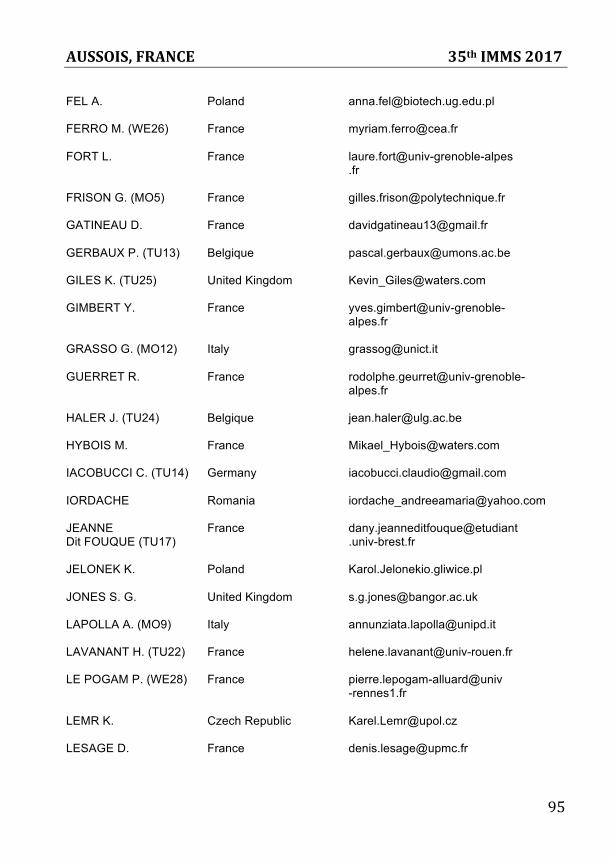
AUSSOIS, FRANCE 35th IMMS 2017
95
FEL A. Poland [email protected] FERRO M. (WE26) France [email protected] FORT L. France laure.fort@univ-grenoble-alpes .fr FRISON G. (MO5) France [email protected] GATINEAU D. France [email protected] GERBAUX P. (TU13) Belgique [email protected] GILES K. (TU25) United Kingdom [email protected] GIMBERT Y. France yves.gimbert@univ-grenoble- alpes.fr GRASSO G. (MO12) Italy [email protected] GUERRET R. France rodolphe.geurret@univ-grenoble- alpes.fr HALER J. (TU24) Belgique [email protected] HYBOIS M. France [email protected] IACOBUCCI C. (TU14) Germany [email protected] IORDACHE Romania [email protected] JEANNE France dany.jeanneditfouque@etudiant Dit FOUQUE (TU17) .univ-brest.fr JELONEK K. Poland Karol.Jelonekio.gliwice.pl JONES S. G. United Kingdom [email protected] LAPOLLA A. (MO9) Italy [email protected] LAVANANT H. (TU22) France [email protected] LE POGAM P. (WE28) France pierre.lepogam-alluard@univ -rennes1.fr LEMR K. Czech Republic [email protected] LESAGE D. France [email protected]
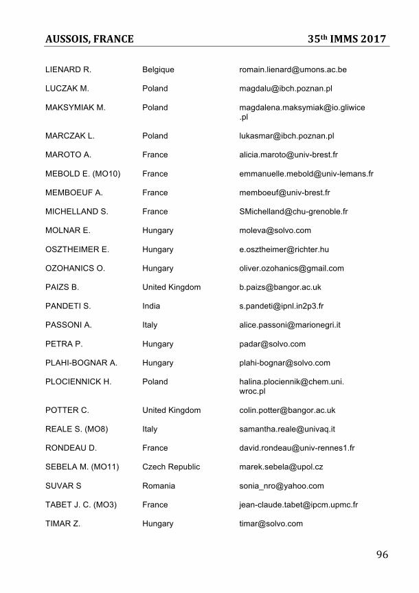
AUSSOIS, FRANCE 35th IMMS 2017
96
LIENARD R. Belgique [email protected] LUCZAK M. Poland [email protected] MAKSYMIAK M. Poland [email protected] .pl MARCZAK L. Poland [email protected] MAROTO A. France [email protected] MEBOLD E. (MO10) France [email protected] MEMBOEUF A. France [email protected] MICHELLAND S. France [email protected] MOLNAR E. Hungary [email protected] OSZTHEIMER E. Hungary [email protected] OZOHANICS O. Hungary [email protected] PAIZS B. United Kingdom [email protected] PANDETI S. India [email protected] PASSONI A. Italy [email protected] PETRA P. Hungary [email protected] PLAHI-BOGNAR A. Hungary [email protected] PLOCIENNICK H. Poland [email protected]. wroc.pl POTTER C. United Kingdom [email protected] REALE S. (MO8) Italy [email protected] RONDEAU D. France [email protected] SEBELA M. (MO11) Czech Republic [email protected] SUVAR S Romania [email protected] TABET J. C. (MO3) France [email protected] TIMAR Z. Hungary [email protected]
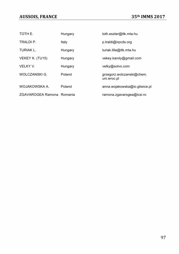
AUSSOIS, FRANCE 35th IMMS 2017
97
TOTH E. Hungary [email protected] TRALDI P. Italy [email protected] TURIAK L. Hungary [email protected] VEKEY K. (TU15) Hungary [email protected] VELKY V. Hungary [email protected] WOLCZANSKI G. Poland grzegorz.wolczanski@chem. uni.wroc.pl WOJAKOWSKA A. Poland [email protected] ZGAVAROGEA Ramona Romania [email protected]
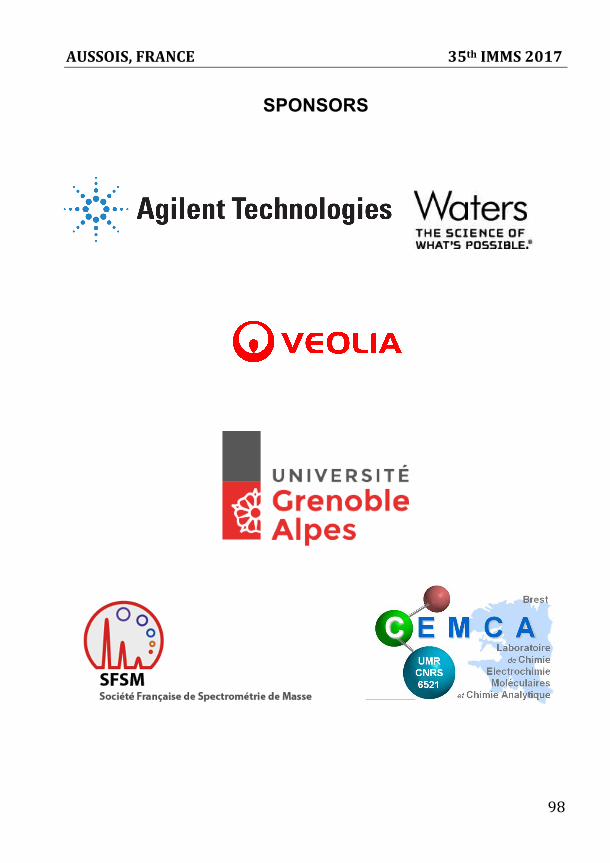
AUSSOIS, FRANCE 35th IMMS 2017
98
SPONSORS