Embed Size (px)
Citation preview

Issue 2
www.oteurope.com
Products inPRACTICEReal world experience of
leading ophthalmic products

3
Imaging & Diagnostics4 The diagnostic power of MultiColor imaging for SPECTRALIS OCT MultiColor Scanning Laser Imaging is the new colour fundus imaging
option for the SPECTRALIS OCT family of products from Heidelberg Engineering. The basics of MultiColor imaging are outlined in this feature, highlighting its advantages and presenting the latest experiences.
6 Zero-dilation scanning laser ophthalmoscopy for the early diagnosis of diabetic retinopathy: Results from the DRIVE study
A confocal line scanning laser ophthalmoscope (cSLO) has been developed by i-Optics for non-mydriatic diagnosis of diabetic retinopathy. Comparative results between zero-dilation cSLO and conventional fundus imaging are revealed.
7 Case study on a dry eye patient using the new OCULUS Keratograph 5M
In this case study, the author describes how to use the OCULUS Keratograph 5M for diagnosis and follow up of a patient after they have undergone Toyos Intense Pulse Light Treatment for Dry Eye.
Laser Surgery9 PRESBYOND Laser Blended Vision: An approach to presbyopia with
little or no compromise Dan Reinstein discusses PRESBYOND Laser Blended Vision (Carl Zeiss
Meditec) as a solution for presbyopia that meets all the goals of good binocular vision at all distances with little or no compromise.
10 Refractive lenticule extraction: Will ReLEx replace LASIK in the near future?
Professor Sekundo reveals why he believes ReLEx (Carl Zeiss Meditec) is an attractive alternative to LASIK and offers great future potential for further developments.
Surgical Instruments11 Microincisions optimize refractive results of cataract surgery In this piece, Dr Hoffman discusses his experiences with post-limbal
2.2 mm incisions and the newly developed sets for coaxial mini phaco available from Geuder.
12 Product profiles Heidelberg Engineering i-Optics OCULUS Carl Zeiss Meditec Geuder
Fuse
/Get
ty Im
ages
Products inPRACTICE
www.oteurope.com
Contents
Imaging & Diagnostics
The diagnostic power of MultiColor imaging for SPECTRALIS OCTBy Professor Sebastian Wolf, MD, PhD, University of Bern, Bern, Switzerland.
media opacities, for example with a cataract. The MultiColor images are automatically colour-balanced to match the overall appearance of a traditional colour fundus photograph, with the exception that the optic disc colour does not match its natural appearance. Users can choose to display a MultiColor image simultaneously with an OCT cross sectional image, or view the combined colour image together with the images of the individual laser colours to gain a better understanding of anatomic and pathologic detail at different depths within the retina. These options allow the user to identify abnormalities quickly and reliably. (Case 1).
Benefits across a range of diseasesUsers can acquire an OCT scan directly on the live MultiColor image. This allows to quickly analyze abnormal structures. MultiColor images can be acquired without dilating the pupils of the patient, which makes MultiColor a useful standard imaging modality in every setting. Camera alignment and acquisition of MultiColor images is easy because of the continuously scanning laser system that delivers a live fundus image.
The benefit of MultiColor in clinical practice is its capability of highlighting diseases found to be more difficult to recognize in traditional colour fundus photographs.
MultiColor imaging is particularly helpful with diabetic patients, for detecting and managing retinovascular diseases and for
MultiColor Scanning Laser Imaging is the new colour fundus imaging option for the SPECTRALIS OCT family of
products. It adds a new dimension to the multimodality imaging platform by combining simultaneous SD-OCT and selective colour fundus imaging.
All SPECTRALIS models (OCT, OCTPlus, HRA and HRA+OCT) can now be delivered with this new feature — or be ordered with an Advanced Upgrade Package that allows an upgrade to MultiColor at a later point.
MultiColor imaging uses multiple laser colours simultaneously to selectively capture and display diagnostic information originating from different retinal structures within a single examination, revealing diagnostically relevant details that are often hard to spot in corresponding classic colour fundus photographs.
This article will outline the basics of MultiColor imaging, highlighting advantages and presenting latest experiences.
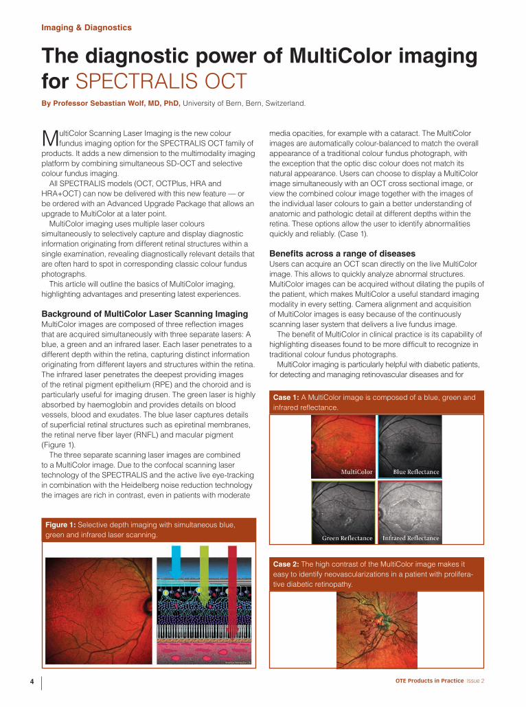
Background of MultiColor Laser Scanning ImagingMultiColor images are composed of three reflection images that are acquired simultaneously with three separate lasers: A blue, a green and an infrared laser. Each laser penetrates to a different depth within the retina, capturing distinct information originating from different layers and structures within the retina. The infrared laser penetrates the deepest providing images of the retinal pigment epithelium (RPE) and the choroid and is particularly useful for imaging drusen. The green laser is highly absorbed by haemoglobin and provides details on blood vessels, blood and exudates. The blue laser captures details of superficial retinal structures such as epiretinal membranes, the retinal nerve fiber layer (RNFL) and macular pigment (Figure 1).
The three separate scanning laser images are combined to a MultiColor image. Due to the confocal scanning laser technology of the SPECTRALIS and the active live eye-tracking in combination with the Heidelberg noise reduction technology the images are rich in contrast, even in patients with moderate
Figure 1: Selective depth imaging with simultaneous blue, green and infrared laser scanning.
Case 1: A MultiColor image is composed of a blue, green and infrared reflectance.
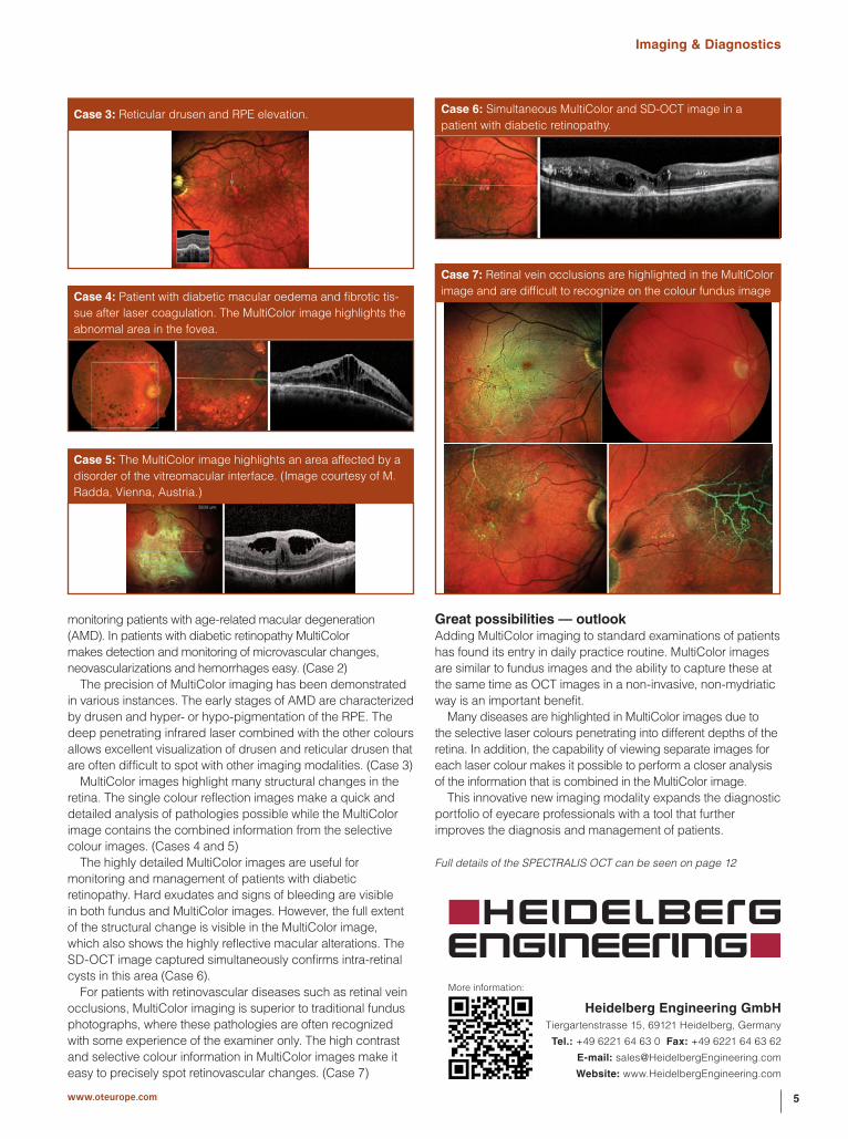
Case 2: The high contrast of the MultiColor image makes it easy to identify neovascularizations in a patient with prolifera-tive diabetic retinopathy.
OTE Products in Practice Issue 24
Imaging & Diagnostics
Great possibilities — outlookAdding MultiColor imaging to standard examinations of patients has found its entry in daily practice routine. MultiColor images are similar to fundus images and the ability to capture these at the same time as OCT images in a non-invasive, non-mydriatic way is an important benefit.
Many diseases are highlighted in MultiColor images due to the selective laser colours penetrating into different depths of the retina. In addition, the capability of viewing separate images for each laser colour makes it possible to perform a closer analysis of the information that is combined in the MultiColor image.
This innovative new imaging modality expands the diagnostic portfolio of eyecare professionals with a tool that further improves the diagnosis and management of patients.
Full details of the SPECTRALIS OCT can be seen on page 12
Heidelberg Engineering GmbHTiergartenstrasse 15, 69121 Heidelberg, Germany
Tel.: +49 6221 64 63 0 Fax: +49 6221 64 63 62
E‑mail: [email protected]
Website: www.HeidelbergEngineering.com
monitoring patients with age-related macular degeneration (AMD). In patients with diabetic retinopathy MultiColor makes detection and monitoring of microvascular changes, neovascularizations and hemorrhages easy. (Case 2)
The precision of MultiColor imaging has been demonstrated in various instances. The early stages of AMD are characterized by drusen and hyper- or hypo-pigmentation of the RPE. The deep penetrating infrared laser combined with the other colours allows excellent visualization of drusen and reticular drusen that are often difficult to spot with other imaging modalities. (Case 3)
MultiColor images highlight many structural changes in the retina. The single colour reflection images make a quick and detailed analysis of pathologies possible while the MultiColor image contains the combined information from the selective colour images. (Cases 4 and 5)
The highly detailed MultiColor images are useful for monitoring and management of patients with diabetic retinopathy. Hard exudates and signs of bleeding are visible in both fundus and MultiColor images. However, the full extent of the structural change is visible in the MultiColor image, which also shows the highly reflective macular alterations. The SD-OCT image captured simultaneously confirms intra-retinal cysts in this area (Case 6).
For patients with retinovascular diseases such as retinal vein occlusions, MultiColor imaging is superior to traditional fundus photographs, where these pathologies are often recognized with some experience of the examiner only. The high contrast and selective colour information in MultiColor images make it easy to precisely spot retinovascular changes. (Case 7)
Case 3: Reticular drusen and RPE elevation.
Case 4: Patient with diabetic macular oedema and fibrotic tis-sue after laser coagulation. The MultiColor image highlights the abnormal area in the fovea.
Case 5: The MultiColor image highlights an area affected by a disorder of the vitreomacular interface. (Image courtesy of M. Radda, Vienna, Austria.)
Case 6: Simultaneous MultiColor and SD-OCT image in a patient with diabetic retinopathy.
Case 7: Retinal vein occlusions are highlighted in the MultiColor image and are difficult to recognize on the colour fundus image
More information:
5www.oteurope.com

Imaging & Diagnostics
Zero-dilation scanning laser ophthalmoscopy for the early diagnosis of diabetic retinopathy: Results from the DRIVE studyDirk De Brouwere,1 Peter van Etten2 and Jose Martinez,31i-Optics bv, The Hague, The Netherlands 2Rotterdam Eye Institute, Rotterdam, The Netherlands 3Rotterdam Eye Hospital, Rotterdam, The Netherlands
technique for DR diagnosis which demonstrated improved sensitivity over traditional fundus imaging and no requirement for time-consuming pharmacologic mydriasis. Zero-dilation cSLO can be performed relatively quickly and has the potential to improve efficiency and reduce appointment times, with consequent benefits for both patients and healthcare providers. cSLO imaging may facilitate screening of DR in primary care centres and thereby promote early diagnosis and treatment.
Reference1. S. Garg and R.M. Davis, Clin. Diabetes, 2009;27:140–145.2. Nederlands Oogheelkundig Gezelschap. Richtlijn: Diabetische
retinopathie. Screening, diagnostiek en behandeling. Accessed at http://www.oogheelkunde.org/uploads/VS/qr/VSqrAwWsMsiAu4bXQLv1uQ/Richtlijnen-Diabetische-Retinopathie-.pdf, 15 November 2011.
Full details of the EasyScan can be found on page 12
i-OpticsMauritskade 35 2514 HD, The Hague, The Netherlands
Tel.: +31 70 3993 112
Website: www.i-optics.com
The incidence of diabetic retinopathy (DR) is projected to escalate as the number of people diagnosed with
diabetes increases. Regular DR screening, often performed using a traditional fundus camera, enables early intervention and significantly reduces the risk of vision loss.1 However, fundus imaging requires pharmacologic mydriasis for small pupil diameters, which is time-consuming, expensive and often uncomfortable for patients. A confocal line scanning laser ophthalmoscope (cSLO) has been developed by i-Optics (The Netherlands) for non-mydriatic diagnosis of DR. In the Diabetic Retinopathy Initial Validation Enrolment (DRIVE) study, performed at the Rotterdam Eye Hospital, the clinical performance and ophthalmologist referral rates obtained following DR screening by zero-dilation cSLO and conventional fundus imaging were compared.
MethodsSequential eye examinations were conducted in 100 patients with diabetes (age 60 ± 11 years) using the cSLO imaging device (EasyScan v1.2, i-Optics, The Netherlands) and a standard fundus camera (Topcon TRC-NW6) (Figure 1). cSLO imaging used green (532 nm) and infrared (785 nm) laser light to illuminate a narrow line on the retina through a 2.0 mm pupil and across a 45- by 45-degree retinal central and nasal field. Following cSLO imaging, mydriasis was performed to allow imaging with the fundus camera. Acquisition time was recorded for EasyScan. Referral for DR was assessed, based on The Netherlands Guidelines,2 by two licensed graders using images from both devices.
ResultsGradeability of cSLO images was in line with the criteria for diabetic diagnosis devices specified by the British Diabetic Association. Median image acquisition time using cSLO was less than 3 minutes. 33% of patients imaged with the cSLO had a pupil diameter of less than 3.3 mm and would require mydriasis before fundus imaging. DR referral rates obtained using cSLO and fundus imaging showed strong agreement (88.6%) after device-blinded evaluation. Normalized sensitivity for detection of DR lesion characteristics was greater with cSLO imaging than with fundus imaging (100% versus 85%). Normalized specificity was equivalent for the two techniques (96%).
ConclusionsThe DRIVE study found cSLO imaging to be an efficient
Figure 1: Zero-dilation cSLO and conventional fundus imaging
by i-Optics.com
OTE Products in Practice Issue 26

Imaging & Diagnostics
A print out will show you graphically where the break up occurred, time of the first break up, and average break up time (Figure 1).
The advantages of using the NIKBUT is that a technician can do the scan, which frees up physician time, no drops are applied to the patient’s eyes, the results are objective and reproducible, and visual representation of the problem is available for the doctor to discuss with the patient.
In this patient it is obvious that he has a poor NIKBUT making it more likely that he has Meibomian Gland Dysfunction causing evaporative dry eye.
Now the technician can measure tear meniscus height which can be used as a measure of tear production (Figure 2). A Schirmer Test can still be done if needed. In our patient the tear meniscus height was normal.
The next test is Meibography (Figure 3) where an infrared light source and video camera are used on the inverted lid to show the Meibomian Glands (MG). This test is useful to record MG dropout. Studies have shown that MG dropout can lead to a poor lipid layer. We have found that lower lid drop out is more common than upper lid drop out. Some doctors have reported that increase tortuosity may also be an indication of poor function. In a patient with evaporative dry eye, poor functioning glands could be the direct cause of their DED. In this patient we see that he has some dropout in the inferior lid.
Dry eye patients consistently complain about bulbar redness. The Keratograph 5M uses a Bulbar Redness classification scale to help us objectively measure one of their chief complaints (Figure 4).
Case Study on a Dry Eye Patient using the new OCULUS Keratograph 5MStudy performed by Dr Rolando Toyos, MD, Medical Director and Founder of Toyos Clinic, USA
Dry Eye Disease (DED) is one of the most common problems that we deal with in our clinics. We have seen our dry eye
clinic grow exponentially as the baby boomers become older. We completed a 3 year retrospective study showing that over 75% of our dry eye disease patients were over the age of 45. These patients suffer irritation and pain due to their ocular surface disease that compels them to visit their doctor for treatment and relief.
Our clinic provides many treatment modalities that can help patients improve. We also have many tools to objectively measure severity and progress of DED.
One tool that has integrated nicely into our programme is the new OCULUS Keratograph 5M, which is a topographer and DED analyser. The high-resolution colour camera and the integrated magnification changer offer a new perspective to the tear film assessment procedure. In this case study we will show how we use the Keratograph 5M to diagnose and follow a patient after they have had the Toyos Intense Pulse Light Treatment for Dry Eye (Dermamed).
Case study60 yr old white male complaining of red, irritated eyes for 3 years. He had been seen in several clinics where he tried 100 mg doxycycline, artificial tears, restasis, warm compresse, and lid scrubs. As we knew beforehand that the patient might be a possible dry eye evaluation when he came to our clinic we had our technician screen him using the Keratograph 5M.
The first part of the screening was the Non Invasive Keratograph Break Up Time (NIKBUT). Conventional TBUT requires the placement of a fluorescein drop on the eye and then for a physician to check the eye at the slit lamp to monitor the first break up. This method of measuring can be time consuming for the doctor and often times can lead to inconsistent results.
The Keratograph 5M uses placido ring illumination measuring thousands of points throughout the surface of the cornea. As the margins of the rings become displaced a high resolution camera records the break up in tenths of a second.
Figure 1: Example of Keratograph 5M TF-Scan (evaluation of the tear film break-up time)
Figure 2: Example of Keratograph 5M Tear Meniscus Height Measurement.
Figure 3: Example of Keratograph 5M Meibography.
7www.oteurope.com

Imaging & Diagnostics
found the picture and video feature invaluable in our practice not only for the clinician but as a tool to help the patient understand their disease and improvement. Here we see the patient shows all the signs of lid margin disease that accompany MGD.
The Keratograph 5M is an incredible piece of technology that has the advantage of being a topographer as well as a specific Dry Eye Disease Analyser. It allows the technician to perform some of the tests in a more objective way freeing up the physician to spend more time with the patient to discuss their disease and treatment options. The Keratograph 5M captures pictures and videos that can be used by the clinician for assessment and to educate the patient. We believe that it has been an invaluable part of our dry eye clinic success.
Full details of the OCULUS Keratograph 5M can be found on page 13
OCULUS Optikgeräte GmbHMünchholzhäuser Str. 29, 35582 Wetzlar, Germany
Tel.: +49 641 2005 0
E-mail: [email protected] Website: www.oculus.de
Once the patient has undergone these tests by the technician then I can begin my exam. If I determine that the patient has a poor tear film I can use the Keratograph 5M to assist me further by performing a lipid layer scan and photographing lid margin pathology. The Keratograph 5M uses white light interferometry and specular reflection to give you a coloured assessment of the lipid layer. I find this lipid view reaffirms my diagnosis.
All of these patients have lid margin pathology whether it is erythema, thickening of the lid, loss of lashes, scurf, scalloping of the glands or telangiectasias. The Keratograph 5M can take a picture of this lid pathology so that you have a baseline as comparison when the patient undergoes treatment. We have
Figure 4: Example of Keratograph 5M Bulbar Redness Scan.
It’s not tricky. Clicky, clicky! Join OTEurope’s growing on-line community… …via LinkedIn to discuss issues that matter to you and your peers www.oteurope.com/discuss�…via twitter to get regular industry news and article updateswww.twitter.com/OTEurope
…via the web for regular featureswww.oteurope.com
OTE Products in Practice Issue 28

Laser Surgery
PRESBYOND Laser Blended Vision: An approach to presbyopia with little or no compromiseBy Dan Z. Reinstein, MD, MA(Cantab), FRCSC, DABO, FRCOphth, FEBO, London Vision Clinic, London, UK.
longer able to fully ‘process’ the spherical aberration and will result in aberration-related quality of vision symptoms. Therefore, this spherical aberration method cannot be used to correct presbyopia by itself.
Combining this increased depth of field with monovision enables good near vision to be achieved, but with a lower degree of anisometropia than in traditional monovision. This strategy creates a blend zone of vision between the two eyes at intermediate distances meaning that much less suppression is required and there is no dissociation between the eyes. Patients retain a functional level of uncorrected stereoacuity — proving that they have binocular function.
In PRESBYOND Laser Blended Vision a number of factors are considered including age, accommodative amplitude, preoperative wavefront, tolerance to anisometropia and the amount of refractive error. The software then combines these factors to generate an ablation profile with the aim of leaving the patient with an appropriate level of spherical aberration to maximize the depth of field without compromising contrast sensitivity, stereoacuity or night vision.
At 1 year after Laser Blended Vision, binocular UDVA was 20/20 or better and UNVA was J2 or better in 95% of 136 myopic patients (≤–8.50 D), 77% of 111 hyperopic patients (≤+5.75 D), and 95% of 148 emmetropic patients (within ±0.88 D). The safety was the same as for standard LASIK with no eyes losing more than one line CDVA and contrast sensitivity was either the same or slightly better than pre-op.
In summary, Laser Blended Vision is a solution for presbyopia that meets all the goals of good binocular vision at all distances, no compromise in safety, contrast sensitivity, or night vision, and retention of functional stereoacuity. The procedure is immediately reversible by wearing spectacles or a simple retreatment can be done using a standard ablation with the advantage of keeping the depth of field. All this is achieved while simultaneously correcting a wide range of refractive errors and astigmatism levels. The key to this approach was to base it on the natural mechanisms of spherical aberration processing and binocular fusion, unlike multifocal approaches, which require the patient to adjust to the unnatural situation of having to differentiate between two images in the same eye.
Dan Z. Reinstein, MD, practices at the London Vision Clinic, London, UK, and is
affiliated with the Department of Ophthalmology, Columbia University Medical
College, New York, USA, and the Centre Hospitalier National d’Ophtalmologie,
Paris, France. He has financial interests with Carl Zeiss Meditec (Jena, Germany)
and ArcScan Inc. (Morrison, Colorado, USA).
Full details of this offering can be found on page 13
Carl Zeiss Meditec AGGoeschwitzer Str. 51–52, 07745 Jena, Germany
Tel.: +49 3641 220 0
Website: www.meditec.zeiss.com
The ideal solution for presbyopia would be to restore accommodation, however, no procedure up to now has
been able to restore the natural focusing mechanism of the eye. Current treatments for presbyopia therefore rely on splitting the refractive power for far and near distances either within the same eye (multifocality) or between eyes (monovision), but all treatments require some compromise from the patient.
The challenge is to achieve good binocular vision at far, intermediate and near distances while also maintaining optical quality, contrast sensitivity, night vision, stereoacuity and as a bonus the procedure should be reversible. This was the goal we set when developing Laser Blended Vision with Carl Zeiss Meditec. Our approach was to take advantage of the natural mechanisms within our optical system and minimize the need for the patient to adapt.
All multifocal approaches require the patient to adjust to the unnatural situation of having to differentiate between two images in the same eye, so it is no surprise that these procedures are associated with loss of CDVA, contrast sensitivity and night vision disturbances. There have been significant improvements over the years, however, multifocality will always rely on the patient’s ability to adapt to this new and unnatural intra-ocular rivalry. Multifocal treatments are also usually limited to a small range of refractive error (usually low hyperopic patients).
The well-established principles of contact lens monovision have been used in laser refractive surgery, however, many of the limitations of contact lens monovision also affected laser refractive surgery induced monovision. These limitations include loss of fusion due to anisometropia, poor intermediate vision, poor distance vision in the near eye, reduced binocular contrast sensitivity, and reduced (or even broken) stereoacuity. However, monovision is based upon the natural process of binocular fusion (interocular rivalry, not intraocular) and recent studies have demonstrated that many of these limitations are avoided by limiting the anisometropia to 1.25–1.50 D. But, this level of anisometropia does not always give the patient enough near vision.
With Laser Blended Vision we incorporated another natural visual process — filtering of spherical aberration — to increase the depth of field in each eye and achieve good binocular vision at all distances. Introducing some spherical aberration disseminates the retinal focal point meaning that there is a wider range of distances where the focus is equivalent, although slightly reduced, but the perceived image is still sharp due to the natural ability of the visual cortex to ‘process’ spherical aberration. This range is the depth of field and can be demonstrated by the better-than-expected distance vision in the near eye (the mean visual acuity is about 20/45 whereas 20/80 would be expected for –1.50 D).
This concept is simply an extension of the eye’s natural state as everyone has some naturally occurring spherical aberration, and the brain is pre-programmed to do this filtering. If there is too much spherical aberration, however, the visual cortex is no
9www.oteurope.com
Laser Surgery
Refractive lenticule extraction: Will ReLEx replace LASIK in the near future?By Walter Sekundo, MD, Prof. of Ophthalmology, Philipps University of Marburg, Germany
Shah et al., Vestergaard, Hjortdal et al. and Kamiya et al.).
ReLEx smile: Minor changes, major successReLEx receives a further boost from the ongoing refinement of ReLEx smile. The key to the success of ReLEx smile lies partly in the use of flapless technology, which eliminates induced complications and impairs corneal stability to a lesser extent than flap-based methods. Recently, Wiltfang (Munich, Germany) and Meyer (Cologne, Germany) developed new laser energy settings. This has further reduced the duration of the procedure, facilitated the dissection of the tissue connections and accelerated visual acuity recovery. Special ReLEx instruments have now also been developed. In the past 6 months, I have not only been using the new settings, but also the spatula developed by Dr Chansue from Bangkok, Thailand. With this equipment even in eyes with –8 or –10 D, I achieve unaided distance visual acuity of 0.8 to 1.25 with a postoperative foreign body sensation lasting a maximum of 2 hours.
Summary and outlookIn my view the above mentioned benefits, both clinical and economic, make this method an attractive alternative to (Femto-)LASIK, with the result that I currently use it for about 50% of all my refractive laser patients.
ReLEx also offers great future potential for further developments. At the beginning of 2013, for example, a second hyperopia study is starting in Erfurt and Marburg with the goal of expanding the range of indications. The subject of ‘enhancement’ — although not common – can currently only be addressed with the aid of an excimer laser.
To sum up, I would say that the new development that was still eyed with skepticism six years ago has now attained a degree of maturity that opens up outstanding opportunities to this surgical approach in competition to the proven technology and is gaining increasing popularity on a daily basis.
Prof. Sekundo has financial interests with Carl Zeiss Meditec (Jena, Germany).
Full details of this offering can be found on page 14
Carl Zeiss Meditec AGGoeschwitzer Str. 51–52, 07745 Jena, Germany
Tel.: +49 3641 220 0
Website: www.meditec.zeiss.com
Refractive lenticule extraction for the correction of myopia and myopic astigmatism has developed at a rapid pace all over
the world since the introduction of the minimally invasive version ReLEx smile. This surgical technique is based on cutting tissue using the VisuMax femtosecond system (Carl Zeiss Meditec, Germany), instead of ablation with an excimer laser.
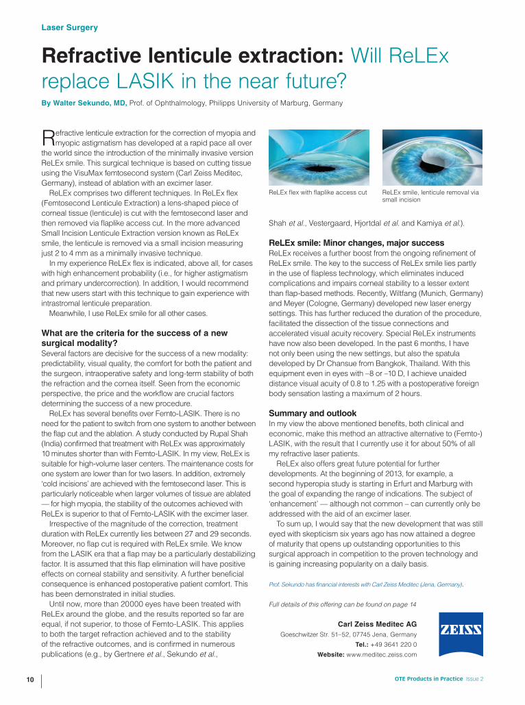
ReLEx comprises two different techniques. In ReLEx flex (Femtosecond Lenticule Extraction) a lens-shaped piece of corneal tissue (lenticule) is cut with the femtosecond laser and then removed via flaplike access cut. In the more advanced Small Incision Lenticule Extraction version known as ReLEx smile, the lenticule is removed via a small incision measuring just 2 to 4 mm as a minimally invasive technique.
In my experience ReLEx flex is indicated, above all, for cases with high enhancement probability (i.e., for higher astigmatism and primary undercorrection). In addition, I would recommend that new users start with this technique to gain experience with intrastromal lenticule preparation.
Meanwhile, I use ReLEx smile for all other cases.
What are the criteria for the success of a new surgical modality?Several factors are decisive for the success of a new modality: predictability, visual quality, the comfort for both the patient and the surgeon, intraoperative safety and long-term stability of both the refraction and the cornea itself. Seen from the economic perspective, the price and the workflow are crucial factors determining the success of a new procedure.
ReLEx has several benefits over Femto-LASIK. There is no need for the patient to switch from one system to another between the flap cut and the ablation. A study conducted by Rupal Shah (India) confirmed that treatment with ReLEx was approximately 10 minutes shorter than with Femto-LASIK. In my view, ReLEx is suitable for high-volume laser centers. The maintenance costs for one system are lower than for two lasers. In addition, extremely ‘cold incisions’ are achieved with the femtosecond laser. This is particularly noticeable when larger volumes of tissue are ablated — for high myopia, the stability of the outcomes achieved with ReLEx is superior to that of Femto-LASIK with the excimer laser.
Irrespective of the magnitude of the correction, treatment duration with ReLEx currently lies between 27 and 29 seconds. Moreover, no flap cut is required with ReLEx smile. We know from the LASIK era that a flap may be a particularly destabilizing factor. It is assumed that this flap elimination will have positive effects on corneal stability and sensitivity. A further beneficial consequence is enhanced postoperative patient comfort. This has been demonstrated in initial studies.
Until now, more than 20 000 eyes have been treated with ReLEx around the globe, and the results reported so far are equal, if not superior, to those of Femto-LASIK. This applies to both the target refraction achieved and to the stability of the refractive outcomes, and is confirmed in numerous publications (e.g., by Gertnere et al., Sekundo et al.,
ReLEx flex with flaplike access cut ReLEx smile, lenticule removal via small incision
OTE Products in Practice Issue 210

Surgical Instruments
Microincisions optimize refractive results of cataract surgeryBy Dr med. P. Hoffmann, Ophthalmic- and Laser Clinic, Castrop-Rauxel, Germany.
more narrow, which allows for a larger vacuum but not one that is so large it destabilizes the anterior chamber. Currently we are working with 550 mmHg in conjunction with the Venturi version of the Geuder Megatron S4. Through the large opening with internal tiers, quick and controlled emulsification of even very hard cores poses no problem. The newly-developed sleeve allows for good mobility in the small incision. We are convinced that coaxial micro-phaco with 2.2 mm incision width is performed as quickly and securely as conventional phaco. Due to the sleeve’s better sealing ability and less stress to the wound, coaxial is preferential to biaxial. This is particularly true because with incision widths significantly below 2 mm, compromises have to be made regarding the lenses to be implanted. Refractive predictability, especially for toric and aspherical premium lenses, is significantly improved through aberration-neutral incisions.
In our opinion, coaxial micro-phaco will also become the method of choice for the currently-emerging femtosecond laser-assisted surgical technique, because only this method allows the potential precision advantages of the laser to be fully used.
Full details of the Pure Efficiency Phaco Sets can be found on page 14
Geuder AGHertzstrasse 4, 69126 Heidelberg, Germany
Tel.: +49 6221 3066 Fax: +49 6221 303122
E‑mail: [email protected]
Website: www.geuder.de
In today’s cataract surgery the refractive component has become increasingly significant. For patients and referring
eye specialists uncorrected visual acuity and emmetropia in particular are the decisive factors for success. In the last few years, aspherical and toric lenses have further improved optical quality. To take full advantage of these lenses, the calculation and refraction prediction has to be very accurate. Prediction accuracy mainly depends on four variables:• Measurement technique (biometer, keratometer, topography)• Positioning of the lens (centring, rotation)• Surgically-induced aberrations• Postoperative refraction accuracy
To achieve the best results possible, all four variables should be kept as small as possible. Much has already been written about measurement and surgical techniques. Postoperative refraction mechanics can, of course, not be influenced.
Surgically-induced astigmatism — or better: surgically-induced aberration because comas and higher-order astigmatisms are also caused — is often assumed to be a constant value in common IOL calculations for toric lenses. Thus, a flattening of 0.25 D. in the astigmatic axis is, for example, included. Unfortunately, unsystematic deviations are added to this systematic change, representing no more than a mean value, which worsen the result in total. Not only is induced astigmatism stronger the wider the incision and the closer to the optical centre of the cornea but so are the higher-order aberrations and unsystematic dispersion.
The only way to avoid this dilemma is by making an aberration-neutral incision. We want to get as close to this ideal as possible and it can only be achieved with the smallest incisions possible, which should additionally be far away from the optically-active part of the cornea, meaning temporal and far outside (post-limbal).
An incision width of 2.2 mm seems to be the best compromise for us. Most standard lenses made of hydrophobic or hydrophilic acrylate can be implanted through this incision width using suitable injection systems. Likewise, capsulorhexis surgery with utrata forceps is still possible. Coaxial phaco with special mini-tips allows surgeons to retain their individual favourite surgical techniques without loss of efficiency compared to large tips with 3.0 mm incisions. We have re-examined the induced astigmatisms in 550 eyes, which were operated with 2.2 mm post-limbal incisions, and found a median close to zero (Figure 1 left boxplot) and an unsystematic dispersion that complies with the measurement accuracy of the keratometer (Figure 1 right boxplot = two preoperative measurements with IOLMaster and Lenstar). These incisions are thus to be viewed as astigmatism-neutral.
We have been routinely applying 2.2 mm coaxial phaco since 2008. In a pilot study on 202 eyes in 2009, we were able to show that the effective phaco time (median 1.7 s vs 1.5 s) and the real time in which the phaco tip is in the eye (57 s vs 53 s) are nearly identical. Thus, no efficiency loss is present. In the current version of the needle, the shaft has again been made
Figure 1: Box-Whisker-Plots illustrating that 2.2 mm post-limbal incisions are astigmatism-neutral
11www.oteurope.com
Product Pro� les
Heidelberg Engineering GmbHTiergartenstr. 15, 69121, Heidelberg, Germany
Tel.: +49 6221 6463 0
E-mail: [email protected]
Website: www.HeidelbergEngineering.com
i-OpticsMauritskade 35, 2514 HD The Hague,
The Netherlands
Tel.: +31 7039 93112
Website: www.i-optics.com/DRIVE
SPECTRALIS OCT
EasyScan
What an OCT should have: SPECTRALIS OCT
For accurate diagnosis and individualized treatment of patients, the precise detection of even smallest structural changes is vital.
All SPECTRALIS models offer the unique AutoRescan function that enables the system to track retinal changes with high precision. The
Smart retinal imaging based on scanning laser ophthalmoscope (SLO) technology
EasyScan is a breakthrough zero-dilation retinal imaging system that is ideal for the diagnosis of retinal pathologies, for example, diabetic retinopathy, age-related macular degeneration and glaucoma.
EasyScan makes retinal imaging fast, easy and patient-friendly. It is the first to pack high contrast confocal SLO into a compact, affordable and patient-friendly device. And it weighs just 7 kg, so it opens up high-end eyecare to patients all over the world.
Check the benefits of confocal imaging in our clinical atlas www.i-optics.com/library
Active Live Eye Tracker positions the follow-up OCT cross sectional scans accurately on the very same spot of the retina every time a patient is imaged. This way, the system ensures that the changes detected in morphology and thickness are associated with true pathology and not motion artifact. Changes in retinal nerve fibre layer as small as 1 µm can be reliably detected, aiding the early diagnosis of glaucoma.
MultiColor Laser Scanning Imaging and Non-Contact Ultra-Widefield Angiography are the latest options added to the modular SPECTRALIS product family.
With MultiColor laser fundus imaging, the specific laser colours capture information from different depths and structures of the retina and create detailed, high-contrast fundus images. MultiColor is available for all SPECTRALIS models.
Features:• Optical engine: Confocal SLO• Capture mode: Green (532 nm), near
infrared (785 nm) and combined, (pseudo colour)
• Field angle: 60 degrees horizontal, 45 degrees vertical
• Minimum pupil size: 2.0 mm• Alignment help: ‘See what you get’
with IR live imaging• Fixation targets: 3 internal targets • Autofocus and autocapture: YES• Flash settings: Auto Exposure• Emmetropia compensation: ±10 D • Unique movie loop feature showing
multiple frames• Easy switching between capture,
review, archive and export• Networking capabilities including
telediagnosis• Image formats: TIFF, JPEG, PNG,
BMP, DICOM, PDF • Compact, portable, USB-powered • Weight < 7 kg
The new Ultra-Widefield Angiography Module captures images contact-free. The result is distortion-free, high contrast FA and ICGA images that are evenly illuminated out into the far periphery — all with the simple exchange of a lens. All HRA2 and SPECTRALIS angiography models can be upgraded with the new module — a cost-effective alternative to separate widefield imaging devices.
i-Optics pioneers smart and superior eye diagnosis solutions. Our innovations also include Cassini Color Led Topography and EyePrevent, retinal screening programs.
SEE THINGS DIFFERENT.
by i-Optics.com
OTE Products in Practice Issue 212
Product Profiles
OCULUS Optikgeräte GmbHMünchholzhäuser Str. 29, 35582, Germany
Tel.: +49 641 2005 0
E-mail: [email protected]
Website: www.oculus.de
OCULUS Keratograph 5M
PRESBYOND Laser Blended Vision
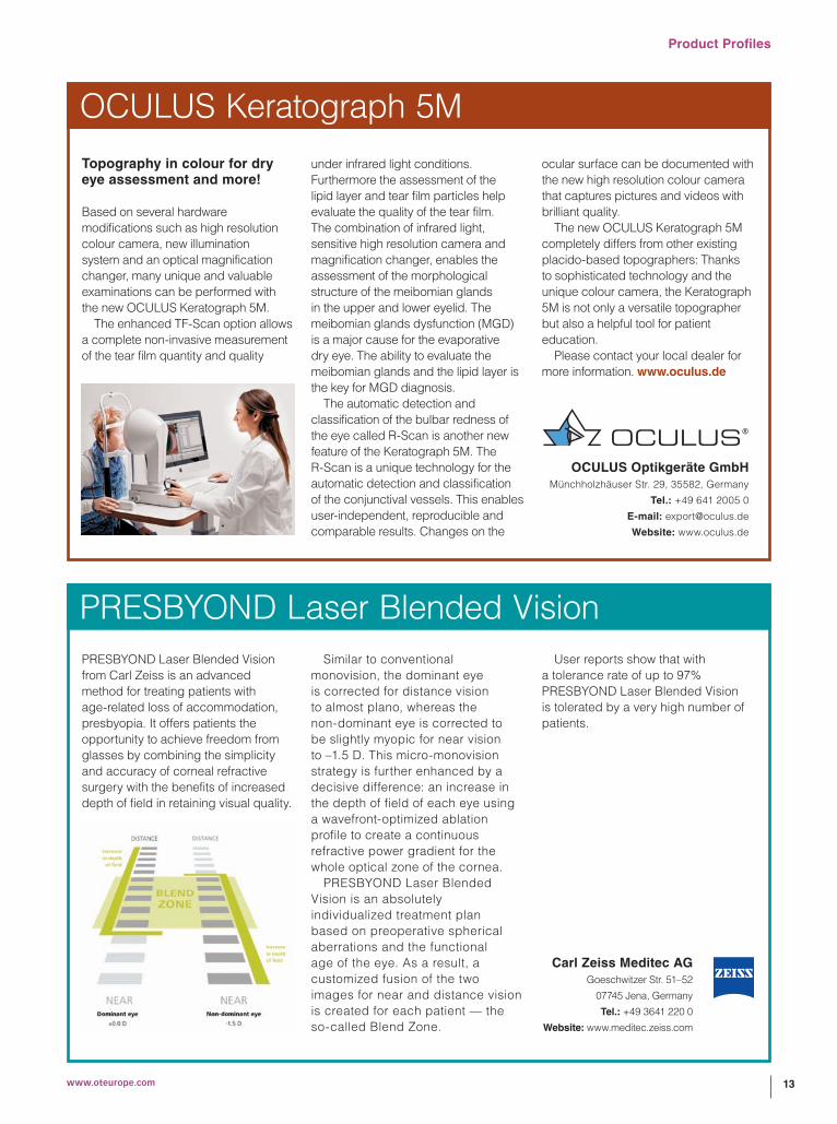
Topography in colour for dry eye assessment and more!
Based on several hardware modifications such as high resolution colour camera, new illumination system and an optical magnification changer, many unique and valuable examinations can be performed with the new OCULUS Keratograph 5M.
The enhanced TF-Scan option allows a complete non-invasive measurement of the tear film quantity and quality
PRESBYOND Laser Blended Vision from Carl Zeiss is an advanced method for treating patients with age-related loss of accommodation, presbyopia. It offers patients the opportunity to achieve freedom from glasses by combining the simplicity and accuracy of corneal refractive surgery with the benefits of increased depth of field in retaining visual quality.
under infrared light conditions. Furthermore the assessment of the lipid layer and tear film particles help evaluate the quality of the tear film. The combination of infrared light, sensitive high resolution camera and magnification changer, enables the assessment of the morphological structure of the meibomian glands in the upper and lower eyelid. The meibomian glands dysfunction (MGD) is a major cause for the evaporative dry eye. The ability to evaluate the meibomian glands and the lipid layer is the key for MGD diagnosis.
The automatic detection and classification of the bulbar redness of the eye called R-Scan is another new feature of the Keratograph 5M. The R-Scan is a unique technology for the automatic detection and classification of the conjunctival vessels. This enables user-independent, reproducible and comparable results. Changes on the
Similar to conventional monovision, the dominant eye is corrected for distance vision to almost plano, whereas the non-dominant eye is corrected to be slightly myopic for near vision to –1.5 D. This micro-monovision strategy is further enhanced by a decisive difference: an increase in the depth of field of each eye using a wavefront-optimized ablation profile to create a continuous refractive power gradient for the whole optical zone of the cornea.
PRESBYOND Laser Blended Vision is an absolutely individualized treatment plan based on preoperative spherical aberrations and the functional age of the eye. As a result, a customized fusion of the two images for near and distance vision is created for each patient — the so-called Blend Zone.
ocular surface can be documented with the new high resolution colour camera that captures pictures and videos with brilliant quality.
The new OCULUS Keratograph 5M completely differs from other existing placido-based topographers: Thanks to sophisticated technology and the unique colour camera, the Keratograph 5M is not only a versatile topographer but also a helpful tool for patient education.
Please contact your local dealer for more information. www.oculus.de
User reports show that with a tolerance rate of up to 97% PRESBYOND Laser Blended Vision is tolerated by a very high number of patients.
Carl Zeiss Meditec AGGoeschwitzer Str. 51–52
07745 Jena, Germany
Tel.: +49 3641 220 0
Website: www.meditec.zeiss.com
13www.oteurope.com
Product Profiles
Carl Zeiss Meditec AGGoeschwitzer Str. 51–52
07745 Jena, Germany
Tel.: +49 3641 220 0
Website: www.meditec.zeiss.com
ReLEx smile and ReLEx flexReLEx smile and ReLEx flex are new proven techniques for laser vision correction of myopia and myopic astigmatism. Introduced in 2011/2010, they are continuously finding their way to worldwide markets. Globally, users report results with equal or even superior results to those of (Femto-)LASIK.
ReLEx smile and ReLEx flex are based on the removal of a tissue disc (called lenticule) instead of tissue ablation, distinguishing them from traditional methods. The refractive
lenticule is created in the intact cornea with the VisuMax femtosecond laser (see Figure 1). The refractive correction is achieved by lenticule removal. There are two different ways to do that. With ReLEx flex the lenticule is removed through a flap-like access cut similar to LASIK, created by the VisuMax. ReLEx smile is the minimally-invasive version of ReLEx. Only a small access cut below 4 mm is created and the lenticule is removed through this small incision.
The ReLEx techniques provide several advantages over traditional refractive techniques, such as (Femto-)LASIK and PRK. Due to the use of femtosecond-cutting instead of ablation, refractive correction is not affected by ambient room conditions and corneal hydration, and leads, amongst other things, to an excellent predictability, including higher corrections. With ReLEx smile the upper corneal layer remains practically unaffected and there is only a small
incision instead of a side cut. This may not only lead to a better biomechanical stability but also suggests that with ReLEx smile fewer nerves are severed in the corneal surface, which positively affects dry eye syndrome by reducing its severity. Furthermore, the small incision lowers the incidence of infection, epithelial ingrowth or flap complications.
Due to its advantages for surgeons and patients, ReLEx is opening a new era by introducing the next generation of laser vision correction and it is increasingly gaining popularity on a daily basis.
Geuder AGHertzstrasse 4, 69126 Heidelberg, Germany
Tel.: +49 6221 3066 Fax: +49 6221 303 122
E-mail: [email protected]
Website: www.geuder.de
Pure Efficiency Phaco setsHighest efficiency for smallest incisions: New sets for coaxial mini phacoFor 2.2 mm and 2.4 mm microincision surgery, Geuder has designed new ‘Pure Efficiency Phaco’ sets.
One of the highlights of the sets is the high-performance titanium PEP ultrasonic tip. Due to the special
3-step design and the 40° angle the active surface for ultrasound emulsification has been maximized. The new tip has a smaller edge geometry so that entry of a hard lens nuclei is easier and ultrasound times can be shortened. Additionally, with smooth transition of the sleeve to the tip, insertion is more gentle.
The newly developed sleeve that is harder and more rigid prevents corneal burns and allows good mobility in the small incision as well as providing a more stable infusion. Increased followability and efficient holdability can be realized due to improved fluidics. In this ultrasonic needle the shaft has again been made slimmer, which allows for a larger vacuum.
The combination of the Geuder Megatron S4 with its perfectly adapted parameters for the PEP ultrasonic tips enables the user to perform highly efficient phaco with reduced ultrasonic power.
For further details please visit www.geuder.com/PEPSets
Figure 1: VisuMax Femtosecond System
OTE Products in Practice Issue 214