Embed Size (px)
Citation preview

Production of Libraries to Study Biopolymer Metabolism in Arabidopsis
thaliana and Tylosema esculentum
by
Corné Swart
Dissertation presented in partial fulfilment of the requirements for the degree Master of
Science in Plant Biotechnology at the University of Stellenbosch
Supervisor: Dr. James Richard Lloyd
Co-supervisor: Prof. Jens Kossmann
Faculty of Agricultural Sciences
Department of Genetics
University of Stellenbosch
Institute for Plant Biotechnology
December 2013
2
Declaration
By submitting this thesis/dissertation electronically, I declare that the entirety of the work
contained therein is my own original work, that I am the sole author thereof (unless to the
extent explicitly otherwise stated), that reproduction and publication thereof by Stellenbosch
University will not infringe any third party rights and that I have not previously in its entirety
or in part submitted it for obtaining any qualification.
Date: September 2013
Copyright © 2013 Stellenbosch University
All rights reserved
Stellenbosch University http://scholar.sun.ac.za

3
Abstract
Biopolymers and bio-degradable polymers are of utmost importance to ensure a sustainable
economy. Industry depends on raw material, which in many cases are derived from fossil
fuels, but in light of looming energy crises and green revolutions attention is being directed at
cellulose and starch biopolymers. This study was therefore set forth to investigate novel
genetic key elements of cell wall metabolism in Arabidopsis thaliana and starch synthesis in
an under-utilized southern African crop plant, Tylosema esculentum.
In the first section of the study a cDNA library of good quality was constructed from
regenerating A. thaliana protoplasts as it was expected to be enriching for genes involved in
cell wall biosynthesis. Small scale EST sequencing of the library confirmed that a few
sequences were similar to genes identified to be highly expressed during protoplast
regeneration. The library was to be screened by expression in a microalgae as it is anticipated
that cell wall metabolising genes would change the wall structure and visibly alter the colony
morphology. An attempt was made at establishing a high-throughput transformation system
in the unicellular algae Chlorella protothecoides in which the library was proposed to be
screened. Conventional microalgal transformation techniques do not appear to be effective in
this strain as the study produced no transgenic algae. Alternative studies into a screening
system within another species could still lead to the identification of cell wall biosynthetic
genes, which was the first objective in the study.
The second objective in the study was to investigate the potential of the orphan crop T.
esculentum as starch-producing cash-crop in developing southern African countries. In this
section of the study a cDNA library of good quality was produced form the tuber of T.
esculentum. The library was transferred to an expression vector and screened functionally in
E. coli for the presence of sequences with starch synthase activity. No sequences have been
identified yet and screening procedures are still on-going. The starch content in the tuber has
also been determined for the first time. The relatively high starch content in combination with
low agricultural inputs indicate the potential of the plant as an industrial starch source.
Further investigations into the nature of the starch are proposed to identify prospective buyers
within the industry.
Stellenbosch University http://scholar.sun.ac.za

4
Opsomming
Biopolimere en bio-afbreekbare polimere is van kardinale belang om ‘n volhoubare
ekonomie te ontwikkel. Industriële toepassings maak op die oomblik hoofsaaklik staat op
fossielbrandstof verwante bronne, maar met die oog op ‘n groen revolusie en energie krissise
wat dreig word meer belangstelling getoon in sellulose en stysel biopolimere. Hierdie studie
is daarom onderneem om genetiese elemente te identifiseer wat betrokke is by die sintese van
die selwand in Arabidopsis thaliana en stysel sintese in die suider Afrikaanse gewas
Tylosema esculentum wat grotendeels onderbenut is.
In die eerste deel van die studie is ‘n cDNA biblioteek, van goeie kwaliteit, geskep vanuit A.
thaliana protoplaste wat besig was om hulle selwande te herbou. Dit word verwag dat die
protoplaste gedurende die tydperk aktief besig sal wees om gene uit te druk wat betrokke is
by selwandsintese. DNA volgordebepaling het bevestig dat ‘n klein aantal volgordes
ooreengestem het met gene wat voorheen gevind was om in ‘n oormaat uitgedruk te word
tydens die herbou van protoplas-selwande. Daar was beoog om die biblioteek in ‘n mikroalge
uit te druk en sodoende die morfologie op kolonievlak waar te neem vir verandering wat in
die selwand meegebring is. Om hierdie rede was die doel om ‘n hoë opbrengs transformasie
sisteem te ontwikkel in die mikroalge Chlorella protothecoides. Algemene mikroalge
transformasie tegnieke blyk om nie effektief in die spesie te wees nie aangesien geen
transgeniese alge waargeneem is nie. Die ontwikkeling van ‘n soortgelyke proses in ‘n ander
spesie kan steeds lei na die ontdekking van gene betrokke by selwandsintese in A. thaliana
wat die eerste uitkoms van die projek as geheel was.
Die tweede uitkoms van die projek was om te ondersoek wat die waarskynlikheid was om T.
esculentum te kommersialiseer as ‘n stysel gewas en sodoende ‘n inkomste te skep vir arm
boere in ontwikkelende lande in suider Afrika. In hierdie gedeelte van die projek was daar ‘n
goeie cDNA biblioteek geskep uit die knol van T. esculentum. Die biblioteek is oorgedra na
‘n plasmied waarop dit aktief uitgedruk kon word in Escherischia coli G6MD2 en daar is
gesoek na volgordes wat lei na die sintese van stysel in hierdie bakterieë. Tot op hede is geen
sulke volgordes gevind nie, maar die ondersoek gaan steeds voort. Die styselinhoud van die
knol is ook vir die eerste keer bepaal in hierdie ondersoek. ‘n Styselinhoud wat relatief hoog
is en die lae moeite wat geverg word om die gewas te verbou toon dat die plant potensieel het
as ‘n kommersiële bron van stysel. Verdere ondersoeke in die aard van die stysel word ook
voorgestel om toekomstige industriële kopers te identifiseer.
Stellenbosch University http://scholar.sun.ac.za

5
Acknowledgements
I would like to thank my supervisor, Dr. James R Lloyd, for his patience, guidance, research
inputs and incredible editing speed. I really appreciate your hard work and support.
I would also like to thank Professor Jens Kossmann (co-supervisor) for valuable inputs and
suggestions regarding my project.
Many thanks go out to Dr. Shaun W Peters, Dr. Paul N Hills, Dr. Inonge Mulako, Dr.
Christelle van der Vyver, Bianke Loedolff, Stanton Hector, Ebrahim Samodien and Pedri
Claassens for technical advice and suggestions which were incredibly useful in my project. I
also want to thank the tissue culture team for the assistance they have provided me
throughout this study. IPB students and Staff, thank you for the friendly working
environment.
My gratitude is also expressed to the Donald Danforth Plant Science Center for providing the
Chlorella protothecoides UTEX25 strain and Emmanuele Nepolo for providing Tylosema
esculentum seeds and assistance with the Marama bean project.
I am absolutely blessed to have the support of my family. My mother, father and sister have
been with me every step of the way. In addition to the support from my family, Déhan
Dempers, Charl W King and Katrien Grobbelaar, I want to thank you for the incredible
friendship we have shared thus far. And a very special thanks to Yolandi Bester for your kind
words of inspiration and motivation throughout the writing process.
My appreciation and thanks to the IPB and the University of Stellenbosch for providing
financial assistance for this study.
A final and very important thanks to the NRF for funding. The financial assistance of the
National Research Foundation (NRF) towards this research is hereby acknowledged.
Opinions expressed and conclusions arrived at, are those of the author and are not necessarily
to be attributed to the NRF.
Stellenbosch University http://scholar.sun.ac.za

6
Table of Contents
Description Page number
Title 1
Declaration 2
Abstract 3
Opsomming 4
Acknowledgements 5
Table of Contents 6
List of Figures 9
List of Tables 9
Abbreviations 10
Chapter 1: General introduction and literature review 11
1.1 Biopolymers challenges synthetic polymers in an economic tug of war 11
1.2 The plant cell wall, a highly complex resource containing the most abundant 12
biopolymer on earth
1.3 Starch, a biopolymer in a booming industry 17
1.4 Development of underutilized crops for economic security in developing countries 20
1.5 Aim of the project 22
Chapter 2: Construction of an Arabidopsis thaliana cDNA library enriched for 23
cell wall biosynthesis genes and an investigation into the establish-
ment of a high-throughput transformation system in Chlorella
protothecoides for screening applications
2.1 Introduction 23
2.2 Materials and methods 24
Stellenbosch University http://scholar.sun.ac.za

7
2.2.1 Plant material and growth conditions 24
2.2.2 Protoplast isolation 24
2.2.3 Protoplast regeneration 24
2.2.4 RNA extraction from regenerating protoplasts 25
2.2.5 Precipitation of RNA 25
2.2.6 Isolation of mRNA 26
2.2.7 Construction of a cDNA library 26
2.2.8 Bacterial culture and maintenance 26
2.2.9 Small scale isolation of plasmid DNA 26
2.2.10 Restriction digests 26
2.2.11 Sequencing of inserts selected at random from the library 27
2.2.12 Large scale isolation of library plasmid DNA 27
2.2.13 Algal strains and cultivation methods 27
2.2.14 Determination of antibiotic sensitivity for C. protothecoides 28
2.2.15 Electroporation of C. protothecoides 28
2.2.16 Biolistic bombardment of C. protothecoides 28
2.2.17 Chemical transformation of C. protothecoides 29
2.2.18 Glass bead transformation of C. protothecoides 29
2.2.19 Agrobacterium mediated transformation of C. protothecoides 30
2.3 Results and discussion 31
2.3.1 Protoplast isolation and regeneration 31
2.3.2 Construction of a cDNA library enriched for genes related to cell wall 32
synthesis
2.3.3 Analysis of ESTs selected at random from the library 33
2.3.4 Attempts to establish a high-throughput transformation system in 34
C. protothecoides
2.4 Conclusion 35
Chapter 3: Library construction and screening for the identification of tuber 37
specific starch synthase genes in Tylosema esculentum
3.1 Introduction 37
Stellenbosch University http://scholar.sun.ac.za

8
3.2 Materials and methods 38
3.2.1 T. esculentum seed germination and cultivation 38
3.2.2 RNA extraction from tuber material 38
3.2.3 Precipitation of RNA 38
3.2.4 Isolation of mRNA 39
3.2.5 Construction of a cDNA library 39
3.2.6 Bacterial culture and maintenance 39
3.2.7 Small scale isolation of plasmid DNA 39
3.2.8 Restriction digests 40
3.2.9 Large scale isolation of library plasmid DNA 40
3.2.10 Preparation of chemically competent E. coli cells 41
3.2.11 Heat shock transformation of chemically competent E. coli cells 41
3.2.12 Construction of pBluescript SK(-)::DEST 41
3.2.13 Library transfer reaction 41
3.2.14 Preparation of electrocompetent E. coli G6MD2::pACAG cells 42
3.2.15 Electroporation of electrocompetent E. coli G6MD2::pACAG cells 42
3.2.16 Screening of expression library 42
3.2.17 Sequencing of plasmid inserts 43
3.2.18 Tuber starch analysis 43
3.3 Results and discussion 43
3.3.1 Construction of a cDNA library from Marama bean tubers 43
3.3.2 Transfer of cDNA library to an expression vector and qualitative 44
analysis of the expression library
3.3.3. Screening of the T. esculentum tuber expression library 45
3.3.4. Analysis of tuber starch content and composition 46
3.4 Conclusion 47
Chapter 4: General Discussion 48
References 51
Stellenbosch University http://scholar.sun.ac.za

9
List of Figures
Figure 1.1 Primary Cell Wall Structure and Synthesis 14
Figure 1.2 Higher plant starch synthesis 19
Figure 2.1 Filtered protoplast isolation 31
Figure 2.2 Electrophoresis profile of total RNA extracted from A. thaliana 31
protoplasts
Figure 2.3 Confirmation of partial cell wall regeneration 32
Figure 2.4 Functional distribution of ESTs represented within the library 35
Figure 2.5 Antibiotic sensitivity of C. protothecoides 34
Figure 3.1 Electrophoresis profile of total RNA extracted from the tuber of 43
T. esculentum
Figure 3.2 Iodine vapour staining of E. coli G6MD2 45
List of Tables
Table 1.1 Genes identified in cell wall synthesis 15
Table 1.2 Major under-utilized crops species of Africa 21
Table 2.1 A. thaliana regenerating protoplast library insert size and 33
sequencing of ESTs
Table 3.1 T. esculentum primary library insert size 44
Table 3.2 T. esculentum expression library insert size 45
Stellenbosch University http://scholar.sun.ac.za

10
Abbreviations
ADP Adenosine diphosphate
ATP Adenosine triphosphate
cfu colony forming unit
CTAB Cetyltrimethylammonium bromide
DDPSC Donald Danforth Plant Science Center
DEPC Diethylpyrocarbonate
EDTA Ethylenediaminetetraacetitc acid
EST Expressed sequence tag
FAO Food and Agriculture Organisation
G1P Glucose 1 phosphate
G6P Glucose 6 phosphate
GDP Guanosine diphosphate
kb kilobase
MP Modified Proteose
PCW Primary Cell Wall
PEG Polyethylene glycol
RACE-PCR Rapid amplification of cDNA ends – polymerase chain reaction
SCW Secondary Cell Wall
SDS Sodium dodecyl sulfate
SS Starch Synthase
T-DNA Transfer-DNA
TRIS Trisaminomethane
Stellenbosch University http://scholar.sun.ac.za

11
Chapter 1: General introduction and literature review
1.1 Biopolymers challenge synthetic polymers in an economic tug of war
The modern world has been growing at an astounding rate, reaching a population of 7 billion
in 2011 and predicted to reach 9 billion by 2050 (United Nations, 2011). Accompanying this
will be an increased demand that consumers will place on natural resources, thereby reducing
sustainability, damaging the environment and inducing climate change (Barnosky et al.,
2012). Industry currently depends principally on man-made plastics and polymers for
production. Properties that favour them above natural polymers are that they are chemically
inert, versatile, lightweight, strong, and damage resistant. These traits, however, complicate
their disposal and allow them to accumulate in ecosystems, particularly those that are aquatic
(Glover, 1993; Jayasekara et al., 2005 Moore, 2008). The challenges in disposal are not the
only hindrance, as a number of synthesis procedures produce toxic by-products that also need
to be managed (Bay et al., 2003; Israel et al., 2008). Research and development divisions
have been emerging in the majority of market-leading companies with the aim of providing
sustainable solutions, but financial implications and technological limitations impede the
implementation of bio-degradable polymers (Casale, 2008). Comparisons in the value of
biopolymers and their non-degradable polymeric counterparts indicate that where there was
once a 100 fold difference in production cost, this has now decreased to a more appealing 3
or 4 fold difference (Festel et al., 2005). Biopolymers, backed by numerous government
incentives and legislative rulings are gaining a foothold in industry and, with diminishing
fossil fuel reserves and ever-increasing crude oil prices, production costs will increasingly
favour bio-degradable polymers.
The responsibility to address environmental difficulties is not exclusively that of industry and
academic institutions have been working on similar projects for years in the umbrella field of
biotechnology. This can be described as a series of enabling technologies that involve the
applications of organisms to manufacturing and service industries in order to achieve
environmental sustainability and stability. Biotechnology includes the fields of biochemistry,
genetics, molecular biology, biomedical engineering, chemistry and chemical engineering
and could, in future, be the foundation of “Green” environmentally friendly technological
platforms (Rao, 2010). Another favourable characteristic that most commercially valuable
biopolymers share is that they are harvested from plants. As they are perfectly renewable,
Stellenbosch University http://scholar.sun.ac.za

12
plant based biopolymers may strengthen the primary agricultural sector and lead to extensive
job creation across all involved industries.
1.2 The plant cell wall, a highly complex resource containing the most abundant
biopolymer on earth
Industrial sectors responsible for manufacturing textiles, lumber, thickeners, paper and films,
already depend primarily on plant based polymers that are found in the plant cell wall.
Composed primarily of the most abundant biopolymer on earth, cellulose, plant cell walls are
composed of intricate and complex structures. They are deposited in two separate events,
with the primary cell wall (PCW) being synthesised during expansion while, afterwards, the
secondary cell wall (SCW) is manufactured within the PCW when cell enlargement ceases
(Taylor, 2008; Sánchez-Rodríguez et al., 2010). The PCW, consisting of cellulose,
hemicellulose, pectin and glycosylated proteins, is known to be flexible and thin, yet strong,
and capable of extension (Baskin, 2005; Sánchez-Rodríguez et al., 2010). SCWs are not
present in all cells and, comprised of cellulose, hemicellulose and lignin, they play important
roles in providing structural support to tissues that require extreme rigidity and strength
(Taylor, 2008).
The majority of externally recognizable plant features are determined by the shape and size of
the cell wall (Fry, 2004). Molecular continuity is maintained with the cytoskeleton and
plasma membrane, and the physical connection between the plasma membrane and cell wall
is determined by responses to changes in osmotic pressure (Szymanski & Cosgrove, 2009).
The aforementioned structural roles are complemented with biochemical functions in the
regulation of development as numerous surface markers, predicting both developmental
patterns and indicating positions, exist within the wall (Knox et al., 1989; Knox, 1990;
Pennel & Roberts, 1990; Pien et al., 2001). Furthermore development is aided by the delivery
of nutrients to adjacent cells by pathways made possible by the continuous symplastic
extension through the plasmodesmata (Cilia & Jackson, 2004). These pathways are also
involved in intercellular signalling, whereby RNA molecules, hormones, regulatory proteins
and other developmental messengers are delivered to their targets (Oparka, 2004; Kim,
2005). Cell walls are also the site at which biochemical defence pathways are induced by
predation and pathogenic infection (Hématy et al., 2009). Responses to bacterial and fungal
invasion include, but are not limited to, production of lignin- and protein shells, defensively
restructuring the morphology of the wall and the secretion of antimicrobial peptides as a
Stellenbosch University http://scholar.sun.ac.za

13
counter-attack mechanism (Lipka et al., 2005; Field et al., 2006; Hückelhoven, 2007;
Underwood & Somerville, 2008).
Because of these complex functions, the cell wall has a specialised structure (Figure 1.1)
comprised of intricately weaved polysaccharides similar to a fibreglass- like arrangement
(Carpita & Gibeaut, 1993). Hemicelluloses and pectins form the matrix of elaborately
arranged polysaccharides that embed the crystalline cellulose rods to construct a strong, yet
resilient, wall structure. The major polysaccharide fraction in the wall is represented by
cellulose, an unbranched (1,4)-linked β-D-glucan (Taylor, 2008). This is able to form a
scaffold, within which a multitude of parallel glucans are arranged to function as a structural
backbone with an impressive resistance against enzymatic digestion (McCann & Carpita,
2008). In general each of the cellulose rods contributing to the structural scaffold has a width
of 2-4 nm (Thimm et al., 2002).
Several other components are present alongside cellulose and these will be briefly outlined
below. The first of these, hemicelluloses, display a high level of similarity to cellulose, but
are unable to form microfibrils as a result of branching and modification in the molecular
structure of the (1,4)-β-D-linked glycan backbone (Cosgrove, 2005; Scheller & Ulvskov,
2010). Xyloglucan, the most abundant hemicellulose in most species and arabinoxylan,
usually present in smaller amounts, have distinct variation within their backbone and are
suggested to be involved in the complex networks formed in conjunction with cellulose
(Figure 1.1). Theories about the formation of these network have been presented in a number
of studies (Cosgrove, 2000; Cosgrove, 2005) and involve ideas such as hemicelluloses
binding spontaneously to the cellulose microfibril surfaces, anchoring adjacent units into a
scaffold (Fry, 1989; Hayashi, 1989), or that cellulose microfibrils are covered with
xyloglucans which are, in turn, bound to other matrix polysaccharides, thereby removing
direct linkages between cellulose rods (Talbot & Ray, 1992).
Receiving special attention in recent years, pectins have been described as being possibly the
most complex polysaccharides in living systems (Willats et al., 2001, Vincken et al., 2003).
Extreme heterogeneity within the group and characteristic domains, with suggested linkages
enabled by covalent bonding, are traits that characterise this group of wall polysaccharides
(Ridley et al., 2001; Willats et al., 2001, Vincken et al., 2003). Covalent linkages are not only
believed to maintain domain positions within pectin molecules, but also to attach pectin to the
hemicellulose xyloglucan, thereby entering the network of wall polysaccharides (Rizk et al.,
Stellenbosch University http://scholar.sun.ac.za

14
2000; Cumming et al., 2005). Pectic polysaccharides interact with hemicelluloses, but
evidence has been presented that they also bind to cellulose, thereby participating in the load-
bearing ability of the cell wall (Dick-Pérez et al., 2011, 2012). Pectins assist cell growth by
developing into hydrated gels which physically separate microfibrils, allowing them mobility
until finally fixing them into place when growth ceases (Stolle-Smits et al., 1999; Morris et
al., 2000 Cosgrove, 2005). Furthermore they are involved in determining wall thickness and
porosity and are responsible for intercellular cell adhesion by the formation of the glue-like
middle lamella (Iwai et al., 2001, 2002). The flexibility in the wall is proposed to be aided by
neutral pectin polysaccharides which appear to bind to cellulose surfaces (Jones et al., 2003;
Zykwinska et al., 2005). The main pectin domains are highlighted in figure 1.1.
Figure 1.1. Primary Cell Wall Structure and Synthesis. A simplified diagram of the PCW is presented in the figure. Cellulose and
hemicellulose interactions are highlighted without pectin on the left-hand side. The right hand side displays the integration of the major
polysaccharides that constitute the cell wall. The cellulose synthase complex, membrane bound, synthesises cellulose, whilst the other
matrix polysaccharides are manufactured in the Golgi apparatus and transported in vesicles to the cell membrane where they are exported to
the cell wall. (modified from Cosgrove 2005)
The synthesis of cellulose and matrix polysaccharides, are executed via distinct pathways.
Cellulose in both the PCW and SCW are synthesized by cellulose synthase (CESA) proteins,
a group of membrane bound hexameric rosette structures which are functional only when
expressed by at least three independent CESA genes (Kimura et al., 1999; Burn et al., 2002;
Taylor et al., 2003; Burton et al., 2004; Scheible & Pauly, 2004). Matrix polysaccharides
originate from the plant’s secretory network, with their synthesis localized in the Golgi
apparatus, followed by transport to the cell membrane in tiny vesicles that deliver them to the
Stellenbosch University http://scholar.sun.ac.za

15
wall (Richmond & Somerville, 2000; Hazen et al., 2002). Unlike cellulose which is
synthesized by extrusion from the plasma membrane, the matrix polysaccharides can diffuse
into the wall to reach their final destination facilitated by the energy gradient generated by
cell turgor pressure (Ray, 1967; Proseus & Boyer, 2005).
Table 1.1 Genes identified in cell wall synthesis. A non-exhaustive list of genes identified to have varying levels of involvement in the
biosynthesis of the plant cell wall.
Cellulose synthesis
CESA Superfamily
Family of genes encoding cellulose synthase catalytic subunit
Doblin et al., 2002; Scheible & Pauly, 2004 Somerville, 2006
KORRIGAN β-1,4-glucanase Lane et al., 2001; Szyjanowicz et al., 2004
CYT1 Gene product involved in biosynthesis of GDP-mannose
Lukowitz et al., 2001
PEANUT Enzymes involved in the biosynthesis of glycosylphosphatidylinositol membrane anchors
Gillmor et al., 2005
KOBIT01 Plant specific gene-unknown function
Pagant et al., 2002
COBRA Protein essential for microfibril organization
Roudier et al., 2005
Hemicellulose synthesis
CSL Superfamily Family of genes possibly encoding Golgi-localized glycan synthases involved in the biosynthesis of these polysaccharides
Richmond & Somerville, 2001; Hazen et al., 2002
FRA8 Involved in xylan biosynthesis, exact function unknown
Baydoun et al., 1989; Zhong et al., 2005
FUT1/MUR2 Encodes Xyloglucan-α-fucosyltransferase
Perrin et al., 1999; Faik et al., 2000; Vanzin et al., 2002
KATAMARI1 (KAM1)/MUR3
Xyloglucan-β-galactosyltransferase Madson et al., 2003
AtXT1 & AtXT2 α-Xylosyltransferase Faik et al., 2002
UGE4 Alters cell specific xyloglucan content
Nguema-Ona et al., 2006
Pectin synthesis
QUA1 (GUAT8) Homogalacturonan galacturonosyltransferase activity
Ridley et al., 2001
NpGUT1 Pectin biosynthesis Iwai et al., 2002
PARVUS/GLZ1 (GATL1)
Pectin biosynthesis Lao et al., 2003; Shao et al., 2004
GATL Superfamily
Galacturonosyltransferase-like proteins
Lao et al., 2003; Shao et al., 2004; Sterling et al., 2006
ARAD1 Arabinan α-1,5-arabinosyltransferase, biosynthesis of arabinan sidechains of RG-1
Harholt et al., 2006
GAUT1 – related Superfamilyy
Homogalacturonan galacturonosyltransferase activity
Sterling et al., 2006
Stellenbosch University http://scholar.sun.ac.za

16
The question then raised is which genes are involved in producing this dynamic structure of
polysaccharides, highly glycosylated proteins and lignin that represent the cell wall? The
truth is that question is largely unanswered. Estimates suggest that 10% of the A. thaliana
genome (approximately 2500 genes) might be involved in cell wall metabolism (McCann &
Carpita, 2008). These include in excess of 730 genes encoding putative glycosyltransferases
or glycosyl hydrolases and several hundred more thought to be involved in wall biosynthesis
or function (Henrissat et al., 2001). In most cases the catalytic activity has been deduced from
studying sequence data, however the exact biological roles and enzymatic activities have not
been identified for most of these candidate proteins (Fry, 2004). One reason for this is the
difficulty encountered when applying traditional biochemical techniques in cell wall studies
(Lerouxel et al., 2006). More recently, the use of genetic and genomic resources have led to
the identification of a number of cell wall biosynthesis genes (Table 1.1). The methods
employed have depended on a mixture of forward and reverse genetic approaches combined
with subsequent genomic analysis by association and transcriptional profiling.
Although the genetic approaches to gene identification have been somewhat successful, a
number of limitations are still apparent. Assigning gene function by forward and reverse
genetics depends on the availability of mutants of interest, or the ability to identify candidate
genes. In some instances difficulty has been encountered with these methods, as mutants have
aberrant phenotypes, deterring growth by inadequate root hair formation, or engender
lethality at early stages of development (Favery et al., 2001; Goubet et al., 2003). Mutants
utilized in a reverse genetics approach may appear phenotypically unaltered for a number of
reasons, for example due to gene redundancy masking the phenotype. Strong evidence to
support these statements has been gathered from genomic approaches, as genes belonging to
the CESA, CSL, GAUT1, GAUT7 and GATL superfamilies exist in multiple copies with high
homology (Richmond & Somerville, 2001; Sterling et al., 2006; Somerville, 2006). The
development of new strategies to investigate genes putatively identified to be involved in
wall synthesis, or to identify previously overlooked genes is, therefore, essential to expand
upon our current knowledge of wall architecture, biosynthesis and regulatory processes
(Lerouxel et al., 2006). A better understanding of these key-processes could also contribute to
reducing current high costs associated with the production of biofuels from lignocellulosic
carbohydrates (da Costa Sousa et al., 2009).
Stellenbosch University http://scholar.sun.ac.za

17
1.3 Starch, a biopolymer in a booming industry
Another versatile glucan-based biopolymer extracted from raw plant material, starch, is
utilized extensively in food, feed and non-food based industries (Marz, 2006). Besides being
the source of over 80% of the human populations calories (Keeling & Myers, 2010), it is
integrally tied to the production of adhesives, agrochemicals, cosmetics, detergents, paper,
board, pharmaceuticals, plastics, textiles and renewable energy. According to the European
Starch Industry (www.aaf-eu.org/european-starch-industry) the market for starch in 2012 was
worth €7.7 Billion in the EU alone. Synthesised in a number of tissues in many species,
starch granules are highly diversified in size and physical properties. The primary interest in
higher plant starches is due to their economic value and this has motivated studies into the
biochemistry, genetics and molecular biology of starch metabolism.
Energy captured by photosynthesis in the chloroplasts leads to production of starch granules
which act as storage molecules for energy in a transitory form in leaves, stems and roots of
plants (Smith et al., 2005), whilst usually maintaining long term reserves in seeds and tubers.
Under normal conditions starch granules are semi-crystalline (Zobel, 1988), forming a white
powder when extracted and dried. Although the innate heterogeneity in shape and size is
remarkable, even amongst starches in the same plant (Kossmann & Lloyd, 2000), those
originating from the same organ in a species are generally similar (Jane et al., 1994). Within
a starch granule, known for over a century, there are two fractions, amylose and amylopectin
(Kossmann & Lloyd, 2000). Amylopectin, which makes up the majority (normally
approximately 75%) of the granule, is a branched molecule with a molecular weight
estimated to be in the range of 107 and 10
9 Daltons (Buléon et al., 1998). Known to be the
determining feature in starches crystallinity and granular structure, amylopectin’s glucosyl
residues extend as chains of between six and an excess of a hundred, linked by α-1,4-bonds
(Zeeman et al., 2010). Branching within the amylopectin structure is facilitated by the
presence of α-1,6 bonds, connecting the α-1,4-linked chains in a highly organized manner
(Kossmann & Lloyd, 2000; Zeeman et al., 2010). These branching points confer a left-hand
helical structure to amylopectin chains. Exact details describing the architecture of
amylopectin at molecular level are not clear at this stage (Zeeman et al., 2010), but the
branching pattern, frequency of branching points and chain lengths amalgamate to bring forth
a dendriform, or treelike structure with a semi-crystalline nature (Hizukuri, 1986; Keeling &
Myers, 2010). These clustered chains enable the higher-order structure of starch to exist as
concentric rings which have been observed microscopically with the light- and scanning
Stellenbosch University http://scholar.sun.ac.za

18
electron microscope (Buttrose, 1960; Zeeman et al., 2002; Pilling & Smith 2003). The other
25% of the starch granule is made up of amylose, a smaller molecule composed of glucose
units linked by α-1,4-bonds (Buléon et al., 1998). Amylose structure has been reported to be
influenced by the presence of a tiny proportion of α-1,6 bonds (Peat et al., 1952; Hizukuri et
al., 1981; Buléon et al., 1998).
In the primary stages of starch biosynthesis, the key metabolite, ADP-glucose, is generated
from a reaction between ATP and G-1-P under the catalysis of ADP-glucose
pyrophosphorylase (Espada, 1962; Preiss & Sivak 1998a; Preiss & Sivak 1998b; Preiss,
1999). This reaction, the first committed step in the pathway of starch synthesis, fuels
additional reactions catalysed by starch synthases and branching enzymes which produce the
final polysaccharide (see Figure 1.2). Higher plant starch synthases are encoded by five
classes of genes, GBSS (granule-bound starch synthase) and SSI to SSIV (Ball & Morrel,
2003). Amylose synthesis takes place via GBSS, an enzyme which is closely bound to the
starch granule itself (Nelson & Rines, 1962; Ball & Morrel, 2003; Zeeman et al., 2010). The
remaining four classes of starch synthases, responsible for amylopectin synthesis are usually
partially soluble in the stroma of the plastid and partially associated with the starch granule
(Zeeman et al., 2010). Analyses of data relating to the biochemical properties of starch
synthase isoforms and their genetic phylogeny indicate that they have different specialized
functions within amylopectin synthesis, some producing chains of differing lengths within
amylopectin and others involved in initiating granule synthesis (Tetlow et al., 2004).
Contributing to amylopectin synthesis branching enzymes add branching points to actively
extending chains (Nielsen et al., 2002). These enzymes are also subdivided into two groups,
of which the first group is involved in the transfer of shorter chains to the C6 position of the
extending glucosyl moiety and the latter in the transfer of longer chains (Ball & Morrel,
2003; Tetlow et al., 2004).
In addition to the two α-glucans, amylose and amylopectin, starch also contains proteins and
lipids. The quantity and attributes of these proteins and lipids in combination with the size
and structure of amylose molecules are responsible for the differences in starches between
species (Takeda et al., 1987; Morrison, 1988; Takeda et al., 1989). Ultimately depending on
the application of the starch, crop-plant species are selected based on the physical properties
of their raw starch extract. Natural variation has been essential to the successful application
of starch economically, but further alterations can be induced by cross-breeding, mutant lines
and transgenic approaches (Davis et al., 2003). Biotechnological approaches to date have
Stellenbosch University http://scholar.sun.ac.za

19
included the overproduction of starch, modification in starch regulatory pathways and
alterations in starch structure (Zeeman et al., 2010). Starch furthermore can be modified
physically or chemically post-harvest and this is often done, to suit specific industrial
requirements (Jobling, 2004).
Figure 1.2 Higher plant starch synthesis. A simplified diagram is presented that demonstrates the partitioning of reactions in starch
synthesis by their location within the plant cell. (Modified from http://www.jic.ac.uk/STAFF/trevor-wang/images/full/starchpath2.jpg)
Recent analyses by the European Starch Industry have indicated that starch production has
increased from 8.7 million tonnes to 10 million tonnes between the period of 2004 and 2011.
This growth in the starch sector had been predicted previously (Ellis et al., 1998). Although
millions of tonnes of starch are harvested annually from wheat, maize and potato in the EU,
only 20% of the starch is used in its native form, whilst 23% is modified, and 57% is used as
starch-derived sweeteners. Identifying additional sources of starch from under-utilized crops
could assist in alleviating poverty by creating new industries.
Stellenbosch University http://scholar.sun.ac.za

20
1.4 Development of underutilized crops for economic security in developing countries
Comparing the contribution of crop plants in the agricultural sector, it has been observed that
a number of species have been underutilized or neglected. Dependence on a limited number
of crops exists around the world in both developed and developing countries. Globally more
than 7000 species are harvested as wild or cultivated plants, but only 150 of these crops are
produced on a commercially significant scale (Jaenicke & Höschle-Zeledon, 2006). These
statistics have been met with a number of strategic frameworks set forth to promote interest
in orphan crops, by organization such as the International Centre for Underutilized Crops, the
Global Facilitation Unit for Underutilized Species and the International Plant Genetic
Resources Institute (Williams & Haq, 2002; Gündel et al., 2003; Jaenicke & Höschle-
Zeledon, 2006). In the most recent framework, the developing regions identified as high-
priority for the implementation and improvement of under-utilized crops are Asia, the Pacific
and Sub-Saharan Africa (Jaenicke & Höschle-Zeledon, 2006). These are generally seen as the
most disadvantaged and are in dire need of solutions to the problems encountered with
agricultural security and downstream revenue generating applications of cultivated crops
(Naylor et al., 2004).
Within these developing regions, significant interest in has been taken in Sub-Saharan Africa,
as this represents the region most likely to fail in meeting the first Millenium Development
Goal (MDG1), which aims at a 50% reduction in extreme hunger and poverty by 2015
(Bartel, 2009). Asia and Latin America have increased agricultural productivity over the last
three decades, whilst similar progress in Africa has been stagnant (Bartel, 2009). Orphan
crops are particularly popular in Africa as they are better suited to extreme soil and
environmental conditions than major crop species (Tadele, 2009). Taking into account the
surface area of cultivation, or their contribution to feeding the population, the main orphan
crops in Africa are highlighted in Table 1.2 (Tadele, 2009). Of these, Cassava, a food source
for over a billion people worldwide has its highest production in Africa (FAO, 2010;
Okogbenin et al., 2013; DDPSC, 2011). It is also useful as a cash crop for poor farmers, with
transferable applications to industry, producing amongst many others biofuels, animal feed
and starch (Okogbenin et al., 2013). The cassava starch industry represents an unlikely
success story, one where large scale modern factories now depend upon an under-utilized
crop species (Sriroth et al., 2000). This development was in part possible because of the low
technology required to extract starch from the cassava roots (Sriroth et al., 2000). Other
contributing factors include the high starch content of the roots, the superb resilience of the
Stellenbosch University http://scholar.sun.ac.za

21
plant under suboptimal conditions, and the communal farming methods employed by millions
of farmers to cultivate the crops (Cock, 1982; Sriroth et al., 2000).
Considering the traits that make under-utilized crops so favourable the developing regions
and the successes that have been reported as a source of income, or feeding of populations,
the identification of previously overlooked species should receive more attention (Williams
& Haq, 2002; Gündel et al., 2003; Jaenicke & Höschle-Zeledon, 2006). Not only should this
be a priority for both developed and developing countries, but research to domesticate these
plants, or modify them by breeding or biotechnology should be pursued as well (Naylor et al.,
2004). One such tropical orphan crop with unexploited potential in Southern Africa is the
Marama bean. Displaying agronomic traits favouring its survival in dry and hot regions, the
plant has in recent times received significant interest as a cash crop (Jackson et al., 2010)
Although primary investigations have revolved around the oleaginous protein rich seeds, the
plant also maintains a relatively under-studied subterranean tuber with an unknown starch
content (Biesele & Murray, 1983; Müseler, 2005; Jackson et al., 2010) Currently no revenue
is being generated from the tuber of the plant and local farmers could benefit financially if
industrial applications were developed, as in the case of cassava.
Table 1.2 Major under-utilized crop species of Africa. Crops are selected on the basis of their contribution to feeding the population or
surface of cultivation (Tadele, 2009).
Botanical Name Common Name Type of
Crop
Reference
Eleusine coracana Finger Millet Cereal Savitha et al., 2013
Eragrostis tef Tef Cereal Tatham et al., 1995
Digitaria exilis Fonio Cereal De Lumen et al., 1993
Vignia unguiculata Cowpea Legume Sprent et al., 2010
Vigna subterranea Bambara groundnut Legume Sprent et al., 2010
Lathyrus sativus Grass pea Legume Grela et al., 2010
Manihot esculentum Cassava Root Babaleye, 2005
Dioscorea spp Yam Root Williams & Haq, 2002
Ensete ventricosum Enset Trunk/root Birmeta et al., 2004
Stellenbosch University http://scholar.sun.ac.za

22
1.5 Aim of the project
The outcome of this project was to shed light on the metabolism of cell wall synthesis and
starch synthesis in higher plants. The first aim of the project was to identify genes involved in
cell wall synthesis in the model plant A. thaliana. The approach was to construct a cDNA
library, enriched for cell wall synthesis genes, from regenerating protoplasts. The
aforementioned library was then to be screened in a unicellular algae for which a genetic
transformation protocol would be established. The second aim of the project was to identify
starch synthesis genes from the tuber of the orphan crop T. esculentum. The experimental
design involved the construction of a tuber-specific cDNA library, which was to be
transferred to a bacterial expression vector and screened for starch synthase activity in E. coli.
Stellenbosch University http://scholar.sun.ac.za

23
Chapter 2: Construction of an Arabidopsis thaliana cDNA library enriched
for cell wall biosynthesis genes and an investigation into the establishment
of a high-throughput transformation system in Chlorella protothecoides for
screening applications
2.1 Introduction:
The complexities of synthesising the plant cell wall, the most abundant renewable resource
on earth (Pauly & Keegstra, 2008) remains relatively poorly understood. Reasons for this
include the difficulty of isolating the structural components (Cosgrove, 2005), as well as
problems encountered with a number of forward and reverse genetics approaches as
discussed in Chapter 1. Considering that it is estimated that 10% of A. thaliana genes are
involved in cell wall metabolism, with the function of hundreds of these genes still unknown,
the need for novel experimental approaches to identification their roles clearly exists
(Lerouxel et al., 2006; Carpita & McCann, 2008).
In 2001, Hicks et al. evaluated the potential uses of unicellular algae to investigate
carbohydrate metabolism in vascular plants. Sharing a number of similarities to higher plants,
the algae in the division Chlorophyta offer a unique experimental platform. They often
contain genomes that are considerably smaller than those in higher plants and are thought to
have less genetic redundancy. Due to the lack of developmental complexity (such as organ
differentiation) localization of expression patterns could also be eliminated as a reason for
overlooking genes. Additionally many species contain a vascular plant-like cell wall, a short
life-cycle and simple culturing procedures which further contribute to the value of these
organisms as a scientific platform.
In this chapter I aimed to set up a novel screening system designed to identify genes involved
in plant cell wall metabolism in a two-staged approach. The first aim of the project was to
construct a cDNA library from regenerating A. thaliana protoplasts, which would be expected
to be enriching for genes involved in cell wall synthesis (Gipman, 2001). The second aim was
to develop a high-throughput transformation system in the unicellular algae Chlorella
protothecoides and evaluate its potential for screening a vascular plant library. Taking into
account previous reports which indicate that some unicellular algae display a visibly altered
colony morphology when the cell-wall structure is modified (Hicks et al., 2001) and the
success in transforming Chlorella species (Coll, 2006). It would therefore be hoped that when
Stellenbosch University http://scholar.sun.ac.za

24
higher plant cell wall metabolising genes are expressed in this algae they will alter the cell
wall and this in turn will alter colony morphology. Misshapen colonies could then be isolated
and the transgene within them identified.
2.2 Materials and methods:
2.2.1 Plant material and growth conditions
Arbidopsis thaliana ecotype Col-O were grown at 80 µE.ms-2
.s-1
on peat discs (Jiffy, Efekto)
with a 16 hr light and 8 hr dark photoperiod at approximately 25 ºC for 6 weeks.
2.2.2 Protoplast isolation
A. thaliana plants were incubated overnight in a humid dark environment prior to protoplast
isolation. Leaves (length: 5cm; width: 2 cm, total weight: 35g) harvested from A. thaliana
plants had the lower epidermal layer disrupted by rubbing with a fine grit (p600) sandpaper
(Endler et al., 2006). Protoplasts were released by incubating the leaves in a protoplast
digest/wash solution (Sigma Aldrich) with 1.5% w/v Cellulase Onozuka R-10 (from
Trichoderma viride, Sigma Aldrich) and 0.4% Macerozyme R-10 (from Trichoderma reesei,
Apollo Scientific) at 30˚C with occasional gentle swirling by hand. Before the addition of
leaves the solution was preheated to 55 ˚C to ensure complete solubilisation of enzymes and
filtered through a 0.22 µm filter. Incubation was seen as sufficient after approximately 6
hours, when the solution turned green and disappearance of tissue clumps was observed. The
solution containing protoplasts was collected and filtered through a 50 µm mesh (Falcon) and
the protoplast number and integrity assessed by light-microscopy. Protoplasts were pelleted
by centrifugation in a swinging bucket rotor at 400 x g for 10 minutes after which the
supernatant was removed and the protoplast gently washed in a volume of protoplast
wash/digest solution (Sigma Aldrich).
2.2.3 Protoplast regeneration
Washed protoplasts were collected by centrifugation in a swinging bucket rotor at 400 x g for
10 minutes and, after aspiration of the supernatant, re-suspended in B5 (Sigma Aldrich)
regeneration media (0.4M glucose; 3.1 g/l B5 salts; 0.112 % w/v B5 vitamins; pH 5.8; 1.0
mg/l 2,4-Dichlorophenoxyacetic acid; 0.15 mg/l 6-Benzylaminopurine) before being
incubated in the dark for 3 or 6 hours. After incubation the regenerating protoplasts were
observed by light microscopy and harvested by centrifugation at 400 x g for 10 minutes.
Stellenbosch University http://scholar.sun.ac.za
25
2.2.4 RNA extraction from regenerating protoplasts
RNA was extracted by means of a CTAB RNA extraction procedure. CTAB buffer (2% w/v
cetyl trimethylammonium bromide; 2% w/v Polyvinylpyrrolidone; 100 mM TRIS-HCl, pH
8.0; 25 mM EDTA; 2 M NaCl) was prepared and autoclaved. β-mercaptoethanol was added
to a final concentration of 2% (v/v) directly before use. RNA extractions were performed by
the addition of 10 ml CTAB buffer to approximately 1.42 x 10⁷
protoplasts immediately after
centrifugation and removal of the supernatant. The samples were vigorously mixed by
vortexing for 30 seconds and separated into microcentrifuge tubes. The samples were then
incubated at 65 °C for 30 minutes, and vortexed for 30 seconds at 5 minute intervals.
Centrifugation was performed at 16 000 x g for 10 minutes at room temperature before the
supernatant was transferred to a fresh tube and one volume of chloroform/isoamylalcohol
(24:1) was added. The sample was vortexed for 30 seconds and then centrifuged at 16 000 x g
for ten minutes at 4 °C before the supernatant was collected and the
chloroform/isoamylalcohol extraction repeated. RNA was precipitated by the addition of LiCl
to the collected supernatant at a final concentration of 2 M and incubation overnight at 4 °C.
The sample was centrifuged at 16 000 x g for 1 hour at 4 °C, the supernatant removed and the
pellet washed with 70% ethanol. This was centrifuged at 16 000 x g for ten minutes at 4 °C,
and the ethanol removed. The resulting RNA pellet was allowed to dry for 30 minutes before
30 μl of DEPC-treated dH20 was used to re-suspend it.
2.2.5 Precipitation of RNA
The RNA concentration in each sample was determined spectrophotometrically. The samples
were combined into a RNAse free 1.5 ml microcentrifuge tube and 1/10 volume of DEPC-
treated 3 M Sodium acetate (pH 4.8) was added along with RNA grade glycogen (Thermo
Scientific) at a final concentration of 0.1μg/μl. 2.5 volumes of ethanol were mixed gently
with the solution before being incubated at -80 °C overnight. The mixture was centrifuged at
16 000 x g at 4 °C for one hour, the supernatant removed and the pellet washed with 70%
(v/v) ethanol. RNA was recovered by centrifugation for 30 minutes at 16 000 x g and 4 °C.
Following removal of the supernatant the pellet was centrifuged for 20 minutes at 16 000 x g
at 4 °C and the remaining ethanol was also removed. RNA was re-suspended in DEPC-
treated dH20 and the concentration was determined spectrophotometrically. Approximately 2
μg of the re-suspended precipitated RNA was separated on a 1% (w/v) agarose gel to analyse
the quality of the RNA
Stellenbosch University http://scholar.sun.ac.za

26
2.2.6 Isolation of mRNA
mRNA was isolated from the total RNA using a commercially available kit (GenEluteTM
mRNA miniprep kit, Sigma Aldrich). All procedures were performed according to
manufacturer’s protocol.
2.2.7 Construction of a cDNA library
The cDNA library was constructed with approximately 1 μg of starting mRNA. Library
construction was accomplished by using the CloneMiner TM
II cDNA library kit (Invitrogen).
All procedures were performed according to the manufacturer’s protocol.
2.2.8 Bacterial culture and maintenance
Bacterial colonies were cultivated on solid LB media (10 g/l Tryptone; 5 g/l Yeast Extract
powder; 10 g/l NaCl; 15 g/l bacto agar) and subsequently inoculated into liquid LB broth (10
g/l Tryptone; 5 g/l Yeast Extract powder; 10 g/l NaCl).
2.2.9 Small scale isolation of plasmid DNA
Bacterial colonies were inoculated into 2 ml LB broth containing 50 μg/ml kanamycin and
incubated overnight at 37 °C with shaking at 200 rpm. The culture was decanted into a 2 ml
microcentrifuge tube which was centrifuged at 16 000 x g for two minutes. The pellet was re-
suspended in 200μl of 50 mM TRIS-HCl pH 8.0; 10 mM EDTA; 0.1 g/l RNAse A by
vortexing. 200 μl of 200 mM NaOH; 1% (w/v) SDS was added and the tube mixed gently by
inversion followed by the addition of 200 μl of 3 M KAc; pH 5.5. The tube was mixed gently
by inversion and placed on ice for five minutes prior to centrifugation at 16 000 x g for 10
minutes. The supernatant was transferred to a tube containing 0.7 volumes of isopropanol and
mixed by inversion. This was left at room temperature for 5 minutes and then centrifuged at
16 000 x g for 10 minutes. After removal of the supernatant, the DNA pellet was washed with
70% (v/v) ethanol. Following removal of the supernatant the pellet was left to air-dry on the
bench after which it was re-suspended in TE buffer (10 mM TRIS-HCL, pH 8.0; 1 mM
EDTA). The concentration of the plasmid in solution was determined by spectrophotometry.
2.2.10 Restriction digests
DNA was digested by restriction enzymes BsrG I, XhoI and NcoI (Fermentas) according to
the manufacturer’s instructions.
Stellenbosch University http://scholar.sun.ac.za

27
2.2.11 Sequencing of inserts selected at random from the library
Plasmid DNA was sequenced by the Central Analytical Facility of the University of
Stellenbosch.
2.2.12 Large scale isolation of library plasmid DNA
The library was transformed into E. coli and were plated onto solid LB media containing
appropriate antibiotics and incubated overnight at 37 °C. Colonies were re-suspended by
washing with LB broth and then pelleted by centrifugation at 4000 x g for 20 minutes at room
temperature. The cells were re-suspended in 8 ml of 50 mM glucose; 25 mM TRIS-Cl, pH
8.0; 10 mM EDTA and incubated on ice for 5 minutes. Following incubation, 16 ml of 0.2 M
NaOH; 1% (w/v) SDS was added and incubated on ice for 5 minutes. After that 24 ml of 3M
KAc pH 4.8, was added and the samples incubated on ice for a further 5 minutes. The
reaction mixture was filtered through four layers of Miracloth (Fisher Scientific) and diluted
with 0.7 volumes of isopropanol. Precipitation of plasmid DNA took place for 30 minutes at
room temperature, following which the DNA was isolated by centrifugation at 4000 x g for
30 minutes at room temperature. The DNA was re-suspended in 1 ml of dH20 and mixed with
1 ml of 5 M LiCl. RNA was precipitated at room temperature for 10 minutes and the tubes
centrifuged at room temperature for 10 minutes and 14 000 x g. 1 volume of
chloroform:isoamylalcohol:phenol (24:1:25) was added and the sample vortexed after which
it was centrifuged for three minutes at 14 000 x g. The upper phase was removed and 0.1
volumes of 8 M LiCl and 2.5 volume of ethanol was added before the sample was mixed by
inversion. Samples were incubated at room temperature for 10 minutes before being
centrifuged for 10 minutes at 14 000 x g. The supernatant was removed and each pellet
washed with 70% ethanol. After removal of ethanol, DNA was air dried for 5 minutes and re-
suspended in TE buffer (10 mM TRIS-CL, pH 8.0; 1 mM EDTA).
2.2.13 Algal strain and cultivation methods:
Cultures of Chlorella protothecoides were maintained on solid modified proteose (MP)
media (2.94 mM NaNO3; 0.17 mM CaCl2.2H2O; 0.3 mM MgSO4.7H2O; 0.43 mM K2HPO4;
1.29 mM KH2PO4; 0.43 mM NaCl; 1 g/l peptone powder; casein 0.5 g/l and Bacto agar 12
g/l) in a controlled environment (16:8 day:night cycle, 50 µE.ms-2
.s-1
light and 25 °C).
Subculturing was performed once a month to maintain axenic cultures. Liquid cultures were
Stellenbosch University http://scholar.sun.ac.za

28
routinely initiated by means of a pregrowth culture and growth monitored by cell count with
an improved Neubauer Brightline haemocytomer.
2.2.14 Determination of antibiotic sensitivity for C. protothecoides
Solid MP media and stock solutions of antibiotics and herbicides were prepared. Solid MP
media was supplemented with either antibiotics or herbicides ranging from 5 μg/ml to 1000
μg/ml final concentration. Liquid cultures in the logarithmic growth phase were harvested
and plated onto SMP media and monitored for growth. Antibiotic selection was determined
by the lowest minimum inhibitory concentration required to inhibit algal growth completely.
2.2.15 Electroporation of C. protothecoides
Electroporation procedures were performed as described by Chow and Tung (1999) and Chen
et al (2001). Cultures were grown in liquid MP media to a density of 1 x 10⁷
cells/ml (early
stationary phase), collected by centrifugation at 2000 x g for 2 minutes and re-suspended in
an equal volume of washing media (0.2M Mannitol; 0.2M Sorbitol). Cells were incubated on
ice for an hour, collected by centrifugation at 2000 x g at 4 ˚C for 2 minutes and taken up in
electroporation buffer (0.08 M KCl; 0.005 M CaCl2; 0.01 M HEPES-KOH, pH 7.0; 0.2 M
Mannitol; 0.2 M Sorbitol; Chow and Tung, 1999). To an aliquot of 40 µl (4 x 10⁶ cells), 0.5-
1.0 µg of pCAMBIA2301 (www.cambia.org) was added prior to electroporation. An addition
of 2.5 µg of herring sperm DNA was also tested. Electroporation of samples were done in 0.2
cm electro-cuvettes at 1500, 1800 and 2000 V/cm (25 µF; 200 Ohm) and plated onto
selective (35 µg/ml of G418) and non-selective solid MP media. Plates were monitored for 3-
4 weeks.
2.2.16 Biolistic bombardment of C. protothecoides
Biolistic bombardment was performed similar to a method used successfully for
Chlamydomonas reinhardtii transformation by Boynton et al (1988). Liquid cultures were
grown to reach an approximate density of 5 x 10⁶ cells/ml, which were then plated onto
sterile cellophane discs (A.A. Packaging Limited, UK) on top of solid MP media. The algae
were allowed to form a lawn over a period of 7 days in preparation for biolistic
bombardment. 5 mg of tungsten powder (0.7 µm per particle, Bio-Rad) was sterilised in 400
µl of ethanol, washed three times with sterile dH2O and re-suspended in 50 µl of Milli-Q
water (MQ) (Millipore). Approximately 10µg of pCAMBIA 2301 (1 µg/µl) was precipitated
onto the tungsten by the concurrent addition of 50 µl CaCl2 (2.5 M) and 20 µl of Spermidine
Stellenbosch University http://scholar.sun.ac.za
29
(0.1 M) during incubation on ice. A custom built gene gun was utilized for the bombardment
procedure. Before use the chamber was disinfected with 70% (v/v) ethanol, the helium
cylinder outlet pressure set to 1000 kPa and the solenoid timer adjusted to 0.05 seconds.
From the precipitation mixture a volume of 100 μl of supernatant was aspirated before re-
suspension, followed by the transfer of 5 μl into the centre of the support grid of a sterile
syringe filter holder (stainless steel, 13 mm diameter). The MP media, covered with a lawn of
algae was placed in the central circle of the gene gun before the door was closed. A vacuum
was created within the compartment and the tungsten was fired at the lawn of algae when the
pressure reached 80 kPa. The cellophane discs were carefully removed, each washed with a 1
ml of liquid MP media, and plated thinly onto selective (35 µg/ml of G418) and non-selective
plates. Plates were monitored for the presence of growth for 3-4 weeks.
2.2.17 Chemical transformation C. protothecoides
A chemical transformation method for yeast (Chen et al., 1992) was tested on C.
protothecoides. Cultures were harvested in the logarithmic phase (excess of 1 x 10⁷ cells/ml)
by centrifugation at 2000 x g for 2 minutes at room temperature. For each millilitre of
centrifuged culture, the pellet was re-suspended in 100 µl of One-step buffer (8% w/v
polyethylene glycol (PEG) 4000; 100 mM dithiothreitol (DTT); 1 µg pCAMBIA 2301; 50 µg
herring sperm carrier DNA). After vigorous vortexing the cell mixture was incubated at 45˚C
for 30 minutes before being plated directly onto non-selective and selective (35 µg/ml G418)
solid MP media. Plate growth was monitored for 3-4 weeks.
2.2.18 Glass bead transformation of C. protothecoides
Glass bead agitation transformation of C. protothecoides was performed by means of a
procedure routinely used for Chlamydomonas reinhardtii (Kindle, 1990). Cells were cultured
(1 x 10⁶
cells/ml) before 100 ml was centrifuged for 2 minutes at room temperature at 2000 x
g. The pellet was taken up in 2 ml of fresh liquid MP media and incubated with gentle
shaking (100 rpm) for 2 hours at room temperature. Transformations were performed by
adding 300 μl of cells to microfuge tubes containing 300 mg of sterile glass beads (425-600
μm, acid washed, Sigma Aldrich), 100 μl of 20 % w/v PEG 4000, 1-2 μg of plasmid DNA
(pCAMBIA 2301, pBK-CMV; Agilent Technologies, or pEmuKN; Franks & Birch, 1991)
and vortexing at maximum speed for 30 seconds. The transformation mixture was diluted into
liquid MP media, cultured overnight, plated thinly onto selective (35 μg/ml G418) and non-
selective solid MP media, and growth monitored for 3-4 weeks.
Stellenbosch University http://scholar.sun.ac.za

30
2.2.19 Agrobacterium mediated transformation of C. protothecoides
Cells of C. protothecoides were utilized in a transformation procedure in a manner similar to
that used by Kathiresan & Sarada (2009) on Haematococcus pluvialis and by Pratheesh et al.
(2012) on C. reinhardtii. A total of 1 x 10⁶ cells were spread over solid MP plates and
allowed to develop a lawn of cellular growth for a week. Agrobacterium tumefaciens strains
EHA 105 (L,L succinamopine type, A vir helper, also maintains a supervirulant Ti plasmid, a
T-DNA deletion derivative of pTiBo542; Hood et al., 1986) and LBA 4404 (Octopine type
with A vir helper maintaining the disarmed pAL4404 Ti plasmid, a derivative of pTiAch5
with a T-DNA deletion; Hoekema et al., 1983) with pCAMBIA 2301 were cultured at 28 °C
overnight with shaking in liquid YEP media (10 g/l Peptone powder; 10 g/l Yeast extract
powder; 5 g/l NaCl) with appropriate antibiotics. When the Agrobacterium cultures reached
an OD600 of 0.5, the bacteria were collected by centrifugation at 2800 x g for 10 minutes. The
pellet was washed with sterile dH20, centrifuged again for 10 minutes at 2800 x g before the
washing step was repeated. After the final centrifugation step, the Agrobacterium cells were
taken up in 3 ml of liquid MP before additions of acetosyringone to final concentrations of 0
μM, 100 μM and 250 μM. 200 μl of the Agrobacterium solution was spread onto each MP
plate with C. protothecoides and co-cultivated in the dark for 48 hours at 25 °C. Liquid MP
media (600 μg/ml Cefotaxime) was used to wash the co-cultivated cultures from the solid
media, and allowed to shake for one hour. The algae were pelleted by centrifugation at 100 x
g for 5 minutes, washed 3 times with dH20 containing 600 μg/ml cefotaxime and plated thinly
onto selective (35 μg/ml G418) and non-selective solid MP media (600 μg/ml cefotaxime).
Growth and possible recurring Agrobacterium infections were monitored for 3-4 weeks.
Stellenbosch University http://scholar.sun.ac.za

31
2.3 Results and discussion:
2.3.1 Protoplast isolation and regeneration
Approximately 2.84 x 10⁷ protoplasts were isolated from A. thaliana rosette leaves which
appeared to be generally intact as confirmed by microscopic investigation (Fig. 2.1).
Figure 2.1 Filtered protoplast isolation. Filtered A. thaliana protoplasts visualized at 100x magnification. Bar represents 50 μm.
The protoplast isolation was separated into two equal volumes of B5 regeneration media and
allowed to partially regenerate their cell walls for 3 or 6 hours respectively before the total
RNA was extracted (Fig. 2.2). These time points were selected as the expression of known
cell wall biosynthetic genes start 3 hours after hydrolytic enzymes have been removed and
peak when regeneration has taken place for 6 hours (Gipman, 2001). Microscopic
investigation into deposition of cell wall components confirmed that partial cell wall
regeneration after a period of 6 hours took place (Fig. 2.3).
Figure 2.2 Electrophoresis profile of total RNA extracted from A. thaliana protoplasts. RNA extracted from protoplasts (1) after 3
hours of regeneration and (2) after 6 hours of regeneration was separated by non-denaturing agarose gel electrophoresis (1% w/v).
Stellenbosch University http://scholar.sun.ac.za

32
Figure 2.3 Confirmation of partial cell wall regeneration. A. thaliana protoplasts visualized at 400x magnification. A. Protoplasts in B5
regeneration media after 0 Hrs of regeneration. B. Protoplasts in B5 regeneration media after 6 Hrs of regeneration. Arrows highlight the
cell membrane where the deposition of cell wall components can be seen. Bars represent 50 μm each.
2.3.2 Construction of a cDNA library enriched for genes related to cell wall synthesis
A cDNA library from regenerating protoplasts was constructed in pENTRTM
222 with the
CloneMinerTM
II cDNA library kit. The library contained a titer of approximately 1.07 x 10⁶
cfu which exceeded the general guideline of 1 x 10⁶ clones. It is therefore expected that the
library is representative as the number of clones should ensure sufficient transcript coverage.
The library was qualitatively analysed by selecting 22 random clones, digesting the
pENTRTM
222 entry plasmid with BsrG I and consequently separating the fragments by
agarose gel electrophoresis.
The average insert size (Table 2.1) was determined by comparison to a known DNA ladder (λ
DNA/PstI digest, Sigma Aldrich). Sizes were maintained within the range of 0.4 and 2.4 kb,
and the average insert size was found to be 1.16 kb (Table 2.1). This is close to the theoretical
average size of eukaryotic cDNA’s which has been estimated as 1.35 kb (Xu et al., 2006).
Each plasmid contained an insert indicating a recombination rate of 100% and approximately
59% of the sequences exceeded 1 kb.
Stellenbosch University http://scholar.sun.ac.za

33
Table 2.1 A. thaliana regenerating protoplast library insert size and sequencing of ESTs. Average insert size of 22 randomly selected
clones determined by digestion of plasmid DNA with BsrG I and separation by agarose gel electrophoresis. Samples were also sequenced
from the M13 forward primer to discern the identity of inserts by Blastn analysis. Predicted size of full-length cDNA is also presented.
2.3.3 Analysis of ESTs selected at random from the library
22 Clones selected at random from the library were sequenced from the M13 Forward primer
and the sequences obtained used in Blastn homology searches on NCBI
(www.ncbi.nlm.nih.gov). The ESTs identified were compared to loci upregulated during cell
wall regeneration of rice (Sharma et al., 2011) and cotton (Yang et al., 2008) protoplasts, as
well as the proteins isolated from the apoplastic region of regenerating A. thaliana protoplasts
Clone
Insert
size
(kb)
EST
Full-length
cDNA (kb)
Accession
Number
1 1.30 Glutamate dehydrogenase 2 1.52 NM_001125711.1
2 0.90 Peptidase S24/S26A/S26B/S26C family protein 0.87 NM_104138.4
3 1.35 C2H2 zinc finger protein FZF 1.54 NM_128011.4
4 0.85 HSP20 family protein 0.72 NM_128504.3
5 1.70 Ribosome biogenesis co-factor 1.66 NM_102901.3
6 0.80 Putative zinc finger (AN1-Like) family protein 0.95 NM_113740.5
7 1.60 Putative glucosyltransferase 1.76 NM_119575.1
8 1.95 Global transcription factor group e8 3.17 NM_001203056.1
9 1.65 Polyadenylate binding protein RBP45B 1.57 NM_101037.3
10 1.20 Arginine decarboxylase 2 2.77 NM_202955.1
11 0.40 Class 1 heat shock protein 0.81 NM_100614.2
12 1.05 Elongation factor EF-2 2.96 NM_179487.1
13 0.65 Uncharacterized protein 0.16 NM_001126022.1
14 0.95 Glutathione S-transferase TAU 11 1.05 NM_105661.4
15 1.40 Heat shock transcription factor 4 1.78 NM_119862.3
16 2.40 Nitrate transporter 1.1 2.14 NM_101083.3
17 1.45 Heat shock protein 70B 2.33 NM_101471.2
18 0.75 Ribosomal protein s21e 0.60 NM_122652.6
19 1.05 Unknown protein (NC domain containing related) 1.08 NM_111138.3
20 0.45 Hypothetical protein 0.74 AK221826.1
21 0.96 Unknown protein, response to low sulfur 2 0.56 NM_122375.2
22 1.00 Gram domain containing protein 1.11 NM_121323.3
Stellenbosch University http://scholar.sun.ac.za

34
(Kwon et al., 2005). The functional distributions of this small scale EST survey are displayed
in Figure 2.4 and Table 2.1.
Figure 2.4 Functional distribution of ESTs represented within the library. Clustered representation of functions of 22 ESTs isolated
from the library.
During cell wall reconstruction a number of genes are up-regulated (Sharma et al., 2011) and
the groups highly up-regulated were compared to the ESTs obtained from the library. It was
found that the majority (68%) of the ESTs in the library were in functional groups that
represented the largest groupings of up-regulated genes during regeneration of the cell wall of
protoplasts (Sharma et al., 2011). A large proportion of the ESTs also represent unknown,
uncharacterized and hypothetical proteins, which has also been observed in other studies
(Yang et al., 2008; Sharma et al., 2011). Sequences coding for a protein belonging to the
HSP20 family (NM_128504.3) and a putative glucosyl transferase (NM_119575.1) from the
library were also found to be highly expressed in rice protoplasts after 12 hours of
regeneration (Sharma et al., 2011). Glutamate dehydrogenase (NM_001125711.1) a protein
found to be loosely associated with the cell wall of A. thaliana protoplasts after 1 and 3 hours
of regeneration (Kwon et al., 2005) was also present amongst the library ESTs. This small
number of sequenced clones indicate that the library is possibly representative of regenerating
protoplasts and would, therefore, likely contain a higher proportion of cell wall biosynthetic
genes as has been demonstrated previously (Gipman, 2001).
2.3.4 Attempts to establish a high-throughput transformation system in C. protothecoides
A range of antibiotics were tested to determine their potential as a selective agent for
transformation studies in C. protothecoides. It was determined that 35 µg/ml of G418 was the
minimum concentration required to completely inhibit algal growth (Fig 2.5 B).
Stellenbosch University http://scholar.sun.ac.za

35
Transformation studies based on an agrobacterium-mediated approach required a lack of
sensitivity to cefotaxime (Kathiresan & Sarada, 2009) and it was observed that C.
protothecoides was insensitive to cefotaxime at concentrations of 1mg/ml (Fig 2.5 D).
Figure 2.5 Antibiotic sensitivity of C. protothecoides. Liquid cultures of C. protothecoides spread onto solid MP media with: A. No
antibiotics; B. 35 µg/ml G418; C. No antibiotics; D. 1 mg/ml cefotaxime.
Being the most widely used promoter in plant transformation and successfully driving
expression in a number of unicellular algae, the CaMV 35S was the promoter of choice in
this study (Brown et al., 1991; Jarvis and Brown, 1991; Maruyama et al., 1994; El-Sheekh,
1999; Hawkins & Nakamura, 1999; Kim et al., 2002). The pCAMBIA2301 vector which
confers resistance to G418 by means of expression of NPTII (neomycin phosphotransferase
II) under the CaMV35S (Cauliflower Mosaic Virus) promoter was selected for
transformation procedures. Unfortunately, despite numerous attempts none of the
transformation approaches attempted yielded a single transgenic C. protothecoides colony.
Controls on non-selective media, displaying expected growth, confirmed that procedures
were non-lethal. Previous reports have indicated that the algae is transformable by means of
the glass-bead transformation procedure (Donald Danforth Plant Science Center, unpublished
results). To test if the problem was due to a lack of expression using the CaMV35S promoter
I additionally utilised the plasmid pBK-CMV (where expression of the NPTII gene is under
control of the eukaryotic SV40 promoter that is active in Chlamydomonas reinhardtii;
Butanaev, 1994) as well as pEmuKN (Where NPTII expression is driven by the synthetically
modified Adh1 promoter; Franks & Birch, 1991). These were utilized in glass bead
transformation attempts on C. protothecoides, however, no transformed colonies were
observed with the use of these additional transformation vectors either.
2.4 Conclusion:
This section of the study was aimed at the identification of cell wall biosynthetic genes,
which were not obtained as significant difficulties were encountered in the development of a
Stellenbosch University http://scholar.sun.ac.za

36
suitable screening system. The study did result in the production of a good cDNA library
which appears to be enriched for genes expressing actively during the regeneration of
protoplasts. The current approaches taken to transform C. protothecoides have not been
successful and this system could be replaced by the use of another strain of Chlorella in
which an established transformation procedure exists.
Stellenbosch University http://scholar.sun.ac.za
37
Chapter 3: Library construction and screening for the identification of
tuber-specific starch synthase genes in Tylosema esculentum
3.1 Introduction
Tylosema esculentum, the Braaiboontjie or Marama bean, is an underutilized crop that occurs
natively in the arid and semiarid regions of Namibia, South Africa and Botswana. This
perennial species of plant is drought-tolerant and a popular nutrient source for both wild
animals and indigenous human populations. Exhibiting annual prostrate runners, which grow
well upon dry and sandy soil, the plant belongs to the legume family and bears seedpods
containing normally between one and three edible seeds (Keith & Renew, 1975; Müseler,
2005). A subterranean tuber, capable of storing up to 90% (w/v) water maintains the plant
during winter months when the runners die off (Keith & Renew, 1975). The seeds, rich in
both lipids and proteins, have been studied thoroughly and represent the main economic
revenue generated by this crop. For the interest of this project, the focus will be shifted to the
tuber that remains largely unstudied (Biesele & Murray, 1983, Müseler, 2005; Jackson et al.,
2010). The T. esculentum tuber, at the young age of 5 months, has a nutritional content as
follows: water, 92.1%; ash (unknown mineral content), 0.42%; carbohydrate, 4.38%; fat,
0.14%; and protein 2.1% (Biesele & Murray, 1983). The composition might change through
the aging process as it has been reported that the tuber becomes more fibrous and difficult to
chew (Keith & Renew, 1975). Literature also mentions that the tuber starch content is high
(Percy et al., 2010), but to my knowledge no studies have been presented on the starch
content and composition.
Considering the success of one underutilized crop, cassava, that developed into an
internationally competitive starch industry in Thailand (Sriroth et al., 2000), the opportunity
exists to investigate the potential of T. esculentum as a cash crop in Sub-Saharan Africa. In
order to help in developing this, more knowledge is required about the starch in the tuber and
the molecular biology of its synthesis. The study set forth in this chapter was performed to
identify novel genes involved in starch synthesis in the tuber of T. esculentum. The approach
taken was to construct (1) a tuber-specific cDNA library from mRNA isolated from the
legume, T. esculentum, and (2) to screen the aforementioned library to identify genes
involved in starch synthesis.
Stellenbosch University http://scholar.sun.ac.za

38
3.2 Materials and methods:
3.2.1 T. esculentum seed germination and cultivation
T. esculentum seeds were a gift of Emmanuel Nepolo (University of Namibia). Seeds were
placed in a mixture of 50% sand and 50% potting soil and allowed to grow for approximately
three months in a glasshouse between September and December.
3.2.2 RNA extraction from tuber material
T. esculentum tubers were removed from the soil, sliced into smaller discs, and immediately
frozen in liquid nitrogen. Material was ground into a fine powder with a mortar and pestle
and stored at -80 °C. RNA was extracted by means of a modified CTAB RNA extraction
procedure. CTAB buffer (2% w/v cetyl trimethylammonium bromide; 2% (w/v)
Polyvinylpyrrolidone; 100 mM TRIS-HCl, pH 8.0; 25 mM EDTA; 2 M NaCl) was prepared
and autoclaved. β-mercaptoethanol was added to a final concentration of 2% (v/v) directly
before use. RNA extractions were performed by the addition of 1.5 ml CTAB buffer to
approximately 200 mg of ground material. Mixing was accomplished by vortexing vigorously
for 30 seconds. The samples were then incubated at 65 °C for 30 minutes, and vortexed for
30 seconds at 5 minute intervals. Centrifugation was performed at 16 000 x g for 10 minutes
at room temperature before the supernatant was transferred to a fresh tube and one volume of
chloroform/isoamylalcohol (24:1) was added. The sample was vortexed for 30 seconds and
then centrifuged at 16 000 x g for ten minutes at 4 °C before the supernatant was collected
and the chloroform/isoamylalcohol extraction repeated. Precipitation by the addition of LiCl
to the collected supernatant to a final concentration of 2 M and incubation overnight at 4 °C
followed. The sample was centrifuged at 16 000 x g for 1 hour at 4 °C, the supernatant
removed and the pellet washed with 70% ethanol. This was centrifuged at 16 000 x g for ten
minutes at 4 °C, and the ethanol removed. The resulting RNA pellet was allowed to dry for
30 minutes before 30 μl of DEPC-treated dH20 was used to re-suspend it.
3.2.3 Precipitation of RNA
The RNA concentration in each sample was determined spectrophotometrically. The samples
were combined together into separate RNAse free 1.5 ml microcentrifuge tube and 1/10
volume of DEPC-treated 3 M Sodium acetate (pH 4.8) was added along with RNA grade
glycogen (Thermo Scientific) at a final concentration of 0.1μg/μl. 2.5 volumes of ethanol
were mixed gently with the solution before being incubated at -80 °C overnight. The mixture
Stellenbosch University http://scholar.sun.ac.za

39
was centrifuged at 16 000 x g at 4 °C for one hour, the supernatant removed and the pellet
washed with 70% (v/v) ethanol. RNA was recovered by centrifugation for 30 minutes at
16 000 x g and 4 °C. Following removal of the supernatant the pellet was centrifuged for 20
minutes at 16 000 x g at 4 °C and the remaining ethanol was also removed. RNA was re-
suspended in DEPC-treated dH20 and the concentration was determined
spectrophotometrically. Approximately 2 μg of the re-suspended precipitated RNA was
separated on a 1% (w/v) agarose gel to analyse the quality of the RNA
3.2.4 Isolation of mRNA
mRNA was isolated from the total RNA using a commercially available kit (GenEluteTM
mRNA miniprep kit, Sigma Aldrich). All procedures were performed according to
manufacturer’s protocol.
3.2.5 Construction of a cDNA library
The T. esculentum cDNA library was constructed with approximately 1 μg of starting
mRNA. Library construction was accomplished by using the CloneMiner TM
II cDNA library
kit (Invitrogen). All procedures were performed according to the manufacturer’s protocol.
3.2.6 Bacterial culture and maintenance
Bacterial colonies were cultivated on solid LB media (10 g/l Tryptone; 5 g/l Yeast Extract
powder; 10 g/l NaCl; 15 g/l bacto agar) and subsequently inoculated into liquid LB broth (10
g/l Tryptone; 5 g/l Yeast Extract powder; 10 g/l NaCl). E. coli strain G6MD2 (CGSC# 5080)
cells were maintained on solid LB media supplemented with DAPA (diaminopimelate) to a
final concentration of 0.01% (w/v).
3.2.7 Small scale isolation of plasmid DNA
Bacterial colonies were inoculated into 2 ml LB broth containing 50 μg/ml kanamycin and
incubated overnight at 37 °C with shaking at 200 rpm. The culture was decanted into a 2 ml
microcentrifuge tube which was centrifuged at 16 000 x g for two minutes. The pellet was re-
suspended in 200μl of 50 mM TRIS-Cl pH 8.0; 10 mM EDTA; 0.1 g/l RNAse A by
vortexing. 200 μl of 200 mM NaOH; 1% (w/v) SDS was added and the tube mixed gently by
inversion followed by the addition of 200 μl of 3 M KAc, pH 5.5. The tube was mixed gently
by inversion and placed on ice for five minutes prior to centrifugation at 16 000 x g for 10
minutes. The supernatant was transferred to a tube containing 0.7 volumes of isopropanol and
Stellenbosch University http://scholar.sun.ac.za

40
mixed by inversion. This was left at room temperature for 5 minutes and then centrifuged at
16 000 x g for 10 minutes. After removal of the supernatant, the DNA pellet was washed with
70% (v/v) ethanol. Following removal of the supernatant the pellet was left to air-dry on the
bench after which it was re-suspended in TE buffer (10 mM TRIS-HCL, pH 8.0; 1 mM
EDTA). The concentration of the plasmid in solution was determined by spectrophotometry.
3.2.8 Restriction digests
DNA was digested by restriction enzymes, EcoRV, PvuII and MluI (Fermentas) according to
the manufacturer’s instructions.
3.2.9 Large scale isolation of library plasmid DNA
The library was transformed into E. coli and were plated onto solid LB media containing
appropriate antibiotics and incubated overnight at 37 °C. Colonies were re-suspended by
washing with LB broth and then pelleted by centrifugation at 4000 x g for 20 minutes at room
temperature. The cells were re-suspended in 8 ml of 50 mM glucose; 25 mM TRIS-Cl, pH
8,0; 10 mM EDTA and incubated on ice for 5 minutes. Following incubation, 16 ml of 0.2 M
NaOH; 1% (w/v) SDS was added and incubated on ice for 5 minutes. After that 24 ml of 3M
KAc pH 4.8, was added and the samples incubated on ice for a further 5 minutes. The
reaction mixture was filtered through four layers of Miracloth (Fisher Scientific) and diluted
with 0.7 volumes of isopropanol. Precipitation of plasmid DNA took place for 30 minutes at
room temperature, following which the DNA was isolated by centrifugation at 4000 x g for
30 minutes at room temperature. The DNA was re-suspended in 1 ml of dH20 and mixed with
1 ml of 5 M LiCl. RNA was precipitated at room temperature for 10 minutes and the tubes
centrifuged at room temperature for 10 minutes and 14 000 x g. 1 volume of
chloroform:isoamylalcohol:phenol (24:1:25) was added and the sample vortexed after which
it was centrifuged for three minutes at 14 000 x g. The upper phase was removed and 0.1
volume of 8 M LiCl and 2.5 volume of ethanol was added before the sample was mixed by
inversion. Samples were incubated at room temperature for 10 minutes before being
centrifuged for 10 minutes at 14 000 x g. The supernatant was removed and each pellet
washed with 70% ethanol. After removal of ethanol, DNA was air dried for 5 minutes and re-
suspended in TE buffer (10 mM TRIS-CL, pH 8.0; 1 mM EDTA).
Stellenbosch University http://scholar.sun.ac.za

41
3.2.10 Preparation of chemically competent E. coli cells
E. coli was inoculated in 2 ml liquid LB broth and incubated at 37 °C overnight. 1 ml of this
was inoculated into 125 ml of liquid SOB media (5 g/l Yeast Extract Powder; 20 g/l
Tryptone; 10 mM NaCl; 2,5mM KCl; 20 mM MgSO4; pH 7.5) and grown to an OD600 of 0.3.
The cells were placed on ice and centrifuged for 10 minutes at 1500 x g and 4 °C. The
supernatant was removed and the cells gently re-suspended in 40 ml of ice cold CCMB80
buffer (10 mM KAc, pH 7.0; 80 mM CaCl2.2H20; 20 mM MnCl2.4H20; 10 mM MgCl2.6H20;
10% (v/v) glycerol; pH 6.4) and incubated on ice for 20 minutes. The re-suspended cells were
centrifuged for 10 minutes at 1500 x g and 4 °C and re-suspended in 10 ml of CCMB80
buffer (10 mM KAc, pH 7.0; 80 mM CaCl2.2H20; 20 mM MnCl2.4H20; 10 mM MgCl2.6H20;
10% (v/v) glycerol; pH 6.4). The re-suspended cells were diluted with CCMB80 buffer until
a mixture of 50 μl of the re-suspended cells and 200 μl LB broth yielded an OD600 of between
1.0-1.5. Aliquots of 50 μl of re-suspended cells were frozen in liquid nitrogen and stored at -
80 °C.
3.2.11 Heat shock transformation of chemically competent E. coli cells
Chemically competent E. coli cells were thawed on ice for 5 minutes. Plasmid DNA or
ligation products were added to the cells and incubated on ice for a further 20 minutes. The
reaction was then incubated at 42 °C for a minute before incubation on ice for 2 minutes. 350
μl of LB media was added to the cells and incubated for one hour at 37 °C. The cells were
plated onto solid LB media containing appropriate antibiotics.
3.2.12 Construction of pBluescript SK(-)::DEST
The Gateway® Cassette was ligated into the EcoRV site of pBluescript SK(-) in sense
orientation with respect to the Lac promoter, to generate the pBluescript SK(-)::DEST
Gateway® compatible expression vector.
3.2.13 Library transfer reaction
A sample of the library plasmid DNA was treated with RNase A (Fermentas) at 37 °C for 30
minutes and an aliquot diluted to 25 ng/ul, after which the manufacturer’s instructions were
followed to transfer the library by means of an LR Library Transfer Reaction (Invitrogen).
Stellenbosch University http://scholar.sun.ac.za
42
3.2.14 Preparation of electrocompetent E. coli G6MD2 ::pACAG cells
The plasmid pACAG (Kossmann et al. 1999), a pACYC-184 deriviative containing E. coli
glgC16, was introduced to chemically competent E. coli G6MD2 cells by heat shock. A
single colony was inoculated into liquid LB containing 0.01% (w/v) DAPA; 35 µg/ml
chloramphenicol and grown overnight. 2 ml of this was added to 250 ml LB and grown to an
OD600 of 0.6. Cells were chilled on ice and collected by centrifugation at 4˚C and 4000 x g
for 15 minutes. The supernatant was discarded and the cells re-suspended in an equal volume
of 250 ml cold 10% glycerol. Centrifugation was repeated and after the supernatant was
discarded, the pellet was re-suspended in half a volume of cold 10% (v/v) glycerol. A third
centrifugation step was performed and the cell pellet was taken up in 2 ml of ice cold 10%
(v/v) glycerol. Aliquots of 50 µl were frozen in liquid nitrogen and stored at -80˚C.
3.2.15 Electroporation of electrocompetent E. coli G6MD2::pACAG cells
Electrocompetent cells were thawed on ice and 20 μl mixed with approximately 1 μl (100 ng)
of expression library plasmid DNA. The cell mixture was transferred a 0.2 cm pre-chilled
electroporation cuvette and electroporated (Capacitance, 25 μF; Resistance, 200 ohm; V, 2.5
kV). Bacterial cells were re-suspended in SOC media (5 g/l Yeast Extract Powder; 20 g/l
Tryptone; 10 mM NaCl; 2,5mM KCl; 20 mM MgSO4; 20 mM glucose; pH 7.5) containing
0.01% (w/v) DAPA immediately following electroporation, and incubated at 37 °C with
shaking at 225 rpm for 1 hour. Cells were diluted in LB media and plated onto solid LB
media (35 μg/ml chloramphenicol; 50 μg/ml ampicillin; 0.01% w/v DAPA; 20 mM glucose)
and grown overnight at 37 °C.
3.2.16 Screening of expression library
E. coli G6MD2::pACAG cells transformed with the expression library and grown overnight
on solid LB media (35 μg/ml chloramphenicol; 50 μg/ml ampicillin; 0.01% w/v DAPA; 20
mM glucose) were stained with vapours from iodine crystals until colonies. Blue colonies
were selected and the plasmids extracted on small scale. The isolated plasmids were diluted
10-1
and electroporated into E. coli G6MD2::pACAG cells and stained with iodine vapours to
confirm the presence of blue colonies. This procedure was repeated until re-transformation
yielded only blue colonies. Plasmids responsible for producing blue colonies were
transformed into chemically competent E. coli DH5α cells.
Stellenbosch University http://scholar.sun.ac.za

43
3.2.17 Sequencing of plasmid inserts
Plasmid DNA was sequenced by the Central Analytical Facility of the University of
Stellenbosch.
3.2.18 Tuber starch analysis
Tuber starch was analysed using a commercially available kit (Megazyme) based on the
selective precipitation of amylopectin using Concanavalin a, according to the manufacturer’s
instructions.
3.3 Results and discussion
3.3.1 Construction of a cDNA library from Marama bean tubers
Total RNA of good quality (Fig. 3.1) from which mRNA was obtained, was extracted from
the tuber of T.esculentum. The CloneMinerTM
II cDNA library kit (Invitrogen) was then
utilized to construct a tuber-specific cDNA library from the T. esculentum mRNA.
Figure 3.1 Electrophoresis profile of total RNA extracted from the tuber of T. esculentum. RNA extracted from the tuber was separated
by non-denaturing agarose gel electrophoresis (1% w/v).
The primary library, constructed in pENTRTM
222, contained a titer of approximately 1.5 x
106 cfu. This should, therefore, be representative as it exceeds the general guideline of 1 x 10
6
clones which ensures sufficient transcript coverage. Qualitative analysis of the library was
performed by isolating plasmid DNA from 20 colonies at random and consequent digestion
with MluI. The plasmid digests were separated by agarose gel electrophoresis and the
fragment sizes (Table 3.1) determined by comparison to a known DNA ladder (λ DNA/PstI
digest, Sigma Aldrich)
Stellenbosch University http://scholar.sun.ac.za

44
Table 3.1 T. esculentum primary library insert size. Insert sizes were estimated following MluI digests of plasmid DNA and separation by
agarose gel electrophoresis.
Clone Insert Size (kb) Clone Insert Size (kb)
1 1.20 11 1.40
2 0.40 12 1.20
3 1.10 13 1.30
4 1.30 14 1.50
5 1.50 15 1.70
6 1.20 16 3.00
7 1.40 17 3.30
8 3.40 18 2.00
9 1.55 19 1.40
10 1.20 20 0.50
The average insert size was found to be approximately 1.5 kb and sizes ranged from 0.4 kb to
3.4 kb, with 90% of the sequences exceeding 1 kb (Table 3.1). Each of the isolated plasmids
contained an insert indicating that the recombination rate was close to 100%. The average
insert size of 1.5 kb matches the theoretical average size of eukaryotic cDNA’s (Xu et al.,
2006).
3.3.2 Transfer of cDNA library to an expression vector and qualitative analysis of the
expression library
A Gateway® compatible vector was constructed in pBluescript SK(-). The Gateway®
cassette was ligated in sense orientation with regard to the Lac promoter, and the orientation
confirmed by sequencing. The library was transferred from pENTRTM
222 to pBluescript
SK(-)::DEST by means of a LR library transfer reaction to generate a bacterial primary
expression library for functional screening. The final expression library was produced from
the plasmids contained within 1 x 10⁶ colonies from the primary expression library. 20
Colonies from the expression library were selected at random and their plasmid DNA
digested with PvuII. The digested plasmid DNA was separated by agarose gel electrophoresis
and the average insert size (Table 3.2) calculated by comparison to a known DNA ladder (λ
DNA/PstI digest, Sigma Aldrich). Analysis of the fragments indicated that the average insert
size was maintained at approximately 1.7 kb. Of these fragments, ranging between 0.4 kb and
5.7 kb, 60% appeared to exceed the size of 1 kb. Recombination rate was close to 100%.
Stellenbosch University http://scholar.sun.ac.za

45
Table 3.2 T. esculentum expression library insert size. Insert sizes were estimated following PvuII digests of plasmid DNA and separation
by agarose gel electrophoresis.
Clone Insert Size (kb) Clone Insert Size (kb)
1 2.40 11 0.80
2 1.80 12 0.85
3 1.30 13 1.80
4 0.70 14 0.45
5 1.90 15 0.45
6 0.95 16 4.75
7 0.40 17 2.2
8 0.95 18 1.65
9 3.10 19 1.50
10 1.85 20 5.70
3.3.3 Screening of the T. esculentum tuber expression library
Screening of the expression library was performed in the mutant E. coli G6MD2, a strain in
which the glg operon has been deleted and is unable to manufacture glycogen (Schwartz,
1966). The bacteria was first transformed with the pACAG plasmid (Kossmann et al., 1999),
which confers the expression of glgC16, This encodes a mutant, unregulated, form of E. coli
ADP-glucose pyrophosphorylase which produces large amounts of ADP-glucose (Creuzat
Sigal et al., 1972). Bacteria grown on a glucose rich media accumulate linear glucans when
an active form of starch synthase is expressed and stain dark blue (Figure 3.1) when exposed
to iodine (Kossmann et al., 1999).
Figure 3.2 Iodine vapour staining of E. coli G6MD2. E. coli G6MD2::pACAG cells stain a pale golden yellow in the presence of iodine
vapours, whilst E. coli G6MD2::pACAG expressing Solanum tuberosum soluble starch synthase I stain dark blue.
Stellenbosch University http://scholar.sun.ac.za
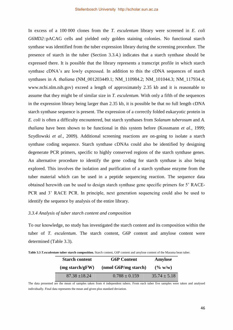
46
In excess of a 100 000 clones from the T. esculentum library were screened in E. coli
G6MD2::pACAG cells and yielded only golden staining colonies. No functional starch
synthase was identified from the tuber expression library during the screening procedure. The
presence of starch in the tuber (Section 3.3.4.) indicates that a starch synthase should be
expressed there. It is possible that the library represents a transcript profile in which starch
synthase cDNA’s are lowly expressed. In addition to this the cDNA sequences of starch
synthases in A. thaliana (NM_001203449.1; NM_110984.2; NM_101044.3; NM_117934.4;
www.ncbi.nlm.nih.gov) exceed a length of approximately 2.35 kb and it is reasonable to
assume that they might be of similar size in T. esculentum. With only a fifth of the sequences
in the expression library being larger than 2.35 kb, it is possible be that no full length cDNA
starch synthase sequence is present. The expression of a correctly folded eukaryotic protein in
E. coli is often a difficulty encountered, but starch synthases from Solanum tuberosum and A.
thaliana have been shown to be functional in this system before (Kossmann et al., 1999;
Szydlowski et al., 2009). Additional screening reactions are on-going to isolate a starch
synthase coding sequence. Starch synthase cDNAs could also be identified by designing
degenerate PCR primers, specific to highly conserved regions of the starch synthase genes.
An alternative procedure to identify the gene coding for starch synthase is also being
explored. This involves the isolation and purification of a starch synthase enzyme from the
tuber material which can be used in a peptide sequencing reaction. The sequence data
obtained herewith can be used to design starch synthase gene specific primers for 5’ RACE-
PCR and 3’ RACE PCR. In principle, next generation sequencing could also be used to
identify the sequence by analysis of the entire library.
3.3.4 Analysis of tuber starch content and composition
To our knowledge, no study has investigated the starch content and its composition within the
tuber of T. esculentum. The starch content, G6P content and amylose content were
determined (Table 3.3).
Table 3.3 T.esculentum tuber starch composition. Starch content, G6P content and amylose content of the Marama bean tuber.
Starch content
(mg starch/gFW)
G6P Content
(nmol G6P/mg starch)
Amylose
(% w/w)
87.38 ±18.24 0.788 ± 0.159 35.74 ± 5.18
The data presented are the mean of samples taken from 4 independent tubers. From each tuber five samples were taken and analysed
individually. Final data represents the mean and given plus standard deviation.
Stellenbosch University http://scholar.sun.ac.za

47
In comparison to industrially important starch sources, the T. esculentum tuber with a starch
content of approximately 9% is lower than that of potato tuber (15.44% w/w; USDA),
cassava roots (18.1-23.4% w/w, 6 months; Sriroth et al., 1999) and cereal seeds (65% w/w,
maize, Li et al., 2011).
Although Marama bean tubers have lower starch content than traditional starch crop plants it
is important to consider the low agricultural input required for their cultivation. They grow
wild, need little maintenance and the soil area can be shared with other revenue generating
crops. The tuber at this stage has no economic value, but studies on the starch properties
could possibly identify market segments where it would be of value. A possible application
could exist in the food industry, as the viscosity indicates that it has double the gelling
property of potato starch (Pers. Comm., Dr. James Lloyd, Stellenbosch University). More
studies will need to be performed on the nature of the starch before its potential as an orphan
crop in Southern Africa can be realized.
3.5. Conclusion
This study aimed at identifying genes involved in starch synthesis in the tuber of T.
esculentum. Although this was not achieved a tuber-specific library of good quality was
produced, transferred into an expression vector and a screening procedure was established for
the identification of a functional starch synthase. Unfortunately this approach did not confirm
the presence of a starch synthase within the expression library.
Investigations into the properties of the tuber confirmed the presence of starch. The average
starch content, G6P content and amylose content of the tuber was determined and this data
will likely lead to additional studies into the nature of the tuber starch and its potential
industrial applications.
Stellenbosch University http://scholar.sun.ac.za

48
Chapter 4: General Discussion
Biopolymers are of great importance to industry, and investigations into their metabolism
could identify aspects that could further contribute to their economic value. Therefore the
anticipated outcomes of the project as a whole were to identify genes involved in biopolymer
synthesis in higher plants, with specific focus on cell wall synthesis in A. thaliana and starch
synthesis in T. esculentum. The study did not result in the identification of any novel genes,
but was successful in producing data that provides a fundamental structure for the future
continuation of such projects.
Great advances have been made in understanding the complex process of wall synthesis, but
it is believed that the majority of genes that participate in these activities remain unknown
(Lerouxel et al., 2006; Carpita & McCann, 2008). This verifies the importance in developing
new experimental platforms wall-metabolism studies. In the first experimental chapter (See
Chapter 2) the potential of a new system to investigate cell wall biosynthesis was explored. A
cDNA library was constructed from regenerating protoplasts, as it was expected to enrich for
cell wall-related genes (Gipman, 2001). The cDNA library was found to be of good quality
and a few ESTs with sequence similarity to genes previously discovered in protoplast
regeneration studies confirmed that it was representing a population of sequences that are
expected to be present during wall regeneration. The difficulty was encountered in
establishing a screening method for the produced library.
Unicellular algae offer a unique opportunity to investigate carbohydrate metabolism in
vascular plants (Hicks et al., 2001) and therefore it was decided to screen the library in a
microalgal species. Screening of the library was to be performed by transferring the library
into C. protothecoides by transformation and studying colony morphology for apparent
changes induced by the expression of higher plant genes. The development of a high-
throughput transformation procedure was thus essential. Most of the conventional approaches
previously found to be successful in producing transgenic microalgae were tested on C.
protothecoides and no transformants were obtained. The effect of promoter influence on
transformation was also tested by introducing the same transgene under the control of
different promoters into C. protothecoides. Considering these findings experiments are
underway to develop a screening method in another strain of unicellular algae with an
established transformation system. It is expected that an established screening system in
combination with the produced cDNA library will lead to the identification of genes involved
Stellenbosch University http://scholar.sun.ac.za

49
in cell wall metabolism and possibly contribute to a better understanding of cell wall
synthesis in higher plants.
In the second experimental chapter (See Chapter 3) an investigation was performed into the
potential of commercialization of the southern African under-utilized crop plant, T.
esculentum. This section of the project was aimed at the identification of genes involved in
the synthesis of tuber starch. The study did produce a tuber-specific cDNA library of good
quality from T. esculentum. Additionally a screening procedure used in previous
identification of a starch synthase gene from S. tuberosum (Kossmann et al., 1999) was
further developed to be used in conjunction with the commercially available Gateway®
(Invitrogen) system which was essential for library screening purposes. Despite screening
more than 100 000 plasmids, none were found that led to starch synthase activity. The library
could perhaps be sequenced using next generation technology in order to find not just starch
synthases, but other starch metabolic genes as well. Such a sequencing project would also
provide genetic resources for scientists producing a genetic map for Marama bean.
The data generated in the analysis of the tuber starch are the first known information about T.
esculentum tuber starch content and composition and indicates that it has potential to be a
starch crop. Low agricultural input and relatively high yields of tuber starch from this crop
might lead to its development as a revenue generating crop for poor farmers in developing
regions of southern Africa. Further research endeavours are now directed at studying the
nature of the Marama bean tuber starch to identify industries that would serve as prospective
buyers. The possibility of a use in the food industries has been suggested due to its high
viscosity, with gelling properties approximately twice that of potato starch (Pers. Comm., Dr.
James Lloyd, University of Stellenbosch). More studies are bound to follow soon, as the
interest of the scientific community especially those working on orphan crops are redirected
to the potential value of T. esculentum as a starch producing cash-crop.
Essentially the data, experimental components and procedures developed in this study will
provide a stable platform for continued studies on the metabolism of biopolymer production
in higher plants. To identify the initially proposed cell wall biosynthetic genes from the
regenerating protoplast library now depends on the successful establishment of a screening
system in an alternative transformable microalgae which then needs to be optimized for high
yields of transgenic colonies. The identification of sequences coding for functional starch
synthases are still underway by additional rounds of screening within the established system.
Stellenbosch University http://scholar.sun.ac.za

50
Both of the produced cDNA libraries in addition to their current on-going studies, are also in
the process of being utilized in studies beyond the scope of their initial proposed use.
Stellenbosch University http://scholar.sun.ac.za

51
References
Babaleye T (2005) Can Cassava Solve Africa’s Food Crisis? African Business 314: 24-25
Ball SG, Morell MK (2003) From bacterial glycogen to starch: understanding the biogenesis
of the plant starch granule. Annu Rev Plant Biol 54: 207-233
Barnosky AD, Hadly EA, Bascompte J, Berlow EL, Brown JH, Fortelius M, Getz WM,
Harte J, Hastings A, Marquet PA (2012) Approaching a state shift in Earth/'s biosphere.
Nature 486: 52-58
Bartel C (2009) Enhancing food security in Africa through science, technology and
innovation. ATDF J 6: 4-8
Baskin T (2005) Anisotropic expansion of the plant cell wall. Annu Rev Cell Dev Biol 21:
203-222
Bay S, Jones BH, Schiff K, Washburn L (2003) Water quality impacts of stormwater
discharges to Santa Monica Bay. Mar Environ Res 56: 205-223
Baydoun E, Waldron KW, Brett CT (1989) The interaction of xylosyltransferase and
glucuronyltransferase involved in glucuronoxylan synthesis in pea (Pisum sativum) epicotyls.
Biochem J 257: 853-858
Biesele M, Murry R (1983) Alternative food plants for arid regions. Final project report,
morama and other plant foods of Kalahari foragers: An applied ethnobotanical study. Center
for the Study of Human Adaptation, Univ.Texas, Austin
Birmeta G, Nybom H, Bekele E (2004) Distinction between wild and cultivated enset
(Ensete ventricosum) gene pools in Ethiopia using RAPD markers. Hereditas 140: 139-148
Boynton JE, Gillham NW, Harris EH, Hosler JP, Johnson AM, Jones AR, Randolph-
Anderson BL, Robertson D, Klein TM, Shark KB (1988) Chloroplast transformation in
Chlamydomonas with high velocity microprojectiles. Science 240: 1534-1538
Brown LE, Sprecher S, Keller L (1991) Introduction of exogenous DNA into
Chlamydomonas reinhardtii by electroporation. Mol Cell Biol 11: 2328-2332
Stellenbosch University http://scholar.sun.ac.za

52
Buléon A, Colonna P, Planchot V, Ball S (1998) Starch granules: structure and
biosynthesis. Int J Biol Macromol 23: 85-112
Burn JE, Hocart CH, Birch RJ, Cork AC, Williamson RE (2002) Functional Analysis of
the Cellulose Synthase GenesCesA1, CesA2, and CesA3 in Arabidopsis. Plant Physiol 129:
797-807
Burton RA, Shirley NJ, King BJ, Harvey AJ, Fincher GB (2004) The CesA gene family
of barley. Quantitative analysis of transcripts reveals two groups of co-expressed genes. Plant
Physiol 134: 224-236
Butanaev AM (1994) Use of the hygromycin phosphotransferase gene as the dominant
selective marker for Chlamydomonas reinhardtii transformation. Mol Biol (Mosk) 28: 1061-
1068
Buttrose MS (1960) Submicroscopic development and structure of starch granules in cereal
endosperms. J Ultrastruct Res 4: 231-257
Carpita NC, Gibeaut DM (1993) Structural models of primary cell walls in flowering
plants: consistency of molecular structure with the physical properties of the walls during
growth. Plant J 3: 1-30
Casale A (2008) Biopolymers: Market potential and challenging research. JEC Group,
Knowledge and Networking, http://www.jeccomposites.com/news/composites-
news/biopolymers-market-potential-and-challenging-research
Chen D, Yang B, Kuo T (1992) One-step transformation of yeast in stationary phase. Curr
Genet 21: 83-84
Chen Y, Wang Y, Sun Y, Zhang L, Li W (2001) Highly efficient expression of rabbit
neutrophil peptide-1 gene in Chlorella ellipsoidea cells. Curr Genet 39: 365-370
Chow K, Tung W (1999) Electrotransformation of Chlorella vulgaris. Plant Cell Rep 18:
778-780
Cilia ML, Jackson D (2004) Plasmodesmata form and function. Curr Opin Cell Biol 16:
500-506
Stellenbosch University http://scholar.sun.ac.za
53
Cock JH (1982) Cassava: a basic energy source in the tropics. Science 218: 755-762
Coll J (2006) Review. Methodologies for transferring DNA into eukaryotic microalgae. Span
J Agric Res 4: 316-330
Cosgrove DJ (2000) Expansive growth of plant cell walls. Plant Physiol Bioch 38: 109-124
Cosgrove DJ (2005) Growth of the plant cell wall. Nat Rev Mol Cell Bio 6: 850-861
Creuzat-Sigal N, Latil-Damotte M, Cattaneo J, Puig J (1972) Genetic analysis and
biochemical characterization of mutants impairing glycogen metabolism in Escherichia coli
K-12. In: Piras R, Pontis HG (eds) Biochemistry of the glycoside linkage. Academic Press,
New York, pp 647-680
Cumming CM, Rizkallah HD, McKendrick KA, Abdel-Massih RM, Baydoun EA, Brett
CT (2005) Biosynthesis and cell-wall deposition of a pectin–xyloglucan complex in pea.
Planta 222: 546-555
da Costa Sousa L, Chundawat SP, Balan V, Dale BE (2009) ‘Cradle-to-grave’assessment
of existing lignocellulose pretreatment technologies. Curr Opin Biotechnol 20: 339-347
Davis JP, Supatcharee N, Khandelwal RL, Chibbar RN (2003) Synthesis of novel
starches in planta: opportunities and challenges. Starch‐Stärke 55: 107-120
de Lumen BO, Thompson S, Odegard WJ (1993) Sulfur amino acid-rich proteins in acha
(Digitaria exilis), a promising underutilized African cereal. J Agric Food Chem 41: 1045-
1047
Dick-Pérez M, Zhang Y, Hayes J, Salazar A, Zabotina OA, Hong M (2011) Structure and
interactions of plant cell-wall polysaccharides by two-and three-dimensional magic-angle-
spinning solid-state NMR. Biochemistry 50: 989-1000
Dick‐Pérez M, Wang T, Salazar A, Zabotina OA, Hong M (2012) Multidimensional solid‐
state NMR studies of the structure and dynamics of pectic polysaccharides in uniformly 13C‐
labeled Arabidopsis primary cell walls. Magn Reson Chem 50: 539-550
Stellenbosch University http://scholar.sun.ac.za

54
Doblin MS, Kurek I, Jacob-Wilk D, Delmer DP (2002) Cellulose biosynthesis in plants:
from genes to rosettes. Plant Cell Physiol 43: 1407-1420
DDPSC (2011) Cassava Research, Donald Danforth Plant Science Center,
http://www.danforthcenter.org/news-media/Cassava-Research-
Ellis RP, Cochrane MP, Dale MFB, Duffus CM, Lynn A, Morrison IM, Prentice RDM,
Swanston JS, Tiller SA (1998) Starch production and industrial use. J Sci Food Agric 77:
289-311
El-Sheekh M (1999) Stable transformation of the intact cells of Chlorella kessleri with high
velocity microprojectiles. Biol Plant 42: 209-216
Endler A, Meyer S, Schelbert S, Schneider T, Weschke W, Peters SW, Keller F,
Baginsky S, Martinoia E, Schmidt UG (2006) Identification of a vacuolar sucrose
transporter in barley and Arabidopsis mesophyll cells by a tonoplast proteomic approach.
Plant Physiol 141: 196-207
Espada J (1962) Enzymic synthesis of adenosine diphosphate glucose from glucose 1-
phosphate and adenosine triphosphate. J Biol Chem 237: 3577-3581
Faik A, Bar-Peled M, DeRocher AE, Zeng WQ, Perrin RM, Wilkerson C, Raikhel NV,
Keegstra K (2000) Biochemical characterization and molecular cloning of an alpha-1,2-
fucosyltransferase that catalyzes the last step of cell wall xyloglucan biosynthesis in pea. J
Biol Chem 275: 15082-15089
Faik A, Price NJ, Raikhel NV, Keegstra K (2002) An Arabidopsis gene encoding an α-
xylosyltransferase involved in xyloglucan biosynthesis. Proc Natl Acad Sci USA 99: 7797-
7802
FAO (2010) FAOSTAT: FAO Statistical databases, production data.
http://faostat.fao.org/site/339/default.aspx
Favery B, Ryan E, Foreman J, Linstead P, Boudonck K, Steer M, Shaw P, Dolan L
(2001) KOJAK encodes a cellulose synthase-like protein required for root hair cell
morphogenesis in Arabidopsis. Genes Dev 15: 79-89
Stellenbosch University http://scholar.sun.ac.za

55
Festel G, Kölle S, Sell D (2005) Biopolymers on the way up. European Chemical News,
http://www.founding-angels.com/Biopolymers_ECN_10-05.pdf
Field B, Jordán F, Osbourn A (2006) First encounters–deployment of defence‐related
natural products by plants. New Phytol 172: 193-207
Franks T, Birch R (1991) Gene transfer into intact sugarcane cells using microprojectile
bombardment. Funct Plant Biol 18: 471-480
Fry SC (1989) Cellulases, hemicelluloses and auxin‐stimulated growth: a possible
relationship. Physiol Plantarum 75: 532-536
Fry SC (2004) Primary cell wall metabolism: tracking the careers of wall polymers in living
plant cells. New Phytol 161: 641-675
Gillmor CS, Lukowitz W, Brininstool G, Sedbrook JC, Hamann T, Poindexter P,
Somerville C (2005) Glycosylphosphatidylinositol-anchored proteins are required for cell
wall synthesis and morphogenesis in Arabidopsis. The Plant Cell Online 17: 1128-1140
Gipman M (2001) Molecular Genetic Approaches to study Cell Wall Biosynthesis in
Arabidopsis thaliana. PhD Thesis. University of Potsdam, Potsdam.
Glover R (1993) Markets for degradable plastics. Int Biodeterior Biodegrad 31: 171-178
Goubet F, Misrahi A, Park SK, Zhang Z, Twell D, Dupree P (2003) AtCSLA7, a
cellulose synthase-like putative glycosyltransferase, is important for pollen tube growth and
embryogenesis in Arabidopsis. Plant Physiol 131: 547-557
Grela ER, Rybiński W, Klebaniuk R, Matras J (2010) Morphological characteristics of
some accessions of grass pea (Lathyrus sativus L.) grown in Europe and nutritional traits of
their seeds. Genet Resour Crop Evol 57: 693-701
Gündel S, Höschle-Zeledon I, Krause B, Probst K (2003) Underutilized Plant Species and
Poverty Alleviation. Proceedings of an International Workshop, Leipzig, pp 6-8
Stellenbosch University http://scholar.sun.ac.za
56
Harholt J, Jensen JK, Sørensen SO, Orfila C, Pauly M, Scheller HV (2006) ARABINAN
DEFICIENT 1 is a putative arabinosyltransferase involved in biosynthesis of pectic arabinan
in Arabidopsis. Plant Physiol 140: 49-58
Hawkins RL, Nakamura M (1999) Expression of human growth hormone by the eukaryotic
alga, Chlorella. Curr Microbiol 38: 335-341
Hayashi T (1989) Xyloglucans in the primary cell wall. Ann Rev Plant Biol 40: 139-168
Hazen SP, Scott-Craig JS, Walton JD (2002) Cellulose synthase-like (CSL) genes of rice.
Plant Physiol 128: 336-340
Hématy K, Cherk C, Somerville S (2009) Host–pathogen warfare at the plant cell wall.
Curr Opin Plant Biol 12: 406-413
Henrissat B, Coutinho PM, Davies GJ (2001) A census of carbohydrate-active enzymes in
the genome of Arabidopsis thaliana. Plant Mol Biol 47: 55-72
Hicks GR, Hironaka CM, Dauvillee D, Funke RP, D'Hulst C, Waffenschmidt S, Ball SG
(2001) When simpler is better. Unicellular green algae for discovering new genes and
functions in carbohydrate metabolism. Plant Physiol 127: 1334-1338
Hizukuri S (1986) Polymodal distribution of the chain lengths of amylopectins, and its
significance. Carbohydr Res 147: 342-347
Hizukuri S, Takeda Y, Yasuda M, Suzuki A (1981) Multi-branched nature of amylose and
the action of debranching enzymes. Carbohydr Res 94: 205-213
Hoekema A, Hirsch P, Hooykaas P, Schilperoort R (1983) A binary plant vector strategy
based on separation of vir-and T-region of the Agrobacterium tumefaciens Ti-plasmid.
Nature 303: 179-180
Hood E, Helmer G, Fraley R, Chilton MD (1986) The hypervirulence of Agrobacterium
tumefaciens A281 is encoded in a region of pTiBo542 outside of T-DNA. J Bacteriol 168:
1291-1301
Stellenbosch University http://scholar.sun.ac.za

57
Hückelhoven R (2007) Cell wall-associated mechanisms of disease resistance and
susceptibility. Annu Rev Phytopathol 45: 101-127
Israel A, Obot I, Umoren S, Mkpenie V, Ebong G (2008) Effluents and Solid Waste
Analysis in a Petrochemical Company-A Case Study of Eleme Petrochemical Company Ltd,
Port Harcourt, Nigeria. E-J of Chemistry 5: 74-80
Iwai H, Ishii T, Satoh S (2001) Absence of arabinan in the side chains of the pectic
polysaccharides strongly associated with cell walls of Nicotiana plumbaginifolia non-
organogenic callus with loosely attached constituent cells. Planta 213: 907-915
Iwai H, Masaoka N, Ishii T, Satoh S (2002) A pectin glucuronyltransferase gene is essential
for intercellular attachment in the plant meristem. Proc Natl Acad Sci USA 99: 16319-16324
Jackson JC, Duodu KG, Holse M, Lima de Faria, Margarida D, Jordaan D, Chingwaru
W, Hansen A, Cencic A, Kandawa-Schultz M, Mpotokwane SM (2010) The Morama
Bean(Tylosema esculentum): A Potential Crop for Southern Africa. Adv Food Nutr Res 61:
187-246
Jaenicke H, Höschle-Zeledon I (2006) Strategic Framework for Underutilized Plant
Species: With special reference to Asia and the Pacific, and to Sub-Saharan Africa.
Bioversity International.
Jane J, Kasemsuwan T, Leas S, Zobel H, Robyt JF (1994) Anthology of starch granule
morphology by scanning electron microscopy. Starch‐Stärke 46: 121-129
Jarvis EE, Brown LM (1991) Transient expression of firefly luciferase in protoplasts of the
green alga Chlorella ellipsoidea. Curr Genet 19: 317-321
Jayasekara R, Harding I, Bowater I, Lonergan G (2005) Biodegradability of a selected
range of polymers and polymer blends and standard methods for assessment of
biodegradation. J Polym Environ 13: 231-251
Jobling S (2004) Improving starch for food and industrial applications. Curr Opin Plant Biol
7: 210-218
Stellenbosch University http://scholar.sun.ac.za

58
Jones L, Milne JL, Ashford D, McQueen-Mason SJ (2003) Cell wall arabinan is essential
for guard cell function. Proceedings of the National Academy of Sciences 100: 11783-11788
Kathiresan S, Sarada R (2009) Towards genetic improvement of commercially important
microalga Haematococcus pluvialis for biotech applications. J Appl Phycol 21: 553-558
Keeling PL, Myers AM (2010) Biochemistry and genetics of starch synthesis. Annu Rev
Food Sci Techn 1: 271-303
Keith M, Renew A (1975) Notes on some edible wild plants found in the Kalahari. Koedoe-
African Protected Area Conservation and Science 18: 1-12
Kim D, Kim YT, Cho JJ, Bae J, Hur S, Hwang I, Choi T (2002) Stable integration and
functional expression of flounder growth hormone gene in transformed microalga, Chlorella
ellipsoidea. Mar Biotechnol 4: 63-73
Kim J (2005) Regulation of short-distance transport of RNA and protein. Curr Opin Plant
Biol 8: 45-52
Kimura S, Laosinchai W, Itoh T, Cui X, Linder CR, Brown RM (1999) Immunogold
labeling of rosette terminal cellulose-synthesizing complexes in the vascular plant Vigna
angularis. The Plant Cell Online 11: 2075-2085
Kindle KL (1990) High-frequency nuclear transformation of Chlamydomonas reinhardtii.
Proc Natl Acad Sci USA 87: 1228-1232
Knox JP (1990) Emerging patterns of organization at the plant cell surface. J Cell Sci 96:
557-561
Knox JP, Day S, Roberts K (1989) A set of cell surface glycoproteins forms an early
position, but not cell type, in the root apical carota L. Development 106: 47-56
Kossmann J, Abel GJ, Springer F, Lloyd JR, Willmitzer L (1999) Cloning and functional
analysis of a cDNA encoding a starch synthase from potato (Solanum tuberosum L.) that is
predominantly expressed in leaf tissue. Planta 208: 503-511
Stellenbosch University http://scholar.sun.ac.za

59
Kossmann J, Lloyd J (2000) Understanding and influencing starch biochemistry. Crit Rev
Plant Sci 19: 171-226
Kwon H, Yokoyama R, Nishitani K (2005) A proteomic approach to apoplastic proteins
involved in cell wall regeneration in protoplasts of Arabidopsis suspension-cultured cells.
Plant Cell Physiol 46: 843-857
Lane DR, Wiedemeier A, Peng L, Höfte H, Vernhettes S, Desprez T, Hocart CH, Birch
RJ, Baskin TI, Burn JE (2001) Temperature-sensitive alleles of RSW2 link the
KORRIGAN endo-1, 4-β-glucanase to cellulose synthesis and cytokinesis in Arabidopsis.
Plant Physiol 126: 278-288
Lao NT, Long D, Kiang S, Coupland G, Shoue DA, Carpita NC, Kavanagh TA (2003)
Mutation of a family 8 glycosyltransferase gene alters cell wall carbohydrate composition and
causes a humidity-sensitive semi-sterile dwarf phenotype in Arabidopsis. Plant Mol Biol 53:
687-701
Lerouxel O, Cavalier DM, Liepman AH, Keegstra K (2006) Biosynthesis of plant cell
wall polysaccharides - a complex process. Curr Opin Plant Biol 9: 621-630
Li N, Zhang S, Zhao Y, Li B, Zhang J (2011) Over-expression of AGPase genes enhances
seed weight and starch content in transgenic maize. Planta 233: 241-250
Lipka V, Dittgen J, Bednarek P, Bhat R, Wiermer M, Stein M, Landtag J, Brandt W,
Rosahl S, Scheel D (2005) Pre-and postinvasion defenses both contribute to nonhost
resistance in Arabidopsis. Science 310: 1180-1183
Lukowitz W, Nickle TC, Meinke DW, Last RL, Conklin PL, Somerville CR (2001)
Arabidopsis cyt1 mutants are deficient in a mannose-1-phosphate guanylyltransferase and
point to a requirement of N-linked glycosylation for cellulose biosynthesis. Proc Natl Acad
Sci USA 98: 2262-2267
Madson M, Dunand C, Li X, Verma R, Vanzin GF, Caplan J, Shoue DA, Carpita NC,
Reiter W (2003) The MUR3 gene of Arabidopsis encodes a xyloglucan galactosyltransferase
that is evolutionarily related to animal exostosins. The Plant Cell Online 15: 1662-1670
Stellenbosch University http://scholar.sun.ac.za

60
Maruyama M, Horáková I, Honda H, Xing X, Shiragami N, Unno H (1994) Introduction
of foreign DNA into Chlorella saccharophila by electroporation. Biotechnol Tech 8: 821-826
Marz U (2006) World Market for Starches/Glucose, Emphasizing Cassava. Report Code:
FOD037A
McCann MC, Carpita NC (2008) Designing the deconstruction of plant cell walls. Curr
Opin Plant Biol 11: 314-320
Moore CJ (2008) Synthetic polymers in the marine environment: a rapidly increasing, long-
term threat. Environ Res 108: 131-139
Morris G, Foster T, Harding S (2000) The effect of the degree of esterification on the
hydrodynamic properties of citrus pectin. Food Hydrocoll 14: 227-235
Morrison WR (1988) Lipids in cereal starches: A review. J Cereal Sci 8: 1-15
Müseler DL (2005) Evaluation of the quality characteristics of the marama bean (Tylosema
esculentum), an underutilized grain and tuber producing legume in southern Africa. MSc
thesis. University of Namibia, Windhoek.
Naylor RL, Falcon WP, Goodman RM, Jahn MM, Sengooba T, Tefera H, Nelson RJ
(2004) Biotechnology in the developing world: a case for increased investments in orphan
crops. Food Policy 29: 15-44
Nelson OE, Rines HW (1962) The enzymatic deficiency in the waxy mutant of maize.
Biochem Biophys Res Commun 9: 297-300
Nguema-Ona E, Andème-Onzighi C, Aboughe-Angone S, Bardor M, Ishii T, Lerouge P,
Driouich A (2006) The reb1-1 mutation of Arabidopsis. Effect on the structure and
localization of galactose-containing cell wall polysaccharides. Plant Physiol 140: 1406-1417
Nielsen TH, Baunsgaard L, Blennow A (2002) Intermediary glucan structures formed
during starch granule biosynthesis are enriched in short side chains, a dynamic pulse labeling
approach. J Biol Chem 277: 20249-20255
Stellenbosch University http://scholar.sun.ac.za

61
Okogbenin E, Setter TL, Ferguson M, Mutegi R, Ceballos H, Olasanmi B, Fregene M
(2013) Phenotypic approaches to drought in cassava: review. Front Physiol 4: 1-15
Oparka KJ (2004) Getting the message across: how do plant cells exchange macromolecular
complexes? Trends Plant Sci 9: 33-41
Pagant S, Bichet A, Sugimoto K, Lerouxel O, Desprez T, McCann M, Lerouge P,
Vernhettes S, Höfte H (2002) KOBITO1 encodes a novel plasma membrane protein
necessary for normal synthesis of cellulose during cell expansion in Arabidopsis. The Plant
Cell Online 14: 2001-2013
Pauly M, Keegstra K (2008) Cell‐wall carbohydrates and their modification as a resource
for biofuels. Plant J 54: 559-568
Peat S, Pirt S, Whelan W (1952) 128. Enzymic synthesis and degradation of starch. Part
XV. β-Amylase and the constitution of amylose. J Chem Soc (Resumed) 705-713
Pennell RI, Roberts K (1990) Sexual development in the pea is presaged by altered
expression of arabinogalactan protein. Nature 344: 547-549
Percy C, Isaac M, Pamela C (2010) Understanding the relationship between indigenous
(traditional) knowledge systems (IKS), and access to genetic resources and benefits sharing
(ABS). African Journal of Biotechnology 9: 9204-9207
Perrin RM, DeRocher AE, Bar-Peled M, Zeng W, Norambuena L, Orellana A, Raikhel
NV, Keegstra K (1999) Xyloglucan fucosyltransferase, an enzyme involved in plant cell
wall biosynthesis. Science 284: 1976-1979
Pien S, Wyrzykowska J, McQueen-Mason S, Smart C, Fleming A (2001) Local
expression of expansin induces the entire process of leaf development and modifies leaf
shape. Proc Natl Acad Sci USA 98: 11812-11817
Pilling E, Smith AM (2003) Growth ring formation in the starch granules of potato tubers.
Plant Physiol 132: 365-371
Stellenbosch University http://scholar.sun.ac.za

62
Pratheesh PT, Shonima GM, Jiji T, Abraham CI, Muraleedhara KG (2012) Study on
efficacy of different Agrobacterium tumefaciens strains in genetic transformation of
microalga Chlamydomonas reinhardtii. Adv Appl Science Res 3 (5): 2679-2686
Preiss J (1999) Biosynthesis of bacterial and mammalian glycogen and plant starch synthesis
and their regulation. In: Hechts SM (ed) Biorganic Chemistry: carbohydrates, pp 489-554.
Oxford University Press, Oxford.
Preiss J, Sivak M (1998a) Starch and glycogen biosynthesis. In: Pinto BM (Ed)
Comprehensive Natural Products Chemistry, pp 441-495. Pergamon Press, Oxford.
Preiss J, Sivak M (1998b) Biochemistry, molecular biology and regulation of starch
synthesis. Gen Eng 20: 177-223
Proseus TE, Boyer JS (2005) Turgor pressure moves polysaccharides into growing cell
walls of Chara corallina. Ann Bot-London 95: 967-979
Rao K (2010) The Current State of Biopolymers and Their Potential Future. Omnexus by
Special Chem, http://www.omnexus.com/resources/editorials.aspx?id=25041
Ray PM (1967) Radioautographic study of cell wall deposition in growing plant cells. J Cell
Biol 35: 659-674
Richmond TA, Somerville CR (2000) The cellulose synthase superfamily. Plant Physiol
124: 495-498
Richmond TA, Somerville CR (2001) Integrative approaches to determining Csl function.
Plant Mol Biol 47: 131-143
Ridley BL, O'Neill MA, Mohnen D (2001) Pectins: structure, biosynthesis, and
oligogalacturonide-related signaling. Phytochemistry 57: 929-967
Rizk SE, Abdel-Massih RM, Baydoun EA, Brett CT (2000) Protein-and pH-dependent
binding of nascent pectin and glucuronoarabinoxylan to xyloglucan in pea. Planta 211: 423-
429
Stellenbosch University http://scholar.sun.ac.za
63
Roudier F, Fernandez AG, Fujita M, Himmelspach R, Borner GH, Schindelman G,
Song S, Baskin TI, Dupree P, Wasteneys GO (2005) COBRA, an Arabidopsis extracellular
glycosyl-phosphatidyl inositol-anchored protein, specifically controls highly anisotropic
expansion through its involvement in cellulose microfibril orientation. The Plant Cell Online
17: 1749-1763
Sánchez-Rodríguez C, Rubio-Somoza I, Sibout R, Persson S (2010) Phytohormones and
the cell wall in Arabidopsis during seedling growth. Trends Plant Sci 15: 291-301
Savitha P, Nirmalakumari A, Maheswaran M, Raguchander T (2013) Combining ability
analysis and estimates of heterosis for grain yield and nutritional traits in finger millet
(Eleusine coracana (L.) Geartn). Madras Agricultural J 100: 15-19
Scheible W, Pauly M (2004) Glycosyltransferases and cell wall biosynthesis: novel players
and insights. Curr Opin Plant Biol 7: 285-295
Scheller HV, Ulvskov P (2010) Hemicelluloses. Plant Biol 61: 263
Schwartz M (1966) Location of the maltose A and B loci on the genetic map of Escherichia
coli. J Bacteriol 92: 1083-1089
Shao M, Zheng H, Hu Y, Liu D, Jang J, Ma H, Huang H (2004) The
GAOLAOZHUANGREN1 gene encodes a putative glycosyltransferase that is critical for
normal development and carbohydrate metabolism. Plant Cell Physiol 45: 1453-1460
Sharma R, Tan F, Jung K, Sharma MK, Peng Z, Ronald PC (2011) Transcriptional
dynamics during cell wall removal and regeneration reveals key genes involved in cell wall
development in rice. Plant Mol Biol 77: 391-406
Smith AM, Zeeman SC, Smith SM (2005) Starch degradation. Annu Rev Plant Biol 56: 73-
98
Somerville C (2006) Cellulose synthesis in higher plants. Annu Rev Cell Dev Biol 22: 53-78
Sprent JI, Odee DW, Dakora FD (2010) African legumes: a vital but under-utilized
resource. J Exp Bot 61: 1257-1265
Stellenbosch University http://scholar.sun.ac.za
64
Sriroth K, Santisopasri V, Petchalanuwat C, Kurotjanawong K, Piyachomkwan K,
Oates C (1999) Cassava starch granule structure–function properties: influence of time and
conditions at harvest on four cultivars of cassava starch. Carbohydr Polym 38: 161-170
Sriroth K, Piyachomkwan K, Wanlapatit S, Oates CG (2000) Cassava starch technology:
the Thai experience. Starch‐Stärke 52: 439-449
Sterling JD, Atmodjo MA, Inwood SE, Kolli VK, Quigley HF, Hahn MG, Mohnen D
(2006) Functional identification of an Arabidopsis pectin biosynthetic homogalacturonan
galacturonosyltransferase. Proc Natl Acad Sci USA 103: 5236-5241
Stolle-Smits T, Beekhuizen JG, Kok MT, Pijnenburg M, Recourt K, Derksen J,
Voragen AG (1999) Changes in cell wall polysaccharides of green bean pods during
development. Plant Physiol 121: 363-372
Szydlowski N, Ragel P, Raynaud S, Lucas MM, Roldán I, Montero M, Munoz FJ,
Ovecka M, Bahaji A, Planchot V, Pozueta-Romero J, D’Hulst C, Mérida, Á (2009)
Starch granule initiation in Arabidopsis requires the presence of either class IV or class III
starch synthases. The Plant Cell Online 21: 2443-2457
Szyjanowicz PM, McKinnon I, Taylor NG, Gardiner J, Jarvis MC, Turner SR (2004)
The irregular xylem 2 mutant is an allele of korrigan that affects the secondary cell wall of
Arabidopsis thaliana. Plant J 37: 730-740
Szymanski DB, Cosgrove DJ (2009) Dynamic coordination of cytoskeletal and cell wall
systems during plant cell morphogenesis. Curr Biol 19: R800-R811
Tadele Z (2009) Role of orphan crops in enhancing and diversifying food production in
Africa. ATDF J 6: 9-15
Takeda Y, Hizukuri S, Takeda C, Suzuki A (1987) Structures of branched molecules of
amyloses of various origins, and molar fractions of branched and unbranched molecules.
Carbohydr Res 165: 139-145
Takeda C, Takeda Y, Hizukuri S (1989) Structure of amylomaize amylose. Cereal Chem
66: 22-25
Stellenbosch University http://scholar.sun.ac.za

65
Talbott LD, Ray PM (1992) Molecular size and separability features of pea cell wall
polysaccharides Implications for models of primary wall structure. Plant Physiol 98: 357-368
Tatham A, Fido R, Moore C, Kasarda D, Kuzmicky D, Keen J, Shewry P (1996)
Characterisation of the Major Prolamins of Tef (Eragrostis tef) and Finger Millet (Eleusine
coracana). J Cereal Sci 24: 65-71
Taylor NG (2008) Cellulose biosynthesis and deposition in higher plants. New Phytol 178:
239-252
Taylor NG, Howells RM, Huttly AK, Vickers K, Turner SR (2003) Interactions among
three distinct CesA proteins essential for cellulose synthesis. Proc Natl Acad Sci USA 100:
1450-1455
Tetlow IJ, Morell MK, Emes MJ (2004) Recent developments in understanding the
regulation of starch metabolism in higher plants. J Exp Bot 55: 2131-2145
Thimm JC, Burritt DJ, Sims IM, Newman RH, Ducker WA, Melton LD (2002) Celery
(Apium graveolens) parenchyma cell walls: cell walls with minimal xyloglucan. Physiol
Plantarum 116: 164-171
Turner S, Gallois P, Brown D (2007) Tracheary element differentiation. Annu.Rev.Plant
Biol. 58: 407-433
Underwood W, Somerville SC (2008) Focal accumulation of defences at sites of fungal
pathogen attack. J Exp Bot 59: 3501-3508
United Nations (2011) Department of Economic and Social Affairs, Population division,
data of 2011, http://www.un.org/en/development/desa/population/publications/policy/world-
population-policies-2011.shtml
USDA (2013) United States Department of Agriculture. Full Report (All Nutrients): 11352,
Potato, flesh and skin, raw,
http://ndb.nal.usda.gov/ndb/foods/show/3127?qlookup=11352&format=Full&max=25&man
=&lfacet=&new=1
Stellenbosch University http://scholar.sun.ac.za

66
Vanzin GF, Madson M, Carpita NC, Raikhel NV, Keegstra K, Reiter W (2002) The
mur2 mutant of Arabidopsis thaliana lacks fucosylated xyloglucan because of a lesion in
fucosyltransferase AtFUT1. Proc Natl Acad Sci USA 99: 3340-3345
Vincken J, Schols HA, Oomen RJ, McCann MC, Ulvskov P, Voragen AG, Visser RG
(2003) If homogalacturonan were a side chain of rhamnogalacturonan I. Implications for cell
wall architecture. Plant Physiol 132: 1781-1789
Willats WG, McCartney L, Mackie W, Knox JP (2001) Pectin: cell biology and prospects
for functional analysis. Plant Mol Biol 1-2: 9-27
Willats WG, Knox JP, Mikkelsen JD (2006) Pectin: new insights into an old polymer are
starting to gel. Trends Food Sci Technol 17: 97-104
Williams J, Haq N (2002) Global research on underutilized crops: an assessment of current
activities and proposals for enhanced cooperation. Bioversity International.
Xu L, Chen H, Hu X, Zhang R, Zhang Z, Luo Z (2006) Average gene length is highly
conserved in prokaryotes and eukaryotes and diverges only between the two kingdoms. Mol
Biol Evol 23: 1107-1108
Yang X, Tu L, Zhu L, Fu L, Min L, Zhang X (2008) Expression profile analysis of genes
involved in cell wall regeneration during protoplast culture in cotton by suppression
subtractive hybridization and macroarray. J Exp Bot 59: 3661-3674
Zeeman SC, Tiessen A, Pilling E, Kato KL, Donald AM, Smith AM (2002) Starch
synthesis in Arabidopsis. Granule synthesis, composition, and structure. Plant Physiol 129:
516-529
Zeeman SC, Kossmann J, Smith AM (2010) Starch: its metabolism, evolution, and
biotechnological modification in plants. Annu Rev Plant Biol 61: 209-234
Zhong R, Peña MJ, Zhou G, Nairn CJ, Wood-Jones A, Richardson EA, Morrison WH,
Darvill AG, York WS, Ye Z (2005) Arabidopsis fragile fiber8, which encodes a putative
glucuronyltransferase, is essential for normal secondary wall synthesis. The Plant Cell Online
17: 3390-3408
Stellenbosch University http://scholar.sun.ac.za

67
Zobel H (1988) Molecules to granules: a comprehensive starch review. Starch‐Stärke 40: 44-
50
Zykwinska AW, Ralet MJ, Garnier CD, Thibault JJ (2005) Evidence for in vitro binding
of pectin side chains to cellulose. Plant Physiol 139: 397-407
Stellenbosch University http://scholar.sun.ac.za