Embed Size (px)
Citation preview
Problems Encountered With the Kock lleostomy
William K. Flake, MD,’ Phoenix, Arizona
Michael S. Altman, MD,* Phoenix, Arizona
Andrea M. Cartmill, RNET,’ Phoenix, Arizona
Robert B. Gilsdorf, MD, PhD,’ Phoenix, Arizona
Largely favorable results with the Kock reservoir il- eostomy have been reported since it was described
in 1967. The larger series of patients come from the
Swedish institution where the procedure was devel- oped and from major medical centers in this country [I-3]. Stimulated by such reports and the need for management for a large family with familial polyposis in our clinic population, we began a project of per- forming Kock ileostomy. We began to perform this procedure in patients undergoing total colectomy and in patients needing ileostomy revision. This is a re- port of the problems encountered and the way we handled those problems in the first 12 patients in that series.
Material and Methods
A series of 16 Kock reservoir ileostomies were performed in 12 patients from June 1975 to May 1978. Initially the major portion or all of the procedure was performed by the authors (WKF and RBG) as described in the literature current at the time [2,4]. Additional refinements of the operative techniques and postoperative practices were gained by personal observation of and communication with surgeons at a major medical center where the procedure was being performed frequently. Further minor modifi- cations described herein were introduced as the project continued.
The patients included six men and six women, aged 22 to 53 years. The indications for colectomy were ulcerative colitis in six patients, familial polyposis in five, and un- classified inflammatory bowel disease in one. The Kock ileostomy was the primary ileostomy by choice at the time of colectomy and was created in a one-stage procedure in
Prom the Departments of Surgical Education’ and Enterostomal Therapy,+ Good Samaritan Hospital, and the Phoenix Integrated Surgical Residency Program,’ Phoenix, Arizona.
Presented at the 31st Annual Meeting of the Southwestern Surgical Congress, Las Vegas, Nevada, April 23-26, 1979.
Reprint requests should be addressed to Robert 9. Gilsdorf, MD, PhD. Department of Surgical Education, Good Samaritan Hospital, 1033 East McDowell Road, Phoenix, Arizona 85062.
Volume 139, December 1979
four patients. It was created in five patients because their standard ileostomy required major intraabdominal revision for prolapse and in three patients because their standard ileostomy was in an improper position that precluded se- cure appliance fixation. Four Kock ileostomies were made to replace failed Kock ileostomies created by us earlier in the series. No standard ileostomy was revised for purely subjective reasons. All patients were and continue to be followed up by the same surgical enterostomal services. Each patient has an annual endoscopic and radiologic ex- amination of the reservoir. One patient was lost to follow- up in the immediate postoperative period, and thus 11 patients are included in the long-term follow-up.
After some early difficulties with reduction of the valve that resulted in ileostomy incontinence, the project was terminated for an 8 month period in 1976. In the research laboratory during that period, a method was developed to utilize a prosthetic material as a support for the ileostomy valve. We then resumed the project and used the prosthetic material, expanded polytetrafluoroethylene (Impragrafte), in the next seven reservoirs constructed. The problems encountered in that experience are reported on herein,
Results
The patients studied had a significant series of
problems. Eight of the 11 patients (73 per cent) re- quired further surgery. Four patients required a complete new Kock ileostomy; one of these patients
(9 per cent) required.a new reservoir and three (27 per cent) required valve revision and a new reservoir.
Four patients had and continue to have no trouble with their ileostomy. Two patients (18 per cent) have
had to have only minor stoma1 revisions, and two have had valve revision and removal of the reservoir. Two patients (18 per cent) eventually had the res- ervoir removed and underwent conversion to a con- ventional ileostomy. Nine patients (82 per cent) now have a continent, well functioning ileostomy.
The major problems encountered in this series were as follows: (1) vascular injury during ileostomy
851
Flake et al
creation (one patient); (2) valve reduction producing incontinence (three patients); (3) side fistula at the base of the valve (one patient); (4) unclassified dis- ease that proved to be Crohn’s disease (one patient); (5) enteritis caused by a failure to cannulate (one patient); (6) superficial enteritis in the reservoir (six patients); (7) inability to get a catheter into the res- ervoir (two patients); (8) prolonged hospital stay (six patients); (9) stoma too large, not level, or too low (four patients); and (10) patient lost to follow-up (one). A description of our experiences in managing these problems follows.
Problem 1: Vascular Injury in Reservoir Con- struction. This problem occurred in one patient. Necrosis of the stoma was apparent 48 hours after surgery, and the patient was immediately reoperated on. Necrosis was found not only in the stoma but also in the valve and in half of the reservoir. The vascular injury was believed to be due either to compression of the mesenteric vessels created by passing the res- ervoir through the leaves of the mesentery as de- scribed by Kock [4] or to injury of the vessels on closure of the lateral abdominal gutter. The reservoir was removed and a new one made. At the second procedure we rotated the ileum clockwise rather than using the counterclockwise rotation described by Kock that requires mesenteric pass-through. This left the reservoir oblong at the time of surgery; however, we have since found, on annual examination of that and subsequent reservoirs, that the oblong appearance disappears and that all reservoirs appear to be round with no mesenteric constriction. Fur- thermore, we have since made no major effort to close the defect in the lateral gutter. At reexploration (with which we have had significant experience), the res- ervoir tends to be firmly fixed in the lower quadrant and gutter closure does not seem to be necessary.
Problem 2: Valve Reduction Producing Inconti- nence. Ileostomy leakage plus difficulty in cannu- lating the reservoir are the symptoms of this problem, which developed in three of our early patients, all of whom had these symptoms by 3 months postopera- tively. We first tried prolonged stenting in the hope of getting the valve to heal in a straight position, but this was clearly useless. Then in two patients we tried open correction of the problem. We attempted to tuck the valve back into position without opening the reservoir by using stitches similar to the wrap-around stitches used in a Nissen type hiatal hernia repair. In both of these patients valve incontinence developed again within 6 months. Since that experience we have concluded that valve incontinence, when it develops, can be repaired only by totally remaking the valve,
as has now been described [1,5], or by making a whole new reservoir. We eventually did the latter in all three patients.
As stated earlier, we then stopped making reser- voirs for 8 months and investigated, in dogs, the ef- ficacy and safety of using expanded polytetrafluo- roethylene (PTFE) as a valve support. In brief, the method developed utilized a 3 cm wide belt of PTFE trimmed to size from the material commonly used as an aortic replacement. This belt was wrapped on the serosal side of the efferent bowel. The bowel was then intussuscepted to create the valve in the usual fash- ion. Tabs were made of the PTFE extending beyond the intussuscepted bowel segment, and these were sutured to the abdominal wall to further support the reservoir in a fixed position.
Seven ileostomies were made using the PTFE, and we are continuing to observe the patients. All did very well initially with no infections. The valves were easy to cannulate and completely continent. During the first 18 months, however, sinus tracts developed in two patients, who had to undergo valve revision. In a third patient a small corner of the PTFE is exposed in the reservoir, as seen on endoscopy, but the ileos- tomy is still functioning.
At the time that project was completed, devascu- larization and rotation of the efferent bowel to create the valve had been described by Kock et al [I]. Since then valves have been created in that fashion, and PTFE is no longer used.
Problem 3: Side Fistula at the Base of the Valve. The sign of this problem is incontinence but with continued ease in cannulation. This developed in one patient, who was incontinent by the time she started to eat after surgery. Endoscopy at 6 weeks disclosed a hole at the base of the valve leading to the efferent bowel. The problem was easily corrected by primary closure at reoperation, and the patient has continued to do well.
The problem was caused by a deeply placed stitch passing from the efferent bowel to the reservoir. This problem has been encountered by others [1,5]. Now we do not take large triple bite stitches to affix the abdominal wall, the efferent bowel, and the reservoir. It is much safer to just stitch the reservoir to the ab- dominal wall with smaller bites placed close but not into the efferent bowel. Since we began this practice we have had no problem with fistulas nor any diffi- culty in cannulation.
Problem 4: Unclassified Inflammatory Bowel Disease That Proves to be Crohn’s Disease. One patient in the series had had a diagnosis of unclassi- fied inflammatory bowel disease on histologic section
a52 The American Journal of Surgery

Kock lleostomy
of the colon 6 years before he came to be considered for a Kock ileostomy. He had no evidence of small bowel disease during the 6 years he had a standard ileostomy. He had, however, undergone three at- tempts to repair prolapse of his ileostomy and again presented with a very large prolapse. We hoped his unclassified disease was really ulcerative colitis and, because of his difficulty with recurring prolapse, a reservoir ileostomy was performed. By 1 year the reservoir showed significant changes quite classic of Crohn’s disease. Eventually the reservoir had to be removed, and the patient is now doing well with a standard ileostomy.
Problem .5: Enteritis Caused by Failure to Can- nulate. One patient could not be motivated to handle his continent ileostomy. Despite persistent coun- seling he failed to drain the reservoir at reasonable intervals. In an 18 month period he was admitted to the hospit,al six times for enteritis, dehydration, and electrolyte imbalance. On one of these occasions a severe overgrowth of candida albicans developed in the reservoir. Finally, in frustration, conversion to a standard ileostomy was performed.
Problem 6: Superficial Enteritis in the Reservoir of Patients with Ulcerative Colitis. By endoscopi- tally examining all reservoirs at 1 year, we have ob- served a striking difference in the mucosa of the pa- tients whose original problem was ulcerative colitis compared with the mucosa in those whose original problem was familial polyposis. The mucosa of all reservoirs shows evidence of an inflammatory his- tologic reaction on biopsy. The changes are much more severe in patients with ulcerative colitis. Three of the six patients with ulcerative colitis had multiple shallow ulcers in their reservoir at 1 year. These ul- cers usually caused no symptoms. None of the pa- tients with familial polyposis developed ulcers.
It is our impression that patients with a previous history of ulcerative colitis tend to have more prob- lems with gastroenteritis than do patients with fa- milial polyposis. Their symptoms, if they develop, respond easily to more frequent drainage, Azulfi- dine@, or antibiotics. One patient, interestingly, has had a recurrence of arthritis that had been quiescent during the 5 years she had a standard ileostomy.
Our five patients with familial polyposis have had almost no episodes ofgastroenteritis and have been able to exercise much more freedom in the intervals between cannulations and in their diet. These pa- tients seem to have more sturdy reservoirs.
Problem 7: Inability to Get a Catheter Into the Reservoir. This problem is an emergency that ac- companies valve reduction. Two of our patients who
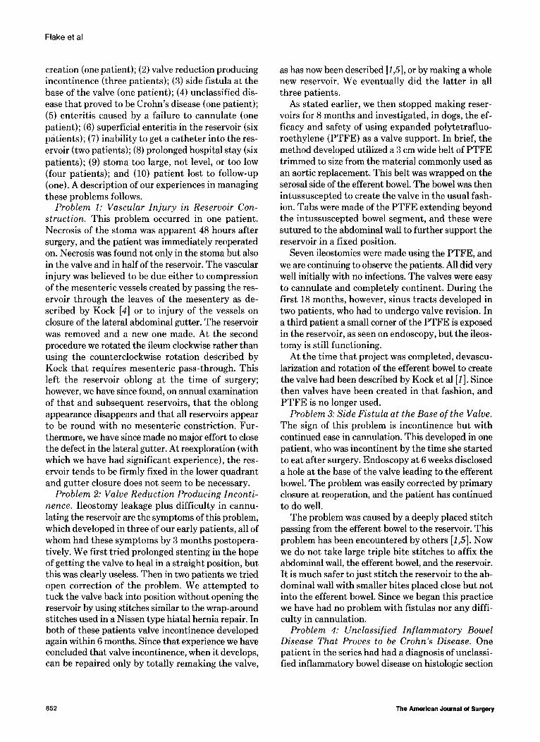
Figure 1. Plastic cannulas kept available for decompressing partially reduced valves. Top, firm plastic douche tip un- altered. Middle, douche tip reformed in heat for cannulating difficult valves. Bottom, standard Kock ileostomy cath- eter.
had sporadically incontinent valves that were diffi- cult to cannulate were doing moderately well while awaiting our decision to operate. They presented with painful overdistension of the reservoir and in- ability to get a catheter of any size through the valve. Retrograde injection of contrast medium with roentgenography was used to indicate the direction of the efferent bowel. In one of these patients a pe- diatric endoscope was threaded into the reservoir to decompress it. We could not see the lumen, but the fixed angle of the scope allowed its passage.
After that experience we developed a set of firm plastic cannulas with varying angles and curves for cannulating such patients (Figure 11. These cannulas are made from heat malleable douche catheters (Surgigator@). After they have been shaped by hand in dry oven heat, a 5 mm hole is drilled near the end. We have been able to cannulate even the most diffi- cult valves with these cannulas, and we keep a set available to all times.
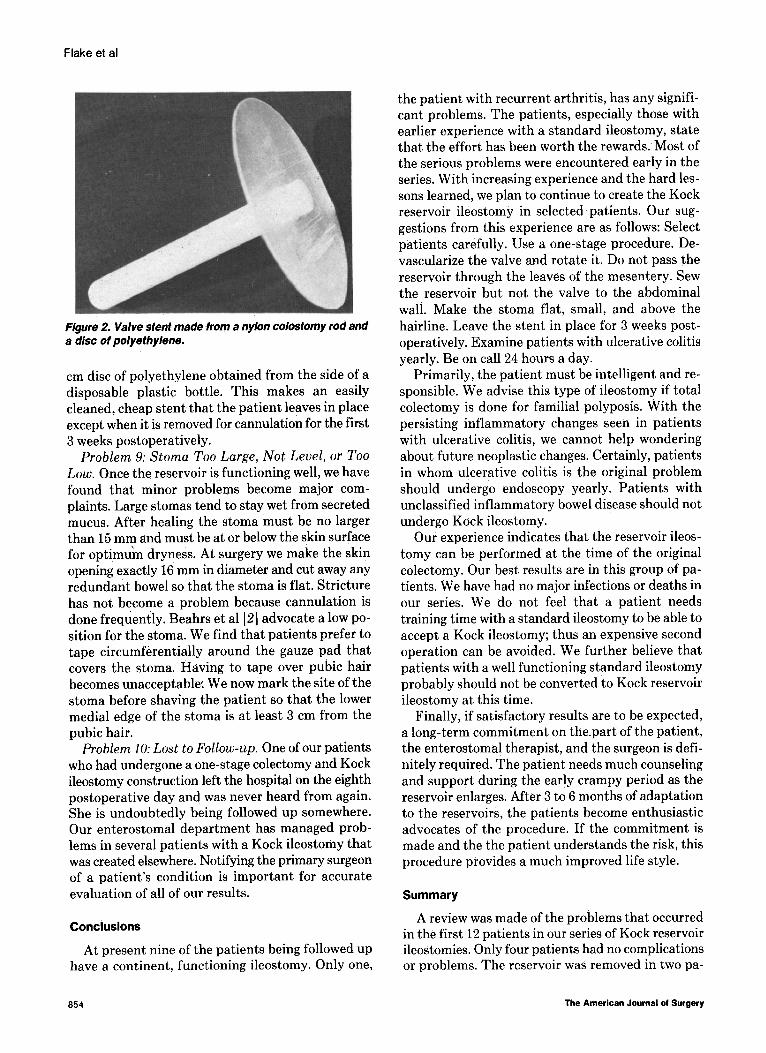
Problem 8: Prolonged Hospital Stay. Convinced of the need for prolonged valve stenting to make the valve heal in a proper position and to prevent valve reduction, as advocated by Gelernt et al [3], we adopted the practice of leaving a stent in place for 3 weeks postoperatively. This required long hospital stays or outpatient wearing of rubber catheters in the reservoir for that period of time. These alternatives were unacceptable to the patient and the financial office, so we developed a stent that could be com- fortably worn by outpatients (Figure 2). The stent is made from a standard nylon colostomy rod shortened to 10 cm. A shallow groove is cut near the end of the rod, which is wedged into a hole in the center of a 6
Volume 138, December 1979 853
Flake et al
Figure 2. Valve sient made from a nylon colostomy rod and a disc of polyethylene.
cm disc of polyethylene obtained from the side of a disposable plastic bottle. This makes an easily cleaned, cheap stent that the patient leaves in place except when it is removed for cannulation for the first 3 weeks postoperatively.
Problem 9: Stoma Too Large, Not Level, or Too Low. Once the reservoir is functioning well, we have found that minor problems become major com- plaints. Large stomas tend to stay wet from secreted mucus. After healing the stoma must be no larger than 15 mm and must be at or below the skin surface for optimu’m dryness. At surgery we make the skin opening exactly 16 mm in diameter and cut away any redundant bowel so that the stoma is flat. Stricture has not become a problem because cannulation is done frequently. Beahrs et al [2] advocate a low po- sition for the stoma. We find that patients prefer to tape circumferentially around the gauze pad that covers the stoma. Having to tape over pubic hair becomes unacceptable: We now mark the site of the stoma before shaving the patient so that the lower medial edge of the stoma is at least 3 cm from the pubic hair.
Problem 10: Lost to Follow-up. One of our patients who had undergone a one-stage colectomy and Kock ileostomy construction left the hospital on the eighth postoperative day and was never heard from again. She is undoubtedly being followed up somewhere. Our enterostomal department has managed prob- lems in several patients with a Kock ileostomy that was created elsewhere. Notifying the primary surgeon of a patient’s condition is important for accurate evaluation of all of our results.
Conclusions
At present nine of the patients being followed up have a continent, functioning ileostomy. Only one,
the patient with recurrent arthritis, has any signifi- cant problems. The patients, especially those with earlier experience with a standard ileostomy, state that the effort has been worth the rewards. Most of the serious problems were encountered early in the series. With increasing experience and the hard les- sons learned, we plan to continue to create the Kock reservoir ileostomy in selected patients. Our sug- gestions from this experience are as follows: Select patients carefully. Use a one-stage procedure. De- vascularize the valve and rotate it. Do not pass the reservoir through the leaves of the mesentery. Sew the reservoir but not the valve to the abdominal wall. Make the stoma flat, small, and above the hairline. Leave the stent in place for 3 weeks post- operatively. Examine patients with ulcerative colitis yearly. Be on call 24 hours a day.
Primarily, the patient must be intelligent and re- sponsible. We advise this type of ileostomy if total colectomy is done for familial polyposis. With the persisting inflammatory changes seen in patients with ulcerative colitis, we cannot help wondering about future neoplastic changes. Certainly, patients in whom ulcerative colitis is the original problem should undergo endoscopy yearly. Patients with unclassified inflammatory bowel disease should not undergo Kock ileostomy.
Our experience indicates that the reservoir ileos- tomy can be performed at the time of the original colectomy. Our best results are in this group of pa- tients. We have had no major infections or deaths in our series. We do not feel that a patient needs training time with a standard ileostomy to be able to accept a Kock ileostomy; thus an expensive second operation can be avoided. We further believe that patients with a well functioning standard ileostomy probably should not be converted to Kock reservoir ileostomy at this time.
Finally, if satisfactory results are to be expected, a long-term commitment on the.part of the patient, the enterostomal therapist, and the surgeon is defi- nitely required. The patient needs much counseling and support during the early crampy period as the reservoir enlarges. After 3 to 6 months of adaptation to the reservoirs, the patients become enthusiastic advocates of the procedure. If the commitment is made and the the patient understands the risk, this procedure provides a much improved life style.
Summary
A review was made of the problems that occurred in the first 12 patients in our series of Kock reservoir ileostomies. Only four patients had no complications or problems. The reservoir was removed in two pa-
854 The American Journal of Surgery
Kock lleostomy
tients and a new reservoir constructed in four. De- spite these problems, at 1 to 3 years all nine patients still being followed up have continent, well func- tioning ileostomies.
A prosthetic material (Impragrafta) was used to support the valve in seven cases. This worked well, but foreign body fistulas requiring stoma revision developed in two patients. Frequently, superficial mucosal ulceration and an inflammatory histologic reaction were observed in the reservoirs of patients who formerly had ulcerative colitis. These conditions were not observed in patients who had had familial polyposis. One patient with ulcerative colitis had a recurrence of arthritis.
Despite these problems, we believe that if one uses good patient selection, some new technical maneu- vers in constructing the reservoir, and some of the postoperative devices and practices developed during this experience, the Kock reservoir ileostomy should be constructed in the appropriate cases.
References
1. Kock NG, Darle N, Hulten L, Kewenter J, Myrvold l-l, Philipson B: Ileostomy. Cur Rob Surg 14: l-52, 1977.
2. Beahrs OH, Kelly KA, Adson MA, Chong GC: lleostomy with ileal reservoir rather than ileostomy alone. Ann Surg 179:634, 1974.
3. delernt IM, Bauer JJ, Kreel I: The reservoir ileostomy: early ex- perience with 54 patients. Ann Surg 185: 179, 1977.
4. Kock NG: Continent ileostomy. hog Surg 12: 180, 1973. 5. Palmu A, Sivula A: Kock’s continent ileostomy: results of 51
operations and experiences with correction of nipple-valve insufficiency. Br J Surg 65: 645, 1978.
Discussion
Kent C. Westbrook (Little Rock, AR): Two aspects of this paper se-m really important to me. First, it points out
the problems encountered when one undertakes a new procedure and second, it presents some innovative tech-
niques for dealing with these problems. In Little Rock, approximately 12 Kock pouches have
been constructed at the University Hospital or affiliated
hospitals. All of the pouches were for ulcerative colitis. The
staple technique was used in constructing about half of the
pouches, with satisfactory results. However, we believe that
the staple technique should not be used for nipple con-
struction. The manufacturer has modified the GIA car- tridge by removing the knife. We have used this cartridge for nipple formation and the staples pulled through, causing nipple failure. The complications we encountered
were essentially the same as those reported in this paper. Four patients were incontinent. and two have been reop-
erated on.
Three patients have had enteritis manifested by non-
specific ulceration in the pouch. This became intolerable
in one patient and the pouch had to be removed. The other two patients were treated with Azulfidine’B and cortico- steroid enemas into the pouch with good results.
Before undertaking this procedure, one absolutely must
look at the movies on technique, read the literature, and
go to the laboratory and work in animals. Even with this
preparation, problems will occur. Proper patient selection
and technique are crucial.
I agree almost entirely with the technique presented by
the authors. The mesenteric flip is confusing, and we do not
worry about it. We simply place the pouch in whatever position seems most appropriate. I see no reason to try to obliterate the gutter; we stopped doing that in colostomies
10 years ago and have had no problems. The intestine is simply brought to the skin and sutured. We do not suture to the fascia, and we have had no problem with sinus tract
formation or retraction. I would like to ask Dr. Flake how he would manage three
patients. First, if a patient with a functioning ileostomy
asks you to create a Kock pouch for him, would you do it?
Second, in a patient with toxic megacolon, what. procedure would you use? Third, in a patient with chronic ulcerative
colitis of 10 years’ duration who is in the hospital for an
elective colectomy, would you create a pouch?
William Flake (closing): In the patient, with a func- tioning ileostomy who just wants the pouch for cosmetic
reasons, we would not provide the Kock reservoir. In the
patient with toxic megacolon, of course, a single procedure
would not be used. If the patient with ulcerative colitis is
sufficiently motivated and intelligent, we would inform him
fully of the nuances of the procedure. If he still strongly requested it, we would perform the Kock ileostomy.
Volume 139, December 1979 a55