Embed Size (px)
Citation preview

www.ejbps.com
Ekundayo et al. European Journal of Biomedical and Pharmaceutical Sciences
32
PREVALENCE AND CHARACTERIZATION OF ENTEROCOCCAL INFECTIONS IN
ENUGU STATE, NIGERIA
1Ekundayo A. O., *
2Ezeah G. A. C.,
1Akpe R. A.,
3Odo O. F.,
4Ugwu M. C.,
5Ike O. C.,
4Amadi N. C. and
4Okuku C. N.
1Department of Microbiology, Ambrose Alli University, Ekpoma, Edo State, Nigeria.
2Department of Microbiology, Enugu State University Teaching Hospital Parklane, Enugu State, Nigeria. 3Department of Histopathology, Enugu State University Teaching Hospital Parklane, Enugu State, Nigeria.
4Department of Medical Laboratory Science, College of Health Sciences, Nnamdi Azikiwe University, PMB 5001
Nnewi, Anambra State, Nigeria. 5Department of Industrial Chemistry, Enugu State University of Science and Technology, Enugu State, Nigeria.
Article Received on 25/11/2018 Article Revised on 15/12/2018 Article Accepted on 05/01/2019
INTRODUCTION Enterococci are gram positive, catalase negative cocci
occurring singly or arranged in pairs or short chains, of
size 0.6-2.5 microns, facultative anaerobic (i.e. they are capable of cellular respiration in both oxygen-rich and
oxygen-poor environments.[1] Though, they are not
capable of forming spores, enterococci are tolerant of
high temperatures (10-45oC) with optimum growth at
35oC, pH (4.5-10) and high sodium chloride
concentrations of up to 6.5%.[2] They hydrolyse aesculin
in the presence of bile salts (Bile-aesculin Test). Because
of their ability to ferment carbohydrates to produce lactic
acid, the enterococci are referred to as typical lactic acid
bacteria. Enterococci typically exhibit gamma
haemolysis on sheep blood agar.[3]
Enterococci are usually perceived as commensal bacteria
that co-exist with their hosts as part of the normal flora.
While not particularly virulent, enterococci can be
significant agents of disease in the hospital setting where
patients’ defenses have been compromised by
catheterization, immune deficiencies or both.[4] Important
clinical infections caused by enterococci include urinary tract infection, bacteremia, bacterial endocarditis,
diverticulitis, meningitis and oesteomyelitis.[1] The
virulence of enterococci is lower than that of organisms
such as Staph. aureus.[5] However, enterococcal
infections often occur in debilitated patients and as part
of polymicrobial infections. These factors limit the
ability of investigators to determine the independent
contribution of enterococcal infections to mortality and
morbidity.
The direct microscopic examination of Gram-stained
smears of normally sterile clinical specimens such as blood may be useful for the diagnosis of enterococcal
infections. Direct examination of certain non-sterile
specimen may also be informative, but should not be
SJIF Impact Factor 4.918 Research Article
ejbps, 2019, Volume 6, Issue 2, 32-49.
European Journal of Biomedical AND Pharmaceutical sciences
http://www.ejbps.com
ISSN 2349-8870
Volume: 6
Issue: 2
32-49
Year: 2019
*Corresponding Author: Ezeah G. A. C.
Department of Microbiology, Enugu State University Teaching Hospital Parklane, Enugu State, Nigeria.
DOI: 10.20959/ejbps20192-6820
ABSTRACT Enterococcus sp. considered a normal commensal of the intestine of man and other animals, is fast emerging as a pathogen, causing serious and life threatening nosocomial infections. A two year progressive study of this
organism was carried out between July, 2012 and June, 2014 in Enugu metropolis and its environs in Eastern
Nigeria. A total of one thousand and eight clinical samples of urine, blood, sputum, stool, aspirates and swabs such
as high vaginal, urethral, ear, nasal, anal and wound samples and forty non-clinical samples of urine, urethral and
high vaginal swabs were examined. Isolation and identification were based on standard procedures and
biochemical tests. Nine organisms were isolated in this study: E. coli 152(24.1%), Streptococcus sp. 95(15%),
Enterococcus sp. 68(10.8%), Staph aureus 64(10.1%), Pseudomonas sp. 63(10.0%), Proteus sp. 63 (10.0%),
Klebsiella sp. 50(7.9%), Candida sp. 45(7.1%) and coagulase negative staph 32(5.1%). The prevalence of
Enterococcus sp was 10.8% and was the third most common cause of infection; the first and second being E. coli
and Streptococcus sp. respectively. Three species of Enterococcus were identified; E. faecium 39(57.4%), E.
faecalis 25(36.8%) and E. avium 4(5.9%). It was observed that the incidence of Enterococcus sp was high and showed multiple drug resistance; it is therefore advised that more attention should be given to this organism if
nosocomial infections and outbreaks are to be contained.
KEYWORDS: Enterococci, infection, prevelance, isolation and identification.

www.ejbps.com
Ekundayo et al. European Journal of Biomedical and Pharmaceutical Sciences
33
over-emphasized. However, only presumptive report of
the presence of gram-positive cocci should be made as
microscopy by itself cannot differentiate the enterococci
from other gram positive cocci. Culture and appropriate
identification techniques can be performed for
confirmation.[5] In the laboratory, enterococci are distinguished by their morphological appearance on
Gram stain and culture (gram-positive cocci that grow in
chains) and their ability to (i) hydrolyze esculin in the
presence of bile, (ii) their growth in 6.5% sodium
chloride, (iii) their hydrolysis of pyrrolidonyl
arylamidase and leucine aminopeptidase, and (iv) their
reaction with group D antiserum. Before they were
assigned their own genus, they were classified as group
D streptococci.[6] The work of Schierl and Blazevic[7]
demonstrated that up to 83% of Enterococcus could be
identified by the rapid litmus milk reduction test. No
false positives were observed. A negative result can be checked by culturing.
Trypticase soy-5% sheep blood agar, brain heart
infusion-5% sheep blood agar or any blood agar base
containing 5% animal blood supports the growth of
enterococci.[6] Enterococci grow well at 35-370C and do
not require an atmosphere containing increased CO2,
although some strains grow better in this atmosphere.[6]
Aesculin, bile and azide containing media are excellent
media for selective isolation of enterococci from
specimens likely to contain gram negative bacteria.
Once isolated, the genus Enterococcus is subjected to
confirmatory tests for speciation. AP1 20 strep
identification system is a tool used in speciation of
enterococci and streptococci. The test systems are stored
in reactive tubes. This involves 21 parameters that are
phenotypic characteristics of the species.
The present study was set out to assess the prevalence
and characterization of enterococcal infection in Enugu
Metropolis and environs in Eastern Nigeria.
MATERIALS AND METHODS
Study area
Samples for this study were sourced from:
Enugu State University of Technology (ESUT) Teaching
Hospital, Parklane in Enugu Metropolis (ancient coal
city) Enugu State, south east of Nigeria: This is a tertiary
health institution that receives direct and referred patients
from all the local government primary and secondary
health facilities in Enugu and environs.
University of Nigeria Teaching Hospital (UNTH), Ituku/Ozalla in Enugu State, Nigeria. The hospital is
situated in a boundary between Ituku in Awgu Local
Government Area of Enugu State and Ozalla in Nkanu
Local Government Area of Enugu State in eastern part of
Nigeria about 50 kilometers from Enugu metropolis. It
receives direct and referred patients from all the local
government areas of the Enugu State and the neighboring
Ebonyi, Abia, Imo, Benue and Kogi states. (see maps
below).
Study design: This is a cross-sectional study. Three
categories of patients were included in the study.
In-patients: 504 in-patients admitted in ESUT Teaching
Hospital and University of Nigeria Teaching Hospital
both in Enugu who submittedtheir samples of urine,
wound swabs, aspirates, sputum, ear swabs, high vaginal
swabs, urethral swabs, semen, CSF and blood to the
Microbiology Departments for microscopy, culture and
sensitivity.
Out-patients: 504 out-patients who visited ESUT
Teaching Hospital, Parklane and University of Nigeria
Teaching Hospital, Ituku/Ozalla and who submitted
clinical samples to the Microbiology Departments for microscopy, culture and sensitivity.
Controls: 20 male and 20 female volunteers who did not
have symptoms of any infection. They were selected
from outside the hospital environment and were used as
controls.
Ethical approval: Ethical approval was obtained from
ESUT Teaching Hospital, Parklane and University of
Nigeria Teaching Hospital, Ituku/Ozalla. Only patients
who gave their informed consent were recruited for the study.
Precautions/Considerations: Patients who were on
antibiotic therapy prior to sample collection were
excluded to avoid the influence of antibiotic on the
growth of the organism.
Samples were cultured within 2 hours of collection to
avoid overgrowth by contaminants.
For blood culture, site of venous puncture was cleaned
thoroughly with 70% alcohol to avoid contamination with skin flora.
For urine samples, patients were advised to collect
midstream urine in order to avoid the isolation of
contaminants from the outer surfaces of the urethra.
Female patients were advised to separate the labia before
passing the urine after proper cleaning with tissue paper
while male patients were also advised to clean the
urethral opening with sterile tissue paper.
Sample collection: Sterile universal containers
containing boric acid preservative were used for urine
sample collection while sputum, stool, aspirates and CSF
were collected with sterile plain universal bottles. Sterile
swabs were used to collect high vaginal, urethral, wound,
nasal, ear, anal sample. For blood culture, five milliliters
of blood was collected with syringe and put aseptically

www.ejbps.com
Ekundayo et al. European Journal of Biomedical and Pharmaceutical Sciences
34
into fifty milliliters of sterile brain heart infusion (BHI)
broth contained in a bijou bottle.
Analytical methods
Macroscopy: The colour and appearance of urine, CSF,
sputum, aspirates and semen were recorded.
Culture/Isolation Considerations The urine samples were inoculated into 5% sheep blood
agar and cystein lactose electrolyte deficient (CLED)
agar plates with quantitative sterile wire loops (0.001
ml). These were incubated for 24 hours at 370C
aerobically and microaerophilically by incubation in a
candle jar. An estimation of colony forming units per
millimeter was made and significant bacteriuria
established using a colony count of 100,000 (105) or
more per millimeter[8] for organisms growing in pure
culture. Sputum, aspirates, CSF, swabs and semen were inoculated in 5% sheep blood agar and cystein lactose
electrolyte deficient (CLED) agar with wire loop,
incubated for 24 hours at 37oC aerobically and
microaerophilically in a candle jar.
Blood culture: This was done by aseptically collecting
five millilters of blood from the patient with syringe and
aseptically inoculating same into fifty milliliters of brain
heart infusion broth incubated for 2 days at 37oC
aerobically and microaerophilically in a candle jar. A
loopful of blood culture was aseptically withdrawn and sub-cultured on 5% sheep blood agar and cystein lactose
electrolyte deficient (CLED) agar incubated for 24 hours
at 37oC aerobically. When applicable, the sub-culturing
was done for three times on 2-days intervals to establish
that there was no bacterial growth.
Microscopy Wet preparations of the urine samples were made by
centrifuging at 1000 rpm for 5 minutes. The deposit was
examined on glass slides covered with cover-slips with
x10 and x40 objectives of the microscope. Pus cells, red
blood cell, casts, crystals, yeast cells were recorded.
Wet preparations of urine deposit and other samples
were, made on glass slides, air dried and stained by
Gram’s Method.[9] Observations were made and
recorded.
Identification of organisms Colonial appearances and gram reactions[9] was the
initial steps taken to identify the isolates. Slightly raised
white colonies with a diameters of I-2 mm, non-
haemolytic (gamma reaction) on blood agar and gram positive cocci that often occured singly or pairs
(diplococci) or short chains were suggestive of
Enterococcus sp.[3] Other organisms were also classified
as gram positive or negative, lactose fermenters
(coliforms) or non-lactose fermenters, haemolytic or
non-haemolytic and speciated accordingly.
Gram Staining Technique Gram staining technique using method described by
Baker et al.[9] was carried out and recorded. This
differentiates bacteria into Gram-positive and Gram
negative based on the physical properties of their cell
walls. Staph aureus and E. coli were used as positive and negative controls respectively.
Motility Test
Hanging Drop slide
A drop of the bacterial suspension was placed on a clean
glass cover-slip as described by Cheesbrough.[11] This
was inverted over the well of a ground glass and sealed
with a ring of Vaseline ointment. The drop was observed
with x10 of the microscope. Motile bacteria were seen
actively moving while non-motile bacteria were not
actively moving. Pseudomonas aeruginosa and Staph
aureus were used as positive and negative controls respectively.
Biochemical Tests
The following distinguishing biochemical tests were
carried out to identify the organisms isolated in this
study.
These tests were carried out in accordance with standard
procedures described by Baker et al.,[9], Cheesbrough[10],
Devriese et al.,[12] and Cheesbrough.[11]
Catalase Test
Catalase test was carried out to differentiate isolates into
catalase positive and catalase negative using a method
described by Cheesbrough.[10] Catalase acts as a catalyst
in the breakdown of hydrogen peroxide to oxygen and
water A drop of hydrogen peroxide was placed on a
sterile microscope slide. With the use of a wire loop, a
bacteria colony was picked and smeared on the hydrogen
peroxide drop. The production of oxygen bubbles
(effervescence) indicated positive result while no
bubbles indicated negative result. Staph aureus and
Streptococcus pyogenes were used as positive and negative controls respectively.
Coagulase Test Coagulase test was used to identify Staph. aureus which
produces coagulase enzyme as described by
Cheesbrough.[12]
Slide Test Method (detects bound coagulase)
A drop of distilled water was placed on an end of a
sterile slide. A colony of the test organism was
emulsified in the drop. A loopful of plasma was added to the suspension and mixed gently. Clumping within 10
seconds indicated presence of bound coagulase. No
clumping within 10 seconds indicated no bound
coagulase present. Staph aureus and Staph epidermidis
were used as positive and negative controls respectively.
Tube Test Method (Detects free coagulase)
Three 5-ml test tubes were labelled:

www.ejbps.com
Ekundayo et al. European Journal of Biomedical and Pharmaceutical Sciences
35
T =Test organism (24 hour broth culture)
Pos = Positive control (24 hours Staph.aureusbroth
culture)
Neg = Negative control (24 hours Staph. epidermidis
broth culture)
With a pipette 0.2ml of plasma was measured into each
tube. Measured 0.8ml of test broth culture was added to
tube T. Measured 0.8ml of Staph. aureus and Staph
epidermidisbroth cultures were added to tubes labelled
‘Pos’ and Neg respectively. The three tubes were
incubated at 37oC after mixing for 24 hours. Clotting of
the T tube contents indicated presence of free coagulase.
No clotting indicated negative test.
Citrate utilization test
Slopes of Simmon’s citrate agar medium in bijou bottles
were prepared as described by Cheesbrough.[10] With a sterile straight wire, the test organism was first streaked
on the slope and then stabbed into the butt. This was
incubated at 37oC for 48 hours and observations
recorded.
Bright blue colour indicated positive citrate test while no
colour change indicated negative citrate test. Klebsiella
pneumoniaeand E. coliwere used as positive and
negative controls respectively.
Indole test This test is important in the identification of
enterobacteria which break down the amino acid
tryptophan with the release of indole. This was a method
described by Baker et al.[9] The test organism was
inoculated into bijou bottle containing 3 ml of tryptone
water, incubated at 37oC for 48 hours. 0.5 ml of Kovac’s
reagents was added and shaken gently.
Red surface layer indicated positive indole test while no
colour change indicated negative indole test. E. coli and
Enterobacter aerogeneswere used as positive and
negative controls respectively.
Oxidase test This test was used to identify Pseudomonas sp. which
produces the enzyme cytochrome oxidase. This was a
method described by Cheesbrough.[11]
A piece of filter paper was placed in a clean petri dish
and three drops of freshly prepared oxidase reagent
(phenylenediamine) was added. Using a piece of glass
rod, a colony of the test organism was removed and
smeared on the filter paper.
Blue purple colour within 30 seconds indicated positive
oxidase test while no blue purple colour indicated
negative oxidase test. Pseudomonas aeruginosa and E.
coli were used as positive and negative controls
respectively.
Voges-Proskauer test This test was used to detect acetoin production during
sugar fermentation in a bacterial broth culture and was
described by Cheesbrough.[11] A tube containing 5 ml of
Voges-Proskauer’s broth was inoculated with a pure
colony of the test organism and incubated at 37oC for 48 hours. After incubation, 1 ml of the VP broth culture was
measured into a clean test tube into which 0.6 ml of 5%
alpha-naphthol was added. This was followed by the
addition of 0.2 ml of 40% potassium hydroxide. The tube
was shaken to expose the medium to atmospheric oxygen
and allowed to stand for 10 to 15 minutes. Red colour
development indicated positive result while no red colour
indicated negative result. Klebsiella pneumoniae and E.
coli were used as positive and negative controls
respectively.
Methyl red test It is used to identify bacteria producing stable acid by
mechanism of mixed acid fermentation of glucose and
was described by Cheesbrough.[11]
The methyl red test is
used to identify enteric bacteria based on their pattern of
glucose metabolism. An isolate was inoculated into a
tube containing 5 ml of one percent glucose with sterile
loop. The tube was incubated at 37oc for 2 days. After
incubation, 2.4 ml of medium was transferred to another
tube. Five drops of the pH indicator methyl red was
added to the tube. The tube was gently rolled between
the palms to disperse the methyl red. The bacteria that metabolized pyruvic acid to other acids lower the pH to
4.2. At this pH, methyl red turned to red. The red colour
represented a positive test. The bacteria that metabolized
pyruvic acid to neutral end- points lower the pH of the
medium to only 6.0. A yellow colour represented a
negative test. E. coli and Enterobacter aerogenes were
used as positive and negative controls respectively.
Sugar fermentation tests The ability of different organisms to ferment certain
carbohydrates is used in their identification.[9] An
aqueous solution containing 1 gram of trypticase, 1 gram of carbohydrate, 0.5 gram of NaCl, and 0.0189 gram of
phenol red all in 100 ml of distilled water was prepared
in conical flask. This was sterilized at 115oc for 15
minutes. This was dispensed in screw capped bottles
containing Durham’s tubes in 5 ml aliquots. The
Durham’s tubes was inverted and filled completely with
the medium. Each broth was aseptically inoculated with
0.1 ml of 0.5 MacFarland standard of bacterial
suspension and incubated at 37oc for 24 hours. A change
in colour from red to yellow indicated organic acid
production but no colour change indicated no fermentation and no acid production. Any trapped bubble
of gas indicated gas production.
Litmus Milk Decolorization Test This was carried out as described by Cheesbrough[11] and
colour change from mauve to white or pale yellow was
suggestive of enterococci.
www.ejbps.com
Ekundayo et al. European Journal of Biomedical and Pharmaceutical Sciences
36
Measured 0.5ml of sterile litmus milk was inoculated
with heavy inoculums of the test organism and incubated
at 37oC for up to 4 hours.
White or pale yellow-pink colour indicated litmus milk
reduction while no colour change indicated negative litmus milk test. Enterococcus faecalis and
Streptococcus pyogenes were used as positive and
negative controls respectively.
Aesculin Hydrolysis
Bile aesculin test is based on the hydrolysis of aesculin
into glucose and esculetin (6, 7- dihydroxy-coumarin) by
a micro-organism that produces an enzyme aesculinase.
aesculetin reacts with an iron salt (ferric citrate) in the
medium to form a phenolic iron complex which produces
a dark brown or black/grey colour. This was performed
by a method described by Cheesbrough[11] by streaking the organism on a plate of bile-aesculin agar incubated at
37oC overnight. Colonies of organisms and their
surroundings turning black/grey indicates positive test.
No colour change means a negative test. Enterococcus
faecalis and Staph. aureus were used as positive and
negative controls respectively.
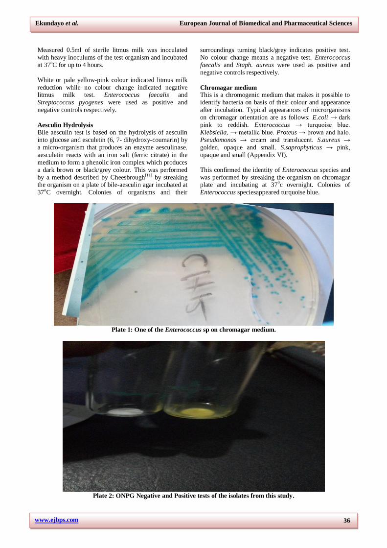
Chromagar medium This is a chromogenic medium that makes it possible to
identify bacteria on basis of their colour and appearance
after incubation. Typical appearances of microrganisms
on chromagar orientation are as follows: E.coli → dark
pink to reddish. Enterococcus → turquoise blue.
Klebsiella, → metallic blue. Proteus → brown and halo.
Pseudomonas → cream and translucent. S.aureus →
golden, opaque and small. S.saprophyticus → pink,
opaque and small (Appendix VI).
This confirmed the identity of Enterococcus species and
was performed by streaking the organism on chromagar plate and incubating at 370c overnight. Colonies of
Enterococcus speciesappeared turquoise blue.
Plate 1: One of the Enterococcus sp on chromagar medium.
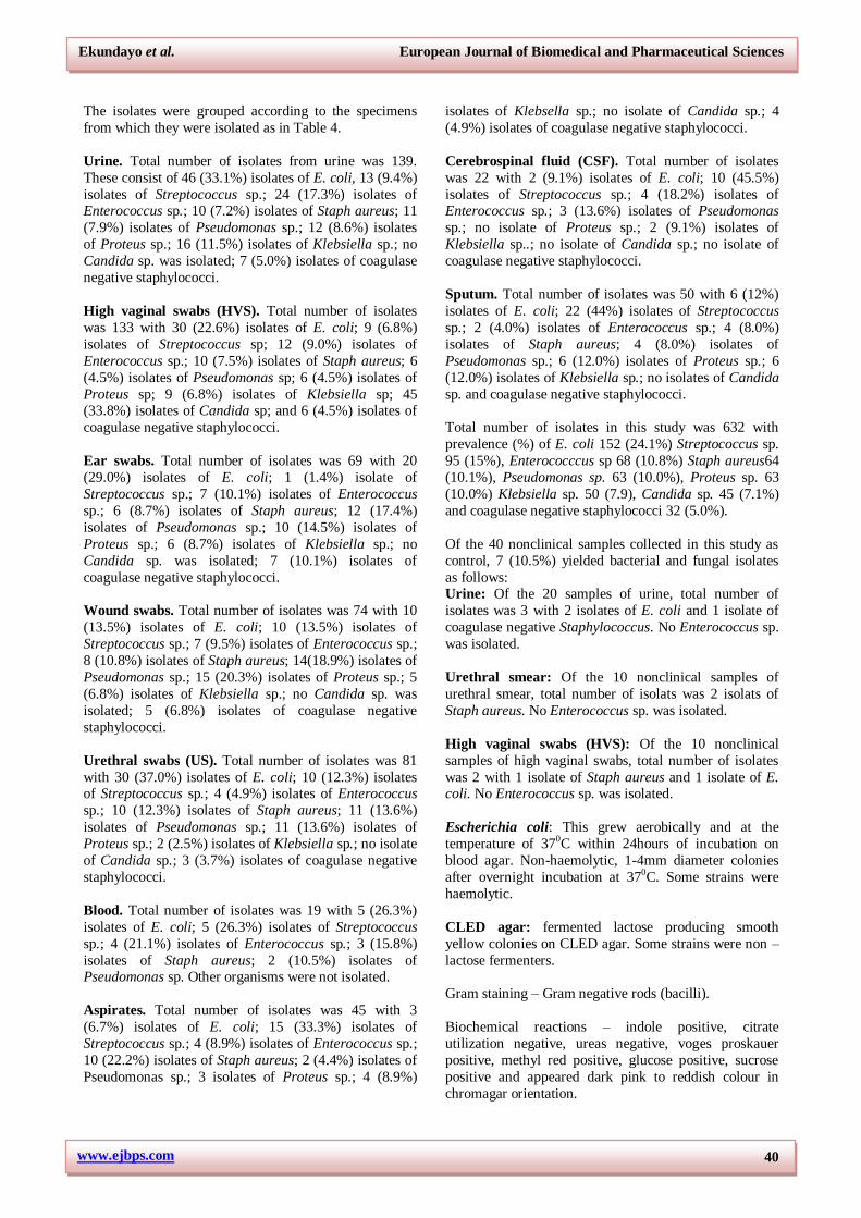
Plate 2: ONPG Negative and Positive tests of the isolates from this study.

www.ejbps.com
Ekundayo et al. European Journal of Biomedical and Pharmaceutical Sciences
37
Beta (β)-GALACTOSIDASE (ONPG) TEST
This is a test to identify the ability of the bacteia to
produce the enzyme beta-galactosidase and was
described by Cheesbrough.[11] Lactose utilization
requires a couple of enzymes, one of which is beta-
galactosidase. In this test, a molecular decoy called ONPG (Ortho-nitrophenyl-b-D-galactopyranoside) that
will turn to a yellow color in the presence of this enzyme
is used. ONPG is an anologue of lactose that the enzyme
can break down to produce a yellow coloured end-
product, O-nitrophenol. Since this enzyme is made only
in the presence of the lactose substrate, there was need to
be sure to grow this organism on media high in lactose.
E. coli and Proteus mirabilis were used as positive and
negative controls respectively.
Organism was made to be growing on lactose broth (to
induce the production of the galactosidase enzyme). Into a sterile tube, 0.5ml of saline was pipetted. Measured 0.1
ml of 0.5 Macfarlane standard suspension of the bacteria
was aseptically inoculated and the ONPG disc added
with sterile forceps to the tube. This was incubated at
37oC for 4 hours. The fluid and disc will turn any shade
of yellow if positive for galactosidase enzyme.
API 20 Strep by Biomerieux
This was for species identification[13] using the
identification software. Reference type E. faecalis strain
(ATCC 29212) was used as control. The API 20 strep strip consists of 20 microtubes containing dehydrated
substrates for the demonstration of enzymatic activity or
the fermentation of sugars. The enzymatic test strips
were inoculated with a dense suspension of organisms,
made from a pure culture, which was used to reconstitute
the enzymatic substrates. During incubation, metabolism
produced colour changes that were either spontaneous or
revealed by the addition of reagents. The fermentation
tests were inoculated with an enriched medium which
rehydrates the sugar substrates. Fermentation of
carbohydrates was detected by colour changes. The
reactions were read according to the reading table provided by Biomereieux and the identification was
obtained by using the identification software.
Interpretation Determination of the numerical profile: On the result
sheet, the 21 parameters which included the 20 tests and
haemolytic reaction (as the 21st parameter) were
separated serially into groups of three and a value 1, 2 or
4 was indicated for the 1st 2nd or 3rd positive parameter in
each group. By adding together the values corresponding
to positive reactions within each group, a seven digit profile number called numerical profile was obtained.
Identification: The identification was done using
database (V.7.0) with APIWEB identification software
provided by Biomereieux. The 7 digit numerical profile
was manually entered via the keyboard. The name of the
specie appeared on the screen as the specie identification.
This was recorded.
Conventional Biochemical Characterization
/identification of the isolates
Conventional biochemical phenotypic tests were carried
out to challenge and confirm the API 20 strep
identification of the isolates. Reference type E. faecalis
strain (ATCC 29212) was used as control. These include: Tests for sugars such as mannitol, sorbose, arabinose,
sorbitol, raffinose and sucrose. This was as described by
Faklam et al.[15] An aqueous solution containing 1 gram
of trypticase, 1 gram of carbohydrate, 0.5 gram of Nacl,
and 0.0189 gram of phenol red all in 100 ml of distilled
water was prepared in conical flask. This was sterilized
at 115oc for 15 minutes. This was dispensed in screw
capped bottles containing Durham’s tubes in 5 ml
aliquots. The Durham’s tubes was inverted and filled
completely with the medium. Each broth was aseptically
inoculated with 0.1 ml of 0.5 MacFarland standard of
bacterial suspension and incubated at 37oc for 24 hours. A change in colour from red to yellow indicated organic
acid production but no colour change indicated no
fermentation and no acid production. Any trapped bubble
of gas indicated gas production.
Arginine dehydrolase test: An inoculum of 0.1 ml of
0.5 MacFarland standard bacterial suspension from a
pure culture was transferred aseptically to 5ml sterile
arginine dehydrolase broth as described by Faklam et
al.[15] This was incubated for 24 hours at 37oc and re-
incubated for another 24 hours at 37oc. A change in colour from purple to yellow after the first incubation
and back to purple from yellow after the second
incubation indicated positive test for arginine
dehydrolase. Failure to turn yellow at 24 hours or to
revert back to purple at 48 hours indicated negative
result. Enterococcus sp. and Streptococcus sp. were used
as positive and negative controls respectively.
Motility test: This was carried out by hanging drop
method. A drop of the bacterial suspension was placed
on a clean glass cover-slip. This was inverted over the
well of a ground glass and sealed with a ring of Vaseline ointment. The drop was observed with x10 of the
microscope. Motile bacteria were seen actively moving
while non-motile bacteria were not actively moving.
Pseudomonas sp. and Enterococcus sp. were used as
positive and negative controls respectively.
Pigment production: This was observed on the solid
blood and CLED agar plates as colour change around the
colonies of the isolates as described by Faklam et al.[15]
Statistical analysis The results obtained from this work were analyzed
statistically using a computer program SPSS version 18.
The values were expressed in percentages.
RESULTS Wet preparation microscopic result of clinical specimens
that yielded growth of micro-organisms processed in this
study was displayed in table 1. Of the 139 samples of

www.ejbps.com
Ekundayo et al. European Journal of Biomedical and Pharmaceutical Sciences
38
urine, 125 (89.9%) had pus cells; 21 (15.1%) had red
blood cells; 34 (24.5%) had casts; 48 (34.5%) had
crystals; 89 (64.0%) had epithelial cells. There was none
(0%) with ova of schistosomes.
Of the 133 samples of high vaginal swabs (HVS), 108
(81.2%) had pus cells; none had red blood cells; 133
(100%) had epithelial cells; 45 (33.8%) had yeast cells.
Of the 22 sample of cerebrospinal fluid (CSF), 22
(100%) had pus cells; 5 (22.7%) had red blood cells.
Table 1: Result of wet preparation microscopy of the clinical samples that yielded organisms.
Clinical samples Pus cells RBCS Casts Crystals Epith
cells Yeast
cells Ova of
schistosomes Urine(n=139) 125(89.9) 21(15.1) 34(24.5) 48(34.5) 89(64) 0 (0) 0 (0) HVS (n=133) 108(81.2) 0 (0) NA NA 133(100) 45(33.8) NA CSF (n=22) 20(90.9) 2(22.7) NA NA NA NA NA
Key: RBCS= Red blood cells
Epith cells= Epithelial cells.
HVS= High vaginal swabs.
CSF= Cerebrospinal fluids.
NA= Not applicable.
Clinical samples of urine and CSF were grouped
according to the number of pus cells per high power field
(x40 objective lens) of light as shown in table 2. Of the
139 urine samples, 14 (10%) had pus cells 0-1 per high power field (/HPF); 26 (18.7%) had 2-3 pus cells PHF;
21 (15.1%) had 4-6 pus cells/HPF; 14 (10%) had 7-9 pus
cells/HPF; 20 (14. microscope 4%) had 10-13 pus
cells/HPF; 21 (15.1%) had many pus cells/HPF; 23
(16.5%) had numerous pus cells/HPF.
Table 2: Number of pus cells per high power field (X40 objective lens of light microscope) of the urine and CSF
wet mount.
Clinical samples 0 1-2 2-3 4-6 7-9 10-`14 15-20 21 and above Urine PC (n=139) 20(14.4) 14 (10) 26 (18.7) 21 (15.1) 14(10) 20 (14.4) 21 (15.1) 23 (16.5) Urine EC (n=139) 50(36.0) 10(7.3) 10 (7.3) 13 (9.3) 12 (8.6) 16 (11.5) 15 (10.8) 13 (9.3) CSF (n=22) 2(9.0) 1(4.5) 5(22.7) 3(13.6) 3(13.6) 4(18.2) 3(13.6) 1(4.5)
Key: CSF= Cerebrospinal fluid. PC= Pus cell. EC= Epithelial cell
Out of one thousand and eight (1008) samples processed
in this study, six hundred and thirty two (632) (58.5%)
yielded different species of bacteria and fungi, the
prevalence of which are displayed in Table 3 as follows:
E. coli 152 (24.1%), Streptococcus sp 95 (15%),
Enterococcus sp 68 (10.8%), Staph aureus 64 (10.1%),
Pseudomonas sp.63 (10.0%), Proteus sp. 63 (10.0%), Klebsiella sp 50 (7.9%), Candida sp. 45 (7.1%) and
Coagulase negative staphylococci 32 (5.1%).

www.ejbps.com
Ekundayo et al. European Journal of Biomedical and Pharmaceutical Sciences
39
Table 3: Characterization of organisms isolated from clinical samples during this study.
Gram rxn Catalase Coagulase LM AH Oxidase Citrate Urease VP Methyl
Red Indol Motility ONPG Glucose surose lactose co
6.5%
NACL Indentification total %
-rods NA NA NA NA NA V - + + + + + + + + Dark pink to
reddish - Escherichia sp 152 24.1
+cocci in
long chain - NA - - NA NA NA NA NA NA - NA NA NA + NA - Streptococcus sp 95 15
+cocci in
short chain - NA + + NA NA NA + NA NA - NA NA
+ Torquoise + Enterococcus sp 68 10.8
+cocci in
clusters + + NA NA NA NA NA NA NA NA - NA NA NA +
Golden
opaque small - Staph.aureus 64 10.1
-Rods NA NA NA NA + NA NA NA NA NA + NA NA NA - NA - Pseudomonas sp 63 10 -Rods NA NA NA NA - NA + NA NA + + NA NA NA - Brown halo - Proteus sp 50 10
-Rods NA NA NA NA - + + + - - - + + + + Dark pink to
reddish - Kiebsiella sp 45 7.9
+cocci in
clusters + - NA NA NA NA NA NA NA NA - NA NA NA -
Pink to
opaque small - Coagulase staph 32 7.1
+budding
yeast cells NA NA NA NA NA - NA NA NA NA - NA NA NA NA NA - Candida sp.
5.1
NA= Not applicable. + =Positive. - = Negative.
www.ejbps.com
Ekundayo et al. European Journal of Biomedical and Pharmaceutical Sciences
40
The isolates were grouped according to the specimens
from which they were isolated as in Table 4.
Urine. Total number of isolates from urine was 139.
These consist of 46 (33.1%) isolates of E. coli, 13 (9.4%)
isolates of Streptococcus sp.; 24 (17.3%) isolates of Enterococcus sp.; 10 (7.2%) isolates of Staph aureus; 11
(7.9%) isolates of Pseudomonas sp.; 12 (8.6%) isolates
of Proteus sp.; 16 (11.5%) isolates of Klebsiella sp.; no
Candida sp. was isolated; 7 (5.0%) isolates of coagulase
negative staphylococci.
High vaginal swabs (HVS). Total number of isolates
was 133 with 30 (22.6%) isolates of E. coli; 9 (6.8%)
isolates of Streptococcus sp; 12 (9.0%) isolates of
Enterococcus sp.; 10 (7.5%) isolates of Staph aureus; 6
(4.5%) isolates of Pseudomonas sp; 6 (4.5%) isolates of
Proteus sp; 9 (6.8%) isolates of Klebsiella sp; 45 (33.8%) isolates of Candida sp; and 6 (4.5%) isolates of
coagulase negative staphylococci.
Ear swabs. Total number of isolates was 69 with 20
(29.0%) isolates of E. coli; 1 (1.4%) isolate of
Streptococcus sp.; 7 (10.1%) isolates of Enterococcus
sp.; 6 (8.7%) isolates of Staph aureus; 12 (17.4%)
isolates of Pseudomonas sp.; 10 (14.5%) isolates of
Proteus sp.; 6 (8.7%) isolates of Klebsiella sp.; no
Candida sp. was isolated; 7 (10.1%) isolates of
coagulase negative staphylococci.
Wound swabs. Total number of isolates was 74 with 10
(13.5%) isolates of E. coli; 10 (13.5%) isolates of
Streptococcus sp.; 7 (9.5%) isolates of Enterococcus sp.;
8 (10.8%) isolates of Staph aureus; 14(18.9%) isolates of
Pseudomonas sp.; 15 (20.3%) isolates of Proteus sp.; 5
(6.8%) isolates of Klebsiella sp.; no Candida sp. was
isolated; 5 (6.8%) isolates of coagulase negative
staphylococci.
Urethral swabs (US). Total number of isolates was 81
with 30 (37.0%) isolates of E. coli; 10 (12.3%) isolates of Streptococcus sp.; 4 (4.9%) isolates of Enterococcus
sp.; 10 (12.3%) isolates of Staph aureus; 11 (13.6%)
isolates of Pseudomonas sp.; 11 (13.6%) isolates of
Proteus sp.; 2 (2.5%) isolates of Klebsiella sp.; no isolate
of Candida sp.; 3 (3.7%) isolates of coagulase negative
staphylococci.
Blood. Total number of isolates was 19 with 5 (26.3%)
isolates of E. coli; 5 (26.3%) isolates of Streptococcus
sp.; 4 (21.1%) isolates of Enterococcus sp.; 3 (15.8%)
isolates of Staph aureus; 2 (10.5%) isolates of Pseudomonas sp. Other organisms were not isolated.
Aspirates. Total number of isolates was 45 with 3
(6.7%) isolates of E. coli; 15 (33.3%) isolates of
Streptococcus sp.; 4 (8.9%) isolates of Enterococcus sp.;
10 (22.2%) isolates of Staph aureus; 2 (4.4%) isolates of
Pseudomonas sp.; 3 isolates of Proteus sp.; 4 (8.9%)
isolates of Klebsella sp.; no isolate of Candida sp.; 4
(4.9%) isolates of coagulase negative staphylococci.
Cerebrospinal fluid (CSF). Total number of isolates
was 22 with 2 (9.1%) isolates of E. coli; 10 (45.5%)
isolates of Streptococcus sp.; 4 (18.2%) isolates of Enterococcus sp.; 3 (13.6%) isolates of Pseudomonas
sp.; no isolate of Proteus sp.; 2 (9.1%) isolates of
Klebsiella sp..; no isolate of Candida sp.; no isolate of
coagulase negative staphylococci.
Sputum. Total number of isolates was 50 with 6 (12%)
isolates of E. coli; 22 (44%) isolates of Streptococcus
sp.; 2 (4.0%) isolates of Enterococcus sp.; 4 (8.0%)
isolates of Staph aureus; 4 (8.0%) isolates of
Pseudomonas sp.; 6 (12.0%) isolates of Proteus sp.; 6
(12.0%) isolates of Klebsiella sp.; no isolates of Candida
sp. and coagulase negative staphylococci.
Total number of isolates in this study was 632 with
prevalence (%) of E. coli 152 (24.1%) Streptococcus sp.
95 (15%), Enterococccus sp 68 (10.8%) Staph aureus64
(10.1%), Pseudomonas sp. 63 (10.0%), Proteus sp. 63
(10.0%) Klebsiella sp. 50 (7.9), Candida sp. 45 (7.1%)
and coagulase negative staphylococci 32 (5.0%).
Of the 40 nonclinical samples collected in this study as
control, 7 (10.5%) yielded bacterial and fungal isolates
as follows: Urine: Of the 20 samples of urine, total number of
isolates was 3 with 2 isolates of E. coli and 1 isolate of
coagulase negative Staphylococcus. No Enterococcus sp.
was isolated.
Urethral smear: Of the 10 nonclinical samples of
urethral smear, total number of isolats was 2 isolats of
Staph aureus. No Enterococcus sp. was isolated.
High vaginal swabs (HVS): Of the 10 nonclinical
samples of high vaginal swabs, total number of isolates
was 2 with 1 isolate of Staph aureus and 1 isolate of E. coli. No Enterococcus sp. was isolated.
Escherichia coli: This grew aerobically and at the
temperature of 370C within 24hours of incubation on
blood agar. Non-haemolytic, 1-4mm diameter colonies
after overnight incubation at 370C. Some strains were
haemolytic.
CLED agar: fermented lactose producing smooth
yellow colonies on CLED agar. Some strains were non –
lactose fermenters.
Gram staining – Gram negative rods (bacilli).
Biochemical reactions – indole positive, citrate
utilization negative, ureas negative, voges proskauer
positive, methyl red positive, glucose positive, sucrose
positive and appeared dark pink to reddish colour in
chromagar orientation.

www.ejbps.com
Ekundayo et al. European Journal of Biomedical and Pharmaceutical Sciences
41
Streptococcus sp
Morphology: These were gram positive cocci occurring
singly, in pairs, in short chains and also in long chains.
They are non- motile. Some species are capsulated.
Culture: Blood agar: produced beta haemolytic colonies on sheep blood agar. Colonies were small (0.5-1mm),
colourless, dry, shiny or mucoid some species are alpha
haemolytic or gamma haemolytic.
Biochemical tests: catalase negative, litmus milk
decoloration negative, ascending hydrolysis negative and
no growth at 6.5% sodium chloride concentration, PYR
(pyrrodonyl test) positive.
Enterococcus sp Morphology: gram positive cocci occurring in short
chains, singly or arranged in pairs. They are non-capsulate and non-motile.
Culture: Enterococci isolated were aerobic organisms
and grew optimally at 370C.
Blood agar: enterococci isolates were non haemolytic,
colonies appeared grey and mucoid about 2mm in
diameter.
CLED agar: enterococcal isolates appeared as small
yellow colonies on cysteine lactose electrolyte deficient agar. They grew in the presence of 6.5% sodium
chloride.
Bile Aesculin agar: black colonies were produced after
4 hours of incubation at 370C. Aesculin was hydrolysed
to aesculatein.
Biochemical tests: lactose fermentation positive aesculin
hydrolysis positive, litmus milk decoloration positive.
Chromagar orientation: turquoise blue colonies grew
on this plate. This confirms the identity of Enterococcus sp.
Staphylococcus aureus
Morphology: gram positive cocci occurring mostly in
clusters, non-motile and non-capsulate.
Culture: Staph aureus isolated grew well aerbically and
microaerophilically. Blood agar: yellow to cream and
occasionally white colonies of diameter 1-2mm size
produced after overnight incubation at 37oc. Thay are
non-haemoltic when grown aerobically and microaerophilically. Colonies were slightly raised and
easily emulsified. CLED agar: deep yellow colonies
were produced. Biochemical test: catalase positive,
coagulase positive.
Pseudomonas sp Morphology: these were gram negative, non-sporing
motile rods. Some strains were capsulate.
Culture: these grew aerobically and produced blue-
green pigments (pyocyanin) at the temperature of 370c.
Blood agar: the isolate yielded large, flat, spreading
colonies with serrated edges, haemolysis and dark
greenish blue pigment.
CLED agar: green colonies were produced on CLED
medium with less pigmentation than in blood agar.
Biochemical reactions: isolates of this organism were
oxidase positive. Produced acid only with glucose (no
gas).
Proteus sp Morphology: Isolates were actively motile, non-
capsulate, gram negative, pleomorphic rods.
Culture: Isolates were aerobic and grew well at 37oc
overnight.
Blood agar: Swarming (no discrete colonies) was
observed with characteristics ‘fishy’ smell.
CLED agar: Blue-grey translucent colonies were
observed. No swarming.
Biochemical test: Lactose fermentation negative, urea
positive, β-galactosidase (ONPG) negative, indole negative.
Klebsiella sp Morphology: These isolates were Gram negative, non-
motile and capsulated rods.
Culture: These isolates were aerobic and grew well at
37oc.
Blood agar: Colonies of Klebsiella sp. were large, grey-
white and mucoid.
CLED agar: Isolates appeared as large, yellow (lactose
fermenting) and mucoid colonies after 24 hours
incubation at 37oc.
Biochemical reactions: Lactose fermentation positive,
indole negative. Dark pink to reddish colour on
chromagar orientation.
Candida sp Morphology: Isolates were small, oval, often with
budding cells and gram positive.
Culture: Isolates were aerobic and grew well at 37oc.
Blood agar: cream coloured pasty colonies were
observed after 24 hours of incubation at 37oc. The
colonies had distinctive yeast smelling and budding cells
were easily seen by direct microscopy in stained or
unstained preparations.

www.ejbps.com
Ekundayo et al. European Journal of Biomedical and Pharmaceutical Sciences
42
Coagulase negative staph
Morphology: gram positive cocci occurring mostly in
clusters, non-motile, non-capsulate.
Culture: coagulase negative staph isolated grew well
aerobically and microaerophilically.
Blood agar: the colonies appeared white and non
haemolytic.
CLED medium: some colonies appeared yellow white
others appeared white.
Biochemical tests: catalase positive, coagulase negative.
Table 4: Specimens and number of organisms isolated (% in brackets).
Clinical samples TOTAL
SAMPLE
NO (n) E.coli Strept Entero
Staph. Aureus
Pseud Proteussp. Kleb.sp. Candidasp. CONS
Urine 139 46(33.1) 13(9.4) 24(17.3) 10(7.2) 11(7.9) 12(8.6) 16(11.5) 0(0) 7(5) HVS 133 30(22.6) 9(6.8) 12(9.0) 10(7.5) 6(4.5) 6(4.5) 9(6.8) 45(33.9) 6(4.5) Ear swab 69 20(29.0) 1(1.5) 7(10.1) 6(8.7) 12(17.4) 10(14.5) 6(8.7) 0(0) 7(10.1) Wound swab 74 10(13.5) 10(13.5) 7(9.5) 8(10.8) 14(18.9) 15(20.3) 5(6.8) 0(0) 5(6.8) U/S 81 30(37) 10(12.3) 4(4.9) 10(12.3) 11(13.6) 11(13.6) 2(2.5) 0(0) 3(3.7) Blood 19 5(26.3) 5(26.3) 4(21.1) 3(15.9) 2(10.5) 0(0) 0(0) 0(0) 0(0) Aspirates 45 3(6.7) 15(33.3) 4(8.9) 10(22.2) 2(4.4%) 3(6.7) 4(8.9) 0(0) 4(8.9) CSF 22 2(9.1) 10(45.5) 4(18.2) 3(13.6) 1(4.5) 0(0) 2(9.1) 0(0) 0(0) Sputum 50 6(12) 22(44) 2(4) 4(8) 4(8) 6(12) 6(12) 0(0) 0(0) Total 632 152 95 68 64 63 63 50 45 32 Prevalence(%) 100 24.1 15.0 10.8 10.1 10.0 10.0 7.9 7.1 5.0
Key:
HVS= High vaginal swab. U/S= Urethral swab. CSF= Cerebrospinal fluid.
E. coli = Escherichia coli. Sptrept= Sptreptoccocus sp. Enterococcus sp. Pseud= Pseudomonas sp.
CONS= Coagulase Negative Staphyloccoci sp. Kleb= Klebsiella sp.
Speciation using API 20 strept identification system
by Biomereux
API 20 strept result displayed on the Table 5 and
interpreted using the reading table provided by
Biomereux and control ATCC 29212 showed that three
species of Enterococcus were isolated in this study.
These are Enterococcus faecalis, E. faecium and E.
avium.
Enterococcus faecalis
The 21 parameters that were used in the table of identification and their results for E. faecalis were as
follows: Voges proskauer positive, Hippurate positive.
aesculin hydrolysis positive, pyrrodonyl arylamidase
positive, α-galactosidase negative, β-glucuronidase
negative, β- galactosidase positive, alkaline phosphatase
positive, leucine amino peptidase positive, arginine
dehydrogenase positive, D-ribose positive, L-arabinose
negative, D-mannose positive, D-sorbose positive, D-
lactose, D-trehalose positive, inulin negative, D-raffinose
negative, starch (amidon) positive, glycogen negative, β-
haemolysis negative. These parameters were separated
into groups of three according to manufacturer’s label and a value 1, 2 or 4 was indicated for each positive
reaction. By adding together the values corresponding to
positive reactions within each group, a seven digit profile
number 7173711 was obtained. The identification was
done using database (V.7.0) with APIWEB identification
software. The 7 digit numerical profile was manually
entered via the keyboard. The name of the specie
Enterococcus faecalis appeared on the screen as the
specie identification. This identification was supported
by conventional biochemical characteristics of the
isolates as displayed in Table 4 as follows: mannitol
positive, sorbose negative, arginine positive, arabinose
negative, sorbitol positive, raffinose negative, motility
negative, pigment negative, sucrose positive.
Enterococcus faecium
The 21 parameters that were used in the table of
identification and their results for Enterococcus faecium were as follows (see Table 5): voges proskauer positive,
hyppurate hydrolysis positive aesculin hydrolysis
positive, pyrrodonyl Aryalamidase positive, α-
galactosidase positive, β-glucuronidase negative, β-
galactosidase positive, alkaline phosphatase positive,
leucine amino peptidase positive, arginine
dehydrogenase positive, D-ribose positive, L-arabinose
positive, D-mannose positive, D- sorbose positive, D-
lactose positive, D-trehalose positive, inulin negative, D-
raffinose negative, starch (amidon) positive, glycogen
negative, β-haemolysis negative.
The numerical profile is 7377511. This was keyed into
the APIWED software and the identification was
Enterococcus faecium.
This identification was supported by conventional
biochemical characteristics of the isolates as displayed in
table 4 as follows: mannitol positive, sorbose negative,
www.ejbps.com
Ekundayo et al. European Journal of Biomedical and Pharmaceutical Sciences
43
arginine negative, arabinose positive, sorbitol positive,
raffinose negative, motility negative, pigment negative,
sucrose positive.
Enterococcus avium
The 21 parameters that were used in the table of identification and their results were as follows (see Table
5) voges proskauer positive, hyppurate hydrolysis
negative, aesculin hydrolysis positive, pyrrodonyl
arylamidase positive, α-galactosidase negative, β-
glucuronidase negative, β-galactosidase negative,
alkaline phosphatase positive, leucine amino peptidase
positive, arginine dehydrogenase negative, D-ribose
positive, L-arabinose positive, D-mannose positive, D-
sorbose positive, D-lactose positive, D-trehalose
positive, inulin negative, D-raffinose negative, starch
(amidon) positive, glycogen negative, β-haemolysis
negative.
The numerical profile is 5166710. This was keyed into
the APIWEB software and the identification was Enterococcus avium.
This identification was supported by conventional
biochemical characteristics of the isolates displayed in
Table 6 as follows: mannitol positive, sorbose positive,
arginine negative, arabinose positive, sorbitol positive,
raffinose negative, motility negative, pigment negative,
sucrose positive.
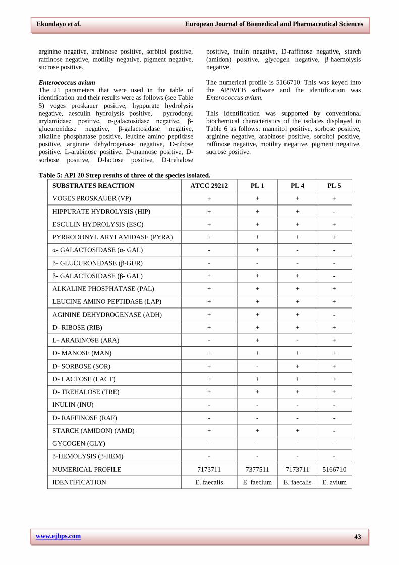
Table 5: API 20 Strep results of three of the species isolated.
SUBSTRATES REACTION ATCC 29212 PL 1 PL 4 PL 5
VOGES PROSKAUER (VP) + + + +
HIPPURATE HYDROLYSIS (HIP) + + + -
ESCULIN HYDROLYSIS (ESC) + + + +
PYRRODONYL ARYLAMIDASE (PYRA) + + + +
α- GALACTOSIDASE (α- GAL) - + - -
β- GLUCURONIDASE (β-GUR) - - - -
β- GALACTOSIDASE (β- GAL) + + + -
ALKALINE PHOSPHATASE (PAL) + + + +
LEUCINE AMINO PEPTIDASE (LAP) + + + +
AGININE DEHYDROGENASE (ADH) + + + -
D- RIBOSE (RIB) + + + +
L- ARABINOSE (ARA) - + - +
D- MANOSE (MAN) + + + +
D- SORBOSE (SOR) + - + +
D- LACTOSE (LACT) + + + +
D- TREHALOSE (TRE) + + + +
INULIN (INU) - - - -
D- RAFFINOSE (RAF) - - - -
STARCH (AMIDON) (AMD) + + + -
GYCOGEN (GLY) - - - -
β-HEMOLYSIS (β-HEM) - - - -
NUMERICAL PROFILE 7173711 7377511 7173711 5166710
IDENTIFICATION E. faecalis E. faecium E. faecalis E. avium

www.ejbps.com
Ekundayo et al. European Journal of Biomedical and Pharmaceutical Sciences
44
Table 6: Conventional biochemical characteristics of the isolates.
Phenotypic characteristics ATCC 29212 PL 1 PL 4 PL 5
Manitol + + + +
Sorbose - - - +
Arginine + + + -
Arabinose - + - +
Sorbitol + + + +
Sucrose + + + +
Motility - - - -
Pigment - - - -
Identification E.faecalis (control) E. faecium E. faecalis E. avium
Key: PL1… Parklane sample 1
PL4… Parklane Sample 4
PL5… Parklane sample 5
ATCC 29212 …American Type Culture Collection number 29212
Table 7 showed that 68 isolates of Enterococcus were speciated into 3 species using the API 20 Strept
Identification system.
1. E. faecium with numerical profile of 7377511 had
39 isolates and percentage of 57.35%.
2. E. faecalis with numerical profile of 7173711 had
25 isolates and percentage 36.76%
3. E. aviumwith numerical profile of 5166710 had 4 isolates and 5.88%.
These three isolates were confirmed, using conventional
biochemical methods of characterization (table 6).
Table 7: Summary of the API 20 strep identification of the isolates.
NUMERICAL PROFILE IDENTIFICATION NO OF ISOLATES PERCENTAGES
7377511 E. faecium 39 57.35
7173711 E. faecalis 25 36.76
5166710 E. avium 4 5.88
TOTAL 68 100
Figure 1: Shows the number and prevalence of organisms isolated from the study.

www.ejbps.com
Ekundayo et al. European Journal of Biomedical and Pharmaceutical Sciences
45
DISCUSSION Enterococci have evolved from normal commensal to
leading pathogens that cause infections in humans.[17]
Infections caused by enterococci are endocarditis,
bacteremia, urinary tract, hospital, infants and central
nervous system infections.[16, 18]
The presence of pus cells in 125 (89.4%) urine samples
out of 139 urine samples that yielded bacterial growth is
typical of urinary tract infections. Al-Saimary et al.
(2006)[19] reported a higher percentage of 93.3% in a
cytological examination of 105 urine specimens from
patients with urinary tract infections. These reports agree
with the report of Fowler (1986)[20] which states that the
clinical features of urinary tract infections include
bacteriuria, pyuria, haematuria, burning sensation on
urination frequent and urgent urination. Haematuria was
also noted in 21 (15.1%) urine samples out of the 139 urine samples that yielded bacteria. Al- Saimary et
al.(2006)[19] reported a higher percentage of 39% urine
samples with red blood cells.
Granular casts were noted in 34 (24.5%) urine samples
out of 139 samples. Al- Saimary et al.(2006)[19] reported
a close but higher percentage of 28.8% urine samples
with granular casts out of 105 urine specimens of
patients with urinary tract infections. This usually occurs
when urinary tract infection has progressed from the
urinary bladder through the urethras to the pelvis and calyces of the kidneys. This leads to a case of
pyelonephritis which is associated with morbidity.[20]
Complications of pyelonephritis include papillary
necrosis with possible development of urethral
obstructions, septic shock, perinephric abscess, scarring
with diminished renal function or renal failure.[20]
Calcium oxalate crystals were also present in 48 (34.5%)
urine samples out of 139 urine samples. This agrees
roughly with 35.2% urine samples reported by Al-
Saimary et al.(2006).[19] Calcium oxalate and calcium
sulphates were found to be associated with kidney stones of both humans and rats (Kahn, 1997).[21]
Epithelial cells were present in 89 (64.0%) urine samples
out of 139 samples. Al- Saimary et al.(2006)[19] reported
a higer percentage of 81.9% urine specimens of patients
with urinary tract infections. Of the 139 urine samples in
this study, 13(9.4%) had epithelial cells in the range of
21 and above per high power field. Epithelial cells are
cells that form thin surface coating on the outside of a
body structure. The normal range of epithelial cells in
urine 15-20 per high power field and if they are higher in urine they can signal a problem with the kidneys or an
infection in the urinary system.[22] They established that
the presence of epithelial cells has significance in
relation to urethritis and Chlamidia trachomatisand
Neisseria gonorrhoeaeinfections.
The wet mount microscopy of urine included a search for
ova of Schistosoma haematobiumbecause the study areas
involved tertiary institutions that received patients from
neighbouring Ebonyi State which is endemic for
schistosomiasis. However, no ova of schistosomes were
seen and recorded during the study. In contrast,
prevalence rates of S. haematobiumamong school
children in Alaukwu and Onicha communities of Ebonyi State, South East Nigeria was 47.9% and 11.0%
respectively.[23]
The wet mount of high vaginal swab (HVS) showed a
high level of pus cells signaling infection. Of the 133
HVS samples, 108 (81.2%) had pus cells and all the 133
(100%) had epithelial cells, 45 (33.8%) showed yeast
cells and eventually yielded Candida sp. This is
comparable to the work of Sevitha et al. (2012).[24] who
recorded pus cells in 85% of cases of vaginal discharge,
epithelial cells in 100% of cases of vaginal discharge and
yeast cells in 35% of cases.
Clinical samples of urine and CSF were grouped
according to the number of pus cells per high power field
to establish significant values. Normal range of pus cells
in urine is 5-8 in males and up to 10 in females.[20] The
data showed that 64 (46%) of 139 urine samples had
significant pyuria. Al-Saimary et al. (2006)[19] reported a
higher percentage of 93.3% in a cytological examination
of 105 urine specimens from patients with urinary tract
infections.
Of the 22 samples of cerebrospinal fluid (CSF), 20
(90.9%) had pus cells; 5 (22.7%) had red cells which
could be due to traumatic tapping during lumber
puncture since the samples were not xanthochromatic.
Normal range of pus cells in CSF is 0-1/hpf[25], meaning
that 20 (90.9%) of the 22 CSF samples had significant
number of pus cells/hpf. This agrees approximately with
the work of Kulkarni (2016).[26] who established that 65
(80.2%) of 81 children who had bacterial meningitis had
pus cells range of 2-25 per high power field.
Isolation and characterization A total of 1008 samples were processed in this study and
632 (58.5%) yielded different species of bacteria and
yeast cells. Using Gram staining and other biochemical
methods these isolates were characterized into nine (9)
genera of bacteria and yeast cells. These organisms and
their prevalence are as follows: E. coli 152 (24.1%),
Streptococcus sp. 95 (15%), Enterococcus sp. 68
(10.8%), Staphylococcus aureus 64 (10.1%),
Pseudomonas sp. 63 (10.0%), Proteus sp. 63 (10.0%),
Klebsiella sp. 50 (7.9%), Candida sp. 45 (7.1%) and
coagulase negative staphylococci 32 (5.1%).
The 40 non-clinical samples collected from volunteers
which included 20 urine samples, 10 urethral smear
samples and 10 high vaginal swab samples did not yield
any Enterococcus sp. However, Streptococcus sp., Staph
aureus, coagulase negative staphylococci, E. coli and
Candida sp. were isolated. The absence of Enterococcus
sp. could be because these samples were not from the

www.ejbps.com
Ekundayo et al. European Journal of Biomedical and Pharmaceutical Sciences
46
sites where they exist as normal flora. The organism is
known to be normal flora of the alimentary canal of man
and animals.[27]
The isolates were grouped according the clinical samples
from which they were isolated.
URINE: 139 clinical samples of urine yielded different
bacteria as follows: E. coli 46 (38%), Streptococcus sp.
13 (9.4%), Enterococcus sp. 24 (17.3%), Staphylococcus
aureus 10 (7.2%), Pseudomonas sp. 11 (7.9%), Proteus
sp. 12 (8.6%), Klebsiella sp. 16 (11.5%), coagulase
negative staphylococci 7 (5.0%) and no Candida sp. was
isolated from urine. This result is in line with the work of
Nwadioha et al., (2010)[28] who undertook a prevalence
study in Nguru, Northern Nigeria of Uropathogenic
microorganisms. The same set of organisms were
isolated by them except that there were minor differences in their prevalence and that Salmonella was isolated in
theirs but not isolated in this study. A similar study was
carried out in India which also showed E. coli as
predominant 50% among the isolates.[29] Another study
that was carried out in Sagamu, Western Nigeria among
pregnant and non-pregnant women recorded 26.6% and
5.6% prevalence respectively of significant bacteriuria
(Olusanya et al., 1992).[30] Similar findings were also
observed by Onyemelukwe et al. (2003)[31] in Enugu,
Eastern Nigeria. From the above research works, it is
obvious that urinary tract infection is a serious disease that deserves attention by researchers since it is
encountered in virtually all levels of health care
provision in Nigeria and beyond. It is interesting to note
that Enterococcus sp. is the second leading cause of
urinary tract infection in this study just as reported by
Malani et al. (2002)[31] that Enterococcus sp. has become
the second leading cause of urinary tract infection,
wound infections and bacteremia in USA.
High vaginal swabs (HVS). 133 clinical samples of
HVS yielded isolates as follows: 30 (22.6%) isolates of
E. coli; 9 (6.8%) isolates of Streptococcus sp.; 12 (9.0%) isolates of Enterococcus sp., 10 (7.5%) isolates of Staph
aureus; 6 (4.5%) isolates of Pseudomonas sp.; 6 (4.5%)
isolates of Proteus sp.; 9 (6.8%) isolates of Klebsiella
sp.; 45 (33.8%) isolates of Candida sp.; and 6 (4.5%)
isolates of coagulase negative staphylococci. Candidiasis
is the most predominant vaginal infection in this study.
This agrees with the report of Corsello, (2003)[33] that in
the second half of the twentieth century, the incidence of
vaginal candidiasis increased dramatically and that it is
estimated that 75% of women experience at least one
episode of vulvovaginal candidiasis during their childbearing age, and approximately 40% experience a
second attack. The second leadingcause of vaginal
infection in this study is E. coli (22-.6%) followed by
Enterococcus sp. (9.0%).
Ear swabs. Total number of isolates was 69 with 20
(29.0%) isolates of E. coli; 1 (1.4%) isolate of
Streptococcus sp.; 7 (10.1%) isolates of Enterococcus
sp.; 6 (8.7%) isolates of Staph aureus; 12 (17.4%)
isolates of Pseudomonas sp.; 10 (14.5%) isolates of
Proteus sp.; 6 (8.7%) isolates of Klebsiella sp.; no
Candida sp. was isolated; 7 (10.1%) isolates of
coagulase negative staphylococci. The above result is
comparable to clinical studies conducted from 1998 to 2000, during which microbiology specimens were
collected from 2039 subjects by 101 investigators
throughout the United States. A total of 2838 bacteria, 32
yeast cells, and 17 molds were recovered from 2048 ear.
Of the 202 bacterial species recovered, the species most
frequently isolated was Pseudomonas aeruginosa (38%).
The next 10 species most frequently isolated were:
Staphylococcus epidermidis, 9.1%; Staphylococcus
aureus, 7.8%; Microbacterium otitidis, 6.6%;
Microbacterium alconae, 2.9%; Staphylococcus caprae,
2.6%; Staphylococcus auricularis, 2.0%; Enterococcus
faecalis, 1.9%; Enterobacter cloacae, 1.6%; Staphylococcus capitissub sp. ureolyticus, 1.4%; and
Staphylococcus haemolyticus, 1.3%.[34]
Wound swabs. Total number of isolates was 74 with 10
(13.5%) isolates of E. coli; 10 (13.5%) isolates of
Streptococcus sp; 7 (9.5%) isolates of Enterococcus sp.;
8 (10.8%) isolates of Staph aureus; 14(18.9%) isolates of
Pseudomonas sp.; 15 (20.3%) isolates of Proteus sp.; 5
(6.8%) isolates of Klebsiella sp.; no Candida sp. was
isolated; 5 (6.8%) isolates of coagulase negative
staphylococci. Wound infection with enterococci is by direct contamination of wound with infected materials.
The clinical features include inflammation and pus
production.[35] In a 3 years study in Delhi, India, the
prevalence of wound infections due to Enterococcus sp.
was 8.6%[36] which is lower than the 10.8% (prevalence)
of this study.
Urethral swabs (US). Total number of isolates was 81
with 30 (37.0%) isolates of E. coli; 10 (12.3%) isolates
of Streptococcus sp; 4 (4.9%) isolates of Enterococcus
sp; 10 (12.3%) isolates of Staph aureus; 11 (13.6%)
isolates of Pseudomonas sp; 11 (13.6%) isolates of Proteus sp; 2 (2.5%) isolates of Klebsiella sp; no isolate
of Candida sp; 3 (3.7%) isolates of coagulase negative
staphylococci. Urethral swabs are used to collect samples
from the urethra to investigate the cause of urethritis
which is inflamation of the urethra. Enterococci have
been implicated in 10% of all urinary tract infections and
up to approximately 16% of nosocomial urinary tract
infections.[32]
Blood. Total number of isolates was 19 with 5 (26.3%)
isolates of E. coli; 5 (26.3%) isolates of Streptococcus sp.; 4 (21.1%) isolates of Enterococcus sp.; 3 (15.8%)
isolates of Staph aureus; 2 (10.5%) isolates of
Pseudomonas sp. Other organisms were not isolated. It is
noteworthy that enterococci are the third leading cause of
bacteraemia in this study and this is in line with the
report that in 2005, there were 7066 reported cases of
bacteraemia caused by Enterococcus species in the UK,
an 8% increase from 2004 and twenty eight per cent of

www.ejbps.com
Ekundayo et al. European Journal of Biomedical and Pharmaceutical Sciences
47
all cases were antibiotic resistant.[37] The risk of death
from vancomycin-resistant enterococci (VRE) is 75%,
compared with 45% for those infected with a susceptible
strain.[37] These figures were the same as in the USA.
Over a 15 year period there was a 20-fold increase in
VRE associated with nosocomial infections reported to CDC’s National Nosocomial Infections Surveillance
(NNIS).[38]
Aspirates. Total number of isolates was 45 with3 (6.7%)
isolates of E. coli; 15 (33.3%) isolates of
Streptococcussp.; 4 (8.9%) isolates of Enterococcus sp.;
10 (22.2%) isolates of Staph aureus; 2 (4.4%) isolates of
Pseudomonas sp.; 3 isolates of Proteus sp.; 4 (8.9%)
isolates of Klebsella sp.;. no isolate of Candida sp.; 4
(4.9%) isolates of coagulase negative staphylococci. In a
study of diabetic feet using needle aspiration, mixed
aerobic and anaerobic organisms were isolated. The most frequently isolated organisms were enterococci (29.8%),
anaerobic streptococci (25.6%) and species of Proteus
(22.5%), Clostridium (9.8%) and Bacteroides (.2%).[39]
Cerebrospinal fluid (CSF). Total number of isolates
was 22 with 2 (9.1%) isolates of E. coli; 10 (45.5%)
isolates of Streptococcus sp.; 4 (18.2%) isolates of
Enterococcus sp.; 3 (13.6%) isolates of Pseudomonas
sp.; no isolate of Proteus sp.; 2 (9.1%) isolates of
Klebsiella sp.; no isolate of Candida sp.; no isolate of
coagulase negative staphylococci. Enterococci are unusual etiologic agents of bacterial meningitis. In a
review of 151 cases of nosocomial meningitis,
enterococci accounted for only 3% of the cases.[40]
Enterococcal meningitis tends to occur in patients with
chronic medical conditions that are often associated with
the use of immunosuppressive therapy, underlying
central nervous system disease (trauma, surgery, and
epidural catheter), gastrointestinal pathology and
Strongyloides species.[41] Also an association of E.
faecium meningitis with Strongyloides hyperinfection
has been reported.[41] The presumed pathogenesis is
enterococcal bacteraemia originating from the gastrointestinal tract with secondary seeding of the
meninges (Cappello and Hotez, 1993).[42]
Sputum. Total number of isolates was 50 with 6 (12%)
isolates of E. coli; 22 (44%) isolates of Streptococcus
sp.; 2 (4.0%) isolates of Enterococcus sp.; 4 (8.0%)
isolates of Staph aureus; 4 (8.0%) isolates of
Pseudomonas sp.; 6 (12.0%) isolates of Proteus sp.; 6
(12.0%) isolates of Klebsiella sp.; no isolates of Candida
sp. and coagulase negative staphylococci. Respiratory
tract infections rarely result from enterococci.[43] Enterococcal pneumonia has been reported as a
nosocomial infection in severely debilitated patients
receiving long-term antibiotic therapy.[44] Empyema due
to E. faecalis is also uncommon. In three separate
retrospective reviews of empyema.[45] and one
prospective study of empyema in cirrhosis[46], 30 cases of
enterococcal empyema were reported. A specific source
of infection was found in two cases of E. faecalis
empyema - one in a patient with endocarditis and splenic
abscess[47] and one related to an esophagopleural fistula
after pneumonectomy.[48]
Prevalence of infection caused by Enterococcus
species The prevalence of infection caused by Enterococcus
species in this study was 10.8% and enterococcal
infection was also the third leading cause of infection.
Among the species identified, E. faecium showed the
highest proportion of 39(57.4%) followed by E. faecalis
25(36.8%) and then E. avium 4(5.88). This is in line with
the work of Murray, (1990)[14] reporting that
Enterococcus faecalis and Enterococcus faecium are the
most prevalent species cultured from humans;
accounting for more than 90% clinical isolates. Hidron et
al. (2008)[49] also reported that between 1978 and 2008,
Enterococcus faecium has emerged as a leading cause of enterococcal infection in the United States. Because they
produce bacteriocins, Enterococcus species have been
used widely over the last decade in the food industry as
probiotics or as starter cultures.[50] However, the
difference between an enterococcal pathogen and an
apparently safe food use strain is unclear and the
potential for the latter to acquire virulent factors by gene
transfer has been demonstrated.[51] Recent studies have
pointed out that E. faecium and E. faecalis might be
potential recipients of vancomycin resistance genes, and
consequently, the FAO/WHO (2001)[52] have recommended that E. faecium should not be considered
as probiotics for human use. Recently, enterococci have
become one of the most common nosocomial pathogens,
with patients having a high mortality rate of up to
61%.[53]
CONCLUSION
It was observed from this study that the incidence of
Enterococcus sp was high. It is therefore, advised that
more attention should be given to this organism.
Laboratories should establish protocols for identification
of these organisms. Public enlightenment should be put in place by public health departments.
ACKNOWLEDGEMENTS
Our unreserved gratitude goes Prof. A.O. Ekundayo, Dr.
A.R Akpe and all those who contributed to the success of
this research and presentation of this manuscript.
REFERENCES
1. Fischetti, V. A., Novick, R. P., Ferretti, J. J.,
Portnoy, D. A., and Rood, J.I. (Ed) (2000). Gram-
Positive pathogens. A. S. M. press, Washington D.C., 453-631.
2. Fisher, K. and Philips, C. (2009). ‘The ecology,
epidemiology and virulence of Enterococcus.
Microbiology, 155(6): 1749 – 1757.
3. Ryan, K. J. and Ray, C. G. (Ed). (2004). Sherris
medical microbiology (4th Ed.). MC Graw Hill,
London, 294-295.

www.ejbps.com
Ekundayo et al. European Journal of Biomedical and Pharmaceutical Sciences
48
4. Anderson, D. J., Goldstein, L. B., and Wilkinson, W.
E. (2003). Stroke location, characterization, severity
and outcome in mitral versus aortic valve
endocarditis. Neurology, 61: 1341-1346.
5. DiazGranados. C. A., Zimmer, S. M. and Klein, M.
(2005). Comparison of mortality Associated with vancomycin-resistant and vancomycin-susceptible
enterococcal bloodstream infections: a meta-
analysis. Clinical Infectious Diseases, 41(3):
327-333.
6. Murray, P. R., Ellen, J. O., Baron James H.,
Jorgensen, M. L. and Michael A. P. (2007). Manual
of clinical microbiology 9th ed. A. S. M. Press
Washington D. C. 20036-29041.
7. Schierl, E, A. and Blazevic, D. J. (1981). Rapid
identification of enterococci by reduction of litmus
milk. Journal of clinical microbiology, 14(2):
227-228. 8. Kass, E. H. (1956). Asymptomatic infections of the
urinary tract. Transactions of The Association of
American Physicians, 69(2): 56-65.
9. Baker, F. J., Silverton and Kilshaw. (1985).
Introduction to medical laboratory Technology 5th
ed. Butterworth. London, 251-289.
10. Cheesbrough, M. (1991). Collection and
transportation of Specimens. Examination of
specimens. 1n: Medical Laboratory Manual for
Tropical Countries. Cambridge University press,
UK., 100 - 156. 11. Cheesbrough, M. (2006). Biochemical tests to
identify bacteria. In: District laboratory practice in
Tropical countries part 2. Cambridge university
press, UK., 62-70.
12. Devriese, L. A., Collins, M. D., Wirth, R. (1992).
The genus Enterococcus: In The prokaryotes: A
handbook on the biology of Bacteria,
Ecophysiology, Isolation, and Identification
Applications. 2nd Edition, 2: 1465-1481.
13. Diana-Roxana, P., Elena, S., Mariana Carmen, C.
Ileana, S., Ana-Maria, N., Ionela, A., Floarea S. and
Tatiana, D. (2009). Isolation and identification of some Lactobacillus and Entercoccus strains by a
polyphasic taxonomical approach. Romanian
Biotechnological Letters, 14(2): 4225-4233.
14. Murray, B. E. (1990). The life and times of
Enterococcus. Clinical Microbiology Review, 3(1):
46–65.
15. Facklam, R. R., Carvalho, M. G. S and Teixara, L.
M. (2002). History, Taxonomy, biochemical
characteristics and antibiotic susceptibility testing of
enterococci. P. 1-54; In: M. S. Gilmore, D. B.
Clewell, P., Courvalin, G.M., Dunny, B.E., Murray and L.B. Rice. (Ed). The enterococci; pathogenesis,
molecular biology and antibiotic resistance. A. S. M.
Press, Washington D.C.
16. Adesida, S.A., Ezenta, C.C., Adagbada, A.O.,
Aladesokan, A.A. and Coker, A.O. (2017): Carriage
of multidrug resistant enterococcus faecium and
enterococcus Faecalis among apparently healthy
humans. African Journal of Infectious Diseases,
11(2): 83-89.
17. Saeidi, S., Mirnejad, R., Zavaryani, S.M. and
Rostamzadeh, S. (2017): molecular Identification of
Pathogenic Enterococci and Evaluation of Multi-
drug Resistance in Enterococcus Species Isolated From Clinical Samples of Some Hospitals in Tehran,
Iran. Modern Medical Laboratory Journal, 1(2):
60-67.
18. Jafari-Sales A, Sayyahi J, Akbari-Layeg F, Mizabi-
Asl M, Rasi-Bonab F, et al. (2017): Identification of
gyrA Gene in Ciprofloxacin-Resistant Enterococcus
Faecalis in Strains Isolated from Clinical Specimens
in Hospitals and Clinics of Tabriz and Marand
Cities. Archives of Clinical Microbiology, 8(5): 63.
19. Al- Saimary, I., Jassim, H., Al-Saimary, A., Al-
Ozousaw, R., Taha, H. and Timini, A. (2006). Role
of Cytological Examination of Urine in the Diagnosis of Urinary Tract Infections. Internet
Journal of Infectious Diseases, 6(1): 53-85.
20. Fowler Jackson, E. Jr. (1986). Urinary tract infection
in women. Urology Clinics of North America., 50:
673-683.
21. Kahn, S. R. (1997). Calcium phosphate/calcium
oxalate crystal association in urinary stones:
implications for interogenous nucleation of calcium
oxalate. Journal of urology., 157(1): 376-383.
22. Wiggins, R. C., Homer, P. J., Whittington, K. and
Holmes, C. H. (2009). Quantitative analysis of epithelial cells from men with and without urethritis:
implications for studying epithelial pathogens
interactions in vivo. Biomedical Central Research
notes., 2: 139.
23. Uneke, C., Oyibo, P., Ugwuoru, C., Nwanokwai, A.
and Iloegbunam, R. (2006). Urinary schistosomiasis
among school age children in Ebonyi State, Nigeria.
The Internet Journal of Laboratory Medicine, 2(1):
54-67.
24. Sevitha, B., Nilica, D. and Shalini S. (2012).
Microbiological profile of vaginal swabs. Journal of
Evolution of Medical and Dental sciences., 1(4): 509 -513.
25. Dean, A., Seehusen, M.D., Mark, M., Reeves, M.D.,
Demitril, A. and Formi, M.D. (2003): Cerebrospinal
fluid analysis. American Family Physician., 68(6):
1103-1109.
26. Kulkarni, G. V. (2016). A case report of acute
paediatric bacterial meningitis due to the rare isolate
of Pseudomonas putida. Neuroimmunology
Neuroinfammation, 3: 215-218.
27. Anderson, D. J., Goldstein, L. B., and Wilkinson, W.
E. (2003). Stroke location, characterization, severity and outcome in mitral versus aortic valve
endocarditis. Neurology, 61: 1341-1346.
28. Nwadioha, S., Nwokedi, E., Jombo, G., Kashibu, E.
and Alao, O. (2010). Antibiotics Susceptibility
Pattern of Uropathogenic Bacterial Isolates from
Community- and Hospital- Acquired Urinary Tract
Infections in a Nigerian Tertiary Hospital. The
Internet Journal of Infectious Diseases, 8(1): 12-18.

www.ejbps.com
Ekundayo et al. European Journal of Biomedical and Pharmaceutical Sciences
49
29. Gupta, V., Yadav, A. and Joshi, R.M. (2002):
Antibiotics resistance in uropathogenes. Indian
Journal of Medical Microbiology, 20(2): 96-98.
30. Olusanya, O., Ogunledun, A. and Fakoya, T. A.
(1992). Asymptomatic significant bacteriuria among
pregnant and nonpregnant women in Sagamu, Nigeria. Central African Journal of Medicine, 38:
297-302.
31. Onyemelukwe, N. F., Obi, S. N., and Ozumba, B. C.
(2003). Significant bacteriuria in pregnancy in
Enugu, Nigeria. Journal of College of Medicine, 8:
20-22.
32. Malani, P. N., kaufunan, C. A. and Zervous M. J.
(2002). Enterococcal disease,epidemiology and
treatment. P.385-488. In: Imore, M. S. W. B.,
Clewell, D. B., Courvalin, P., Dunny, G. M., Muray,
B. E. and Rice, L. B. (Ed). Enterococci:
Pathogenesis molecular biologyand antibiotic resistance. A S M Press Washington D. C.
33. Corsello, S. (2003). An epidemiological survey of
vulvovaginal candidiasis in Italy. Journal of
Obstetrics, Gynecology and Reproductive Biology.,
110(3): 66-72.
34. Roland, P. S. and Strowman, D. W. (2002).
Microbiology of acute otitis externa. Laryngoscope,
112(7 pt 1): 1166-1177.
35. Fisher, K. and Philips, C. (2009). ‘The ecology,
epidemiology and virulence of Enterococcus.
Microbiology, 155(6): 1749 – 1757. 36. Nonica, R., Purva, M. and Mahesh, R. (2014). Soft
tissue and Wound Infections due to Enterococcus
spp. Among Hospitalised Trauma Patients in a
Developing Country. Journal of Global Infectious
Diseases, 6(4): 189-193.
37. Bearman, G. M. L. and Wenzel, R. P. (2005).
Bacteraemias: a leading cause of death. Archives of
Medical Research, 36: 646–659.
38. National Nosocomial Infections Surveillance (2004).
System report,data summary from January 1992
through June 2004, issued October 2004. A report
from the NNIS System. American Journal of Infection Control, 32: 470–485.
39. Sapico, F. L., Canawati, H. N., Witte, J. L.,
Montgomerie, J. Z., Wagner, F. W. and Bessman, A.
N. (1980). Quantitative aerobic and anaerobic
bacteriology of diabetic feet. Journal of Clinical
Microbiology, 12(3): 3413-3420.
40. Durand, M.L., Calderwood, S. B. and Weber, D. J,
(1993). Acute bacterial meningitis in adults: a
review of 493 episodes. New England Journal of
Medicine, 328: 21-29.
41. Stevenson, K. B., Murray, E. W. and Sarubbi, F. A. (1994). Enterococcal meningitis: report of four cases
and review. Clinical and Infectious Diseases, 18:
233-239.
42. Cappello, M. and Hotez, P. J. (1993). Disseminated
Strongyloides. Seminars in Neurology, 13: 169-174.
43. Moellering, R. C. (1992). Emergence of
enterococcus as a significant pathogen. Clinical
Infectious Diseases, 14: 1173-1178.
44. Berk, S. L., Verghese, A. and Holtsclaw, S. A.,
(1983). Enterococcal pneumonia: occurrence in
patients receiving broad-spectrum antibiotic
regimens and enteral feeding. American Journal of
Medicine, 74: 153-154.
45. Alfageme, I., Munol, F. and Pena, N. (1993). Empyema of the thorax in adults: etiology,
microbiologic findings and management. Chest.,
103: 839-843.
46. Xiol, X., Castellvi, J. M. and Guardiola, J (1996).
Spontaneous bacterial empyema in cirrhotic
patients: a prospective study. Hepatology, 23:
719-723.
47. Tornos, M. P., Mayor, G. and Nadal A. (1982).
Empyema and splenic abscess in infective
endocarditis. International Journal of Cardiology, 6:
746-748.
48. Benjamin, I., Olsen, A. M. and Ellis, J. (1969). Esophagopleural fistula: a rare Postpneumonectomy
complication. Annals of Thoracic Surgery, 7:
139-144.
49. Hidron, A.I., Edwards, J. R., Patel, J., Horan, T. C.,
Sievert, D. M. and Pollock D. A. (2008). NHSN
annual update: antimicrobial resistant pathogens
associated with health-associated infections: annual
summary of data reported to the National Health-
care Safety Network at the Centers for Disease
Control and Prevention, 2006-2007. Infection
Control and Hospital Epidemiology, 29(11): 996-1011.
50. Foulquie, M. M. R., Sarantinopoulos, P.,
Tsakalidou, E. and De Vuyst, L. (2006). The role
and application of enterococci in food andhealth.
International Journal of Food Microbiology, 106:
1–24.
51. Tracy, J. E. and Michael, J. G. (2001). Molecular
Screening of Enterococcus Virulence Determinants
and Potential for genetic exchange between Food
and Medical Isolates. Applied and Environmental
Microbiology, 67(4): 1628-1635.
52. FAO/WHO (2001). Report of a Joint FAO/WHO Expert consultation on evaluation of health and
nutritional properties of probiotics in food including
powder milk with live lactic acid bacteria, Còrdoba,
Argentina, 1–33.
53. De Fatima, M., Simoes, A. P., Tenreiro, R.,
Figueiredo Marques, J. J. and Barreto Crespo, M. T.
(2006). Activity and expression of a virulence
factor, gelatinase, in dairy enterococci. International
Journal of Food Microbiology, 112: 208–214.