Embed Size (px)
Citation preview

Enterococcal Sex Pheromones: Evolutionary Pathways to Complex,Two-Signal Systems
Gary M. Dunny,a Ronnie Per-Arne Berntssonb
Department of Microbiology and Immunology, University of Minnesota, Minneapolis, Minnesota, USAa; Department of Medical Biochemistry and Biophysics, UmeåUniversity, Umeå, Swedenb
Gram-positive bacteria carry out intercellular communication using secreted peptides. Important examples of this type of com-munication are the enterococcal sex pheromone systems, in which the transfer of conjugative plasmids is controlled by intercel-lular signaling among populations of donors and recipients. This review focuses on the pheromone response system of the con-jugative plasmid pCF10. The peptide pheromones regulating pCF10 transfer act by modulating the ability of the PrgXtranscription factor to repress the transcription of an operon encoding conjugation functions. Many Gram-positive bacteria reg-ulate important processes, including the production of virulence factors, biofilm formation, sporulation, and genetic exchangeusing peptide-mediated signaling systems. The key master regulators of these systems comprise the RRNPP (RggRap/NprR/PlcR/PrgX) family of intracellular peptide receptors; these regulators show conserved structures. While many RRNPP systemsinclude a core module of two linked genes encoding the regulatory protein and its cognate signaling peptide, the enterococcal sexpheromone plasmids have evolved to a complex system that also recognizes a second host-encoded signaling peptide. Additionalregulatory genes not found in most RRNPP systems also modulate signal production and signal import in the enterococcal pher-omone plasmids. This review summarizes several structural studies that cumulatively demonstrate that the ability of threepCF10 regulatory proteins to recognize the same 7-amino-acid pheromone peptide arose by convergent evolution of unrelatedproteins from different families. We also focus on the selective pressures and structure/function constraints that have driven theevolution of pCF10 from a simple, single-peptide system resembling current RRNPPs in other bacteria to the current complexinducible plasmid transfer system.
BACKGROUND AND SIGNIFICANCE
In 1965, Tomasz (1) described “a new type of regulatory mecha-nism in bacteria,” in which the control of competent cell genetic
transformation in pneumococci was expressed in a density-de-pendent fashion (1). He reported that the culture medium of cellsgrown to the optimal density for maximum competence con-tained a soluble factor capable of inducing competence expressionwhen added to low-density noncompetent cultures. Conceptu-ally, the phenomenon of density-dependent pneumococcal com-petence expression mediated by intercellular signaling moleculesis very similar to the autoinduction of light production in marineVibrio species described a few years later by Nealson (2). Theseseminal studies initiated a paradigm shift in microbial research,changing the concept of normal bacterial behavior from singlecells acting independently to coordinated behaviors of microbialpopulations via communication between individuals. Quorumsensing, in which a single cell type monitors its population densityto coordinate activity (3), is perhaps the best studied mechanismfor the modulation of multicellular behaviors by intercellular sig-naling, which is more broadly termed sociomicrobiology (4).
Enterococcus faecalis is a major cause of opportunistic infec-tions of hospital patients, and E. faecalis clinical isolates are noto-rious for their carriage of antibiotic resistance genes (5, 6). Theseare frequently disseminated by conjugation. In 1978, Dunny et al.(7) reported that donor/recipient clumping and conjugativetransfer of plasmids in Enterococcus (formerly Streptococcus)faecalis could be induced by low-molecular-weight signaling mol-ecules excreted by recipient cells and sensed by plasmid-contain-ing donor cells; it was suggested that these signals served as bacte-rial sex pheromones. A few years later, the Clewell et al. (8) and
Suzuki et al. (9) research groups reported the identification ofseveral different molecules that mediated signaling for variousplasmids; these signals were unmodified hydrophobic peptides 7to 8 amino acid residues in length. These studies were the firstdemonstrations that the prevalent extracellular signaling mole-cules of Gram-positive bacteria were oligopeptides, in contrastto the acyl-homoserine-lactone signals that frequently mediatequorum sensing in Gram-negative microbes (10). Both the pep-tide-mediated signaling mechanisms and the peptide signalsthemselves fall into two categories. Some signals are secreted asunmodified peptides processed from longer precursors, whileothers are both processed and posttranslationally modified (11–13). Likewise, sensing of peptide signals can involve either signaltransduction across the membrane or signal import, followed bybinding to a cytoplasmic receptor protein, which is often a tran-scription factor (14).
The enterococcal sex pheromone systems function by importof a signaling pheromone peptide encoded by the chromosome.For simplicity, we use “C” as an abbreviation for all conjugation/clumping-inducing peptide pheromones, where cCF10 is the pep-tide that specifically induces cells carrying pCF10, cAD1 induces
Accepted manuscript posted online 28 March 2016
Citation Dunny GM, Berntsson RP-A. 2016. Enterococcal sex pheromones:evolutionary pathways to complex, two-signal systems. J Bacteriol198:1556 –1562. doi:10.1128/JB.00128-16.
Editor: W. Margolin, University of Texas Medical School at Houston
Address correspondence to Gary M. Dunny, [email protected].
Copyright © 2016, American Society for Microbiology. All Rights Reserved.
MINIREVIEW
crossmark
1556 jb.asm.org June 2016 Volume 198 Number 11Journal of Bacteriology
on April 13, 2018 by guest
http://jb.asm.org/
Dow
nloaded from

those carrying pAD1, etc. Mature C is processed by host-encodedproteins, and all known members of the sex pheromone family areprocessed from the cleaved signal peptides of secreted lipoproteins(15, 16). Binding of imported C by its cytoplasmic receptor initi-ates the pheromone response in the donor; the presence of C in thegrowth medium of donor cells thus serves as a cue for the presenceof recipients (Fig. 1). Peptide binding modulates the ability of theC receptor (PrgX in the case of pCF10) to regulate the transcrip-tion of an operon containing conjugation genes (17). However,the enterococcal sex pheromone systems have several additionallayers of complexity, including a second plasmid-encoded peptide(inhibitor [I]) that competes directly with C for binding to thesame receptor (17, 18). In addition, several layers of posttranscrip-tional regulation greatly amplify the direct effects of the peptideson the expression of conjugation genes (17). The remainder of this
essay will focus on the tetracycline-resistant pheromone-respon-sive plasmid pCF10 to illustrate the salient features of many sexpheromone plasmids (19) and to explore how the current com-plex systems may have evolved from simpler progenitor systemssimilar to the peptide-regulated RRNPP signaling systems thathave now been implicated in the control of virulence, develop-mental processes, and horizontal gene transfer in numerousGram-positive pathogens (20, 21).
OVERVIEW OF THE PEPTIDE-MEDIATED REGULATION OFpCF10 CONJUGATION
Figure 2 depicts a simplified map of the pheromone-inducibleconjugation genes of pCF10 (22). The prgQ operon confers pro-duction of �30 polypeptides and regulatory RNAs required forregulated expression of conjugation. The pheromone receptor
FIG 1 Diagram of the signaling circuits in the E. faecalis pCF10 conjugation system (adapted from Annual Review of Genetics [19]). Recipient and donor havesimilar chromosomes, but the donor also carries pCF10. The plasmid confers a response to the chromosomally encoded peptide C, which induces conjugation.The plasmid encodes the antagonistic peptide I, which inhibits C competitively. Two constitutively expressed pCF10 gene products, PrgZ and PrgY, function inpheromone import and in reduction of the amount of active C excreted by plasmid-carrying cells, respectively, as detailed in the text. Imported C interacts withPrgX (not shown) in the cytoplasm to induce a conjugation response. Pheromone induction of donor cells results in the synthesis of conjugation-related geneproducts, including surface adhesin proteins, type 4 secretion proteins (T4SS), and DNA transfer proteins (DTR).
FIG 2 Genetic organization of pheromone-inducible conjugation genes found on enterococcal plasmids (approximate size of the entire region indicated at thetop). This map depicts the prg genes of pCF10 with single-letter designations, but similar gene content and organization are found on other well-studied plasmids,such as pAD1 and pPD1 (17). The left portion of the map shows conserved genes involved in pheromone sensing, and the relative locations of the genes of thepheromone-inducible prgQ operon encoding the I peptide, surface adhesin gene module (ABUC), downstream type IV secretion system (T4SS) genes, andconjugative DNA transfer genes (Dtr) are shown. The prgQ gene encodes the production of I, whereas an �1-kb segment between prgQ and prgA encodes twosmall open reading frames (ORFs) and small RNAs (sRNAs) that regulate the expression of downstream genes posttranscriptionally (65). The sizes of theindividual genes are not drawn to scale. I, the putative origin of the system as a surface protein module negatively regulated by quorum sensing through the X/Qcassette; this gene pair resembles RRNPP systems recently identified in numerous Gram-positive pathogens (21, 31). II shows how the system became morecomplex as it acquired the ability to enable its host cell to recognize C as an indicator of close proximity of plasmid-free recipients (mate sensing). At themechanistic level, the C peptide competes with I, which functions as a classic quorum-sensing signal of donor density (self-sensing) (64). Evolution of the abilityto differentially respond to these two antagonistic peptides was accompanied by the acquisition of genes encoding an oligopeptide binding protein, PrgZ, whichbinds both C and I with high affinity and increases their import via the Opp ABC transporter (37, 38), and PrgY, a predicted membrane peptidase that reducesthe production of endogenous C by the host cell (36). III depicts the acquisition of T4SS and Dtr genes conferring conjugative transfer ability. There is highconservation of the regions indicated by I and II among many pheromone plasmids, suggesting that they all arose from a common ancestor, but step III likelyoccurred multiple times to link different conjugation gene cassettes to the pheromone-inducible aggregation module.
Minireview
June 2016 Volume 198 Number 11 jb.asm.org 1557Journal of Bacteriology
on April 13, 2018 by guest
http://jb.asm.org/
Dow
nloaded from
PrgX controls the initiation of transcription of this long operonfrom the prgQ promoter; the interaction of I with PrgX reducestranscription, whereas the interaction of C with PrgX allows forincreased transcription. It is important to note that the direct ef-fects of the peptides on control of the prgQ promoter by PrgX areactually quite modest, but they are greatly amplified by severalposttranscriptional mechanisms, which are described elsewhere(23–27). Determination of the structures of Apo-PrgX and ofPrgX bound to I or C, along with extensive genetic and biochem-ical analyses, indicates that Apo-PrgX and PrgX-I complexes re-press transcription from the prgQ promoter, while PrgX-C com-plexes are impaired in repression (28, 29). It was originallysuggested that the replacement of I by C in PrgX-DNA complexesdisrupts PrgX tetramers within repressing complexes, allowingRNA polymerase to access the prgQ promoter (28–30). Very re-cent data (Y. Chen, A. Bandyopadhyay, B. K. Kozlowicz, H. A. H.Haemig, A. Tai, W.-S. Hu, and G. Dunny, unpublished data) sug-gest that PrgX forms tetramers when complexed with either pep-tide, but conformational differences of the DNA-bound tetramersaccount for differential repression. In both models, the ultimateinduction state of a donor cell is dependent on the relative intra-cellular levels of I and C in donor cells. Interestingly, all of thepeptide-controlled transcription factors of the RRNPP family ap-pear to have a structure that is very similar to that of PrgX (20, 21,31), and in most cases, the gene organization of the determinantsfor regulatory protein and the cognate regulatory peptide is simi-lar to that of prgX and prgQ. Below, we focus on the evolutionaryprocesses that likely shaped the emergence of the dual-peptide-controlled pCF10 system and how it may have evolved from asimple RRNPP-like system to its present complex state.
HOW AND WHY DID THE pCF10 SYSTEM BECOME SOCOMPLEX?
The key functional components of the pCF10 system, which arealso found in other pheromone plasmids (17, 32), are illustrated inFig. 2. It is likely that current pheromone-inducible conjugationsystems originated from a system with a single I-regulated Q-X-like module. This module likely controlled the expression of ad-jacent genes for surface adhesins, as similar surface adhesion genecontent and organization are conserved with other pheromone-controlled systems (17). Contemporary pheromone plasmids mayhave a common ancestor that includes contiguous genes corre-sponding to prgZ through prgQ and extending through the down-stream cassette of LPXTG-anchored cell surface protein genes(prgABC) and the small regulatory prgU gene (22, 33); this genecluster is indicated by the Roman numeral II in Fig. 2. Prior1 to theacquisition of the ability to recognize the peptide signal C, theI-autoregulated X-Q surface protein cluster may have functionedto increase the ability of the host bacterium to attach to otherbacterial or metazoan host cells at low density while reducingthese interactions at high bacterial density to enable escape fromstagnant communities and recolonization of new niches.
The next major event in the evolution of the system was prob-ably the ability to recognize the host-encoded C peptide as anindicator of the presence of plasmid-free enterococci in closeproximity. In strict evolutionary biology parlance (34, 35), Cwould be classified as a cue rather than a signal, since the sensingsystem seems to have hijacked this molecule produced from a genenot linked to the sensing genes in physical proximity or in func-tion. In the case of pCF10, the C peptide is produced by processing
the cleaved signal peptide of a predicted secreted lipoproteinCcfA, whose function has not been demonstrated (15); likewise,all known pheromone-responsive plasmids analyzed to date en-code a response to a specific peptide encoded by one of the �50potential lipoprotein genes in the organism (16). As indicated instep II, the system acquired additional components that recognizeC; PrgY prevents self-induction of donors by decreasing theamount of mature C released (36), and PrgZ binds both C and Iand facilitates their import into the cell via a chromosomally en-coded peptide transporter (37, 38). PrgX also needed to evolve torecognize C and I. These 3 proteins are all from different familiesand share only 9 to 13% sequence identity and no significant ho-mology at the structural level. We have structural data on theinteractions of PrgZ with C (37) and of PrgX with both C and I (28,29), but to date, there are no structural data available on PrgY.
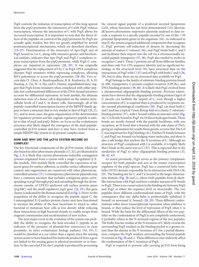
PrgZ belongs to the family of substrate-binding proteins foundin ABC transporters, G protein-coupled receptors (GPCRs), andDNA binding proteins (39, 40). It is likely that PrgZ evolved froma chromosomal oligopeptide-binding protein. Previous experi-ments have shown that the oligopeptide-binding protein OppA ofE. faecalis can facilitate the import of C, even though a higherconcentration of C is required than is produced by recipients un-der normal physiological conditions (38). PrgZ can bind both Cand I and has a typical Venus flytrap fold of a cluster C substrate-binding protein (37), with C bound within an internal cavity (Fig.3a). C is firmly bound to PrgZ via 10 direct hydrogen bonds. Thesebonds are mostly formed with the peptide backbone, with oneexception, an H-bond that is formed with the side chain of Thr3,giving an explanation for results from genetic screens that Thr3 ofC was important for PrgZ binding (41). Further H-bonds betweenC and PrgZ are formed via bridging water molecules, and there isalso a salt bridge that anchors the N terminus of C. Although nostructure of PrgZ complexed with I is available, it is highly likelythat I binds in the same way as C (37). This is expected due to thesimilarities of PrgZ to other oligopeptide-binding proteins (37,40, 42, 43).
As noted previously, PrgX serves as the primary cytoplasmicreceptor for both peptides and acts as the master transcriptionregulator of the prgQ operon. PrgX has a conserved helix-turn-helix (HTH) domain responsible for its interaction with DNA (28,29). The binding site for C and I is located in the larger dimeriza-tion domain (Fig. 3b and c), where both peptides form �-sheet-like interactions with PrgX and have a similar amount of H-bondsto PrgX. There is no conservation in the binding site between PrgXand PrgZ at either the sequence level or structurally. The twopeptides favor different conformational states of PrgX, with theconsequence that one alpha-helix is preferentially stabilized (Ibound) or unwound (C bound) (28, 29). These different confor-mations either favor transcriptional repression when inhibitor isbound, or they reduce the level of repression of PrgX when C isbound. While the basis for the differential effects of the two pep-tides on the conformation of PrgX is not completely understood,it probably relates to the N-terminal regions of the two peptides.The bulky leucine residue at the N terminus of C likely crowds thesurrounding PrgX residues in the binding pocket to a greater ex-tent than the alanine at the N terminus of I (for a partial illustra-tion, compare the PrgX residues surrounding the bound peptideN termini in Fig. 3b and c). These differences may indirectly affectthe conformation of the C terminus of PrgX.
PrgY is required to prevent cells carrying pCF10 from being
Minireview
1558 jb.asm.org June 2016 Volume 198 Number 11Journal of Bacteriology
on April 13, 2018 by guest
http://jb.asm.org/
Dow
nloaded from
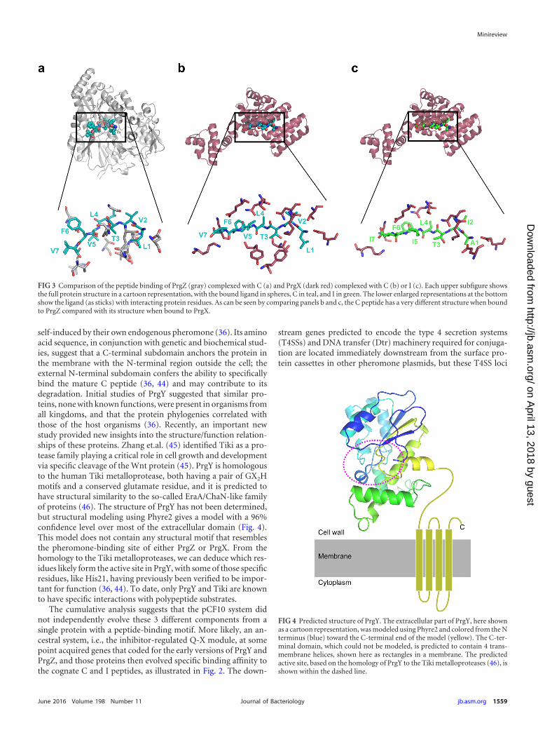
self-induced by their own endogenous pheromone (36). Its aminoacid sequence, in conjunction with genetic and biochemical stud-ies, suggest that a C-terminal subdomain anchors the protein inthe membrane with the N-terminal region outside the cell; theexternal N-terminal subdomain confers the ability to specificallybind the mature C peptide (36, 44) and may contribute to itsdegradation. Initial studies of PrgY suggested that similar pro-teins, none with known functions, were present in organisms fromall kingdoms, and that the protein phylogenies correlated withthose of the host organisms (36). Recently, an important newstudy provided new insights into the structure/function relation-ships of these proteins. Zhang et.al. (45) identified Tiki as a pro-tease family playing a critical role in cell growth and developmentvia specific cleavage of the Wnt protein (45). PrgY is homologousto the human Tiki metalloprotease, both having a pair of GX2Hmotifs and a conserved glutamate residue, and it is predicted tohave structural similarity to the so-called EraA/ChaN-like familyof proteins (46). The structure of PrgY has not been determined,but structural modeling using Phyre2 gives a model with a 96%confidence level over most of the extracellular domain (Fig. 4).This model does not contain any structural motif that resemblesthe pheromone-binding site of either PrgZ or PrgX. From thehomology to the Tiki metalloproteases, we can deduce which res-idues likely form the active site in PrgY, with some of those specificresidues, like His21, having previously been verified to be impor-tant for function (36, 44). To date, only PrgY and Tiki are knownto have specific interactions with polypeptide substrates.
The cumulative analysis suggests that the pCF10 system didnot independently evolve these 3 different components from asingle protein with a peptide-binding motif. More likely, an an-cestral system, i.e., the inhibitor-regulated Q-X module, at somepoint acquired genes that coded for the early versions of PrgY andPrgZ, and those proteins then evolved specific binding affinity tothe cognate C and I peptides, as illustrated in Fig. 2. The down-
stream genes predicted to encode the type 4 secretion systems(T4SSs) and DNA transfer (Dtr) machinery required for conjuga-tion are located immediately downstream from the surface pro-tein cassettes in other pheromone plasmids, but these T4SS loci
FIG 3 Comparison of the peptide binding of PrgZ (gray) complexed with C (a) and PrgX (dark red) complexed with C (b) or I (c). Each upper subfigure showsthe full protein structure in a cartoon representation, with the bound ligand in spheres, C in teal, and I in green. The lower enlarged representations at the bottomshow the ligand (as sticks) with interacting protein residues. As can be seen by comparing panels b and c, the C peptide has a very different structure when boundto PrgZ compared with its structure when bound to PrgX.
FIG 4 Predicted structure of PrgY. The extracellular part of PrgY, here shownas a cartoon representation, was modeled using Phyre2 and colored from the Nterminus (blue) toward the C-terminal end of the model (yellow). The C-ter-minal domain, which could not be modeled, is predicted to contain 4 trans-membrane helices, shown here as rectangles in a membrane. The predictedactive site, based on the homology of PrgY to the Tiki metalloproteases (46), isshown within the dashed line.
Minireview
June 2016 Volume 198 Number 11 jb.asm.org 1559Journal of Bacteriology
on April 13, 2018 by guest
http://jb.asm.org/
Dow
nloaded from
show considerable divergence (22). This suggests that phero-mone-inducible aggregation cassettes became linked to the addi-tional components required for conjugation on multiple occa-sions (Fig. 2, step III). Interestingly, the available data suggest thatthe downstream conjugation functions for all known plasmids aretranscriptionally regulated by the peptide signals even though theybecame linked to the upstream regions in multiple events (22).
REMAINING QUESTIONS AND FUTURE DIRECTIONS
While significant questions about the molecular mechanisms ofpheromone-mediated control of conjugation remain, the mostcompelling areas for future study may be the analysis of structure/function relationships of the key regulatory components and in-vestigations of how these systems function in the natural environ-ment, including their impacts on maintenance and disseminationof the plasmid itself, and on the fitness of the bacterial hosts. Thesignificance of such studies is heightened by the fact that consid-erable experimental and theoretical investigations of the evolu-tionary aspects of the well-studied acyl-homoserine-lactone auto-inducer systems in Gram-negative alphaproteobacteria have beencarried out (reviewed in reference 47). For example, a recent studyby Cornforth et al. (48) provided evidence that combining twoseparate quorum-sensing systems allowed for greater resolutionof the local environment of a bacterium, both in terms of sensingsocial (microbial cell population density) and physical (diffusion,etc.) parameters. In contrast, much less attention has been di-rected toward the naturally occurring and more complex intercel-lular communication system represented by pCF10. While pCF10was discovered because of its role in the transmission of antibioticresistance (49), enterococcal pheromone-responsive plasmids fre-quently do not carry resistance genes (50, 51), suggesting that theymay encode other traits that increase the fitness of their host cells,relative to the costs of plasmid maintenance. It is interesting toconsider how the expression of the pheromone-inducible conju-gation genes may impact host fitness and how this relates to theregulatory properties of the system. Published data from ourgroup (52–57) and others (58–61) suggest that the expression ofaggregation substance proteins, such as PrgB, can increase colo-nization and virulence by promoting biofilm formation and at-tachment to host tissues, and by increasing resistance to phago-cytic killing. Notably, there are still no direct data on how PrgB orother inducible proteins might impact fitness in the gut. On theother hand, the overexpression of these genes likely has very highcosts for the induced cell, including the energy required for syn-thesizing conjugation proteins, the likely inhibition of growth incells trapped in large aggregates, and cell death and lysis due totoxic effects of overexpressed gene products on highly inducedcells (62). Interestingly, clusters of genes related to the plasmid-encoded pheromone-inducible adhesins/transfer determinantshave been identified within genomic islands in the chromosomesof some strains, but these chromosomal determinants are not ca-pable of transfer unless a coresident pheromone plasmid inte-grates and mobilizes them via an Hfr-like mechanism (63).
Numerous studies have documented the extremely tight regu-lation of the pheromone system (19). The system not only avoidsspurious induction but also limits the duration of induction dueto the fact that the induction process itself dramatically increasesinhibitor production, leading to rapid shut off of the responseafter a short period of induction (64). Furthermore, the inhibitorcan function as a classic quorum sensor of donor density; at high
donor densities, donors are poorly induced even by high concen-trations of C (64). These cumulative effects of the inhibitor appar-ently limit the extent of induction in mixed populations of donorsand recipients. This raises the question of whether the system mayhave evolved to maintain mixed populations of donors and recip-ients in shared niches in the natural environment of the bacteria,e.g., the intestinal tract. The maintenance of recipient populationsby limiting their conversion to donors should result in a steadysupply of C within the niche, whose inducing capacity is limited bythe inhibitor. In this scenario, basal levels of expression of theinducible genes could be maintained within the mixed popula-tion, providing the previously described benefits (note that induc-tion of a few donors can coaggregate recipients and uninduceddonors in close proximity) while minimizing costs of overexpres-sion. The pheromone system has thus evolved under strong con-flicting selective pressures for an extremely sensitive detection sys-tem to induce expression while simultaneously limiting the extentand duration of induction. This may have driven convergent evo-lution of the three unrelated proteins with vital, but distinct, reg-ulatory functions to recognize the same peptide. Direct experi-mental testing of these ideas in the mammalian gastrointestinal(GI) tract, along with further mechanistic and structural studies ofregulatory components, is in progress and might yield insightsinto more effective approaches to reduce the spread of antibioticresistance and to impair the ability of resistant strains to overgrowand disrupt the gut microbiota of hospital patients.
ACKNOWLEDGMENTS
The research for this review was supported by U.S. PHS grant GM49530 toG.M.D. and by the Kempe Foundations grant JCK-1524 to R.P.-A.B.
FUNDING INFORMATIONThis work, including the efforts of Gary M. Dunny, was funded by HHS |National Institutes of Health (NIH) (GM49530). This work, including theefforts of Ronnie Per-Arne Berntsson, was funded by Kempestiftelserna(Kempe Foundations) (JCK-1524).
REFERENCES1. Tomasz A. 1965. Control of the competent state in Pneumococcus by a
hormone-like cell product: an example for a new type of regulatorymechanism in bacteria. Nature 208:155–159. http://dx.doi.org/10.1038/208155a0.
2. Nealson KH. 1999. Early observations defining quorum-dependent geneexpression, p 277–290. In Dunny GM, Winans SC (ed), Cell-cell signalingin bacteria. ASM Press, Washington, DC.
3. Fuqua WC, Winans SC, Greenberg EP. 1994. Quorum sensing in bac-teria: the LuxR-LuxI family of cell density-responsive transcriptional reg-ulators. J Bacteriol 176:269 –275.
4. Parsek MR, Greenberg EP. 2005. Sociomicrobiology: the connectionsbetween quorum sensing and biofilms. Trends Microbiol 13:27–33. http://dx.doi.org/10.1016/j.tim.2004.11.007.
5. Kristich CJ, Rice LB, Arias CA. 6 February 2014. Enterococcal infec-tion—treatment and antibiotic resistance. In Gilmore MS, Clewell DB, IkeY, Shankar N (ed), Enterococci: from commensals to leading causes ofdrug resistant infection. Massachusetts Eye & Ear Infirmary, Boston, MA.http://www.ncbi.nlm.nih.gov/books/NBK190420/.
6. Miller WR, Munita JM, Arias CA. 2014. Mechanisms of antibiotic resis-tance in enterococci. Expert Rev Anti Infect Ther 12:1221–1236. http://dx.doi.org/10.1586/14787210.2014.956092.
7. Dunny GM, Brown BL, Clewell DB. 1978. Induced cell aggregation andmating in Streptococcus faecalis: evidence for a bacterial sex pheromone.Proc Natl Acad Sci USA 75:3479 –3483. http://dx.doi.org/10.1073/pnas.75.7.3479.
8. Mori M, Sakagami Y, Narita M, Isogai A, Fujino M, Kitada C, Craig RA,Clewell DB, Suzuki A. 1984. Isolation and structure of the bacterial sex
Minireview
1560 jb.asm.org June 2016 Volume 198 Number 11Journal of Bacteriology
on April 13, 2018 by guest
http://jb.asm.org/
Dow
nloaded from

pheromone, cAD1, that induces plasmid transfer in Streptococcus faecalis.FEBS Lett 178:97–100. http://dx.doi.org/10.1016/0014-5793(84)81248-X.
9. Suzuki A, Mori M, Sakagami Y, Isogai A, Fujino M, Kitaga C, Craig RA,Clewell DB. 1984. Isolation and structure of bacterial sex pheromone,cPD1. Science 226:849 – 850. http://dx.doi.org/10.1126/science.6436978.
10. Fuqua C, Parsek MR, Greenberg EP. 2001. Regulation of gene expressionby cell-to-cell communication: acyl-homoserine lactone quorum sensing.Annu Rev Genet 35:439 – 468. http://dx.doi.org/10.1146/annurev.genet.35.102401.090913.
11. Chandler JR, Dunny GM. 2004. Enterococcal peptide sex pheromones:synthesis and control of biological activity. Peptides 25:1377–1388. http://dx.doi.org/10.1016/j.peptides.2003.10.020.
12. Dunny GM, Leonard BAB. 1997. Cell-cell communication in Gram-positive bacteria. Annu Rev Microbiol 51:527–564.
13. Dunny GM, Winans SC. 1999. Cell-cell signaling in bacteria. ASM Press,Washington, DC.
14. Waters CM, Bassler BL. 2005. Quorum sensing: cell-to-cell communica-tion in bacteria. Annu Rev Cell Dev Biol 21:319 –346. http://dx.doi.org/10.1146/annurev.cellbio.21.012704.131001.
15. Antiporta MH, Dunny GM. 2002. ccfA, the genetic determinant for thecCF10 peptide pheromone in Enterococcus faecalis OG1RF. J Bacteriol184:1155–1162. http://dx.doi.org/10.1128/jb.184.4.1155-1162.2002.
16. Clewell DB, An FY, Flannagan SE, Antiporta M, Dunny GM. 2000.Enterococcal sex pheromone precursors are part of signal sequences forsurface lipoproteins. Mol Microbiol 35:246 –248. http://dx.doi.org/10.1046/j.1365-2958.2000.01687.x.
17. Clewell DB, Weaver KE, Dunny GM, Coque TM, Francia MV, Hayes F.9 February 2014. Extrachromosomal and mobile elements in enterococci:transmission, maintenance, and epidemiology. In Gilmore MS, ClewellDB, Ike Y, Shankar N (ed), Enterococci: from commensals to leadingcauses of drug resistant infection. Massachusetts Eye & Ear Infirmary,Boston, MA. http://www.ncbi.nlm.nih.gov/books/NBK190430/.
18. Nakayama J, Ruhfel RE, Dunny GM, Isogai A, Suzuki A. 1994. The prgQgene of the Enterococcus faecalis tetracycline resistance plasmid pCF10encodes a peptide inhibitor, iCF10. J Bacteriol 176:7405–7408.
19. Dunny GM. 2013. Enterococcal sex pheromones: signaling, social behav-ior, and evolution. Annu Rev Genet 47:457– 482. http://dx.doi.org/10.1146/annurev-genet-111212-133449.
20. Cook LC, Federle MJ. 2014. Peptide pheromone signaling in Streptococ-cus and Enterococcus. FEMS Microbiol Rev 38:473– 492. http://dx.doi.org/10.1111/1574-6976.12046.
21. Parashar V, Aggarwal C, Federle MJ, Neiditch MB. 2015. Rgg proteinstructure-function and inhibition by cyclic peptide compounds. ProcNatl Acad Sci U S A 112:5177–5182. http://dx.doi.org/10.1073/pnas.1500357112.
22. Hirt H, Manias DA, Bryan EM, Klein JR, Marklund JK, Staddon JH,Paustian ML, Kapur V, Dunny GM. 2005. Characterization of the pher-omone response of the Enterococcus faecalis conjugative plasmid pCF10:complete sequence and comparative analysis of the transcriptional andphenotypic responses of pCF10-containing cells to pheromone induction.J Bacteriol 187:1044 –1054. http://dx.doi.org/10.1128/JB.187.3.1044-1054.2005.
23. Bae T, Kozlowicz BK, Dunny GM. 2004. Characterization of cis-actingprgQ mutants: evidence for two distinct repression mechanisms by QaRNA and PrgX protein in pheromone-inducible enterococcal plasmidpCF10. Mol Microbiol 51:271–281.
24. Chatterjee A, Johnson CM, Shu CC, Kaznessis YN, Ramkrishna D,Dunny GM, Hu WS. 2011. Convergent transcription confers a bistableswitch in Enterococcus faecalis conjugation. Proc Natl Acad Sci U S A108:9721–9726. http://dx.doi.org/10.1073/pnas.1101569108.
25. Dunny GM, Johnson CM. 2011. Regulatory circuits controlling entero-coccal conjugation: lessons for functional genomics. Curr Opin Microbiol14:174 –180. http://dx.doi.org/10.1016/j.mib.2011.01.008.
26. Johnson CM, Haemig HH, Chatterjee A, Wei-Shou H, Weaver KE,Dunny GM. 2011. RNA-mediated reciprocal regulation between two bac-terial operons is RNase III dependent. mBio 2(5):e00189-11. http://dx.doi.org/10.1128/mBio.00189-11.
27. Johnson CM, Manias DA, Haemig HA, Shokeen S, Weaver KE, HenkinTM, Dunny GM. 2010. Direct evidence for control of the pheromone-inducible prgQ operon of Enterococcus faecalis plasmid pCF10 by a coun-tertranscript-driven attenuation mechanism. J Bacteriol 192:1634 –1642.http://dx.doi.org/10.1128/JB.01525-09.
28. Kozlowicz BK, Shi K, Gu ZY, Ohlendorf DH, Earhart CA, Dunny GM.
2006. Molecular basis for control of conjugation by bacterial pheromoneand inhibitor peptides. Mol Microbiol 62:958 –969. http://dx.doi.org/10.1111/j.1365-2958.2006.05434.x.
29. Shi K, Brown CK, Gu ZY, Kozlowicz BK, Dunny GM, Ohlendorf DH,Earhart CA. 2005. Structure of peptide sex pheromone receptor PrgX andPrgX/pheromone complexes and regulation of conjugation in Enterococ-cus faecalis. Proc Natl Acad Sci U S A 102:18596 –18601. http://dx.doi.org/10.1073/pnas.0506163102.
30. Kozlowicz BK. 2005. The molecular mechanism and peptide signalingresponse of PrgX used to control pheromone-induced conjugativetransfer of pCF10. Ph.D. dissertation. University of Minnesota, Min-neapolis, MN.
31. Declerck N, Bouillaut L, Chaix D, Rugani N, Slamti L, Hoh F, Lereclus D,Arold ST. 2007. Structure of PlcR: insights into virulence regulation andevolution of quorum sensing in Gram-positive bacteria. Proc Natl Acad SciU S A 104:18490–18495. http://dx.doi.org/10.1073/pnas.0704501104.
32. Clewell DB, Dunny GM. 2002. Conjugation and genetic exchange inenterococci, p 265–300. In Gilmore MS, Clewell DB, Courvalin P, DunnyGM, Murray BE, Rice LB (ed), The enterococci: pathogenesis, molecularbiology and antibiotic resistance. ASM Press, Washington, DC.
33. Kozlowicz BK, Dworkin M, Dunny GM. 2006. Pheromone-inducibleconjugation in Enterococcus faecalis: a model for the evolution of biologicalcomplexity? Int J Med Microbiol 296:141–147. http://dx.doi.org/10.1016/j.ijmm.2006.01.040.
34. Platt TG, Fuqua C. 2010. What’s in a name? The semantics of quorumsensing. Trends Microbiol 18:383–387.
35. Keller L, Surette MG. 2006. Communication in bacteria: an ecologicaland evolutionary perspective. Nat Rev Microbiol 4:249 –258. http://dx.doi.org/10.1038/nrmicro1383.
36. Chandler JR, Flynn AR, Bryan EM, Dunny GM. 2005. Specific control ofendogenous cCF10 pheromone by a conserved domain of the pCF10-encoded regulatory protein PrgY in Enterococcus faecalis. J Bacteriol 187:4830 – 4843. http://dx.doi.org/10.1128/JB.187.14.4830-4843.2005.
37. Berntsson RP, Schuurman-Wolters GK, Dunny G, Slotboom DJ, Pool-man B. 2012. Structure and mode of peptide binding of pheromone re-ceptor PrgZ. J Biol Chem 287:37165–37170. http://dx.doi.org/10.1074/jbc.M112.386334.
38. Leonard BA, Podbielski A, Hedberg PJ, Dunny GM. 1996. Enterococcusfaecalis pheromone binding protein, PrgZ, recruits a chromosomal oligo-peptide permease system to import sex pheromone cCF10 for induction ofconjugation. Proc Natl Acad Sci U S A 93:260 –264. http://dx.doi.org/10.1073/pnas.93.1.260.
39. Berntsson RP, Doeven MK, Fusetti F, Duurkens RH, Sengupta D,Marrink SJ, Thunnissen AM, Poolman B, Slotbloom DJ. 2009. Thestructural basis for peptide selection by the transport receptor OppA.EMBO J 28:1332–1340. http://dx.doi.org/10.1038/emboj.2009.65.
40. Berntsson RP, Smits SH, Schmitt L, Slotbloom DJ, Poolman B. 2010. Astructural classification of substrate-binding proteins. FEBS Lett 584:2606 –2617. http://dx.doi.org/10.1016/j.febslet.2010.04.043.
41. Fixen KR, Chandler JR, Le T, Kozlowicz BK, Manias DA, Dunny GM.2007. Analysis of the amino acid sequence specificity determinants of theenterococcal cCF10 sex pheromone in interactions with the pheromone-sensing machinery. J Bacteriol 189:1399 –1406. http://dx.doi.org/10.1128/JB.01226-06.
42. Levdikov VM, Blagova EV, Brannigan JA, Wright L, Vagin AA, Wilkin-son AJ. 2005. The structure of the oligopeptide-binding protein, AppA,from Bacillus subtilis in complex with a nonapeptide. J Mol Biol 345:879 –892. http://dx.doi.org/10.1016/j.jmb.2004.10.089.
43. Tame JR, Murshudov GN, Dodson EJ, Neil TK, Dodson GG, HigginsCF, Wilkinson AJ. 1994. The structural basis of sequence-independentpeptide binding by OppA protein. Science 264:1578 –1581. http://dx.doi.org/10.1126/science.8202710.
44. Chandler JR, Dunny GM. 2008. Characterization of the sequence speci-ficity determinants required for processing and control of sex pheromoneby the intramembrane protease Eep and the plasmid-encoded proteinPrgY. J Bacteriol 190:1172–1183. http://dx.doi.org/10.1128/JB.01327-07.
45. Zhang X, Abreu JG, Yokota C, MacDonald BT, Singh S, Coburn KL,Cheong SM, Zhang MM, Ye QZ, Hang HC, Steen H, He X. 2012. Tiki1is required for head formation via Wnt cleavage-oxidation and inactiva-tion. Cell 149:1565–1577. http://dx.doi.org/10.1016/j.cell.2012.04.039.
46. Bazan JF, Macdonald BT, He X. 2013. The TIKI/TraB/PrgY family: acommon protease fold for cell signaling from bacteria to metazoa? DevCell 25:225–227. http://dx.doi.org/10.1016/j.devcel.2013.04.019.
Minireview
June 2016 Volume 198 Number 11 jb.asm.org 1561Journal of Bacteriology
on April 13, 2018 by guest
http://jb.asm.org/
Dow
nloaded from
47. Stevens AM, Schuster M, Rumbaugh KP. 2012. Working together for thecommon good: cell-cell communication in bacteria. J Bacteriol 194:2131–2141. http://dx.doi.org/10.1128/JB.00143-12.
48. Cornforth DM, Popat R, McNally L, Gurney J, Scott-Phillips TC, IvensA, Diggle SP, Brown SP. 2014. Combinatorial quorum sensing allowsbacteria to resolve their social and physical environment. Proc Natl AcadSci U S A 111:4280 – 4284. http://dx.doi.org/10.1073/pnas.1319175111.
49. Dunny G, Funk C, Adsit J. 1981. Direct stimulation of the transfer ofantibiotic resistance by sex pheromones in Streptococcus faecalis. Plasmid6:270 –278. http://dx.doi.org/10.1016/0147-619X(81)90035-4.
50. Clewell DB. 1993. Bacterial sex pheromone-induced plasmid transfer.Cell 73:9 –12. http://dx.doi.org/10.1016/0092-8674(93)90153-H.
51. Clewell DB. 1999. Sex pheromone systems in enterococci, p 47– 66. InDunny GM, Winans SC (ed), Cell-cell signaling in bacteria. ASM Press,Washington, DC.
52. Chandler JR, Hirt H, Dunny GM. 2005. A paracrine peptide sex phero-mone also acts as an autocrine signal to induce plasmid transfer and vir-ulence factor expression in vivo. Proc Natl Acad Sci U S A 102:15617–15622. http://dx.doi.org/10.1073/pnas.0505545102.
53. Chuang ON, Schlievert PM, Wells CL, Manias DA, Tripp TJ, DunnyGM. 2009. Multiple functional domains of Enterococcus faecalis aggrega-tion substance Asc10 contribute to endocarditis virulence. Infect Immun77:539 –548. http://dx.doi.org/10.1128/IAI.01034-08.
54. Chuang-Smith ON, Wells CL, Henry-Stanley MJ, Dunny GM. 2010.Acceleration of Enterococcus faecalis biofilm formation by aggregationsubstance expression in an ex vivo model of cardiac valve colonization.PLoS One 5:e15798. http://dx.doi.org/10.1371/journal.pone.0015798.
55. Hirt H, Erlandsen SL, Dunny GM. 2000. Heterologous inducible expres-sion of Enterococcus faecalis pCF10 aggregation substance Asc10 in Lacto-coccus lactis and Streptococcus gordonii demonstrates contribution to cellhydrophobicity and adhesion to fibrin. J Bacteriol 182:2299 –2306. http://dx.doi.org/10.1128/JB.182.8.2299-2306.2000.
56. Olmsted SB, Dunny GM, Erlandsen SL, Wells CL. 1994. A plasmid-encoded surface protein on Enterococcus faecalis augments its internaliza-tion by cultured intestinal epithelial cells. J Infect Dis 170:1549 –1556.
57. Schlievert PM, Gahr PJ, Assimacopoulos AP, Dinges MM, Stoehr JA,Harmala JW, Hirt H, Dunny GM. 1998. Aggregation and binding sub-
stances enhance pathogenicity in rabbit models of Enterococcus faecalisendocarditis. Infect Immun 66:218 –223.
58. Kreft B, Marre R, Schramm U, Wirth R. 1992. Aggregation substance ofEnterococcus faecalis mediates adhesion to cultured renal tubular cells.Infect Immun 60:25–30.
59. Süssmuth SD, Muscholl-Silberhorn A, Wirth R, Susa M, Marre R,Rozdzinski E. 2000. Aggregation substance promotes adherence, phago-cytosis, and intracellular survival of Enterococcus faecalis within humanmacrophages and suppresses respiratory burst. Infect Immun 68:4900 –4906. http://dx.doi.org/10.1128/IAI.68.9.4900-4906.2000.
60. Chow JW, Thal LA, Perri MB, Vazquez JA, Donabedian SM, ClewellDB, Zervos MJ. 1993. Plasmid-associated hemolysin and aggregationsubstance production contribute to virulence in experimental enterococ-cal endocarditis. Antimicrob Agents Chemother 37:2474 –2477. http://dx.doi.org/10.1128/AAC.37.11.2474.
61. Rakita RM, Vanek NN, Jacques-Palaz K, Mee M, Mariscalco MM,Dunny GM, Snuggs M, van Winkle WB, Simon SI. 1999. Enterococcusfaecalis bearing aggregation substance is resistant to killing by human neu-trophils despite phagocytosis and neutrophil activation. Infect Immun67:6067– 6075.
62. Bhatty M, Cruz MR, Frank KL, Gomez JA, Andrade F, Garsin DA,Dunny GM, Kaplan HB, Christie PJ. 2015. Enterococcus faecalis pCF10-encoded surface proteins PrgA, PrgB (aggregation substance) and PrgCcontribute to plasmid transfer, biofilm formation and virulence. Mol Mi-crobiol 95:660 – 677. http://dx.doi.org/10.1111/mmi.12893.
63. Manson JM, Hancock LE, Gilmore MS. 2010. Mechanism of chromo-somal transfer of Enterococcus faecalis pathogenicity island, capsule, anti-microbial resistance, and other traits. Proc Natl Acad Sci U S A 107:12269 –12274. http://dx.doi.org/10.1073/pnas.1000139107.
64. Chatterjee A, Cook LCC, Shu C-C, Chen Y, Manias DA, Ramkrishna D,Dunny GM, Hu W-S. 2013. Antagonistic self-sensing and mate-sensingsignaling controls antibiotic-resistance transfer. Proc Natl Acad Sci U S A110:7086 –7090. http://dx.doi.org/10.1073/pnas.1212256110.
65. Bensing BA, Manias DA, Dunny GM. 1997. Pheromone cCF10 andplasmid pCF10-encoded regulatory molecules act post-transcriptionallyto activate expression of downstream conjugation functions. Mol Micro-biol 24:285–294. http://dx.doi.org/10.1046/j.1365-2958.1997.3301710.x.
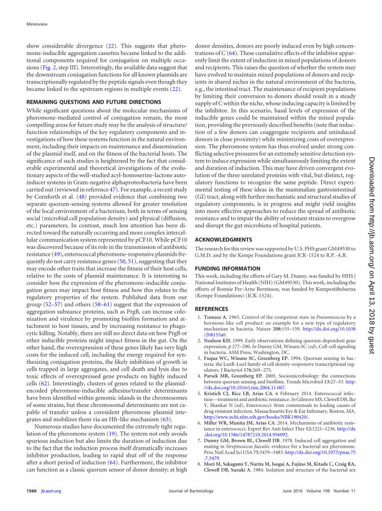
Gary M. Dunny received his B.S. and Ph.D.from the University of Michigan and spent 11years at Cornell University as a postdoctoral fel-low and as a faculty member before moving tothe University of Minnesota in 1989, where he iscurrently professor of microbiology. He hasstudied conjugation, cell signaling, and adapta-tion in enterococci using genetics, biochemis-try, and microscopic imaging for his entirecareer.
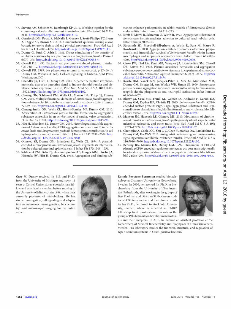
Ronnie Per-Arne Berntsson studied biotech-nology at Chalmers University in Gothenburg,Sweden. In 2010, he received his Ph.D. in bio-chemistry from the University of Groningen,the Netherlands, after working in the groups ofBert Poolman and Dirk-Jan Slotboom on stud-ies of ABC transporters and their domains. Af-ter his Ph.D., he moved to Stockholm Univer-sity, Sweden, where he received an EMBOfellowship to do postdoctoral research in thegroup of Pål Stenmark on botulinum neurotox-ins and their receptors. In 2015, he became an assistant professor at theDepartment of Medical Biochemistry and Biophysics at Umeå University,Sweden. His laboratory studies the function, structure, and regulation oftype 4 secretion systems in Gram-positive bacteria.
Minireview
1562 jb.asm.org June 2016 Volume 198 Number 11Journal of Bacteriology
on April 13, 2018 by guest
http://jb.asm.org/
Dow
nloaded from