Embed Size (px)
Citation preview
The Plant Cell, Vol. 8, 1821-1831, October 1996 0 1996 American Society of Plant Physiologists
Preformed Antimicrobial Compounds and Plant Defense against Funga1 Attack
Anne E. Osbourn The Sainsbury Laboratory, John lnnes Centre, Colney, Norwich NR4 7UH, United Kingdom
INTRODUCTION
Plants produce a diverse array of secondary metabolites, many of which have antifungal activity. Some of these compounds are constitutive, existing in healthy plants in their biologically active forms. Others, such as cyanogenic glycosides and glucosinolates, occur as inactive precursors and are activated in response to tissue damage or pathogen attack. This activa- tion often involves plant enzymes, which are released as a result of breakdown in cell integrity. Compounds belonging to the latter category are still regarded as constitutive because they are immediately derived from preexisting constituents (Mansfield, 1983). VanEtten et al. (i994) have proposed the term “phytoanticipin” to distinguish these preformed an- timicrobial compounds from phytoalexins, which are synthesized from remote precursors in response to pathogen attack, prob- ably as a result of de novo synthesis of enzymes. In recent years, studies of plant disease resistance mechanisms have tended to focus on phytoalexin biosynthesis and other active responses triggered after pathogen attack (Hammond-Kosack and Jones, 1996, in this issue). In contrast, preformed inhibi- tory compounds have received relatively little attention, despite the fact that these plant antibiotics are likely to represent one of the first chemical barriers to potential pathogens.
The distribution of preformed inhibitors within plants is of- ten tissue specific (e.g., Price et ai., 1987; Poulton, 1988; Davis, 1991; Fenwick et ai., 1992; Bennett and Wallsgrove, 1994), and
r there is a tendency for these compounds to be concentrated in the outer cell layers of plant organs, suggesting that they may indeed act as deterrents to pathogens and pests. Some diffusible preformed inhibitors, such as catechol and pro- tocatechuic acid (which are found in onion scales), may influence fungal growth at the plant surface. In general, how- ever, preformed antifungal compounds are commonly sequestered in vacuoles or organelles in healthy plants. There- fore, the concentrations that are encountered by an invading fungus will depend on the extent to which that fungus causes tissue damage. Biotrophs may avoid the release of preformed inhibitors by minimizing damage to the host, whereas necro- trophs are likely to cause substantial release of these compounds. The nature and leve1 of preformed inhibitors to which a potential pathogen is exposed will also vary, depend- ing on factors such as host genotype, age, and environmental conditions (Price et ai., 1987; Davis, 1991).
There have been numerous attempts to associate natural variation in levels of preformed inhibitors in plants with resis-
tance to particular pathogens, but often these attempts have failed to reveal any positive correlation. However, whereas preformed inhibitors may be effective against a broad spec- trum of potential pathogens, successful pathogens are likely to be able to circumvent the effects of these antibiotics either by avoiding them altogether or by tolerating or detoxifying them (Schonbeck and Schlosser, 1976; Fry and Myers, 1981; Bennett and Wallsgrove, 1994; VanEtten et al., 1995; Osbourn, 1996). The isolation of plant mutants defective in the biosynthesis of preformed inhibitors would allow a direct genetic test of the importance of these compounds in plant defense. However, in most cases this approach is technically difficult because of the problems associated with screening for lossof the com- pounds. In the absence of plant mutants, a complementary approach involving the study of fungal mechanisms of resis- tance to preformed inhibitors, and of the contribution of this resistance to fungal pathogenicity to the relevant host plants, offers another route toward investigating the importance of these inhibitors in plant defense.
A large number of constitutive plant compounds have been reported to have antifungal activity. Well-known examples in- clude phenols and phenolic glycosides, unsaturated lactones, sulphur compounds, saponins, cyanogenic glycosides, and glucosinolates (reviewed in Ingham, 1973; Schonbeck and Schlosser, 1976; Fry and Myers, 1981; Mansfield, 1983; Ku6, 1992; Bennett and Wallsgrove, 1994; Grayer and Harborne, 1994; Osbourn, 1996). More recently, 5-alkylated resorcinols and dienes have been associated with disease resistance, in this case, resistance of subtropical fruits to infection by Col- lefotrichum gloeosporioides (Prusky and Keen, 1993). However, only a few classes of preformed inhibitor have been studied in detail to determine their possible roles in plant defense against fungal pathogens. This review focuses on three of these classes-saponins, cyanogenic glycosides, and glu- cosinolates-and summarizes our current knowledge of the role of these preformed inhibitors in determining the outcome of encounters between plants and phytopathogenic fungi.
SAPONINS
Because many saponins exhibit potent antifungal activity and are often present in relatively high levels in healthy plants, these
1822 The Plant Cell
molecules have been implicated as determinants of a plant's resistance to funga1 attack (Osbourn, 1996). A number of other properties are also associated with these compounds, includ- ing piscicidal, insecticidal, and molluscicidal activity; allelopathic action; and antinutritional effects (Price et al., 1987; Fenwick et al., 1992; Hostettmann and Marston, 1995).
Saponins are glycosylated compounds that are widely distributed in the plant kingdom and can be divided into three major groups, depending on the structure of the agly- cone, which may be a triterpenoid, a steroid, or a steroidal glycoalkaloid. Triterpenoid saponins are found primarily in di- cotyledonous plants but also in some monocots, whereas steroid saponins occur mainly in monocots, such as the Lilia- ceae, Dioscoraceae, and Agavaceae, and in certain dicots, such as foxglove, which contains the saponin digitonin (Hostettmann and Marston, 1995). Oats (genus Avena) are unusual because they contain both triterpenoid and steroid saponins (Price et al., 1987). Steroidal glycoalkaloids are found primarily in mem- bers of the family Solanaceae, which includes potato and tomato, but also in the Liliaceae (Hostettmann and Marston, 1995). The saponins produced by oats and tomato have been studied in the greatest detail in relation to their potential role in the defense of plants against phytopathogenic fungi (Osbourn, 1996). These saponins are considered in depth below.
Avenacins and Avenacosides
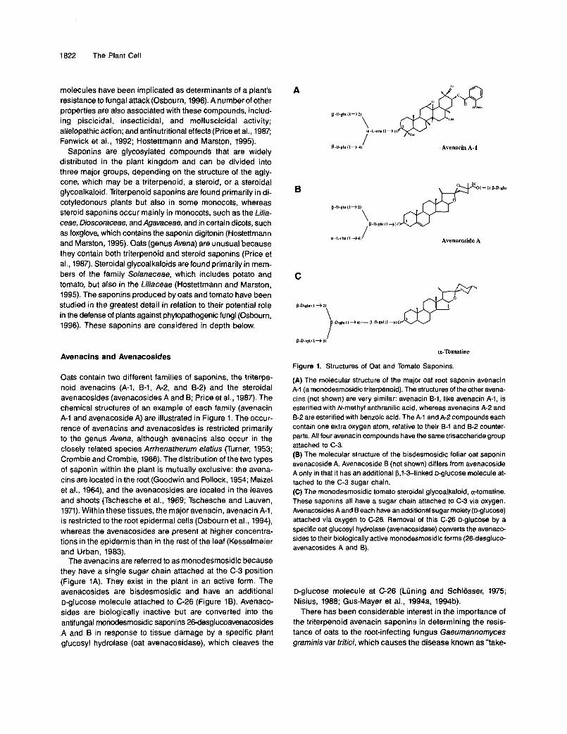
Oats contain two different families of saponins, the triterpe- noid avenacins (A-1, 6-1, A-2, and 6-2) and the steroidal avenacosides (avenacosides A and B; Price et al., 1987). The chemical structures of an example of each family (avenacin A-1 and avenacoside A) are illustrated in Figure 1. The occur- rence of avenacins and avenacosides is restricted primarily to the genus Avena, although avenacins also occur in the closely related species Arrhenatherum elafius (Turner, 1953; Crombie and Crombie, 1986). The distribution of the two types of saponin within the plant is mutually exclusive: the avena- cins are located in the root (Goodwin and Pollock, 1954; Maizel et al., 1964), and the avenacosides are located in the leaves and shoots (Tschesche et al., 1969; Tschesche and Lauven, 1971). Within these tissues, the major avenacin, avenacin A-1, is restricted to the root epidermal cells (Osbourn et al., 1994), whereas the avenacosides are present at higher concentra- tions in the epidermis than in the rest of the leaf (Kesselmeier and Urban, 1983).
The avenacins are referred to as monodesmosidic because they have a single sugar chain attached at the C-3 position (Figure 1A). They exist in the plant in an active form. The avenacosides are bisdesmosidic and have an additional o-glucose molecule attached to C-26 (Figure 1B). Avenaco- sides are biologically inactive but are converted into the antifungal monodesmosidic saponins 26-desglucoavenacosides .A and B in response to tissue damage by a specific plant glucosyl hydrolase (oat avenacosidase), which cleaves the
A
I B-D-gluIl+ 4) Avenacin A-1
B
Avenacoside A
a-Tomatine
Figure 1. Structures of Oat and Tomato Saponins.
(A) The molecular structure of the major oat root saponin avenacin A-I (a monodesmosidic triterpenoid). The structures of the other avena- cins (not shown) are very similar: avenacin 8-1, like avenacin A-1, is esterified with N-methyl anthranilic acid, whereas avenacins A-2 and 8-2 are esterified with benzoic acid. The A-I and A-2 compounds each contain one extra oxygen atom, relative to their 8-1 and 8-2 counter- parts. All four avenacin compounds have the same trisaccharide group attached to C-3. (B) The molecular structure of the bisdesmosidic foliar oat saponin avenacoside A. Avenacoside B (not shown) differs from avenacoside A only in that it has an additional P,1-3-linked o-glucose molecule at- tached to the C-3 sugar chain. (C) The monodesmosidic tomato steroidal glycoalkaloid, a-tomatine. These saponins all have a sugar chain attached to C-3 via oxygen. Avenacosides A and B each have an additional sugar moiety (D-glucose) attached via oxygen to C-26. Removal of this C-26 o-glucose by a specific oat glucosyl hydrolase (avenacosidase) converts the avenaco- sides to their biologically active monodesmosidic forms (26-desgluco- avenacosides A and 0).
D-glucose molecule at C-26 (Lüning and Schlosser, 1975; Nisius, 1988; Gus-Mayer et al., 1994a, 1994b).
There has been considerable interest in the importance of the triterpenoid avenacin saponin:; in determining the resis- tance of oats to the root-infecting fungus Gaeumannomyces graminis var tritici, which causes tht? disease known as "take-

Preformed Antimicrobial Compounds 1823
all.” Although G. g. tritici causes severe yield losses in wheat and barley, it is unable to infect oats, and unlike the oat-attacking variety of G. graminis, G. graminis var avenae, it is relatively sensitive to avenacins. Thus, the resistance of oats to G. g. tritici has been attributed to the presence of these saponins in oat roots (Turner, 1953).
Searches for natural variation in avenacin content have iden- tified one diploid oat species (A. longiglumis) as lacking avenacin A-1 (Osbourn et al., 1994). Significantly, this species is susceptible to infection by G. g. tritici. Unfortunately, A. lon- giglumis does not hybridize readily with other diploid oat species that produce avenacins, making it difficult to test whether the ability to synthesize avenacins cosegregates with
. resistance to G. g. tritici. Recently, mutants of the diploid oat species A. stfigosa lacking avenacin A-1 have been isolated after sodium azide mutagenesis (A.E. Osbourn, R.E. Melton, and M.J. Daniels, unpublished data). The mutants show in- creased susceptibility to isolates of both G. g. avenae and G. g. tritici and also to infection by other root-infecting fungi, such as the Fusarium species F: avenaceum, F: culmorum, and F: graminearum. These observations are consistent with a role for avenacin A-1 as a determinant of resistance of oats to a range of phytopathogenic fungi, although further genetic anal- ysis is required to test whether the absence of this saponin and increased disease susceptibility are indeed causally related.
Natural variation in avenacoside content also exists in the genus Avena. Avena species belonging to the C-genome sub- group (A. clauda, A. pilosa, and A. ventricosa) are all known to lack both avenacosides A and 6 (Nisius, 1988), although there is no information in the literature to indicate whether the absence of these saponins is linked with increased suscepti- bility to disease.
The Tomato Saponin, a-Tomatine
The major saponin in tomato is the steroidal glycoalkaloid, a-tomatine. a-Tomatine is monodesmosidic and, like the avena- cins, is present in healthy plants in its biologically active form. The sugar moiety attached to C-3 consists of two molecules of D-glucose and one each of D-galactose and D-xylose, and is known as P-lycotetraose (Figure 1C). The levels of this sapo- nin are particularly high in the leaves, flowers, and green fruits of tomato (Roddick, 1974).
Despite the considerable variation in a-tomatine levels in the genus Lycopersicon (Courtney and Lambeth, 1977; Juvik and Stevens, 1982; Juvik et al., 1982; Rick et al., 1994), rela- tionships between saponin content and disease resistance are not well documented. Attempts have been made to address the contribution of a-tomatine to specific resistance of near- isogenic tomato lines to the vascular wilt fungi Fusarium oxy- sporum f sp lycopersici (Smith and MacHardy, 1982) and Verticillium albo-atrum (Pegg and Woodward, 1986). No preferential accumulation of a-tomatine was seen in resistant
interactions when compared with compatible interactions, lead- ing to the conclusion that a-tomatine does not appear to have a role in variety-specific resistance (although these experiments are complicated by the fact that both of these fungi are able to degrade a-tomatine enzymatically). However, it has been suggested that a-tomatine may play a secondary role in the variety-specific resistance of tomato to incompatible races of the biotroph Cladosporium fulvum and that release of the sapo- nin from leaf cells as a consequence of an incompatible interaction may act to kill or contain the pathogen (Dow and Callow, 1978).
Mechanisms of Resistance of Fungi to Saponins
Fungi that invade saponin-containing plants must have strate- gies for protecting themselves from host saponins. For biotrophic fungi such as the tomato pathogen C. fulvum, which restricts its growth to the intercellular spaces of tomato leaves, this may be achieved simply by avoiding the release of a-tom- atine. However, correlations have been made between the ability of various fungi to infect saponin-containing plants and the resistance of these fungi to the relevant saponins in vitro. These data suggest that for many fungi, saponin resistance may be a prerequisite for successful infection (Osbourn, 1996).
Membrane Composition
The toxic action of saponins to fungi is associated with the ability of these compounds to complex with membrane sterols and cause pore formation (Price et al., 1987; Fenwick et al., 1992). The membraneolytic.action of a-tomatine is pH depen- dent (Roddick and Drysdale, 1984), and some tomato-infecting fungi, such as Alternaria solani, may counter the effects of a-tomatine by lowering the pH at the infection site to levels at which the saponin is ineffective as an antifungal agent (Schonbeck and Schlosser, 1976). However, a major mecha- nism of resistance to saponins is likely to reside in membrane composition (Steel and Drysdale, 1988; Keukens et al., 1995). Plants may protect themselves from their own saponins by com- partmentalizing them in the vacuole or in other organelles, the membranes of which may avoid lysis due to low or altered sterol composition. Similarly, fungi such as the oomycetes Pythium and Phytophthora contain little or no sterols in their membranes and are resistant to saponins (Arneson and Durbin, 1968).
The importance of membrane composition is further em- phasized by the demonstration that sterol-deficient mutants of a tomato-attacking isolate of 17 solanishowed increased re- sistance to the tomato steroidal glycoalkaloid a-tomatine and gained the ability to infect green fruits of tomato, which are particularly rich in a-tomatine (DBfago and Kern, 1983). The wild type was pathogenic only to ripe tomato fruits, which con- tain relatively low levels of a-tomatine. Analysis of the progeny from crosses between mutant and wild-type fungi showed that pathogenicity to green tomato fruits, low sterol content, and
1824 The Plant Cell
insensitivity to a-tomatine were always inherited together, sug- gesting that a-tomatine may be an important disease resistance determinant (D6fago et al., 1983).
Saponin-Detoxifying Enzymes
Whereas modified membrane composition may allow some phytopathogenic fungi to tolerate saponins, a second major mechanism of resistance involves enzymatic detoxification (Osbourn, 1996). A number of fungal pathogens of oats or tomato produce specific glycosyl hydrolases that remove sug- ars from the sugar chain attached to C-3 of the saponin backbone. Removal of these sugars destroys the ability of the saponin to complex with membrane sterols; therefore, the prod- ucts of hydrolysis have little or no antifungal activity. Thus, for at least some phytopathogenic fungi, the production of saponin- detoxifying enzymes may be a determinant of pathogenicity to saponin-containing hosts.
Avenacinase as a Host Range Determinant. The ability to detoxify avenacin A-1 has been shown to be essential for the interaction of G. g. avenae with oats (Bowyer et al., 1995), provid- ing further evidence that avenacin A-1 may play a role in protecting oats against fungal attack. lsolates of G. g. avenae are avenacin resistant and produce the saponin-detoxifying enzyme avenacinase, a p-glucosyl hydrolase that removes p,l- 2- and p,1-4-linked terminal D-glucose molecules from avena- cin A-1 (Figure 1A; Turner, 1961; Crombie et al., 1986; Osbourn et al., 1991). Glycosyl hydrolases have been grouped into ~ 4 0 families on the basis of sequence similarity (Henrissat, 1991), and within this classification, the majority of p-glucosyl hydro- lyses fall into two of these families (family 1 and family 3). G. graminis avenacinase belongs to the latter family (Osbourn et al., 1995), which also includes a number of cellobiose- degrading p-glucosyl hydrolases from yeasts and Trichoderma.
The isolation of the gene encoding avenacinase allowed the generation of specific fungal mutants defective in saponin detoxification by targeted gene disruption (Bowyer et al., 1995). Funga1 mutants lacking avenacinase showed increased sen- sitivity to avenacin A-1 and importantly were no longer able to infect oats, indicating that avenacinase is a determinant of pathogenicity to this host. However, the enzyme is not ageneral pathogenicity factor because the mutants retained full pathoge- nicity to the alternative host, wheat, which does not contain saponins. These experiments indicate that at least for the in- teraction of G. g. avenae with oats, the ability of fungi to detoxify saponins can determine host range.
Detoxification of domatine. A number of tomato-infecting fungi also produce saponin-detoxifying glycosyl hydrolases, which in this case act on a-tomatine. These enzymes are col- lectively referred to as tomatinases, but their mechanisms of action differ. Some remove a single terminal D-glucose mole- cule, whereas others release the intact p-lycotetraose group or its four individual monosaccharide components (Figure 1C;
reviewed in Osbourn, 1996). Two of these tomatinase enzymes (from Septoria lycopersici and E o. lycopersici) have been stud- ied in detail and are described below.
The S. lycopersici tomatinase enzyme removes a single ter- minal o-glucose molecule from P-lycotetraose by hydrolysis of a p,1-2 linkage (Arneson and Durbin, 1967); therefore, its mode of action is similar to that of G. graminis avenacinase. However, the two enzymes are highly specific for their respec- tive host plant saponins and until recently were not believed to be related. Purification of S. lycopersícitomatinase has now revealed that in fact this enzyme shares many properties (in- cluding immunological cross-reactivity) with avenacinase from G. g. avenae (Osbourn et al., 1995; Sandrock et al., 1995). A cDNA clone encoding tomatinase from S. lycopersici was iso- . lated by using an avenacinase cDNA clone as a heterologous probe (Osbourn et al., 1995). The predicted amino acid se- quence of tomatinase revealed that this enzyme is also a family 3 glycosyl hydrolase and that it shares 68% amino acid similar- ity with avenacinase, allowing for conservative substitutions. The potential now exists to test the role of this tomatinase en- zyme in pathogenicity of S. lycopersici to tomato by targeted gene disruption, following the approach that was taken for G. g, avenae avenacinase.
The tomatinase enzyme from F: o. lycopersici has also been purified recently (Lairini et al., 1996). This enzyme has a differ- ent mechanism of action from the S. lycopersici tomatinase, releasing the intact p-lycotetraose group to give the aglycone tomatidine. In contrast to S. lycopersici tomatinase and G. g. avenae avenacinase, which both have molecular masses ,100 kD, this tomatinase has a molecular mass of 50 kD. Determi- nation of the sequence of the first seven N-terminal amino acids was insufficient to indicate any relatedness with the other saponin-detoxifying enzymes described above or with other known glycosyl hydrolases (Lairini et al., 1996).
Detoxification of Oat Leaf Saponins by S. avenae. The foliar pathogen of oats, S. avenae, is also capable of detoxify- ing saponins and produces an enzyme that is capable of sequential hydrolysis of all of the sugars from the sugar chains attached to C-3 of 26-desglucoavenacosides A and B, ultimately to give the corresponding aglycone, nuatigenin (Figure 1B; Wubben et al., 1996). This enzyme shares the trivial name of avenacosidase with the oat enzyme involved in avenacoside activation (Gus-Mayer et al., 1994a, 1994b), because avenaco- sides are substrates for both enzymes. However, the mechanisms of action of the two enzymes are clearly differ- ent because the oat avenacosidase is specific for the D-glucose molecule attached to C-26 and does not remove sugars from the sugar chain at C-3 (Lüning and Schlosser, 1975; Nisius, 1988), whereas the S. avenae avenacosidase removes L-rhamnose, followed by the remaining two (26-desgluco- avenacoside A) or three (26-desglucoavenacoside B) D-glucose molecules (Wubben et al., 1996). S. avenae avenacosidase is highly specific for the steroidal foliar oat saponins and has no detectable activity toward the triterpenoid saponin avenacin A-1 (which is present in oat roots). Amino acid sequence anal-
ysis of purified S. avenae avenacosidase indicates that the enzyme clearly belongs to the same family of p-glycosyl hydro- lases as G. g. avenae avenacinase and the S. l ppers ic i tomatinase (Osbourn et al., 1995; J.P. Wubben, M.J. Daniels, and A.E. Osbourn, unpublished data).
CYANOGENIC GLYCOSIDES
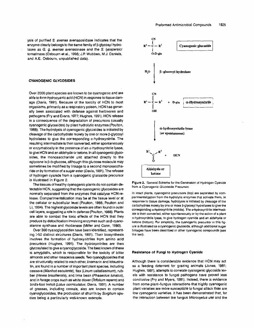
Over 2000 plant species are known to be cyanogenic and are able to form hydrocyanic acid (HCN) in response to tissue dam- age (Davis, 1991). Because of the toxicity of HCN to most organisms, primarily as a respiratory poison, HCN has gener- ally been associated with defense against herbivores and pathogens (Fry and Evans, 1977; Hughes, 1991). HCN release is a consequence of the degradation of precursors (usually cyanogenic glycosides) by plant hydrolytic enzymes (Poulton, 1988). The hydrolysis of cyanogenic glycosides is initiated by cleavage of the carbohydrate moiety by one or more p-glycosyl hydrolases to give the corresponding a-hydroxynitrile. The resulting intermediate is then converted, either spontaneously or enzymatically in the presence of an a-hydroxynitrile lyase, to give HCN and an aldehyde or ketone. In all cyanogenic glyco- sides, the monosaccharide unit attached directly to the aglycone is B-D-glucose, although this glucose molecule may sometimes be modified by linkage to a second monosaccha- ride or by formation of a sugar ester (Davis, 1991). The release of hydrogen cyanide from a cyanogenic glucoside precursor is illustrated in Figure 2.
The tissues of healthy cyanogenic plants do not contain de- tectable HCN, suggesting that the cyanogenic glycosides are normally separated from the enzymes that catalyse HCN re- lesse. Compartmentalization may be at the tissue level or at the cellular or subcellular level (Poulton, 1988; Poulton and Li, 1994). The highest glycoside levels are often found in outer cell layers, suggesting a role in defense (Poulton, 1988). Plants are able to combat the toxic effects of the HCN that they produce by detoxification involving enzymes such as p-cyano- alanine synthase and rhodanese (Miller and Conn, 1980).
Over 300 cyanoglycosides have been identified, represent- ing >50 distinct structures (Davis, 1991). Their biosynthesis involves the formation of hydroxynitriles from amino acid precursors (Hughes, 1991). The hydroxynitriles are then glycosylated to give a cyanoglycoside. The best known of these is amygdalin, which is responsible for the toxicity of bitter almonds and other rosaceous seeds. Two cyanoglycosides that are structurally related to each other, linamarin and lotaustra- lin, are found in a number of important plant species, including cassava (Manihot esculenta), flax (Linum usitatissimum), rub- ber (Hevea brasiliensis), and lima bean (Phaseolus lunatus), and in forage crops such as white clover (Triolium repens) and bird's-foot trefoil (Lotus corniculatus; Davis, 1991). A number of grasses, including cereals, also are known to contain cyanoglycosides, the production of dhurrin by Sorghum spe- cies being a particularly well-known example.
Preformed Antimicrobial Compounds
CN I
R'-c- R' I
O-D-glu
Cyanogenic glucoside
CN
R' - 7 I - Rf + D-glu
OH
a-hydroxynitrile lyase (or spontaneous) 1
1825
Aldehyde or I; ketone
Figure 2. General Scheme for the Generation of Hydrogen Cyanide from a Cyanogenic Glucoside Precursor.
In intact plants, cyanogenic precursors (top) are separated by com- partmentalization from the hydrolytic enzymes that activate them. In response to tissue damage, hydrolysis is initiated by cleavage of the carbohydrate moiety by one or more p-glycosyl hydrolases to give the corresponding a-hydroxynitrile (middle). The a-hydroxynitrile intermedi- ate is then converted, either spontaneously or by the action of a plant a-hydroxynitrile lyase, to give hydrogen cyanide and an aldehyde or ketone (bottom). For simplicity, the cyanogenic precursor in this fig- ure is illustrated as a cyanogenic glucoside, although additional sugar linkages have been described in other cyanogenic compounds (see the text).
Resistance of Fungi to Hydrogen Cyanide
Although there is considerable evidence that HCN may act as a feeding deterrent for grazing animals (Jones, 1981; Hughes, 1991), attempts to correlate cyanogenic glycoside lev- els with resistance to fungal pathogens have proved less conclusive (Fry and Myers, 1981). Indeed, there is evidence from some plant-fungus interactions that highly cyanogenic plant varieties are more susceptible to fungal attack than are low cyanogenic varieties. It has been demonstrated that, for the interaction between the fungus Microcyelus ulei and the
1826 The Plant Cell
rubber tree H. brasiliensis, cyanogenesis inhibits the ability of the plant to produce the phytoalexin scopoletin (Lieberei et ai., 1989). Thus, release of HCN in response to pathogen attack may cause inhibition of active defense responses in highly cyanogenic plants.
Although there is no consistent correlation between cyano- genic glycoside content and fungal disease resistance, there is a clear association belween the ability of fungi to infect cyano- genic plants and their ability to tolerate HCN. For some fungi, such as the rubber tree pathogen M. dei, this tolerance has been attributed to cyanide-resistant respiration (Lieberei, 1988). However, fungi are also reported to detoxify HCN by various means (Fry and Myers, 1981). The majority of successful patho- gens of cyanogenic plants produce the cyanide-inducible enzyme cyanide hydratase (CHT; formerly known as forma- mide hydrolyase), which detoxifies HCN by converting it to formamide (Fry and Evans, 1977; Wang et al., 1992). This en- zyme activity appears to be specific to fungi and has not been found in bacteria or plants. A number of saprophytic fungi as well as some fungi that infect noncyanogenic plants also pro- duce CHT, indicating (not surprisingly) that the ability to metabolize HCN is not the sole determinant of pathogenicity to cyanogenic plants (Fry and Evans, 1977; Wang et ai., 1992).
CHT was first identified in the fungus Stemphylium loti (a pathogen of the cyanogenic plant birds-foot trefoil; Fry and Millar, 1972) but has been studied most extensively in the sor- ghum pathogen Gloeocercospora sofghi (Fry and Myers, 1981; Wang et al., 1992) and also in F: lateritium (a pathogen of sweet potato and other plants; Cluness et al., 1993). The enzymes from G. sorghi and F: lateritium are very similar. They both ex- ist as multimeric aggregates of a single protein with a molecular m a s of ~ 4 5 kD and have relatively high K, values for cya- nide (12 and 43 mM for G. sorghiand E laterifium, respectively). The corresponding genes have been cloned (Wang and VanEtten, 1992; Cluness et al., 1993), and the proteins have been found to share 75% identity at the amino acid levei. The most closely related protein to these CHT enzymes is a nitri- lase from Klebsiella pneumoniae that degrades the herbicide bromoxynil(3,5-dibromo-4-hydroxybenzonitrile; Stalker et al., 1988). The nitrilase shares N35O/o amino acid sequence iden- tity to both the G. sorghi and the F: lateritium CHT enzymes.
Mutants of G. sorghi generated by transformation-mediated disruption of the CHT gene were found to lack CHT activity in vitro and to show the anticipated increase in sensitivity to HCN (I? Wang, R.W. Sandrock, and H.D. VanEtten, personal communication). However, these mutants were still fully patho- genic to the cyanogenic host, sorghum (VanEtten et al., 1995). There was no evidence for CHT activity of the mutants in in- fected plants, eliminating the possibility that G. sofghi may have an alternative CHT enzyme that is expressed in the plant but not in vitro. It appears then that either the ability to tolerate HCN is not important for infection of sorghum by G. sorghi or this fungus has some alternative means of cyanide tolerance (for instance, cyanide-resistant respiration) that is expressed only in the plant. The significance of cyanide resistance in other pathogens of cyanogenic plants (including E lateritium) remains to be tested.
GUCOSINOLATES
Glucosinolates are sulphur-containing glucosides found in members of the family Cruciferae, including the agronomically important genus Brassica and the cruciferous weed Arabidop- sis. The main interest in these compounds relates to their roles as attractants or as feeding deterrents in the interactions of Brassica with vertebrate and invertebrate pests (Chew, 1988; Mithen, 1992; Giamoustaris and Mithen, 1995). In contrast to saponins and cyanogenic glycosides, relatively few studies have been directed toward exploring the significance of glucosinolates in the resistance of plants to fungal attack. How- ever, the increase in interest in this area merits the inclusion of these compounds in this review.
Glucosinolates can be subdivided into three major classes, depending on the nature of their side chains, which may be derived from aliphatic, indolyl, or aralkyl a-amino acids (Fenwick et al., 1983; Chew, 1988; Duncan, 1991; Mithen, 1992). Within individual plants, the distribution of glucosinolates is tissue specific. For instance, in oilseed rape, aliphatic glu- cosinolates predominate in the leaves, but indolyl and phenylethyl glucosinolates are the major glucosinolates in roots and stems (Mithen, 1992). Lines of oilseed rape that have been bred for reduced glucosinolate levels in the seed may retain high levels in the leaves, indicating that leaf and seed glucosinolate levels are under separate genetic control.
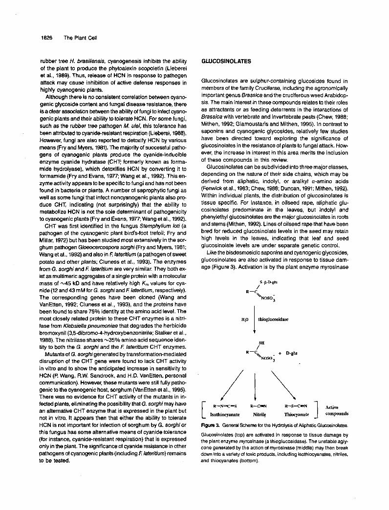
Like the bisdesmosidic saponins and cyanogenic glycosides, glucosinolates are also activated in response to tissue dam- age (Figure 3). Activation is by the plant enzyme myrosinase
R-C
H,O thioglucosidase I R-C + D-glu
h s o ;
Aetive 1 R-N-C-S R-CmN R-S-C-N
Isothiocyanate Nitrile Thiocyanate compounds
Figure 3. General Scheme for the Hydmlysis of Aliphatic Glucosinolates.
Glucosinolates (top) are activated in response to tissue damage by the plant enzyme myrosinase (a thioglucosidase). The unstable agly- cone generated by the action of myrosinase (rniddle) may then break down into a variety of toxic products, including isothiocyanates, nitriles, and thiocyanates (bottom).

Preformed Antimicrobial Compounds 1827
(a thioglucosidase), which in healthy plants is believed to be separated from its glucosinolate substrates by subcellular com- partmentalization. The unstable aglycone generated by the action of myrosinase may then form various products, includ- ing volatile isothiocyanates (mustard oils), nitriles, and thiocyanates, all of which are highly reactive compounds.
Glucosinolate hydrolysis products have been demonstrated to have toxic activity toward a range of fungi in vitro, including both pathogens and nonpathogens of Brassica, although the mechanism of toxicity is not known. Because the nature of the breakdown products depends on the structure of the glucosino- late, the form of myrosinase(s) present, the plant species, and a range of other factors, including pH, temperature, metal ion concentrations, and protein cofactors, it may be difficult, sim- ply by assessing the relative amounts of specific glucosinolates present in the host plant, to predict which toxic products a pathogen is likely to encounter (Fenwick et al., 1983; Chew,
The major breakdown products generated in leaves of Bras- sica are isothiocyanates, the most fungitoxic of which are allyl-(2-propenyl) and 3-butenyl isothiocyanate (Mithen et al., 1986). Breakdown products of indolyl glucosinolates may also be toxic toward fungi (Mithen et al., 1986). In addition, they may be used as precursors for auxin biosynthesis in infection of Brassicas by fungi that cause abnormal tissue growth (such as the root gall fungus Plasmodiophora brassicae; Fenwick et al., 1983; Chew, 1988). It has also been suggested that in- dolyl glucosinolate breakdown products may function as precursors to a class of indole phytoalexins that are induced in Brassica (Rouxel et al., 1989). A number of pathogens of Brassica, such as Lepfosphaeria maculans (Mithen et al., 1986), Peronospora parasitica (Greenhalgh and Mitchell, 1976), Mycosphaerella brassicae (Harthill and Sutton, 1980), and Al- ternaria sp (Milford et al., 1989), have been shown to be sensitive to at least some glucosinolate breakdown products. However, it is unclear whether there is any relationship be- tween resistance of fungi to glucosinolates and ability to cause disease. Glucosinolate breakdown products are also effective against a range of nonpathogens of Brassica, leading to in-
1988).
terest in the use of these compounds as naturally occurring fungicides for the control of cereal diseases (Angus et al., 1994) and postharvest pathogens of fruit and vegetables (Mari et al., 1993).
High glucosinolate levels have been associated with resis- tance of oilseed rape and lndian mustard to L. maculans (Mithen and Magrath, 1992; R. Mithen, personal communica- tions; B. Howlett, personal communication) and with resistance of cabbages to I? parasitica (Greenhalgh and Mitchell, 1976). However, in studies of crosses of B. napus lines with high and low glucosinolate levels in their leaves, resistance to L. macu- lans and glucosinolate profiles did not cosegregate (Mithen and Magrath, 1992). Further studies with B. napus lines that had contrasting glucosinolate profiles indicated that high lev- els of glucosinolates are unlikely to confer greater resistance to L. maculans and Alternaria spp in oilseed rape and may even give enhanced susceptibility (A. Giamoustaris and R. Mithen, personal com m u nication).
CONCWSION AND FUTURE PROSPECTS
Because preformed inhibitors may be harmful to the plants that produce them, they are usually sequestered within plant cells or tissues, often as inactive glycosides. With the excep- tion of the monodesmosidic saponins, the preformed inhibitors considered in this review all require the action of plant hydro- lytic enzymes for release of the corresponding antifungal agent (Table 1). Interestingly, the oat avenacosidase enzyme (Gus- Mayer et al., 1994a, 1994b), linamarase (Oxtoby et al., 1991), and myrosinase (Lenman et al., 1993) (which activate the bis- desmosidic avenacoside saponins, cyanogenic glucosides, and glucosinolates, respectively; Table 1) are all related, sug- gesting that these enzymes may share a common ancestor (Lenman et al., 1993; Gus-Mayer et al., 1994a, 1994b). These three enzymes are all p-glucosyl hydrolases belonging to the family 1 group of glycosyl hydrolases, in contrast to the saponin- detoxifying enzymes, which have been characterized from
Table 1. Preformed Antifungal Compounds in Plants and Resistance Mechanisms in Phytopathogenic Fungi
Major Mechanisms of Preformed Compound Antifungal Agent Plant Activating Enzyme(s) Resistance in Fungi
Monodesmosidic saponins Monodesmosidic saponins None
Bisdesmosidic saponins Monodesmosidic saponins C-26/C-28 specific p-glucosyl hydrolases (e.g., oat avenacosidase)
Cyanogenic glycosides HCN p-glycosyl hydrolase (e.g., linamarase);
Glucosinolates lsothiocyanates Thioglucosidase (e.g., myrosinase) a-hydroxynitrile lyase
Nitriles Thiocyanates
C-3 specific saponin glycosyl hydrolases; membrane composition
glycosyl hydrolases; membrane composition
respiration
C-3 specific saponin
Cyanide hydratase; cyanide-resistant
Unknown

1828 The Plant Cell
G. g. avenae, S. lycopersici, and S. avenae, which all belong to family 3. Another plant p-glucosyl hydrolase, which activates the bisdesmosidic saponin protogracillin, has recently been purified from the herbaceous Asian plant Costus speciosus and also has been shown to be a member of family 1 (Inoue et al., 1996).
Although studies of inhibition of growth of phytopathogenic fungi by isolated plant compounds suggest a role in plant de- fense, such in vitro tests may not always give an accurate indication of the significance of these compounds in restrict- ing fungal growth in the plant (Schonbeck and Schlosser, 1976; Mansfield, 1983). The characterization of mechanisms of re- sistance of phytopathogenic fungi to preformed antifungal compounds offers a means of testing whether antifungal com- pounds play a role in protecting plants against disease. Such resistance mechanisms have been studied in the most detail for fungi that encounter saponins or cyanogenic glycosides, whereas little is known about fungal resistance to glucosino- late breakdown products (Table 1).
A number of fungi actively detoxify preformed inhibitors by producing specific enzymes. Targeted gene disruption has al- lowed stringent genetic tests of the role in pathogenicity of two of these-the saponin-detoxifying enzyme avenacinase, pro- duced by G. g. avenae (which detoxifies the oat root saponin avenacin A-I), and the cyanide-detoxifying enzyme CHT, pro- duced by the sorghum pathogen G. sorghi. These experiments have demonstrated that avenacinase is an essential deter- minant of host range for G. g. avenae, because mutants lacking the enzyme are unable to infect the saponin-containing host, oat (Bowyer et al., 1995). In contrast, CHT does not appear to be required for pathogenicity of G. sorghi to cyanogenic plants (VanEtten et al., 1995). However, the possibility that G. sorghi may have an alternative mechanism of hydrogen cya- nide resistance in the plant (such as cyanide-resistant respiration) cannot be ruled out.
In the future, we are likely to see the continued character- ization of more fungal enzymes involved in the detoxification of preformed inhibitors, especially of saponins. The recent prog- ress in purification and characterization of saponin-detoxifying enzymes from S. lycopersici(0sbourn et al., 1995; Sandrock et al., 1995), F: o. lycopersici (Lairini et al., 1996), and S. ave- nae (Wubben et al., 1996) now presents an opportunity to test the importance of saponin detoxification in pathogenicity, for fungi other than G. g. avenae, by isolating the genes encod- ing these enzymes and generating specific mutants.
The relatedness of at least three out of the four saponin- detoxifying enzymes that have been described in this review suggests that many fungi may employ related enzymes to detoxify host plant saponins. DNA gel blot analyses indicate that DNA sequences related to those encoding avenacinase and S. lycopersici tomatinase exist in a wide range of fungi, including pathogens of plants other than oat and tomato (Osbourn et al., 1995). However, it remains to be seen whether these sequences encode other saponin-detoxifying enzymes or simply S-glucosyl hydrolases required for saprophytic growth. If saponin-detoxifying enzymes do have widespread
significance in fungal phytopathogenicity, they may present attractive targets for inhibitor-based disease control strategies (Osbourn, 1996). Their extracellular location should make them particularly amenable to such an approach.
The demonstration that avenacinase is required for pathoge- nicity of G. g, avenae to oats provides an indirect test of the importance of the oat root saponin avenacin A-í in plant de- fense. Further direct evidence to indicate that the saponin is likely to play a protective role is now emerging from studies of natural variants of oat that lack the saponin and from pre- liminary studies of oat mutants. In the future, it may be possible to address the significance of other preformed antifungal com- pounds in plant defense by generating plant material with altered levels of preformed inhibitors, either by mutagenesis or by manipulation of key steps in the biosynthetic pathways. If such compounds do prove to be important in protecting plants against pathogen attack, as appears to be the case for avena- cin A-I, this approach may pave the way for the development of novel strategies for disease control. Such strategies may be based on interference with mechanisms of resistance of phytopathogenic fungi to preformed inhibitors or on the produc- tion of plants that make novel or altered antifungal compounds. However, in taking the latter approach, the potential effects of the modification of plant secondary metabolites on the re- sistance of plants to pests and grazing animals and on the value of the crop as a food s o m e must also be considered.
ACKNOWLEDGMENTS
I thank all who have shared their unpublished results as well as Richard Mithen and other colleagues for constructive criticism of the manu- script. The Sainsbury Laboratory is supported by the Gatsby Charitable Foundation.
REFERENCES
Angus, J.F., Gardner, P.A., Kirkegaard, J.A., and Desmarchelier, J.M. (1994). Biofumigation: lsothiocyanates released from Brassica roots inhibit growth of the take-all fungus. Plant Soil 162, 107-112.
Arneson, P.A., and Durbin, R.D. (1967). Hydrolysis of tomatine by Septoria lycopersici; a detoxification mechanism. Phytopathology
Ameson, P.A., and Durbin, R.D. (1968). The sensitivity of fungi to a-tomatine. Phytopathology 58, 536-537.
Bennett, R.N., and Wallsgrove, R.M. (1994). Secondary metabolites in plant defence mechanisms. New Phytol. 127, 617-633.
Bowyer, P., Clarke, B.R., Lunness, P., Daniels, M.J., and Osbourn, A.E. (1995). Host range of a plant pathogenic fungus determined by a saponin detoxifying enzyme. Science 267, 371-374.
Chew, F.S. (1988). Biological effects of glucosinolates. In Biologically Active Natural Products-Potential Use in Agriculture. Proceedings of the ACS Symposium 380, H.G. Cutler, ed (Washington, DC: Amer- ican Chemical Society), pp. 155-181.
57, 1358-1360.
Preformed Antimicrobial Compounds 1829
Cluness, M.J., Turner, P.D., Clements, E., Brown, D.T., and OReilly, C. (1993). Purification and properties of cyanide hydratase from Fusar- ium lateritium and analysis of the corresponding chyl gene. J. Gen. Microbiol. 139, 1807-1815.
Courtney, W.H., and Lambeth, V.N. (1977). Glycoalkaloid content of mature fruit of Lycopersicon species. HortScience 12, 550-551.
Cromble, L., Crombie, W.M.L., and Whitlng, D.A. (1984). Structures of the Four Avenacins, Oat Root Resistance Factors to TakeAll Dis- ease. J. Chem. SOC., Chem. Comm. 4, 246-248.
Cromble, W.M.L., and Crombie, L. (1986). Distribution of avenacins A-1, A-2, B-1 and 8-2 in oat roots: Their fungicidal activity toward 'take-all' fungus. Phytochemistry 25, 2069-2073.
Crombie, W.M.L., Cromble, L., Green, J.B., and Lucas, J.A. (1986). Pathogenicity of the take-all fungus to oats: Its relationship to the concentration and detoxification of the four avenacins. Phytochemis- try 25, 2075-2083.
Davis, R.H. (1991). Glucosinolates. In Toxic Substances in Crop Plants, J.P. DMello, C.M. Duffus, and J.H. Duffus, eds (Cambridge, UK: Royal Society of Chemistry), pp. 202-225.
DBfago, G., and Kern, H. (1983). lnduction of Fusarium solani mu- tants insensitive to tomatine, their pathogenicity and aggressiveness to tomato fruits and pea plants. Physiol. MOI. Plant Pathol. 22,2947.
DBfago, G., Kern, H., and Sedlar, L. (1983). Genetic analysis of toma- tine insensitivity, sterol content and pathogenicity for green tomato fruits in mutants of Fusarium solani. Physiol. MOI. Plant Pathol. 22, 39-43.
Dow, J.M., and Callow, J.A. (1978). A possible role for a-tomatine in the varietal-specific resistance of tomato to Cladosporium fulvum. Phytopathol. 2. 92, 211-216.
Duncan, A.J. (1991). Glucosinolates. In Toxic Substances in Crop Plants, J.P. DMello, C.M. Duffus, and J.H. Duffus, eds (Cambridge, UK: Royal Society of Chemistry), pp. 126-147.
Fenwick, G.R., Heaney, R.K., and Mullin, W.J. (1983). Glucosino- lates and their breakdown products in food and food plants. Crit. Rev. Food Sci. Nutr. 18, 123-301.
Fenwick, G.R., Price, K.R., Tsukamota, C., and Okubo, K. (1992). Saponins. In Toxic Substances in Crop Plants, J.P. DMello, C.M. Duffus, and J.H. Duffus, eds (Cambridge, UK: Royal Societyof Chem- istry), pp. 285-327.
Fry, W.E., and Evans, P.H. (1977). Association of formamide hydro- lyase with fungal pathogenicity to cyanogenic plants. Phytopathol-
Fry, W.E., and Millar, R.H. (1972). Cyanide degradation by an enzyme from Stemphylium loti. Arch. Biochem. Biophys. 151, 468-474.
Fry, W.E., and Myers, D.F. (1981). Hydrogen cyanide metabolism by fungal pathogens of cyanogenic plants. In Cyanide in Biology, B. Vennesland, C.J. Knowles, E.E. Conn, J. Westley, and F. Wissing, eds (London: Academic Press), pp. 321-334.
Giamoustaris, A., and Mithen, R. (1995). The effect of modifying the glucosinolate content of leaves of oilseed rape (Brassica napus ssp. oleifera) on its interaction with specialist and generalist pests. Ann. Appl. Biol. 126, 347-363.
Goodwin, R.H., and Pollock, B.M. (1954). Studies on roots. I. Prop- erties and distribution of fluorescent constituents in Avena roots. Am. J. Bot. 4, 516-520.
Grayer, R.J., and Harborne, J.J. (1994). A survey of antifungal com- pounds from higher plants, 1982-1993. Phytochemistry 37, 19-42.
Ogy 67, 1001-1006.
Greenhalgh, J.G., and Mitchell, N.D. (1976). The involvement of flavour volatiles in the resistance to downy mildew of wild and cultivated forms of Brassica oleracea. New Phytol. 77, 391-398.
Gus-Mayer, S., Brunner, H., Schneider-Poetsch, H.A.W., Lottspeich, F., Eckerskorn, C., Grimm, R., and Rüdiger, W. (1994a). The amino acid sequence previously attributed to a protein kinase or a TCP1- related molecular chaperone and co-purified with phytochrome is a 0-glucosidase. FEBS Lett. 347, 51-54.
Gus-Mayer, S., Brunner, H., Schneider-Poetsch, H.A.W., and Rüdiger, W. (1994b). Avenacosidase from oat: Purification, sequence analysis and biochemical characterization of a new member of the BGA family of P-glucosidases. Plant MOI. Biol. 26, 909-921.
Hammond-Kosack, K.E., and Jones, J.D.G. (1996). Resistance gene-dependent plant defense responses. Plant Cell 8,
Harthill, W.F.T., and Sutton, P.G. (1980). lnhibition of germination of Mycosphaerella brassicola ascospores on young cabbage and cauliflower leaves. Ann. Appl. Biol. 96, 153-161.
Henrissat, 6. (1991):P classification of glycosyl hydrolases based on amino acid sequence similarities. Biochem. J. 280, 309-316.
Hostettmann, K.A., and Manton, A. (1995). Saponins. Chemistry and Pharmacology of Natural Products. (Cambridge, UK Cambridge University Press).
Hughes, M.A. (1991). The cyanogenic polymorphism in Tfolium repens L. (white clover). Heredity 66, 105-115.
Ingham, J.L. (1973). Disease resistance in higher plants. The concept of pre-infectional and post-infectional resistance. Phytopathol. Z. 78,
Inoue, K., Shibuya, M., Yamamoto, K., and Ebizuka, Y. (1996). Mo- lecular cloning and bacterial expression of a cDNA encoding furostanol glycoside 26-o-P-glucosidase of Costus speciosus. FEBS Lett. 389, 273-277.
Jones, D.A. (1981). Cyanide and coevolution. In Cyanide in Biology, B. Vennesland, C.J. Knowles, E.E. Conn, J. Westley, and F. Wiss- ing, eds (London: Academic Press), pp. 509-516.
Juvik, J.A., and Stevens, M.A. (1982). lnheritance of foliar a-tomatine content in tomatoes. J. Am. Soc. Hortic. Sci. 107, 1061-1065.
Juvik, J.A., Stevens, M.A., and Rick, C.M. (1982). Survey of the ge- nus Lycopersicon for variability in a-tomatine content. HortScience
Kesselmeier, J., and Urban, B. (1983). Subcellular localization of sape nins in green and etiolated leaves and green protoplasts of oat (Avena sativa L.). Protoplasma 114, 133-140.
Keukens, E.A.J., de Vrije, T., van den Boom, C., de Waard, P., Plasma, H.H., Thiel, F., Chupin, V., Jongen, W.M.F., and de Kruijff, B. (1995). Molecular basis of glycoalkaloid induced mem- brane disruption. Biochim. Biophys. Acta 1240, 216-228.
KuC, J. (1992). Antifungal compounds in plants. In Phytochemical Re- sources for Medicine and Agriculture, H.N. Nigg and D. Seigler, eds (New York: Plenum Press), pp. 159-184.
Lairini, K., Perez-Espinosa, A., Pineda, M., and Ruiz-Rubio, M. (1996). Purification and characterization of tomatinase from Fusar- ium oxysporum f. sp. lycopersici. Appl. Environ. Microbiol. 62,
Lenman, M., Falk, A., Xue, J., and Rask, L. (1993). Characterization of a Bfassica napus myrosinase pseudogene: Myrosinases are mem- bers of the BGAfamily of P-glycosidases. Plant MOI. Biol. 21,463-474.
1773-1791.
314-335.
17, 764-766.
1604-1609.
1830 The Plant Cell
Lieberei, R. (1988). Relationship of cyanogenic capacity of the rub- ber tree Hevea brasiliensis to susceptibility to Microcyclus ulei, the agent causing South American leaf blight. J. Phytopathol. 122,54-67.
Lieberei, R., Biehl, B., Giesemann, A., and Junqueira, N.T.V. (1989). Cyanogenesis inhibits active defense reactions in plants. Plant Phys- iol. 90, 33-36.
Liining, H.U., and Schlosser, E. (1975). Role of saponins in antifun- gal resistance. V. Enzymatic activation of avenacosides. 2. Pflanzenkr. Pflanzenschutz. 82, 699-703.
Maizel, J.V., Burkhardt, H.J., and Mltchell, H.K. (1964). Avenacin, an antimicrobial substance isolated from Avena sativa. 1. lsolation and antimicrobial activity. Biochemistry 3, 424-431.
Mansfield, J.W. (1983). Antimicrobial compounds. In Biochemical Plant Pathology, J.A. Callow, ed (Chichester, UK: John Wiley and Sons),
Mari, M., lori, R., Leoni, O., and Marchi, A. (1993). ln v i m activity of glucosinolate-derived isothiocyanates against postharvest fruit pathogens. Ann. Appl. Biol. 123, 155-164.
Milford, G.F.J., Fieldsend, J.K., Porter, A.J.R., Rawlinson, C.J., Evans, E.J., and Bilsborrow, P. (1989). Changes in glucosinolate concentrations during the vegetative growth of single- and double- low cultivars of winter oilseed rape. In Aspects of Applied Biology, Vol. 23, Production and Protection of Oilseed Rape and Other Bras- sica Crops, M.F.B. Dale, A.M. Dewar, R.J. Froud-Williams, T.J. Hocking, D.G. Jones, and B.L. Rea, eds (Warwick, UK: Association of Applied Biologists), pp. 83-90.
Miller, J.M., and Conn, E.E. (1980). Metabolism of hydrogen cyanide by higher plants. Plant Physiol. 65, 1199-1202.
Mithen, R. (1992). Leaf glucosinolate profiles and their relationship to pest and disease resistance in oilseed rape. Euphytica 63, 71-83.
Mithen, R., and Magrath, R. (1992). Glucosinolates and resistance to Leptosphaeria maculans in wild and cultivated Brassica species. Plant Breed. 108, 60-68.
Mithen, R., Lewis, B.G., Fenwick, G.R., and Heaney, R.K. (1986). ln vitro activity of glucosinolates and their products against Lep- tosphaeria maculans. Trans. Br. Mycol. SOC. 87, 433-440.
Nisius, H. (1988). The stromacentre in Avena plastids: An aggrega- tion of P-glucosidase responsible for the activation of oat-leaf saponins. Planta 173, 474-481.
Osbourn, A.E. (1996). Saponins and plant defence-A soap story. Trends Plant Sci. 1, 4-9.
Osbourn, A.E., Clarke, B.R., Dow, J.M., and Daniels, M.J. (1991). Partia1 characterization of avenacinase from Gaeumannomyces graminis var. avenae. Physiol. MOI. Plant Pathol. 38, 301-312.
Osbourn, A.E., Clarke, B.R., Lunness, P., Scott, P.R., and Daniels, M.J. (1994). An oat species lacking avenacin is susceptible to in- fection by Gaeumannomyces graminis var. tritici. Physiol. MOI. Plant Pathol. 45, 457-467.
Osbourn, A.E., Bowyer, P., Lunness, P., Clarke, B., and Daniels, M. (1995). Funga1 pathogens of oat roots and tomato leaves employ closely related enzymes to detoxify host plant saponins. MOI. Plant- Microbe Interact. 8, 971-978.
Oxtoby, E., Dunn, A., Pancoro, A., and Hughes, M.A. (1991). Nucleo- tide and derived amino acid sequence of the cyanogenic P-glycoside (linamarase) from white clover (Trfoliumrepens L.). Plant MOI. Biol.
pp. 237-265.
17, 209-219.
Pegg, G.F., and Woodward, S. (1986). Synthesis and metabolism of a-tomatine in tomato isolines in relation to resistance to Verticillium albo-atrum. Physiol. MOI. Plant Pathol. 28, 187-201.
Poulton, J.E. (1988). Localisation and catabolism of cyanogenic glyco- sides. In Cyanide Compounds in Biology. Proceedings of the ClBA Foundation Symposium 140, D. Evered and S. Harnett, eds (Chi- chester, UK: John Wiley), pp. 67-91,
Poulton, J.E., and Li, C.P. (1994). Tissue leve1 compartmentation of (R)-amygdalin and amygdalin hydrolase prevents large-scale cyano- genesis in undamaged Prunus seeds. Plant Physiol. 104, 29-35.
Price, K.R., Johnson, I.T., and Fenwick, G.R. (1987). The chemistry and biological significance of saponins in food and feedingstuffs. Crit. Rev. Food Sci. Nutr. 26, 27-133.
Prusky, D., and Keen, N.T. (1993). lnvolvement of preformed antifun- gal compounds in the resistance of subtropical fruits to fungal decay. Plant Disease 77, 114-119.
Rick, C.M., Uhlig, J.W., and Jones, A.D. (1994). High a-tomatine con- tent in ripe fruit of Andean Lycopersicon esculentum var. cerasiforme; developmental and genetic aspects. Proc. Natl. Acad. Sci. USA 91,
Roddick, J.G. (1974). The steroidal glycoalkaloid a-tomatine. Phy- tochemistry 13, 9-25.
Roddick, J.G., and Drysdale, R.B. (1984). Destabilization of liposome membranes by the steroidal glycoalkaloid a-tomatine. Phytochemistry
Rouxel, T., Sarniguet, A., Kollman, A., and Bosquet, J.-F. (1989). Accumulation of a phytoalexin in Brassica ssp. in relation to a hyper- sensitive reaction of Leptosphaeria maculans. Physiol. MOI. Plant Pathol. 34, 507-517.
Sandrock, R.W., DellaPenna, D., and VanEtten, H.D. (1995). Purifi- cation and characterization of P2-tomatinase, an enzyme involved in thedegradation of a-tomatine and isolation of the gene encoding fia-tomatinase from Septoria lycopersici. MOI. Plant-Microbe Inter- act. 8, 960-970.
Schonbeck, F., and Schlosser, E. (1976). Preformed substances as potential protectants. In Physiological Plant Pathology, R. Heitefuss and P.H. Williams, eds (Berlin: Springer-Verlag), pp. 653-678.
Smith, C.A., and MacHardy, W.E. (1982). The significance of toma- tine in the host response of susceptible and resistant tomato isolines infected with two races of Fusarium oxysporum f. sp. lycopersici. Phytopathology 72, 415-419.
Stalker, D.M., Malyj, L.D., and McBride, K.E. (1988). Purification and properties of a nitrilase specific for the herbicide bromoxynil and corresponding nucleotide sequence analysis of the bxn gene. J. Biol. Chem. 263, 6310-6314.
Steel, C.S., and Drysdale, R.B. (1988). Electrolyte leakage from plant and fungal tissues and disruption of liposome membranes by a-tomatine. Phytochemistry 27, 1025-1030.
Tschesche, R., and Lauven, I? (1971). Avenacosid B, ein zweites bis- desmosidisches Steroidsaponin aus Avena sativa. Chem. Ber. 104, 3549-3555.
Tschesche, R., Tauscher, M., Fehlhaber, H.W., and Wulff, G. (1969). Avenacosid A, ein bisdesmosidisches Steroidsaponin aus Avena sativa. Chem. Eer. 102, 2072-2082.
Turner, E.M. (1953). The nature of resistance of oats to the take-all fungus. J. Exp. Bot. 4, 264-271.
12877-12881.
23, 543-547.
Preformed Antimicrobial Compounds 1831
Turner, E.M. (1961). An enzymic basis for pathogen specificity in Ophio- bolus graminis. J. Exp. Bot. 12, 169-175.
VanEtten, H.D., Mansfield, J.W., Bailey, J.A., and Farmer, €.E. (1994). Letter to the editor. Two classes of plant antibiotics: Phytoalexins versus “phytoanticipins.” Plant Cell 6, 1191-1 192.
VanEtten, H.D., Sandrock, R.W., Wasmann, C.C., Soby, S.D., McClusky, K., and Wang, P. (1995). Detoxification of phytoanticipins and phytoalexins by phytopathogenic fungi. Can. J. Bot. 73 (suppl.
Wang, P., and VanEtten, H.D. (1992). Cloning and properties of a cy- anide hydratase gene from the phytopathogenic fungus
I), S518-S525.
Gloeocercospora sorghi. Biochem. Biophys. Res. Commun. 187, 1048-1054.
Wang, P., Matthews, D.E., and VanEtten, H.D. (1992). Purification and characterization of cyanide hydratase from the phytopathogenic fungus Gloeocercospora sorghi. Arch. Biochem. Biophys. 298, 569-575.
Wubben, J.P., Price, K.R., Deniels, M. J., and Osbourn, A.E. (1996). Detoxification of oat leaf saponins by Septufia avenae. Phytopathol- Ogy 86, 986-992.