Embed Size (px)
Citation preview
© 2014 Schlaudraff et al. This work is published by Dove Medical Press Limited, and licensed under Creative Commons Attribution – Non Commercial (unported, v3.0) License. The full terms of the License are available at http://creativecommons.org/licenses/by-nc/3.0/. Non-commercial uses of the work are permitted without any further
permission from Dove Medical Press Limited, provided the work is properly attributed. Permissions beyond the scope of the License are administered by Dove Medical Press Limited. Information on how to request permission may be found at: http://www.dovepress.com/permissions.php
Clinical, Cosmetic and Investigational Dermatology 2014:7 171–183
Clinical, Cosmetic and Investigational Dermatology Dovepress
submit your manuscript | www.dovepress.com
Dovepress 171
O r I g I n a l r e s e a r C h
open access to scientific and medical research
Open access Full Text article
http://dx.doi.org/10.2147/CCID.S59851
Predictability of the individual clinical outcome of extracorporeal shock wave therapy for cellulite
Kai-Uwe schlaudraff1
Maren C Kiessling2
nikolaus BM Császár2
Christoph schmitz2
1Concept Clinic, geneva, switzerland; 2Department of anatomy II, ludwig-Maximilians-University of Munich, Munich, germany
Correspondence: Christoph schmitz Department of anatomy II, ludwig-Maximilians-University, Pettenkoferstrasse, Munich, 80336 germany Tel +49 892 1807 2620 Fax +49 892 1807 2683 email [email protected]
Background: Extracorporeal shock wave therapy has been successfully introduced for the
treatment of cellulite in recent years. However, it is still unknown whether the individual clini-
cal outcome of cellulite treatment with extracorporeal shock wave therapy can be predicted
by the patient’s individual cellulite grade at baseline, individual patient age, body mass index
(BMI), weight, and/or height.
Methods: Fourteen Caucasian females with cellulite were enrolled in a prospective, single-
center, randomized, open-label Phase II study. The mean (± standard error of the mean) cellulite
grade at baseline was 2.5±0.09 and mean BMI was 22.8±1.17. All patients were treated with
radial extracorporeal shock waves using the Swiss DolorClast® device (Electro Medical Sys-
tems, S.A., Nyon, Switzerland). Patients were treated unilaterally with 2 weekly treatments for
4 weeks on a randomly selected side (left or right), totaling eight treatments on the selected side.
Treatment was performed at 3.5–4.0 bar, with 15,000 impulses per session applied at 15 Hz.
Impulses were homogeneously distributed over the posterior thigh and buttock area (resulting in
7,500 impulses per area). Treatment success was evaluated after the last treatment and 4 weeks
later by clinical examination, photographic documentation, contact thermography, and patient
satisfaction questionnaires.
Results: The mean cellulite grade improved from 2.5±0.09 at baseline to 1.57±0.18 after the
last treatment (ie, mean δ-1 was 0.93 cellulite grades) and 1.68±0.16 at follow-up (ie, mean
δ-2 was 0.82 cellulite grades). Compared with baseline, no patient’s condition worsened, the
treatment was well tolerated, and no unwanted side effects were observed. No statistically
significant (ie, P,0.05) correlation was found between individual values for δ-1 and δ-2 and
cellulite grade at baseline, BMI, weight, height, or age.
Conclusion: Radial shock wave therapy is a safe and effective treatment option for cellulite.
The individual clinical outcome cannot be predicted by the patient’s individual cellulite grade
at baseline, BMI, weight, height, or age.
Keywords: acoustic wave therapy, AWT, extracorporeal pulse activation therapy, EPAT, radial
shock wave therapy, RSWT
IntroductionGynoid lipodystrophy, better known as cellulite, is the most common lipodystrophic
disease and is found in 85% of post-adolescent women.1–4 Cellulite usually develops
in particular anatomical areas, such as the thighs, buttocks, abdomen, and upper arms,
and becomes visible through its classical “orange peel” appearance, characterized
by an irregular, dimpled skin surface with thinning of the epidermis/dermis and the
presence of nodular clusters of fat cells.1–4 It represents not only a cosmetic concern

Clinical, Cosmetic and Investigational Dermatology 2014:7submit your manuscript | www.dovepress.com
Dovepress
Dovepress
172
schlaudraff et al
for women, but often becomes a major psychological
problem, impairing sporting activities, choice of clothing,
and social interaction.
The pathophysiology of cellulite is related to various
predisposing factors, such as biotype, heredity, ethnic back-
ground, body weight, age, hormonal changes, smoking, and
genetic predisposition.1,2,4–6 Four main hypotheses regarding
the etiopathogenesis of cellulite have emerged over recent
decades: a different anatomical conformation of the subcuta-
neous tissue in women compared with men;7,8 changes in the
biomechanical properties of epidermal and dermal tissues;8
excessive hydrophilia of the extracellular matrix increasing
interstitial pressure and causing edema of the fatty tissue;9
and alterations in both microvascular and lymphatic circula-
tion resulting in the often painful protrusion of subcutane-
ous adipose tissue into the lower reticular dermis, causing
distinctive mattress-like surface irregularities.10 However,
these hypotheses are mutually conflicting and do not con-
sider recent advances in our understanding of the complex
physiopathology of the adipose organ.10 For instance, one
cannot exclude that inflammation also contributes to the
formation of cellulite.11,12
Nevertheless, various treatments for cellulite have been
developed over recent decades, focusing on skin tightening
with radiofrequency or lasers, improving blood and lym-
phatic circulation using both physical treatments and phar-
macotherapy, and treating deeper deformities with surgical
subcision, laser treatments, ultrasound devices, or liposuction
(summarized in Table 1). However, there is no single treat-
ment of cellulite that is completely effective.13,14
Extracorporeal shock wave therapy (ESWT) and radial
shock wave therapy (RSWT) have been introduced as safe
and effective treatment options for cellulite.15–23 A shock wave
is an acoustic pressure wave that is produced in any elastic
medium, such as air, water, or even a solid substance.24,25
Shock waves differ from sound waves in that the wave
front, where compression takes place, is a region of sudden
change in stress and density.24,25 Both focused shock waves
(ESWT) and radial shock waves (RSWT) are characterized
by a high positive peak pressure (in mPa), a fast initial rise
Table 1 Various therapies for cellulite and their level of evidence based on published studies
Treatment Related studies and their level of evidence*
IB IIA IIB III IV
esWT/rsWT sattler et al15 Knobloch et al16 Russe-Wilflingseder et al17
Braun et al18 angehrn et al19 Christ et al20 Christ et al21 adatto et al22
Kuhn et al23
radiofrequency Mlosek et al50 nootheti et al51 goldberg et al52
sadick and Mulholland53 sadick and Magro54
laser-assisted lipolysis Prado et al55 nagy and Vanek56
Katz et al57 Kim and geronemus58
Topical herbs and retinol lis-Balchin59 Kligman et al60
Topical phosphatidylcholine and leD
saski et al61
esWT + cryolipolysis Ferraro et al44
Focused ultrasound Moreno-Moraga et al62
endermology Collis et al63 Chang et al64
Weight loss smalls et al65 Mauriège et al66
Mesotherapy hexsel et al67 rotunda et al68
Carboxy therapy Brandi et al69 Brandi et al70
liposuction Coleman71
lipolysis with topical phosphatidylcholine injections
Môle72
Cryolipolysis Manstein et al73
subcision hexsel and Mazzuco74
Notes: *level IB: evidence from at least one randomized controlled trial. level IIa: evidence from at least one controlled study without randomization. level IIB: evidence from at least one other type of experimental study. level III: evidence from nonexperimental descriptive studies, such as comparative studies, correlation studies, and case control studies. level IV: evidence from expert committee reports or opinions or clinical experience of respected authorities, or both.75 Abbreviations: esWT, extracorporeal shock wave therapy; rsWT, radial shock wave therapy; leD, light-emitting diode.

Clinical, Cosmetic and Investigational Dermatology 2014:7 submit your manuscript | www.dovepress.com
Dovepress
Dovepress
173
extracorporeal shock wave therapy for cellulite
in pressure (approximately a few microseconds or less),
a diffraction-induced tensile wave following the positive
pressure amplitude that can generate cavitation, and a short
life cycle of approximately 10–20 µseconds (Figure 1).24–29
Extracorporeal shock wave lithotripsy is widely used for
stone management in urology.30 ESWT and RSWT are
byproducts of lithotripter technology. Since the late 1980s,
they have been introduced into treatment for various diseases
of the musculoskeletal system, such as plantar fasciopathy,
Achilles tendinopathy, medial tibial stress syndrome, greater
trochanteric pain syndrome, lateral and medial epicondylitis,
and calcifying tendonitis of the shoulder.27–29,31,32 Shock waves
have both a direct and indirect effect on treated tissues. The
direct effect is the result of the energy of the shock wave
being transferred to the targeted tissues. The indirect effect is
the result of the creation of cavitation bubbles in the treated
tissue.24,25,29 It has been hypothesized that both the direct and
indirect effects produce a biological response in the treated
tissues.24,25,29
ESWT devices share two technical key characteristics of
extracorporeal shock wave lithotripsy devices used for stone
management, namely the electrohydraulic, electromagnetic,
or piezoelectric generation of pressure waves and the genera-
tion of focused or so-called defocused pressure waves.29,33
Radial shock waves are generated ballistically, ie, by
accelerating a bullet that strikes an applicator, transforming
the kinetic energy of the bullet into a radially expanding
pressure wave (Figure 1).29,32,33 In this regard, it is of note
that, in several studies on ESWT/RSWT for cellulite, the
therapy was termed acoustic wave therapy (AWT)15,17,20,22 or
extracorporeal pulse activation therapy (EPAT).21,22 The terms
AWT and EPAT are proprietary names of the manufacturer
of the corresponding devices (Storz Medical, Tägerwillen,
Switzerland; see also Russe-Wilfingseder et al17). AWT is reg-
istered as “… non-medical electric and electronic apparatus
and instruments for the generation and application of shock
waves or pressure waves in the fields of cosmetics and beauty
care”,34 and EPAT as “… electronic apparatus and parts of
the apparatus for generating and applying pressure or shock
waves for use in the fields of cosmetics and beauty care”.35
The similarity between AWT, EPAT, and RSWT has been
addressed in several papers in the literature.21,36,37
Unaddressed in the studies on ESWT/RSWT for cellu-
lite carried out to date15–23 is whether the individual clinical
outcome of the therapy can be predicted by the patient’s cel-
lulite grade at baseline, age, body mass index (BMI), weight,
height, and/or age. This was addressed in the present study
using RSWT. We hypothesized that the individual clinical
outcome of RSWT for cellulite can be predicted by the
patient’s cellulite grade at baseline and the patient’s BMI.
A
B
D
E
F
G
H
I O
N
M
L
K
J
C
10
5
0
0 5 10 15
1
2
3
iii
iv
iii
µseconds
MP
a
−5
Figure 1 Principles of radial shock wave technology.Notes: (A) DolorClast® device (electro Medical systems sa, nyon, switzerland) used in the present study. (B) Power+ hand piece of the swiss DolorClast device with the 36 mm applicator used in the present study. Compressed air (1) is used to fire a projectile within a guiding tube (2) that strikes a 36 mm diameter metal applicator (3) placed on the skin. The projectile generates stress waves in the applicator that transmit pressure waves noninvasively into tissue. (C) Pressure wave generated with the swiss DolorClast device, measured at a distance of 1 mm from the applicator (Power+ hand piece, 36 mm applicator, device operated at 4 bar air pressure and 15 hz impulse frequency as used in the present study). after a delay of approximately 2 µseconds, the pressure wave shows an increase in (positive) pressure (i), followed by a decrease in pressure (ii) with reaching zero at approximately 8 µseconds, a subsequent period of negative pressure (iii) interrupted by a period of positive pressure (iv). (D–O) Cavitation bubbles (black dots) in degassed water generated during the phase of negative pressure of radial shock waves generated with the Power+ hand piece and the 36 mm applicator of the swiss DolorClast device operated at 4 bar air pressure at 15 hz (D–I) as used in the present study or at 1 hz (J–O) either at the center of the applicator (D, E, F, J, K and L) or the edge of the applicator (G, H, I, M, N and O). note that the arrows point to the center of the applicator. Maximum cavitation is shown in (E, H, K and N). The images shown in (D, G, J and M) were taken approximately 1.5 mseconds before the cavitation maximum, and images shown in (F, I, L and O) were taken approximately 1.5 mseconds after the cavitation maximum. Cavitation lasted for approximately one mseconds. The pictures were taken with a high-speed CCD camera (Photron Ultima aPX; Photron, Tokyo, Japan) with a framing rate of 300,000 frames per second and an exposure time of 1/2,700,000 seconds. The scale bar in (O) represents 10 mm. Note that the cavitation field (and thus the pressure field below the applicator) is broader when generating radial shock waves at 15 Hz (D–I) than at 1 hz (J–O). This phenomenon is observed for many radial shock wave devices (Császár et al, submitted for publication).

Clinical, Cosmetic and Investigational Dermatology 2014:7submit your manuscript | www.dovepress.com
Dovepress
Dovepress
174
schlaudraff et al
Materials and methodsstudy designFourteen Caucasian females with cellulite were enrolled in a
prospective, single-center, randomized, open-label Phase II
study. The mean (± standard error of the mean) patient age
was 42.4±2.81 (23–57) years. Mean BMI was 22.8±1.17
(18.7–32.9). The inclusion and exclusion criteria are sum-
marized in Table 2. Informed consent was obtained from each
patient before treatment. The study was approved by the eth-
ics committee of Canton Geneva (Geneva, Switzerland) under
registration number GE 08-40 and by the Swiss Agency for
Therapeutic Products (Swissmedic, Bern, Switzerland) under
registration number 2009-MD-0005. The study is registered
with ClinicalTrials.gov (NCT01974115).38
Determination of cellulite gradeThe mean cellulite grade of the patients at baseline was
2.5±0.09 (range 2–3). Cellulite grades were determined
by clinical inspection of the patients’ skin (documented by
digital photography) and by contact thermography.
Photographs of the patients were taken before the treat-
ment cycle and at each follow-up using a D80 digital camera
system (Nikon, Tokyo, Japan), PocketWizard transceivers
(LPA Design, Burlington, VT, USA), and StudioMax III
lighting equipment (Photogenic Professional Lighting,
Bartlett, IL, USA), with standardized lighting settings and
distance to the patient at each photographic session. Patients
were asked to fully contract the buttock muscles each time
a photograph was taken. This aimed to fully show and stan-
dardize the appearance of the cellulite and thus to avoid any
“softening effects” due to varying muscle tone that might
change the visibility of the cellulite.
Contact thermography was performed using the
Cell-Meter® System Professional Cellulite Thermodetec-
tor (IPS Srl, Milan, Italy) that was applied directly on the
skin of the treated areas. The temperature is displayed in
a color code, with brown-orange-yellow indicating cold
areas (29.5°C–30.5°C) and bluish shades indicating warm
areas (32°C–33.5°C). Cellulite grades, determined by clini-
cal inspection of the skin, correlated well with the contact
thermography data.
TreatmentAll patients were treated with radial extracorporeal shock
waves using the Swiss DolorClast device (Electro Medical
Systems, SA, Nyon, Switzerland) and the Swiss DolorClast
Power+ hand piece with the 36 mm applicator (Figure 1).
Patients were positioned on a treatment table as indicated in
Figure 2 and the areas of the posterior thigh and the anatomi-
cal buttock area were treated. The medial and lateral lines
of the thigh served as borders of the treatment area which
extended superiorly until the buttock crease and inferiorly
5 cm above the popliteal crease.
Patients were treated unilaterally with 2 weekly treatments
for 4 weeks on a randomly selected side (left or right), total-
ing eight treatments on the selected side. After application of
coupling gel, treatment was performed at 3.5–4.0 bar, with
15,000 impulses per session, and applied at 15 Hz. Impulses
were applied homogeneously over the posterior thigh and
buttock area.Table 2 Inclusion and exclusion criteria applied in the present study
Inclusion criteriahealthy women ,60 years of age, with cellulite grade 2–3 Unchanged hormonal treatment for ,6 months Commitment to the study and ability to follow the medical directions during the study signed informed consent formExclusion criteriaPrevious surgery in the treated area (especially liposuction) Medical and/or cosmetic treatment of cellulite ongoing or within the last 3 months Infection and/or tumor disease within the treatment area anticoagulation therapy and/or hemorrhagic disorders Pregnancy Significant weight fluctuations (caused by disease or diet) Modified hormonal treatment Drugs (eg, corticosteroids, nonsteroidal anti-inflammatories) Vascular abnormalities Previous treatment with esWT/rsWT
Abbreviations: esWT, extracorporeal shock wave therapy; rsWT, radial shock wave therapy.
Figure 2 radial shock wave therapy for cellulite.Notes: (A) application of coupling gel. (B) Treatment with the Power+ hand piece of the swiss DolorClast® device (electro Medical systems, nyon, switzerland).

Clinical, Cosmetic and Investigational Dermatology 2014:7 submit your manuscript | www.dovepress.com
Dovepress
Dovepress
175
extracorporeal shock wave therapy for cellulite
evaluation of clinical outcomeThe condition of each patient’s skin was evaluated before
treatment, after the last treatment, and at a follow-up visit
4 weeks after the last treatment. At both the last treatment
and at follow-up, patients completed a detailed question-
naire with scores for treatment comfort, pain intensity, and
satisfaction, while also indicating undesired effects, such
as bruising.
statistical analysisThe mean and standard error of the mean were calculated
for all investigated variables. Dependence of the clinical
outcome of RSWT (calculated as the individual difference
in cellulite grades either between baseline and after the last
treatment [δ-1] or between baseline and follow-up [δ-2]) on
the patients’ initial cellulite grade at baseline, BMI, weight,
height, age, pain during the treatment, feeling of comfort dur-
ing treatment, and satisfaction at the end of treatment (or at
the end of the follow-up period) was tested using Spearman’s
nonparametric rank correlation. Because δ-1 and δ-2 were
each tested against eight variables, an effect was considered
statistically significant if its associated P-value was smaller
than 0.05/8=0.00625 considering the Bonferroni correction
for multiple hypothesis testing.39 Spearman’s nonparametric
rank correlation was also used for testing the relationship
between δ-1 and δ-2. In this case, the effect was consid-
ered to be statistically significant if the associated P-value
was smaller than 0.05. Calculations were performed using
GraphPad Prism version 5.0 for Windows (GraphPad
software, San Diego, CA, USA).
ResultsThe mean cellulite grade improved from 2.5±0.09
(range 2–3) at baseline to 1.57±0.18 (range 0.25–2.75)
at the end of the treatment (ie, the mean δ-1 was 0.93
cellulite grades). At the end of the follow-up period, the
mean cellulite grade was 1.68±0.16, ranging between 0.5
and 2.75 (ie, the mean δ-2 was 0.82 cellulite grades). The
individual δ-1 varied between 0 grades (ie, no improve-
ment) and 1.75 grades, and the individual δ-2 between
0 grades and 1.5 grades (Figure 3). Accordingly, compared
with baseline, no patient’s skin condition worsened during
treatment and follow-up. The treatment was well toler-
ated and no unwanted side effects were observed (note
that discomfort during treatment and reddening of the
skin up to 24 hours after each treatment session are usual
side effects of RSWT and were therefore not considered
unwanted side effects).
No statistically significant (ie, P,0.05/8) correlation was
found between δ-1 or δ-2 and cellulite grade at baseline, BMI,
weight, height, age, pain during treatment, feeling of comfort
during treatment, or satisfaction at the end of treatment (or at
the end of the follow-up period, Figures 4 and 5).
For eleven of the 14 patients, the condition of the skin
further improved or remained constant during the inter-
val between the last treatment and follow-up (Figure 6).
A1 C1 A2 C2
B1 D1 B2 D2
Figure 3 Treatment of two patients (1, 2) with cellulite using radial extracorporeal shock wave therapy.Notes: (A1 and A2) Clinical picture at baseline. (B1 and B2) Contact thermography at baseline. (C1 and C2) Clinical picture 4 weeks after the last treatment (follow-up). (D1 and D2) Contact thermography at follow-up. (A1–D1) a 29-year-old female (body mass index 32.9, weight 84.3 kg, height 160 cm). radial extracorporeal shock wave therapy performed on the left side improved the cellulite from grade 3 at baseline to grade 1–2 at follow-up (ie, δ-2 was 1.5). Despite this objectively substantial treatment success, the patient’s satisfaction was only 5 on a scale ranging from 0 (maximum dissatisfaction) to 10 (maximum satisfaction). (A2–D2) a 51-year-old female (body mass index 20.8; weight 53.3 kg; height 160 cm). radial extracorporeal shock wave therapy performed on the right side improved the cellulite from grade 2–3 at baseline to grade 1–1.5 at follow-up (ie, δ-2 was 1.25). This patient was very satisfied with the treatment (9 on a scale ranging from 0 to 10). Patient consent was obtained to publish the above images.
Clinical, Cosmetic and Investigational Dermatology 2014:7submit your manuscript | www.dovepress.com
Dovepress
Dovepress
176
schlaudraff et al
3
4x 4x
4x2x
2x
2x
2x
2x
3x
2x
3x
5x
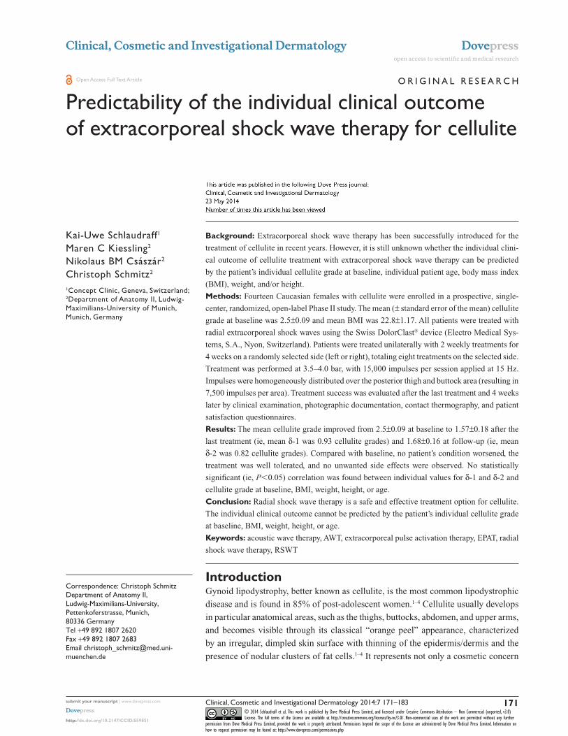
r=−0.294; P=0.308 r=0.196; P=0.502
r=0.042; P=0.888 r=0.116; P=0.693
r=−0.142; P=0.628 r=−0.353; P=0.216
r=−0.009; P=0.975 r=−0.524; P=0.028
r=0.043; P=0.885 r=0.222; P=0.446
2.75
2.25
2.5
2
35
30
25
20
15
90
80
70
60
50
190
180
170
160
150
60
Ag
e [y
ears
]B
od
y le
ng
th [
cm]
Wei
gh
t [k
g]
BM
IIn
itia
l cel
lulit
e st
age
50
40
30
200.0 0.5 1.0 1.5 2.0 0.0 0.5 1.0 1.5 2.0 2.5
A B
C D
E F
G H
I J
δ-1 δ-2
Figure 4 Clinical outcome of radial extracorporeal shock wave therapy for cellulite as a function of the patients’ initial cellulite grade at baseline (A and B), BMI (C and D), weight (E and F), height (G and H) and age (I and J) (calculated as individual difference in cellulite grades either between baseline and after the last treatment [δ-1] or between baseline and at follow-up [δ-2], respectively; the higher δ-1 and δ-2, the better the treatment success).Notes: each dot represents an individual patient; overlapping data are indicated. The Spearman’s nonparametric rank correlation coefficients (r) and the corresponding P-values are provided in red on top of each panel. Abbreviation: BMI, body mass index.
However, there was no statistically significant correlation
between δ-1 and δ-2 (P=0.105).
DiscussionThe results of the present study are generally in line with ear-
lier reports of successful treatment of cellulite with RSWT in
the literature.15,17,20–22 RSWT can improve the clinical picture
by one cellulite grade on average. However, to the authors’
knowledge, the present study is the first to demonstrate that
the individual clinical outcome of RSWT for cellulite can-
not be predicted by the patient’s individual cellulite grade at
baseline, BMI, weight, height, or age. We hypothesize that
the same applies to ESWT for cellulite.
In our clinical experience, the patient’s perception of their
individual cellulite grade and consequently their satisfaction
with the result of treatment for cellulite varies widely from
one patient to another and is truly subjective. Normally,
patients with low cellulite grades are more demanding and
therefore more difficult to manage in their expectations, even
if there is an objectively confirmed clinical improvement.
This was confirmed in our analysis because patient sat-
isfaction, the most important end point of any treatment
for cellulite, did not correlate with δ-1 or δ-2. There were
patients with δ-1=1 (ie, improvement by one cellulite grade)
who were very satisfied, whereas other patients with δ-1=1
were not satisfied at all (Figure 5E). For the clinical setting,
this observation underlines the role of the therapist, who
must correctly evaluate the suitability of the candidate for a
cellulite treatment and must manage the patient’s expecta-
tions accordingly. For studies evaluating existing or new
cellulite treatments, this observation underscores the crucial
importance of applying objective analytical methods, such as
contact thermography and standardized photographic docu-
mentation (in full muscular contraction), because satisfaction
scores may suffer from variations in their consistency. Note
that individual patient satisfaction scores were either not
reported or not correlated with individual objective outcome
measures in the studies of ESWT/RSWT for cellulite pub-
lished to date.15–23 Standardized yet easy clinical analysis of
the severity of cellulite should include easy, effective, and
reproducible measurement tools. In our opinion, clinical
evaluation serves for classification of the cellulite grade,
double contrast photography as applied in the present study
provides a visual contour analysis, and contact thermography
measures the superficial blood perfusion of the skin. Recoil
and elasticity measurements, as applied in some studies of
ESWT/RSWT for cellulite,15,20,21 are helpful in small treat-
ment areas but may considerably vary over the length of a
thigh depending on changing quality and thickness of the
skin in the respective parts.
In recent years, ESWT/RSWT has become the best studied
therapy option for cellulite (Table 1). This is most likely due
to the fact that ESWT/RSWT is noninvasive, does not require
administration of drugs, and can be easily accomplished within
a few minutes per treatment session. It is justified to consider
ESWT (ie, focused shock waves) and RSWT (ie, radial shock
waves) as very similar therapeutic options for cellulite. This is
due to the fact that the energy signatures of ESWT and RSWT
share fundamental physical characteristics, such as high peak
pressure, a fast initial rise in pressure, a low tensile amplitude
Clinical, Cosmetic and Investigational Dermatology 2014:7 submit your manuscript | www.dovepress.com
Dovepress
Dovepress
177
extracorporeal shock wave therapy for cellulite
r=0.093; P=0.752r=0.486; P=0.078
2x
4x
2x2x2x
2x
2x
2x
2x
2x
2x
r=−0.244; P=0.401 r=−0.197; P=0.500
r=0.056; P=0.845 r=0.210; P=0.472
Pai
n d
uri
ng
trea
tmen
tC
om
fort
du
rin
gtr
eatm
ent
Pat
ien
tsa
tisf
acti
on
10
8
6
4
2
0
10
8
4
6
2
0
10
8
4
6
2
00.0 0.5 1.0 1.5 2.0 0.0 0.5 1.0 1.5 2.0 2.5
A B
C D
FE
δ-1 δ-2
Figure 5 Clinical outcome of radial extracorporeal shock wave therapy for cellulite as a function of the patients’ pain during the treatment (A and B), the patients’ feeling of comfort during treatment (C and D), and the patients’ satisfaction with treatment (E and F) (calculated as individual difference in cellulite grades either between baseline and after the last treatment [δ-1] or between baseline and at follow-up [δ-2], respectively; the higher δ-1 and δ-2, the better the treatment success).Notes: each dot represents an individual patient; overlapping data are indicated. Pain was assessed using a visual analog scale ranging from 0 (no pain) to 10 (maximum pain). The feeling of comfort was assessed using a scale ranging from 0 (maximum discomfort) to 10 (maximum comfort), and patients’ satisfaction using a scale ranging from 0 (maximum dissatisfaction) to 10 (maximum satisfaction). The Spearman’s nonparametric rank correlation coefficients (r) and the corresponding P-values are provided in red on top of each panel.
that can generate cavitation, and a short life cycle. Some
authors have offered the following physical definition of “real”
shock waves:26,27 a high positive peak pressure, sometimes
more than 100 mPa, but more often approximately 50–80
mPa; a fast initial rise in pressure during a period of less than
10 nanoseconds; a low tensile amplitude (up to 10 mPa); a short
life cycle of approximately 10 µseconds; and a broad fre-
quency spectrum, typically in the range of 16–20 mHz.
It is well known that radial shock waves do not fulfill the
characteristics set out by this physical definition of real
shock waves (see also Figure 1).29,40 Some ESWT devices
generate pressure waves that fulfill the characteristics set
out by this physical definition of real shock waves, whereas
others do not.29,40,41 Among those ESWT devices that do not
produce real shock waves is the electromagnetic Duolith®
device (Storz Medical)41 that has recently been introduced
into ESWT for cellulite.16 Another device that was used in
several studies for treating cellulite is the D-Actor® 200 (Storz
Medical).15,17,22 The pressure waves generated by this device
are termed “low-energy radial shockwaves” in the literature.42
In contrast, Russe-Wilflingseder et al17 described the D-Actor
200 device as a “vibrating massage system”. Regardless of
these different descriptions in the literature, the D-Actor 200
device is making use of the same construction principle as
the Swiss DolorClast and accelerates a projectile by means
of compressed air. For this reason, the D-Actor 200 device
generates pressure waves that are very similar to the pressure
waves generated by the Swiss DolorClast device, including the
possibility of generating cavitation (Császár et al, submitted
for publication).
Because the studies on ESWT/RSWT for cellulite con-
siderably vary with respect to the level of evidence, shock

Clinical, Cosmetic and Investigational Dermatology 2014:7submit your manuscript | www.dovepress.com
Dovepress
Dovepress
178
schlaudraff et al
wave device used, and treatment protocol, they are discussed
separately, as follows.
In an early pilot study, Braun et al18 treated 20 patients
with “severe cellulite measured with a pinch test”18 using
the electromagnetic DermaSelect® shock wave device
(Storz Medical). The average age of the patients was 37.25
(range 19–56) years and their mean BMI was 29.18 (range
20–41.6). Each patient received six treatment sessions with
2,400 impulses per session on the left leg (the time interval
between treatments, size of the treatment area, and energy
flux density of the shock waves were not provided). According
to the authors’ subjective impressions of the treated leg and
photographic analyses, a significant improvement in skin
surface was shown for more than 70 percent of the patients.
However, treatment success was not expressed according to
changes in cellulite grades.
Angehrn et al19 treated 21 female patients with cel-
lulite (grade 1, n=5; grade 2, n=6; grade 3, n=10) using
defocused shock waves generated with the electrohydraulic
ActiVitor-Derma® device (SwiTech Medical, Kreuzlingen,
Germany). Treatment consisted of 12 sessions at intervals
of 3–4 days, treatment of the skin of the lateral left and right
thigh with 4,000 impulses per thigh per treatment session,
homogeneously distributed over an area of 160 cm2 per side
with an energy flux density of 0.018 mJ/mm2. BMI was
20–24 in ten patients, 25–29 in nine patients, 30–34 in one
patient, and 35–40 in one patient. End points were subjective
opinion of improvement and collagenometry measurements
performed with the high-resolution ultrasound system,
Collagenoson® (Minhorst, Meudt, Germany). At the end
of the treatment period, two patients showed clear worsen-
ing of collagenometry results compared with baseline, five
patients showed some worsening, two patients showed no
change, eight patients showed improvement, and four patients
showed clear improvement compared with baseline. There
was no correlation between the outcome of collagenometry
and individual cellulite grade. Seventeen of the 21 patients
(81%) subjectively assessed their outcome as improved.
Seven patients evaluated the treatment as not suitable (pain
during treatment), six patients assessed it as suitable (no
pain during treatment), and eight patients were indifferent.
The authors concluded that their results provided evidence
that low-energy defocused ESWT caused remodeling of the
collagen within the dermis of the tested region.
Christ et al20,21 treated a total of 59 female patients with
cellulite grade 2 or 3 with planar or radial shock waves
generated with the electromagnetic Cellactor® SC1 device
(Storz Medical). Group 1 (n=15, mean age 44.6 years, mean
BMI 24.4) was treated with planar shock waves generated
with the C-Actor hand piece of the Cellactor SC1 device
(six treatment sessions at intervals of 3–4 days, treatment
of lateral and medial thigh areas as well as the buttocks,
total of 3,200 impulses per treatment session with an energy
flux density of 0.25 mJ/mm2 homogeneously distributed
over a total area of 20×30 cm). Group 2 (n=44, mean age
45.5 years, mean BMI 25.3) was treated identically but
with eight treatment sessions. End points were the elasticity
of the skin measured with the DermaLab® device (Cortex
Technology, Hadsund, Denmark) and the structure of
the connective tissue in the dermis evaluated with the
DermaScan® ultrasound device (Cortex Technology) before
and after treatment. The mean skin elasticity in group 1
patients was improved by 46% after treatment and by 78%
at 3-month follow-up compared with baseline. In group 2,
the mean improvement in skin elasticity was 72% after treat-
ment, 95% at 3-month follow-up, and 105% at 6 months
after baseline. The structure of the connective tissue also
improved between baseline and the 6-month follow-up.
Statistical analysis was not performed to evaluate the impact
of BMI on the results in this study.
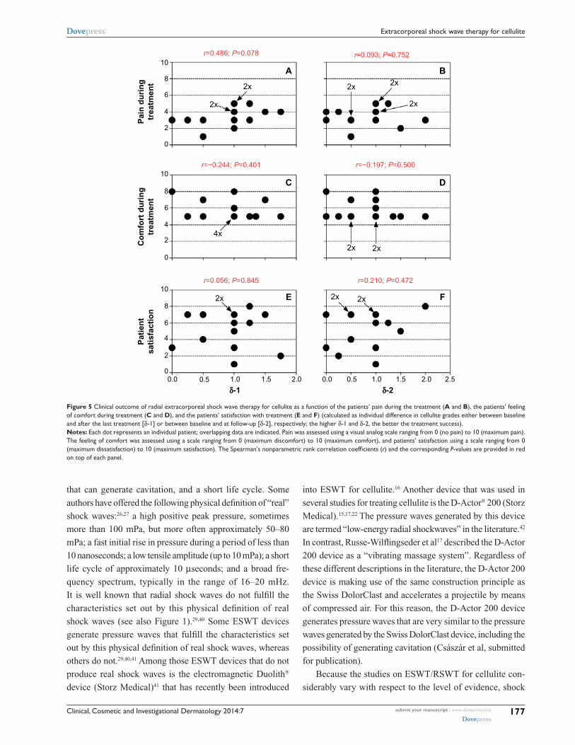
Kuhn et al23 presented a case report concerning a 50-year-
old woman with grade 3 cellulite on her left thigh treated
with the ActiVitor-Derma device (four therapy sessions,
800 impulses per session, energy flux density 0.115 mJ/mm2).
Based on high frequency, high resolution ultrasound
measurements, contact thermography, and histopathologic
2.0
1.5
1.0δ-2
δ-1
0.5
0.0
0.0 0.5 1.0
r=0.452; P=0.105
1.5 2.0
4x
2x
Figure 6 relationship between the individual difference in cellulite grades between baseline and after the last treatment (δ-1) and between baseline and at follow-up (δ-2) after radial extracorporeal shock wave therapy for cellulite (the higher δ-1 and δ-2, the better the treatment success).Notes: each dot represents an individual patient; overlapping data are indicated. red, black, and green dots/asterisks indicate patients whose cellulite grade worsened, remained unchanged, or improved, respectively, during the follow-up period compared with the situation after the last treatment. The spearman’s nonparametric rank correlation coefficient (r) and the corresponding P-value are provided in red on top of the panel.

Clinical, Cosmetic and Investigational Dermatology 2014:7 submit your manuscript | www.dovepress.com
Dovepress
Dovepress
179
extracorporeal shock wave therapy for cellulite
biopsies, the authors reported “some improvement in the
epidermis and the extracellular matrix of the dermis”.23
Sattler et al15 compared three treatments for cellulite.
Group 1 (eleven patients, mean age 40 years, mean BMI 27)
was treated with radial shock waves generated with the
ballistic D-Actor 200 device (a mean of 6.2 treatment ses-
sions, an average of 1,909 impulses per treatment session;
device operated at 2.4–3.0 bar and a frequency of 15 Hz).
Group 2 (eleven patients, of whom nine were included in
the analysis, mean age 34 years, mean BMI 23) was treated
with planar shock waves generated with the C-Actor hand
piece of the electromagnetic Cellactor SC1 (a mean of 6.1
treatment sessions, 1,000 impulses per treatment session
with an energy flux density of 0.35 mJ/mm2). Group 3 (eight
patients, of whom seven were included in the analysis, mean
age 40 years, mean BMI 23) was treated with a combined
radial and planar shock wave protocol (a mean of 6.4 treat-
ment sessions; 2,350 radial pulses on average followed by
an average of 1,925 planar impulses per treatment session;
radial impulses generated by operating the control unit at
2.6–3.0 bar; planar impulses with an energy flux density of
0.35 mJ/mm2). Treatment was focused either on the buttock
and dorsal thigh area or on the ventral thigh area, depending
on the individual clinical picture. End points were visual
impression of the skin (analyzed on photographs), patient
satisfaction, and skin elasticity (measured with the DermaLab
device) 3 months after the last treatment session compared
with baseline. Patients in group 1 had the best result. Analysis
of the photographs showed an optimum treatment result for
five (46%) patients, a satisfactory treatment result for three
(27%) patients, and a not significant treatment result for three
(27%) patients (specific criteria for optimum, satisfactory, and
not significant were not specified). For patients in groups 2
and 3, the corresponding data were: an optimum treatment
result in 1/9 (11%) and 2/7 (29%), respectively; a satisfactory
result in 5/9 (56%) and 4/7 (57%), respectively; and a not
significant result in 3/9 (33%) and 1/7 (14%), respectively.
A statistical analysis was not performed. It is of note that
the authors did not recognize any change in skin elasticity
as a result of shock wave treatment (mean data for group 1,
11.6 mPa at baseline, 10.0 mPa after treatment, and 10.1 mPa
at 3-month follow-up; mean data for group 2, 12.1 mPa at
baseline, 10.8 mPa after treatment, and 12.1 mPa at 3-month
follow-up; mean data for group 3, 10.3 mPa at baseline,
10.4 after treatment, and 10.9 at 3-month follow-up). The
authors discussed the limitations of their study,15 ie, small
numbers of patients, and differences in mean age and mean
BMI between the groups, but concluded that treatment for
cellulite with radial shock waves might be the best choice
(as also performed in the present study).
Adatto et al22 treated 25 women of mean age 42.6 (range
27–63) years with a mean BMI of 24 (range 17–31) on one leg
each with the ballistic D-Actor 200 device (a mean of six treat-
ment sessions within 4 weeks with an average of 3,000 impulses
per treatment session; device operated at 2.6–3.6 bar and with
a frequency of 15 Hz). The authors compared, for each patient,
the treated leg with the untreated leg 1 week and 12 weeks
after the last treatment. The evaluation was performed with
measurements of skin elasticity using the DermaLab device.
Furthermore, three-dimensional images of the skin structure
were recorded using the DermaTOP® system (Eotech, Paris,
France). Adatto et al22 found that skin elasticity, roughness
elevation, and skin depression improved in a statistically sig-
nificant manner on the treated legs compared with the untreated
legs. They concluded that the D-Actor 200 device can be used
effectively to treat cellulite without any side effects.
In a double-blind, randomized controlled trial, Knobloch
et al16 randomly assigned 53 women to either focused shock
waves using the electromagnetic Duolith device (n=25; mean
age 41.4 years, mean BMI 24.2±3.2 kg/m2; six sessions of
ESWT every 1–2 weeks, with 2,000 impulses at 4 Hz, and
an energy flux density of 0.35 mJ/mm2) or sham treatment
(n=28; mean age 45.0 years, mean BMI 25.3±4.5 kg/m2; six
treatment sessions every 1–2 weeks, with 2,000 impulses
and an energy flux density of 0.01 mJ/mm2). In addition
to ESWT or sham-ESWT, all patients underwent specific
gluteal strength exercise training. Among other measure-
ments, the primary end point was score on the photonumeric
Cellulite Severity Scale (CSS) determined by two blinded,
independent assessors. ESWT reduced the mean CSS from
10.9±3.8 at baseline to 8.3±4.1 at 12 weeks after the last
treatment, whereas sham-ESWT did not (CSS at baseline
10.0±3.8; CSS 12 weeks after the last treatment 10.1±3.8).
The difference between the two groups was statistically
significant (P=0.001). The authors concluded that the com-
bination of ESWT and gluteal strength training was superior
to gluteal strength training and sham-ESWT in moderate to
severe cellulite in terms of CSS in a 3-month perspective.
It remains unknown why females with documented cellulite
grade 0 according to Nürnberger and Müller,7 ie, no cellulite,
were eligible for and enrolled in this study. Furthermore, the
authors described that they performed an intention-to-treat
analysis because seven sham-treated women were lost to
follow-up. However, they did not describe which of the vari-
ous available methods for handling missing data in clinical
trials they applied.43
Clinical, Cosmetic and Investigational Dermatology 2014:7submit your manuscript | www.dovepress.com
Dovepress
Dovepress
180
schlaudraff et al
Russe-Wilflingseder et al17 randomly assigned 16 women
with cellulite (mean age 42.7±7.4 years, mean BMI
22.5±1.85 kg/m2) to either radial shock waves using the
D-Actor 200 device (n=11; eight treatments once a week;
1,000 impulses at 2–3 bar air pressure applied using a
DI15 deep impact transmitter (Storz Medical, Tägerwilen,
Switzerland); 2,500 impulses at 3–5 bar applied by the
D-Actor transmitter D20-S; frequency of shock waves not
provided) or sham treatment (n=5; treatment protocol identi-
cal to the RSWT protocol but using a placebo hand piece that
did not emit shock waves). Clinical outcome was assessed
by a patient satisfaction questionnaire, weight control,
measurements of thigh circumference, visual appearance
of the skin in standardized photographs, and an analysis of
images taken with a specially designed three-dimensional
imaging system. Patients were investigated at baseline,
before the last treatment, and at 1 and 3 months after the last
treatment. By combining the results of four efficacy criteria
at the two follow-up visits, the authors found a statistically
significant improvement in the skin of women treated with
radial shock waves but not for those treated with placebo.
The authors concluded that radial shock wave treatment is
safe and efficient for patients with cellulite. This is in line
with the results of the present study.
Finally, a study by Ferraro et al44 warrants mention. The
authors treated 37 women and 13 men with the Proshock Ice®
device (Promoitalia, Milan, Italy) in five different areas: abdo-
men (five women, nine men), ankles (three women, one man),
arms (five women, three men), buttocks (six women), and thighs
(18 women). The authors described the Proshock Ice device as a
combination of a controlled cooling system (“freezing probe”)
and a shock wave generator (“shock probe”) with “pressure vari-
able from 50 to 500 bar, and with impulses that have a duration
of 8 mseconds”.44 Unfortunately, it remains unclear what this
actually means, given that radial shock wave devices are usually
operated with an air pressure of 1–5 bar, have a maximum pres-
sure of 100 bar (10 mPa), and a duration of approximately 20
µseconds.29,40 Ferraro et al44 applied tissue-specific (fat edema-
tous cellulite, fibrous cellulite) treatments (freezing probe,
shock probe) for 20–60 minutes every 15 days for 8 weeks (an
average of 3.73 treatment sessions per patient). In addition to
evaluations of each patient’s individual subjective impression of
the effect and objective clinical data such as skin-fold thickness
and hepatic markers, the authors investigated skin biopsies of
treated and untreated tissue to detect apoptosis, laminin, and
collagen. The results showed statistically significant reductions
in circumference of the treated body regions (abdomen, on
average 6.86 cm; ankles, on average 2.25 cm; arms, on average
2.75 cm; buttocks, on average 5 cm; thighs, on average 5.78 cm)
with no change in body weight. Microscopic investigation of
the skin biopsies showed signs of dying fat cells (adipocytes)
and an inflammatory process in the treated tissue. Ferraro
et al44 discussed their method as a “noninvasive alternative to
conventional liposuction for patients who require only small
or moderate removal of adipose tissue and cellulite or who
are not suitable candidates for surgical approaches to body
contouring”.44
ConclusionSeveral studies have demonstrated that cellulite can be treated
effectively and safely with ESWT and RSWT. The main
conclusion of the present study is that the individual clinical
outcome of treatment with shock waves for cellulite cannot
be predicted by the patient’s cellulite grade at baseline, age,
BMI, weight, or height.
Several questions regarding ESWT/RSWT for cellulite
remain open and should be addressed in future studies. For
instance, the striking difference between the results reported by
Christ et al20,21 and those reported by Sattler et al15, regarding
treatment-related changes in skin elasticity, require an indepen-
dent reanalysis. The higher efficacy of RSWT relative to ESWT
in treating cellulite15 should also be investigated. Presumably,
the most important task will be to unravel the molecular and
cellular mechanisms of shock waves in skin and fat tissue. In
this regard, it is of note that several potential mechanisms have
been proposed in the literature, comprising improved microcir-
culation, apoptosis of fat tissue, and improved lymph circula-
tion (Table 3). Many of these mechanisms may be secondary
to the activation of C nerve fibers in the skin by shock waves
Table 3 Various potential molecular and cellular mechanisms of action of shock waves on skin/fat tissue that have been proposed in the literature
Proposed mechanisms Reference
stimulation of blood and lymph circulation Braun et al18
Increased membrane permeability Braun et al18
stimulation of the exchange of blood lipids Braun et al18
stimulation of metabolism angehrn et al19
reduced oxidative stress Christ et al21
Increased antioxidants (including ascorbic acid) siems et al76
Induction of neocollagenogenesis and neoelastinogenesis
Kuhn et al23
Increased angiogenesis Ferraro et al44
expression of vascular endothelial growth factor, endothelial nitric oxide synthase, and proliferating cell nuclear antigen
angehrn et al19
Apoptosis of fat cells triggered by inflammation Ferraro et al44
Activation of C nerve fibers in the skin and release of substance P
Present study
Clinical, Cosmetic and Investigational Dermatology 2014:7 submit your manuscript | www.dovepress.com
Dovepress
Dovepress
181
extracorporeal shock wave therapy for cellulite
and the release of substance P.45,46 Substance P is one of the
body’s neurotransmitters for pain and heat,47 and is responsible
for causing slight discomfort during and after shock wave
treatment.29 Capsaicin is a neurotoxin that can deplete sensory
nerves of their content of substance P.48 A recent study showed
an age-related decrease in thrombomodulin-positive cells and
vascularity in the skin, and demonstrated that topic applica-
tion of capsaicin to the skin may boost factor XIIIa-positive
dendrocytes, thrombomodulin-positive cells, and the blood
vessel network of the skin.49
AcknowledgmentsThe authors thank Nicholas Angstman for English language
and grammatical technical support.
Author contributionsK-US, MCK, NBMC and CS provided substantial contribu-
tions to conception and design, acquisition of data, or analysis
and interpretation of data; took part in either drafting the article
or revising it critically for important intellectual content; gave
final approval of the version to be published, and have agreed
to be accountable for all aspects of the work in ensuring that
questions related to the accuracy or integrity of any part of the
work are appropriately investigated and resolved.
DisclosureK-US has served as a paid consultant for and received ben-
efits from Electro Medical Systems (Nyon, Switzerland), the
manufacturer and distributor of the Swiss DolorClast radial
shock wave device. MCK and NBMC report no conflicts
of interest. CS serves as a paid consultant for and receives
benefits from Electro Medical Systems.
References1. Rossi AB, Vergnanini AL. Cellulite: a review. J Eur Acad Dermatol
Venereol. 2000;14:251–262.2. Avram MM. Cellulite: a review of its physiology and treatment.
J Cosmet Laser Ther. 2004;6:181–185.3. Khan MH, Victor F, Rao B, Sadick NS. Treatment of cellulite: Part I.
Pathophysiology. J Am Acad Dermatol. 2010;62:361–370.4. de la Casa Almeida M, Suarez Serrano C, Rebollo Roldán J, Jiménez
Rejano JJ. Cellulite’s aetiology: a review. J Eur Acad Dermatol Venereol. 2013;27:273–278.
5. Emanuele E, Bertona M, Geroldi D. A multilocus candidate approach identifies ACE and HIF1A as susceptibility genes for cellulite. J Eur Acad Dermatol Venereol. 2010;24:930–935.
6. Stavroulaki A, Pramantiotis G. Cellulite, smoking and angiotensin-converting enzyme (ACE) gene insertion/deletion polymorphism. J Eur Acad Dermatol Venereol. 2011;25:1116–1117.
7. Nürnberger F, Müller G. So-called cellulite: an invented disease. J Dermatol Surg Oncol. 1978;4:221–229.
8. Rosenbaum M, Prieto V, Hellmer J, et al. An exploratory investigation of the morphology and biochemistry of cellulite. Plast Reconstr Surg. 1998;101:1934–1939.
9. Smalls LK, Lee CY, Whitestone J, Kitzmiller WJ, Wickett RR, Visscher MO. Quantitative model of cellulite: three-dimensional skin surface topography, biophysical characterization, and relationship to human perception. J Cosmet Sci. 2005;56:105–120.
10. Lotti T, Ghersetich I, Grappone C, Dini G. Proteoglycans in so-called cellulite. Int J Dermatol. 1990;29:272–274.
11. Terranova F, Berardesca E, Maibach H. Cellulite: nature and aetiopatho-genesis. Int J Cosmet Sci. 2006;28:157–167.
12. Avram MM, Avram AS, James WD. Subcutaneous fat in normal and dis-eased states: 1. Introduction. J Am Acad Dermatol. 2005;53: 663–670.
13. Khan MH, Victor F, Rao B, Sadick NS. Treatment of cellulite: Part II. Advances and controversies. J Am Acad Dermatol. 2010;62:373–384.
14. Rossi AM, Katz BE. A modern approach to the treatment of cellulite. Dermatol Clin. 2014;32:51–59.
15. Sattler G, Pohl U, Raegener K. [Pilot study acoustic wave therapy (AWT) for cellulite]. Aesthet Dermatol. 2008;2:17–25. German.
16. Knobloch K, Joest B, Krämer R, Vogt PM. Cellulite and focused extracorporeal shockwave therapy for non-invasive body contouring: a randomized trial. Dermatol Ther. 2013;3:143–155.
17. Russe-Wilflingseder K, Russe E, Vester JC, Haller G, Novak P, Krotz A. Placebo controlled, prospectively randomized, double-blinded study for the investigation of the effectiveness and safety of the acoustic wave therapy (AWT®) for cellulite treatment. J Cosmet Laser Ther. 2013;15:155–162.
18. Braun MT, Daser A, Wroblewska KK. [Effects of shock wave therapy on pathological changes in subcutaneous adipose tissue. A pilot study]. Aesthet Dermatol. 2005;4:11–17. German.
19. Angehrn F, Kuhn C, Voss A. Can cellulite be treated with low-energy extracorporeal shock wave therapy? Clin Interv Aging. 2007;2: 623–630.
20. Christ C, Brenke R, Sattler G, Gabriel S, Siems W, Daser A. [Incerase of skin elasticity and revitalization of the dermis in cellulite and connec-tive tissue weakness by extracorporeal acoustic wave therapy]. Aesthet Dermatol. 2008a;1:6–14. German.
21. Christ C, Brenke R, Sattler S, Siems W, Novak P, Daser A. Improvement in skin elasticity and dermal revitalization in the treatment of cellulite and connective tissue weakness by means of extracorporeal pulse activation therapy: EPAT. Aesthet Surg J. 2008;28:538–544.
22. Adatto M, Adatto-Neilson R, Servant JJ, Vester J, Novak P, Krotz A. Controlled, randomized study evaluating the effects of treating cellulite with AWT/EPAT. J Cosmet Laser Ther. 2010;12:176–182.
23. Kuhn C, Angehrn F, Sonnabend O, Voss A. Impact of extracorporeal shock waves on the human skin with cellulite: a case study of an unique instance. Clin Interv Aging. 2008;3:201–210.
24. Ueberle F. Shock wave technology. In: Siebert W, Buch M, editors. Extracorporeal Shock Waves in Orthopaedics. 1st ed. Berlin, Germany: Springer; 1998:59–87.
25. Ueberle F. Einsatz von Stoßwellen in der Medizin [Application of shock waves in medicine]. In: Kramme R, editor. Medizintechnik. 1st ed. Berlin, Germany: Springer; 2007:483–513. German.
26. Schleberger R, Delius M, Dahmen GP, et al. Orthopaedic extracorporeal shock wave therapy (ESWT). Method analysis and suggestion of a pro-spective study design – consensus report. In: Chaussy C, Eisenberger F, Jocham D, Wilbert D, editors. High Energy Shock Waves in Medicine. 1st ed. Stuttgart, Germany: Thieme; 1997.
27. Ogden JA, Tóth-Kischkat A, Schultheiss R. Principles of shock wave therapy. Clin Orthop Relat Res. 2001;387:8–17.
28. Rompe JD, Furia J, Weil L, Maffulli N. Shock wave therapy for chronic plantar fasciopathy. Br Med Bull. 2007;81–82:183–208.
29. Schmitz C, Császár NB, Rompe JD, Chaves H, Furia JP. Treatment of chronic plantar fasciopathy with extracorporeal shock waves (review). J Orthop Surg Res. 2013;8:31.
30. Rassweiler JJ, Knoll T, Köhrmann KU, et al. Shock wave technology and application: an update. Eur Urol. 2011;59:784–796.
31. Gerdesmeyer L, Weil LS. Extracorporeal Shock Wave Therapy. Technologies Basics Clinical Results. 1st ed. Brooklandville, MD, USA: Data Trace Media; 2007.
Clinical, Cosmetic and Investigational Dermatology 2014:7submit your manuscript | www.dovepress.com
Dovepress
Dovepress
182
schlaudraff et al
32. Császár NB, Schmitz C. Extracorporeal shock wave therapy in muscu-loskeletal disorders. J Orthop Surg Res. 2013;8:22.
33. Gerdesmeyer L, Maier M, Haake M, Schmitz C. [Physical- technical principles of extracorporeal shockwave therapy (ESWT)]. Orthopade. 2002;31:610–617. German.
34. Trademark Electronic Search System (TESS) of the United States Patent and Trademark Office. Available from: http://www.uspto.gov/trademarks/index.jsp. Serial Number 79065686; Registration Number 3712310.
35. Trademark Electronic Search System (TESS) of the United States Patent and Trademark Office. Available from: http://www.uspto.gov/trademarks/index.jsp. Serial Number 77249758; Registration Number 3593746.
36. DePace R. Pulsed-activated therapy. J Foot Ankle Surg. 2011;50:783. 37. Saxena A, Ramdath S Jr, O’Halloran P, Gerdesmeyer L, Gollwitzer H.
Pulsed-activated therapy. J Foot Ankle Surg. 2011;50:783–784. 38. Concept Clinic. Extracorporeal Shock Wave Treatment for Cellulite.
Available from: http://clinicaltrials.gov/ct2/show/NCT01974115. NLM identifier: NCT01974115. Accessed May 11, 2014.
39. Shaffer JP. Multiple hypothesis testing. Annu Rev Psychol. 1965;46: 561–584.
40. Cleveland RO, Chitnis PV, McClure SR. Acoustic field of a ballistic shock wave therapy device. Ultrasound Med Biol. 2007;33:1327–1335.
41. Perez C, Chen H, Matula TJ, Karzova M, Khokhlova VA. Acoustic field characterization of the Duolith: measurements and modeling of a clinical shock wave therapy device. J Acoust Soc Am. 2013;134:1663–1674.
42. Saxena A, Ramdath S Jr, O’Halloran P, Gerdesmeyer L, Gollwitzer H. Extra-corporeal pulsed-activated therapy (“EPAT” sound wave) for Achilles tendinopathy: a prospective study. J Foot Ankle Surg. 2011;50: 315–319.
43. European Medicines Agency. Guideline on missing data in confirma-tory clinical trials. 2010. Available from: http://www.ema.europa.eu/docs/en_GB/document_library/Scientif ic_guideline/2010/09/WC500096793.pdf. Accessed May 11, 2014.
44. Ferraro GA, De Francesco F, Cataldo C, Rossano F, Nicoletti G, D’Andrea F. Synergistic effects of cryolipolysis and shock waves for noninvasive body contouring. Aesthetic Plast Surg. 2012;36: 666–679.
45. Maier M, Averbeck B, Milz S, Refior HJ, Schmitz C. Substance P and prostaglandin E2 release after shock wave application to the rabbit femur. Clin Orthop Relat Res. 2003;406:237–245.
46. Klonschinski T, Ament SJ, Schlereth T, Rompe JD, Birklein F. Application of local anesthesia inhibits effects of low-energy extra-corporeal shock wave treatment (ESWT) on nociceptors. Pain Med. 2011;12:1532–1537.
47. Snijdelaar DG, Dirksen R, Slappendel R, Crul BJ. Substance P. Eur J Pain. 2000;4:121–135.
48. Burks TF, Buck SH, Miller MS. Mechanisms of depletion of substance P by capsaicin. Fed Proc. 1985;44:2531–2534.
49. Quatresooz P, Piérard GE. Immunohistochemical clues at aging of the skin microvascular unit. J Cutan Pathol. 2009;36:39–43.
50. Mlosek RK, Wozniak W, Malinowska S, Lewandowski M, Nowicki A. The effectiveness of anticellulite treatment using tripolar radiofrequency monitored by classic and high-frequency ultrasound. J Eur Acad Dermatol Venereol. 2012;26:696–703.
51. Nootheti PK, Magpantay A, Yosowitz G, Calderon S, Goldman MP. A single center, randomized, comparative, prospective clinical study to determine the efficacy of the VelaSmooth system versus the Triactive sys-tem for the treatment of cellulite. Lasers Surg Med. 2006;38:908–912.
52. Goldberg DJ, Fazeli A, Berlin AL. Clinical, laboratory, and MRI analysis of cellulite treatment with a unipolar radiofrequency device. Dermatol Surg. 2008;34:204–209.
53. Sadick NS, Mulholland RS. A prospective clinical study to evaluate the efficacy and safety of cellulite treatment using the combination of optical and RF energies for subcutaneous tissue heating. J Cosmet Laser Ther. 2004;6:187–190.
54. Sadick N, Magro C. A study evaluating the safety and efficacy of the VelaSmooth system in the treatment of cellulite. J Cosmet Laser Ther. 2007;9:15–20.
55. Prado A, Andrades P, Danilla S, Leniz P, Castillo P, Gaete F. A prospective, randomized, double-blind, controlled clinical trial comparing laser-assisted lipoplasty with suction-assisted lipoplasty. Plast Reconstr Surg. 2006;118:1032–1045.
56. Nagy MW, Vanek PF Jr. A multicenter, prospective, randomized, single-blind, controlled clinical trial comparing VASER-assisted lipoplasty and suction-assisted lipoplasty. Plast Reconstr Surg. 2012;129 (4):681e–689e.
57. Katz B, McBean J, Cheung JS. The new laser liposuction for men. Dermatol Ther. 2007;20:448–451.
58. Kim KH, Geronemus RG. Laser lipolysis using a novel 1064 nm Nd:YAG laser. Dermatol Surg. 2006;32:241–248.
59. Lis-Balchin M. Parallel placebo-controlled clinical study of a mix-ture of herbs sold as a remedy for cellulite. Phytother Res. 1999;13: 627–629.
60. Kligman AM, Pagnoni A, Stoudemayer T. Topical retinol improves cellulite. J Dermatol Treat. 1999;10:119–125.
61. Sasaki GH, Oberg K, Tucker B, Gaston M. The effectiveness and safety of topical PhotoActif phosphatidylcholine-based anti-cellulite gel and LED (red and near-infrared) light on Grade II–III thigh cellulite: a randomized, double-blinded study. J Cosmet Laser Ther. 2007;9: 87–96.
62. Moreno-Moraga J, Valero-Altés T, Riquelme AM, Isarria-Marcosy MI, de la Torre JR. Body contouring by non-invasive transdermal focused ultrasound. Lasers Surg Med. 2007;39:315–323.
63. Collis N, Elliot LA, Sharpe C, Sharpe DT. Cellulite treatment: a myth or reality: a prospective randomized, controlled trial of two therapies, endermologie and aminophylline cream. Plast Reconstr Surg. 1999; 104:1110–1114.
64. Chang P, Wiseman J, Jacoby T, Salisbury AV, Ersek RA. Noninvasive mechanical body contouring: (Endermologie) a one-year clinical out-come study update. Aesthetic Plast Surg. 1998;22:145–153.
65. Smalls LK, Hicks M, Passeretti D, et al. Effect of weight loss on cellulite: gynoid lypodystrophy. Plast Reconstr Surg. 2006;118: 510–516.
66. Mauriège P, Imbeault P, Langin D, et al. Regional and gender variations in adipose tissue lipolysis in response to weight loss. J Lipid Res. 1999;40:1559–1571.
67. Hexsel DM, Serra M, Mazzuco R, Dal’Forno T, Zechmeister D. Phosphotidylcholine in the treatment of localized fat. J Drugs Dermatol. 2003;2:511–518.
68. Rotunda AM, Suzuki H, Moy RL, Kolodney MS. Detergent effects of sodium deoxycholate are a major feature of an injectable phosphatidyl-choline formulation used for localized fat dissolution. Dermatol Surg. 2004;30:1001–1008.
69. Brandi C, D’Aniello C, Grimaldi L, et al. Carbon dioxide therapy in the treatment of localized adiposities: clinical study and histopathological correlations. Aesthetic Plast Surg. 2001;25:170–174.
70. Brandi C, D’Aniello C, Grimaldi L, Caiazzo E, Stanghellini E. Carbon dioxide therapy: effects on skin irregularity and its use as a complement to liposuction. Aesthetic Plast Surg. 2004;28:222–225.
71. Coleman WP. Liposuction. In: Coleman WP, Hanke CW, Alt TH, edi-tors. Cosmetic Surgery of the Skin: Principles and Practice. 1st ed. Philadelphia, PA, USA: BC Decker; 1991.
72. Môle B. La lipolyse chimique sous-cutanee par la phosphatidylcholine: une experience de cinq annees [A five years experience of subcutaneous chemical lipolysis with phosphatidylcholine injections]. Ann Chir Plast Esthet. 2011;56:112-119. French.
73. Manstein D, Laubach H, Watanabe K, Farinelli W, Zurakowski D, Anderson RR. Selective cryolysis: a novel method of non-invasive fat removal. Lasers Surg Med. 2008;40:595–604.
74. Hexsel DM, Mazzuco R. Subcision: a treatment for cellulite. Int J Dermatol. 2000;39:539–544.
75. Canadian Task Force on the Periodic Health Examination. The periodic health examination. Can Med Assoc J. 1979;121:1193–1254.
76. Siems W, Grune T, Voss P, Brenke R. Anti-fibrosclerotic effects of shock wave therapy in lipedema and cellulite. Biofactors. 2005;24: 275–282.

Clinical, Cosmetic and Investigational Dermatology
Publish your work in this journal
Submit your manuscript here: http://www.dovepress.com/clinical-cosmetic-and-investigational-dermatology-journal
Clinical, Cosmetic and Investigational Dermatology is an interna-tional, peer-reviewed, open access, online journal that focuses on the latest clinical and experimental research in all aspects of skin disease and cosmetic interventions. All areas of dermatology will be covered; contributions will be welcomed from all clinicians and
basic science researchers globally. This journal is indexed on CAS. The manuscript management system is completely online and includes a very quick and fair peer-review system, which is all easy to use. Visit http://www.dovepress.com/testimonials.php to read real quotes from published authors.
Clinical, Cosmetic and Investigational Dermatology 2014:7 submit your manuscript | www.dovepress.com
Dovepress
Dovepress
Dovepress
183
extracorporeal shock wave therapy for cellulite