Embed Size (px)
Citation preview

Prebiotic Chemistry and the Origin of Life
A thesis submitted to The University of Manchester for the degree of
DOCTOR of PHILOSOPHY in the Faculty of Engineering and Physical Sciences
2010
Lee B. Mullen
The School of Chemistry
Oxford Road Manchester, UK
M13 9PL
2
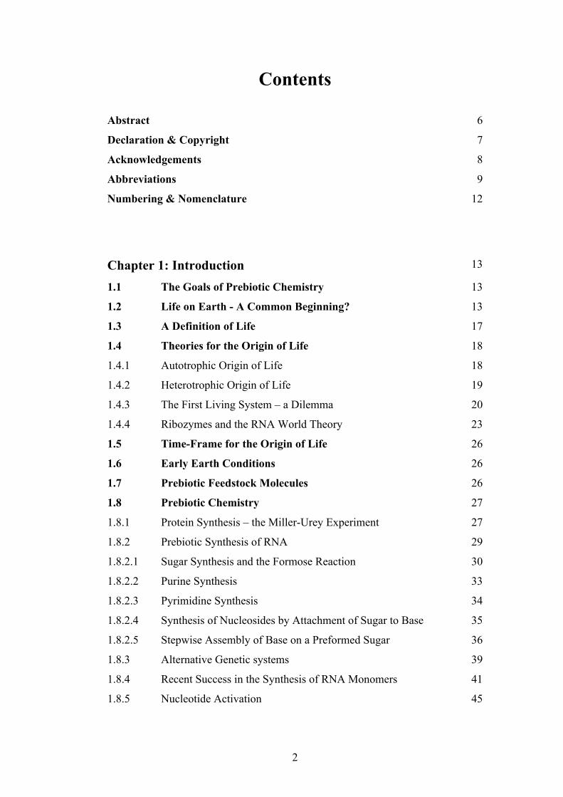
Contents Abstract 6
Declaration & Copyright 7
Acknowledgements 8
Abbreviations 9
Numbering & Nomenclature 12
Chapter 1: Introduction 13
1.1 The Goals of Prebiotic Chemistry 13
1.2 Life on Earth - A Common Beginning? 13
1.3 A Definition of Life 17
1.4 Theories for the Origin of Life 18
1.4.1 Autotrophic Origin of Life 18
1.4.2 Heterotrophic Origin of Life 19
1.4.3 The First Living System – a Dilemma 20
1.4.4 Ribozymes and the RNA World Theory 23
1.5 Time-Frame for the Origin of Life 26
1.6 Early Earth Conditions 26
1.7 Prebiotic Feedstock Molecules 26
1.8 Prebiotic Chemistry 27
1.8.1 Protein Synthesis – the Miller-Urey Experiment 27
1.8.2 Prebiotic Synthesis of RNA 29
1.8.2.1 Sugar Synthesis and the Formose Reaction 30
1.8.2.2 Purine Synthesis 33
1.8.2.3 Pyrimidine Synthesis 34
1.8.2.4 Synthesis of Nucleosides by Attachment of Sugar to Base 35
1.8.2.5 Stepwise Assembly of Base on a Preformed Sugar 36
1.8.3 Alternative Genetic systems 39
1.8.4 Recent Success in the Synthesis of RNA Monomers 41
1.8.5 Nucleotide Activation 45
3
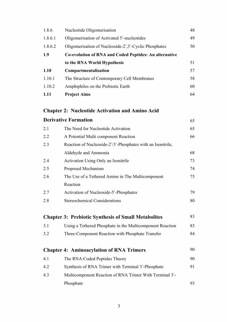
1.8.6 Nucleotide Oligomerisation 48
1.8.6.1 Oligomerisation of Activated 5ʹ′-nucleotides 49
1.8.6.2 Oligomerisation of Nucleoside-2ʹ′,3ʹ′-Cyclic Phosphates 50
1.9 Co-evolution of RNA and Coded Peptides: An alternative
to the RNA World Hypothesis 51
1.10 Compartmentalisation 57
1.10.1 The Structure of Contemporary Cell Membranes 58
1.10.2 Amphiphiles on the Prebiotic Earth 60
1.11 Project Aims 64
Chapter 2: Nucleotide Activation and Amino Acid
Derivative Formation 65
2.1 The Need for Nucleotide Activation 65
2.2 A Potential Multi component Reaction 66
2.3 Reaction of Nucleoside-2ʹ′/3ʹ′-Phosphates with an Isonitrile,
Aldehyde and Ammonia 68
2.4 Activation Using Only an Isonitrile 73
2.5 Proposed Mechanism 74
2.6 The Use of a Tethered Amine in The Multicomponent
Reaction
75
2.7 Activation of Nucleoside-5ʹ′-Phosphates 79
2.8 Stereochemical Considerations 80
Chapter 3: Prebiotic Synthesis of Small Metabolites 83
3.1 Using a Tethered Phosphate in the Multicomponent Reaction 83
3.2 Three-Component Reaction with Phosphate Transfer 84
Chapter 4: Aminoacylation of RNA Trimers 90
4.1 The RNA:Coded Peptides Theory 90
4.2 Synthesis of RNA Trimer with Terminal 3ʹ′-Phosphate 91
4.3 Multicomponent Reaction of RNA Trimer With Terminal 3ʹ′-
Phosphate 93
4
4.4 Synthesis of RNA Trimer with Terminal 5ʹ′-Phosphate 94
4.5 Multicomponent Reaction of RNA Trimer with Terminal 5ʹ′-
Phosphate
97
Chapter 5: Nucleoside Oligomerisation Studies 98
5.1 Oligomerisation of 2ʹ′,3ʹ′-Cyclic Phosphates 98
5.2 Drying Down Experiment of Cytidine-2ʹ′,3ʹ′-Cyclic Phosphate 99
5.3 Synthesis of Cytidine-3ʹ′-Phosphate Ethanolamine Adduct
Standard
101
5.4 Spiking Experiment of the Cytidine-3ʹ′-Phosphate
Ethanolamine Adduct
103
5.5 Drying Down Experiment of Uridine-2ʹ′,3ʹ′-Cyclic Phosphate 104
5.6 Synthesis of Uridine-3ʹ′-Phosphate Ethanolamine Adduct
Standard and Spiking Experiment
105
Chapter 6: Formation of Potentially Prebiotic
Amphiphiles 107
6.1 The Need for Prebiotic Compartmentalisation 107
6.2 A Potentially Predisposed Phosphorylation 109
6.3 Synthesis of β-Amino Alcohols 112
6.4 Reaction of β-Amino Alcohols with Trimetaphosphate 113
6.5 Synthesis of Standard and Spiking Experiment 116
6.6 Rationalisation for the Differing Reactivity 121
Chapter 7: Conclusions 123
Chapter 8: Experimental 125
8.1 General 125
8.2 Experimental Procedures for Chapter 2 128
8.3 Experimental Procedures for Chapter 3 136
8.4 Experimental Procedures for Chapter 4 139
8.5 Experimental Procedures for Chapter 5 152
5
8.6 Experimental Procedures for Chapter 6 165
References 172
Word Count: 39600
6
Abstract
The Sutherland group recently demonstrated the prebiotic synthesis of activated pyrimidine ribonucleotides as their 2ʹ′,3ʹ′-cyclic phosphates, and these species are candidates for oligomerisation to RNA. These species hydrolyse to the corresponding 2ʹ′- and 3ʹ′-monophosphates and there is a need to discover prebiotically plausible ways to re-activate to the cyclic material. Previous methods have suffered from poor yields and/or derivatization of the nucleobase. This study describes a new multicomponent reaction that achieves highly efficient nucleotide activation and at the same time produces amino acid derivatives, also of importance in the origin of life. This reactivity is then further developed and utilised in the prebiotic synthesis of derivatives of glyceric acid 2- and 3-phosphate, used in the glycolysis pathway in contemporary biochemistry. Aminoacyl-RNA trimers are central to the RNA:coded peptides theory by Sutherland, whereby RNA replication and coded peptide synthesis are proposed to have emerged together in the origin of life. The aminoacylation of an RNA trimer is therefore investigated, again using a multicomponent reaction. With the prebiotic synthesis and re-activation of nucleoside-2ʹ′,3ʹ′-cyclic phosphates shown, the oligomerisation of these species is now a major goal. The dry-state oligomerisation of these species using ethanolamine as catalyst is discussed. Key ethanolamine-adduct intermediates are identified, and the preference for the formation of natural [3ʹ′→5ʹ′] linkages produced by this type of oligomerisation is rationalised. The compartmentalisation of a primitive replicating genetic system is considered an important stage in the origin of life in order to overcome the high dilution of the oceans. Previous studies have focussed on long chain carboxylic acids for this purpose but these are unstable to the conditions required for RNA folding and catalysis, and only form bilayer vesicles at a specific pH. The final chapter investigates the prebiotic synthesis of a simple phospholipid amphiphile that has the potential to form more suitable lipid vesicles.
7
Declaration & Copyright No portion of the work referred to in the thesis has been submitted in support of an application for another degree or qualification of this or any other university or other institute of learning. The author of this thesis (including any appendices and/or schedules to this thesis) owns any copyright in it (the “Copyright”) and he has given The University of Manchester the right to use such Copyright for any administrative, promotional, educational and/or teaching purposes. Copies of this thesis, either in full or in extracts, may be made only in accordance with the regulations of the John Rylands University Library of Manchester. Details of these regulations may be obtained from the Librarian. This page must form part of any such copies made. The ownership of any patents, designs, trade marks and any and all other intellectual property rights except for the Copyright (the “Intellectual Property Rights”) and any reproductions of copyright works, for example graphs and tables (“Reproductions”), which may be described in this thesis, may not be owned by the author and may be owned by third parties. Such Intellectual Property Rights and Reproductions cannot and must not be made available for use without the prior written permission of the owner(s) of the relevant Intellectual Property Rights and/or Reproductions. Further information on the conditions under which disclosure, publication and exploitation of this thesis, the Copyright and any Intellectual Property Rights and/or Reproductions described in it may take place is available from the Head of School of The School of Chemistry. Those parts of this thesis having previously been published at the time of writing:
1. L. B. Mullen, J. D. Sutherland. Simultaneous nucleotide activation and synthesis of amino acid amides by a potentially prebiotic multi-component reaction. Angew. Chem. Int. Ed., 2007, 46, 8063.
2. L. B. Mullen, J. D. Sutherland. Formation of potentially prebiotic amphiphiles by reaction of β-hydroxy-n-alkylamines with cyclotriphosphate. Angew. Chem. Int. Ed., 2007, 46, 4166.
3. J. D. Sutherland, L. B. Mullen, F. F. Buchet. Potentially prebiotic Passerini-type reactions of phosphates. Synlett, 2008, 14, 2161.

8
Acknowledgements I would like to begin by thanking my supervisor, John. I am extremely grateful to
have had the opportunity to work under and learn from someone so enthusiastic,
knowledgeable and encouraging.
I am endlessly grateful to the utterly selfless Béatrice for the many hours she
patiently spent proofreading this thesis, you’re a star Béa!
To all the great people I’ve had to pleasure to work with over the years - Lello,
Béatrice, Claire, Matt, Chris, Jesús, Alastair, Fabien, Mikey, Guillaume, Carole,
Basile, Andrew - thanks for all the good memories.
Thanks to all my family and friends who have provided me with support over the
last few years, and especially for putting up with me whilst I was writing up - I
owe you all so much.
It was Stella that persuaded me that this whole thing would be a good idea, so in
many ways all that I have achieved is down to her inspiration, and for that reason
it is to her that I dedicate this thesis. I am also grateful for her recent support
through difficult times.
Finally, thanks to Edith, because I don’t know what I’d do without her.

9
Abbreviations A adenine
Ac acetyl
AICA 5-amino-imidazole-4-carboxamide
AICN 5-amino-imidazole-4-carbonitrile
AmTP amidotriphosphate
ATP adenosine triphosphate
aq. aqueous
APCI atmospheric pressure chemical ionisation
B nucleic acid base tBu tert-butyl
Bn benzyl
°C degrees Celsius
C cytosine
ca. circa
calcd. calculated
cAMP adenosine-3ʹ′,5ʹ′-cyclic phosphate
Celite® high grade diatomaceous earth filtration
agent
CI chemical ionisation
cm-1 wavenumber
conc. concentrated
COSY correlated spectroscopy (NMR)
δ chemical shift
DAMN diaminomaleonitrile
DCM dichloromethane
DMAP 4-(dimethylamino)-pyridine
DMF N,N-dimethylformamide
DMSO dimethylsulfoxide
DNA deoxyribonucleic acid
Eds. editors
ESI electrospray ionisation
10
Et ethyl
et al. et alia
eq. equivalent(s)
G guanine
GC gas chromatography
h hour(s)
hν electromagnetic irradiation (UV)
HPLC high performance liquid chromatography
Hz Hertz
i iso
IR infrared
J NMR coupling constant measured in Hertz
LCA Last Common Ancestor
lit. literature (reference)
m milli
M molar
Me methyl
MHz megahertz
min minute
mL millilitre
mmol millimole
m.p. melting point
MS mass spectrometry
µl microlitre
m/z mass/charge ratio
NMR nuclear magnetic resonance
Ph phenyl
Pi inorganic phosphate
PNA peptide nucleic acid
PPi inorganic pyrophosphate
ppm parts per million
p-RNA pyranosyl ribonucleic acid
py. pyridine
11
quant. quantitative yield
R unspecified group
rac- racemic mixture
RNA ribonucleic acid
r.t. room temperature
sat. saturated
soln. solution
t tertiary
tert tertiary
T thymine
t1/2 half life
TBDMS tert-butyldimethylsilyl
TFA trifluoroacetic acid
THF tetrahydrofuran
TLC thin layer chromatography
TNA L-α-threofuranosyl (3'-2') nucleic acid
U uracil
UV ultraviolet
12
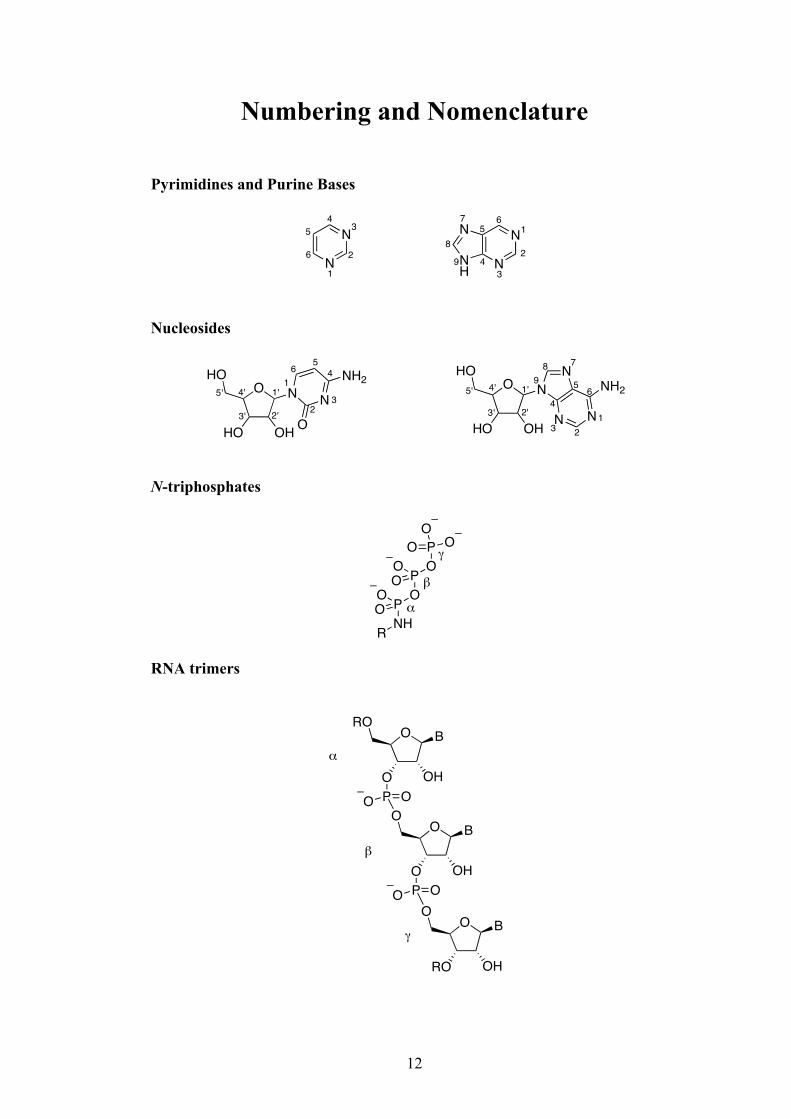
Numbering and Nomenclature
Pyrimidines and Purine Bases
Nucleosides
N-triphosphates
RNA trimers
N
N
N
NH9
8
7 65 1
2
34N
N
1
2
34
5
6
O
HO OH
NHO
N
O
NH2 O
HO OH
NHO N
N N
NH21
23
45
6
1'
2'3'
4'5' 1'
2'3'
4'5'
123
4
5 6
789
NHP OP OPO
O
OO
O
R
OO
!
"
#
O
RO OH
O
O
O OH
O
P OO
O B
O OH
RO
P OO
!
"
#
B
B
13
1. Introduction 1.1 The Goals of Prebiotic Chemistry The precise nature of the emergence of life on Earth is surely one of the most
fundamental puzzles that scientific endeavour can hope to answer. The transition
from a lifeless planet billions of years ago to one that is now occupied by a
species so advanced that it actually has the consciousness to ponder this very
question is a phenomenon that must intrigue scientists of every discipline. Whilst
physicists theorise on the origin of the universe, and biologists demonstrate in
increasing and more incredible detail what life actually is, it is the chemist that
must ultimately discover how life sprung spontaneously from an array of
inanimate molecules. Indeed, in a recent article in Nature entitled ‘What
Chemists Want to Know’, it is no surprise that the question of how life began on
Earth was included.[1]
In the broadest sense, the role of prebiotic chemistry is to carry out reactions that
model as close as possible the chemistry that took place on the early Earth that
gave rise to the emergence of life. Through these experiments it is hoped that an
understanding of precisely what reactions occurred, where they occurred and in
what order they occurred can be gained. Inevitably however, due to the lack of
certainty of a huge number of variables such as planetary conditions and identity
of starting materials, as well as the impossibility of any direct evidence from the
time period concerned, the best a chemist can hope for is a set of experiments that
demonstrate how it may have occurred. Albert Eschenmoser perfectly summed up
this sentiment in saying that “The origin of life cannot be ‘discovered’, it has to be
‘re-invented’”.[2]
1.2 Life on Earth – a Common Beginning?
To theorise on and perform experiments towards understanding the origin of life,
it is absolutely necessary to first have a clear definition of what life actually is.
Due to the vast diversity of species on Earth today, it may seem like the task of
14
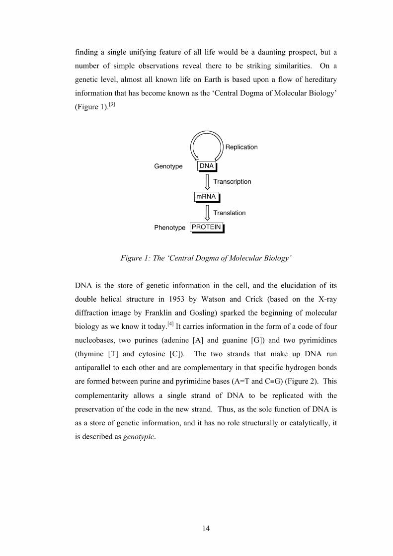
finding a single unifying feature of all life would be a daunting prospect, but a
number of simple observations reveal there to be striking similarities. On a
genetic level, almost all known life on Earth is based upon a flow of hereditary
information that has become known as the ‘Central Dogma of Molecular Biology’
(Figure 1).[3]
Figure 1: The ‘Central Dogma of Molecular Biology’
DNA is the store of genetic information in the cell, and the elucidation of its
double helical structure in 1953 by Watson and Crick (based on the X-ray
diffraction image by Franklin and Gosling) sparked the beginning of molecular
biology as we know it today.[4] It carries information in the form of a code of four
nucleobases, two purines (adenine [A] and guanine [G]) and two pyrimidines
(thymine [T] and cytosine [C]). The two strands that make up DNA run
antiparallel to each other and are complementary in that specific hydrogen bonds
are formed between purine and pyrimidine bases (A=T and C≡G) (Figure 2). This
complementarity allows a single strand of DNA to be replicated with the
preservation of the code in the new strand. Thus, as the sole function of DNA is
as a store of genetic information, and it has no role structurally or catalytically, it
is described as genotypic.
DNA
mRNA
PROTEIN
Replication
Transcription
Translation
Genotype
Phenotype
15
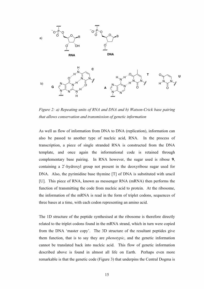
Figure 2: a) Repeating units of RNA and DNA and b) Watson-Crick base pairing
that allows conservation and transmission of genetic information
As well as flow of information from DNA to DNA (replication), information can
also be passed to another type of nucleic acid, RNA. In the process of
transcription, a piece of single stranded RNA is constructed from the DNA
template, and once again the informational code is retained through
complementary base pairing. In RNA however, the sugar used is ribose 9,
containing a 2ʹ′-hydroxyl group not present in the deoxyribose sugar used for
DNA. Also, the pyrimidine base thymine [T] of DNA is substituted with uracil
[U]. This piece of RNA, known as messenger RNA (mRNA) then performs the
function of transmitting the code from nucleic acid to protein. At the ribosome,
the information of the mRNA is read in the form of triplet codons, sequences of
three bases at a time, with each codon representing an amino acid.
The 1D structure of the peptide synthesised at the ribosome is therefore directly
related to the triplet codons found in the mRNA strand, which in turn were copied
from the DNA ‘master copy’. The 3D structure of the resultant peptides give
them function, that is to say they are phenotypic, and the genetic information
cannot be translated back into nucleic acid. This flow of genetic information
described above is found in almost all life on Earth. Perhaps even more
remarkable is that the genetic code (Figure 3) that underpins the Central Dogma is
O B
O OH
OPO
OO B
O
OPO
O
N
NN
NNH H
N
NN
NO
N
H
H
H
N N
O
OH
N N
N
O
H
H
G
C
A
T
N N
O
OH
U
RNA DNA
a)
b)
16
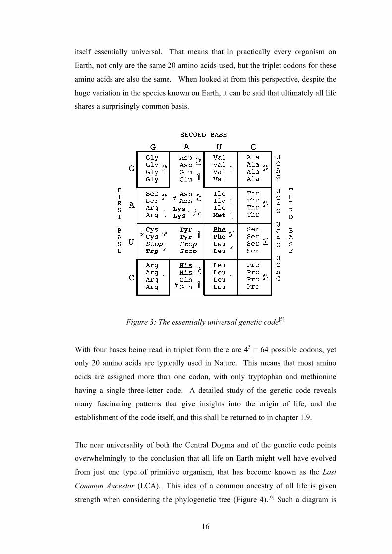
itself essentially universal. That means that in practically every organism on
Earth, not only are the same 20 amino acids used, but the triplet codons for these
amino acids are also the same. When looked at from this perspective, despite the
huge variation in the species known on Earth, it can be said that ultimately all life
shares a surprisingly common basis.
Figure 3: The essentially universal genetic code[5]
With four bases being read in triplet form there are 43 = 64 possible codons, yet
only 20 amino acids are typically used in Nature. This means that most amino
acids are assigned more than one codon, with only tryptophan and methionine
having a single three-letter code. A detailed study of the genetic code reveals
many fascinating patterns that give insights into the origin of life, and the
establishment of the code itself, and this shall be returned to in chapter 1.9.
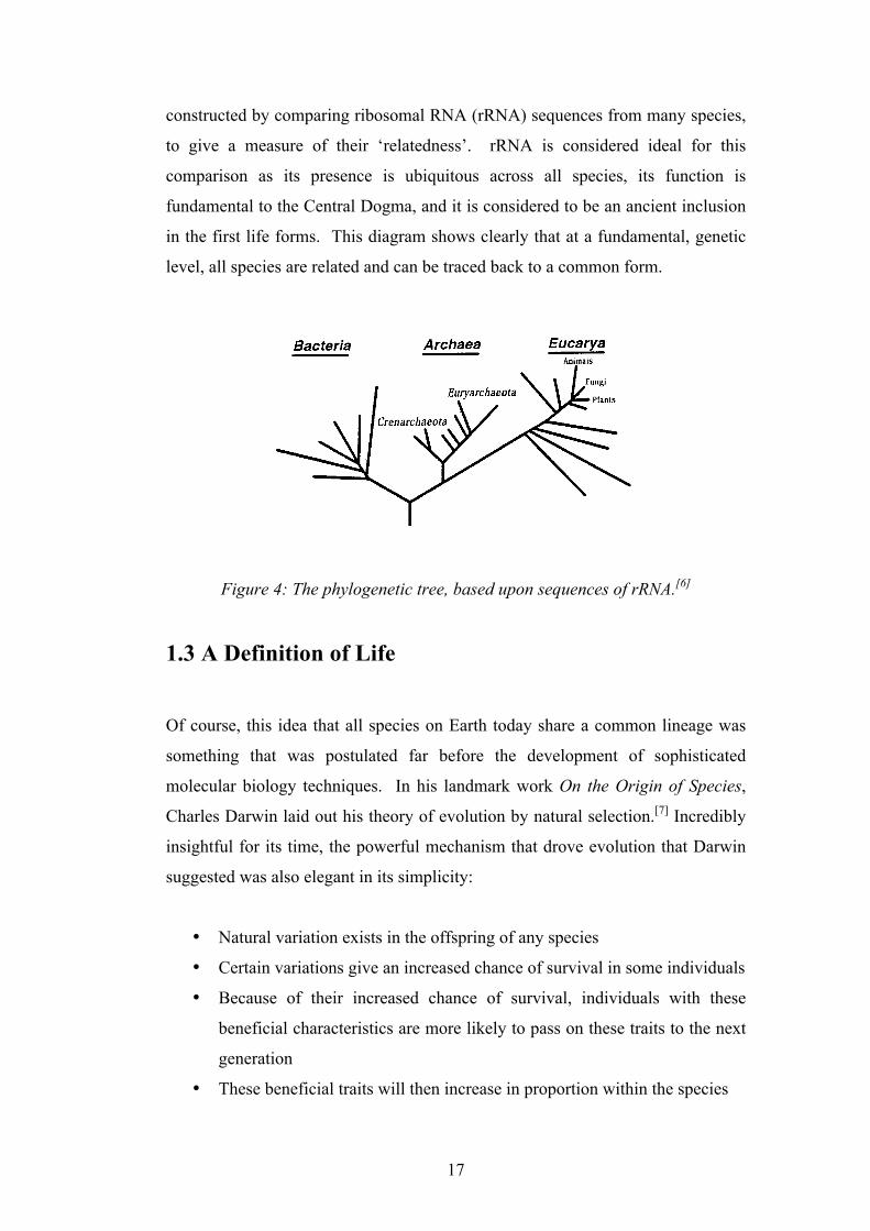
The near universality of both the Central Dogma and of the genetic code points
overwhelmingly to the conclusion that all life on Earth might well have evolved
from just one type of primitive organism, that has become known as the Last
Common Ancestor (LCA). This idea of a common ancestry of all life is given
strength when considering the phylogenetic tree (Figure 4).[6] Such a diagram is
The order of assignment of amino acids to codons has also been considered in thecontext of prebiotic amino acid availabilty. Since translation must have predated anextensive enzyme-mediated metabolism, it is thought that a restricted set of prebioti-cally available amino acids was originally used in translation [22]. Depletion of theseprebiotically available amino acids by incorporation into (coded) peptides would haveprovided a strong driving force for the development of biosynthetic pathways to them.Low catalytic efficiency and restricted scope would most likely have resulted in thepathways being recruited retroacquisitively by catalysis of underlying, predisposedchemistry [28]. According to this hypothesis, the first amino acids must have beenprebiotically available, and other amino acids could not be used until they had becomeavailable for the first time by biosynthesis. Biosynthesis of these later amino acidswould have taken place when a more advanced enzymological repertoire was available,would not have been driven by environmental depletion as discussed above, andconsequently need not (necessarily) have been acquired retroacquisitively [29]. In
Fig. 1. Chemical Analysis of the Genetic Code. In using the genetic code to guide retrosynthetic disconnectionsof RNA :coded peptides, the etiology of the code must be considered. Some of the amino acids have (andrequire) long and complex biosyntheses and appear late additions to the code (bold). An analysis of prebioticavailability suggests that certain amino acids could not have been assigned to the code at the outset(underlined). The allocation of the aminoacyl-tRNA synthetases to either one of two classes (outline) isviolated in one case, and there are certain charging discrepancies (asterisks) which indicate recent assignments.Stop codons (italics) which would otherwise severely limit the length of (random sequence) RNA translationproducts are thought to be late assignments. Finally, parsimony suggests that those amino acids in family boxes(framed in bold) and only coded by the first two bases of the codon are the earliest assignments. When the codeis viewed according to these (chemical) criteria, a distinct pattern emerges with XAZ and UYZ codons
appearing as late assignments.
CHEMISTRY & BIODIVERSITY ± Vol. 1 (2004)210
17
constructed by comparing ribosomal RNA (rRNA) sequences from many species,
to give a measure of their ‘relatedness’. rRNA is considered ideal for this
comparison as its presence is ubiquitous across all species, its function is
fundamental to the Central Dogma, and it is considered to be an ancient inclusion
in the first life forms. This diagram shows clearly that at a fundamental, genetic
level, all species are related and can be traced back to a common form.
Figure 4: The phylogenetic tree, based upon sequences of rRNA.[6]
1.3 A Definition of Life Of course, this idea that all species on Earth today share a common lineage was
something that was postulated far before the development of sophisticated
molecular biology techniques. In his landmark work On the Origin of Species,
Charles Darwin laid out his theory of evolution by natural selection.[7] Incredibly
insightful for its time, the powerful mechanism that drove evolution that Darwin
suggested was also elegant in its simplicity:
• Natural variation exists in the offspring of any species
• Certain variations give an increased chance of survival in some individuals
• Because of their increased chance of survival, individuals with these
beneficial characteristics are more likely to pass on these traits to the next
generation
• These beneficial traits will then increase in proportion within the species
Interpreting the universal phylogenetic treeCarl R. Woese*
Department of Microbiology, University of Illinois at Urbana-Champaign, B103 Chemical and Life Sciences Laboratory, MC-110, 601 South Goodwin Avenue,Urbana, IL 61801-3709
Contributed by Carl R. Woese, May 22, 2000
The universal phylogenetic tree not only spans all extant life, butits root and earliest branchings represent stages in the evolution-ary process before modern cell types had come into being. Theevolution of the cell is an interplay between vertically derived andhorizontally acquired variation. Primitive cellular entities werenecessarily simpler and more modular in design than are moderncells. Consequently, horizontal gene transfer early on was perva-sive, dominating the evolutionary dynamic. The root of the uni-versal phylogenetic tree represents the first stage in cellularevolution when the evolving cell became sufficiently integratedand stable to the erosive effects of horizontal gene transfer thattrue organismal lineages could exist.
Archaea ! Bacteria ! Eucarya ! universalancestor ! horizontal gene transfer
The Grand Challenge
In a letter to T. H. Huxley in 1857, Darwin, with characteristicprescience, foresaw ‘‘[t]he time . . . when we shall have very
fairly true genealogical trees of each great kingdom of nature’’(1), voicing in the terms of his day one of the great, definingchallenges of Biology. Another century would pass, however,before Darwin’s vision became reality. Darwin obviously knewthat the methodologies of the day, paleontology and classicaltaxonomy, were not up to a task this monumental. What couldnot be foreseen, however, was that, as Biology moved to amolecular footing in the following century, evolution wouldcease to be a focus, and what Darwin considered a basic problemwould effectively fade from view. Yet a vision this central, thisessentially biological, cannot remain forever obscured. In the1960s, with the advent of molecular sequencing, gene historiesand organismal genealogies emerged on the molecular stage (2);and with the recent eruption of genomic sequencing, the fullhistory of cellular life on this planet seems now to be unfoldingbefore our eyes.
What molecular sequences taught us in the 1960s was that thegenealogical history of an organism is written to one extent oranother into the sequences of each of its genes, an insight thatbecame the central tenet of a new discipline, molecular evolution(2). The most important distinction between the new molecularapproach to evolutionary relationships and the older classicalones was that molecules ancestral to a group, whose phenotypesare invariant within the group (i.e., plesiomorphies), could nowbe used to infer phylogenetic relationships within the group.Thus, by comparing the sequences of molecules whose functionsare universal, it was possible not only to construct genealogicaltrees for Darwin’s great kingdoms, but also to go beyond this andconstruct a universal phylogenetic tree, one that united all of thekingdoms into a single phylogenetic ‘‘empire.’’
Ribosomal RNA was central to this endeavor. Not only is themolecule ubiquitous, but it exhibits functional constancy, itchanges slowly in sequence, and it is (and was) experimentallyvery tractable. Moreover, as the central component of the highlycomplex translation apparatus, rRNA is among the most refrac-tory of molecules to the vagaries of horizontal gene flow, and sowas considered likely to avoid the phylogenetic hodgepodge ofreticulate evolution and preserve a bona fide organismal trace(3). The rRNA-based universal phylogenetic tree (Fig. 1)
brought Biology to an evolutionary milestone, a comprehensiveoverview of organismal history as well as to the limit of theclassical Darwinian perspective.
The initial and strongest impact of the universal tree has beenin microbiology. For the first time, microbiology sits within aphylogenetic framework and thereby is becoming a compleatbiological discipline: the study of microbial diversity has movedfrom a collection of isolated vignettes to a meaningful study inrelationships. Because niches can now be defined in organismalterms, microbial ecology–long ecology in name only–is becom-ing ecology in the true sense of the word (7). Yet, the ultimateand perhaps most important impact of the universal phyloge-netic tree will be in providing Biology as a whole with a new andpowerful perspective, an image that unifies all life through itsshared histories and common origin, at the same time empha-sizing life’s incredible diversity and the overwhelming impor-tance of the microbial world (historically so, and in terms of thebiosphere).
A New Era, a New PerspectiveIn the 1990s, Biology entered the genomic era. It is ironic that(microbial) genomics, which offers such promise for developingthe universal phylogenetic tree as a basal evolutionary frame-work, has seemed initially to do just the opposite. Now that thesequences of many molecules, whose distributions are phyloge-netically broad if not universal, are known, biologists find thatuniversal phylogenetic trees inferred from many of them do notfundamentally agree with the rRNA-based universal phyloge-netic tree (8). The cause of this incongruity is, of course,reticulate evolution, horizontal gene flow. And the reaction toit–at least according to scientific editorial accounts (9, 10)–hasbeen one of the sky falling. There are grains of truth here. But
*E-mail: [email protected].
The publication costs of this article were defrayed in part by page charge payment. Thisarticle must therefore be hereby marked “advertisement” in accordance with 18 U.S.C.§1734 solely to indicate this fact.
Fig. 1. The basal universal phylogenetic tree inferred from comparativeanalyses of rRNA sequences (4, 5). The root has been determined by using theparalogous gene couple EF-Tu"EFG (6).
8392–8396 ! PNAS ! July 18, 2000 ! vol. 97 ! no. 15
18
Therefore it is simple to understand how, given these basic principles, beginning
from a primitive common ancestor, generation after generation of natural
selection would give rise to huge variation within species and eventually, lead to
new ones. It is astonishing that Darwin could conceive of such an accurate
hypothesis at a time when the molecular details that drive evolution were
completely unknown. We now know how hereditary information is passed on, by
the replication of DNA. Although this replication is catalysed by highly efficient
polymerase enzymes, it is imperfect, so that a small error rate is present, and it is
these errors that give rise to the variation so important to evolution. So, now onto
a definition of life. Clearly, evolution is something that is at the very essence of
what makes something living, and so it must be integral in an accurate definition.
It is now widely accepted that the definition given by Joyce seems to be the most
acceptable:
“Life is a self-sustaining chemical system capable of undergoing Darwinian
evolution”.[8]
Precisely what this ‘chemical system’ might have been that could be considered
the first form of life will be returned to later.
1.4 Theories for the Origin of Life
1.4.1 Autotrophic Origin of Life
So-called autotrophic theories for the origin of life are based around the
assumption that the first life-form could not have been constructed from the
complex organic molecules such as nucleic acids and/or proteins that are at the
centre of contemporary biochemistry. This way of thinking is due to the apparent
difficulties with the de novo synthesis of these compounds from small precursors.
Instead, it is suggested that the first ‘life forms’ were initially based around
energetically favourable metabolic cycles of inorganic molecules that once
established, gave rise to, or were ‘taken over’ by more complex gene-based
19
systems. Wächtershäuser’s “Iron-Sulfur World” suggests that the first organism
derived reducing power from the thermodynamically favourable process by which
mackinawite (FeS) is converted to iron pyrites (FeS2) by hydrogen sulfide.[9] This
energy obtained would then drive the entropically unfavourable process of
constructing an informational genetic system, with the organic material being
produced from the reduction of atmospheric CO2.
Cairns-Smith suggested that the first form of life may have originated within the
patterns of clay minerals.[10] The ability of certain silicate lattices to grow with
imperfections is said to be a source of evolving, primitive genetic information,
and some sort of ‘genetic takeover’ is said to have occurred on the surface of
these minerals whereby the organic genetic system we know today retained the
information contained within the lattice. The major problem with autotrophic
theories of life is that they suffer from a lack of experimental demonstration,
particularly when concerning the intriguing ‘genetic-takeover’ step.
1.4.2 Heterotrophic Origin of Life
Heterotrophic theories for the origin of life propose that the first form of life was
based on a replicating system of organic macromolecules like the ones found in
contemporary biochemistry (chapter 1.2). These molecules were said to have
formed spontaneously from a prebiotic ‘soup’ of small organic molecules
available on the early Earth. Whilst this theory has its own difficulties, not least
the problem of the de novo synthesis of relatively complex molecules, it does not
require the inclusion of vague and undescribed ‘genetic takeover’ steps.
Moreover, there is a solid grounding of experimental demonstration over many
years that makes this type of process seem to be entirely plausible, if not yet
actually demonstrated completely. In considering the nature of the origin of life,
this thesis will assume a heterotrophic origin of life on Earth. With this
assumption having been made, the next step is to consider just how this “self-
sustaining chemical system capable of undergoing Darwinian evolution” emerged
from the prebiotic ‘soup’. As pondered by Darwin himself:
20
“But if we could conceive in some warm pond, with all sorts of ammonia and
phosphoric salts, light, heat, electricity, etc., present that a proteine [sic]
compound was chemically formed.”[11]
1.4.3 The First Living System – a Dilemma
If one looks at the Central Dogma that is present in practically all life on Earth,
and then considers the definition of life put forward in chapter 1.3, one is
immediately confronted by an apparently insoluble paradox when trying to
suggest what sort of single molecule the first form of life was based upon.
Nucleic acids contain the instructions needed for an organism to biosynthesise
proteins. Yet nucleic acids require highly complex and specific proteins
(enzymes) in every stage of their function. In DNA replication, the first stage is
for the double helix to separate into its two single complimentary strands.
Nucleotide monomers (in the form of deoxynucleoside-5ʹ′-triphosphates) are then
added to a growing chain in the [5ʹ′→3ʹ′] direction, with the monomers of the new
chain being matched by Watson-Crick base pairing to the residues on the existing
chain. DNA polymerases catalyse the phosphodiester bond formation between
the 3ʹ′-end of the growing chain and the 5ʹ′-phosphate of the incoming
deoxynucleoside-5ʹ′-triphosphate. The release of pyrophosphate and its hydrolysis
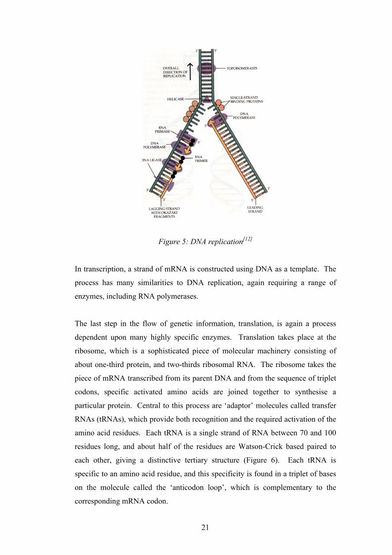
by pyrophosphatase drive the process energetically. This is a simplified account
of DNA replication and in all, over 20 enzymes are required for the process
(Figure 5).
21
Figure 5: DNA replication[12]
In transcription, a strand of mRNA is constructed using DNA as a template. The
process has many similarities to DNA replication, again requiring a range of
enzymes, including RNA polymerases.
The last step in the flow of genetic information, translation, is again a process
dependent upon many highly specific enzymes. Translation takes place at the
ribosome, which is a sophisticated piece of molecular machinery consisting of
about one-third protein, and two-thirds ribosomal RNA. The ribosome takes the
piece of mRNA transcribed from its parent DNA and from the sequence of triplet
codons, specific activated amino acids are joined together to synthesise a
particular protein. Central to this process are ‘adaptor’ molecules called transfer
RNAs (tRNAs), which provide both recognition and the required activation of the
amino acid residues. Each tRNA is a single strand of RNA between 70 and 100
residues long, and about half of the residues are Watson-Crick based paired to
each other, giving a distinctive tertiary structure (Figure 6). Each tRNA is
specific to an amino acid residue, and this specificity is found in a triplet of bases
on the molecule called the ‘anticodon loop’, which is complementary to the
corresponding mRNA codon.
22
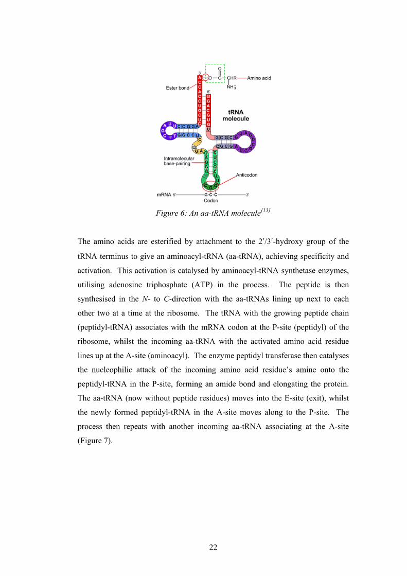
Figure 6: An aa-tRNA molecule[13]
The amino acids are esterified by attachment to the 2ʹ′/3ʹ′-hydroxy group of the
tRNA terminus to give an aminoacyl-tRNA (aa-tRNA), achieving specificity and
activation. This activation is catalysed by aminoacyl-tRNA synthetase enzymes,
utilising adenosine triphosphate (ATP) in the process. The peptide is then
synthesised in the N- to C-direction with the aa-tRNAs lining up next to each
other two at a time at the ribosome. The tRNA with the growing peptide chain
(peptidyl-tRNA) associates with the mRNA codon at the P-site (peptidyl) of the
ribosome, whilst the incoming aa-tRNA with the activated amino acid residue
lines up at the A-site (aminoacyl). The enzyme peptidyl transferase then catalyses
the nucleophilic attack of the incoming amino acid residue’s amine onto the
peptidyl-tRNA in the P-site, forming an amide bond and elongating the protein.
The aa-tRNA (now without peptide residues) moves into the E-site (exit), whilst
the newly formed peptidyl-tRNA in the A-site moves along to the P-site. The
process then repeats with another incoming aa-tRNA associating at the A-site
(Figure 7).

23
Figure 7: Translation at the ribosome[14]
The proteins synthesised at the ribosome then take on a three-dimensional shape
dependent upon the side chains of the amino acid residues, and it is this 3D
structure that gives the protein its function. Thus, the 1D structure of the genetic
code stored within the parent DNA (genotype) has been translated into a
functional 3D structure of a protein (phenotype). This flow of genetic information
that is crucial to life is therefore a process in which nucleic acids and proteins are
highly dependent upon each other, so in the case of the origin of life, how could
one of these types of molecule have possibly arisen first without the other?
1.4.4 Ribozymes and the RNA World Theory
In chapter 1.2 it was pointed out that the sole function of DNA is to store
information as the genetic code, and this code was written in the language of
triplets of bases, or codons. It has no functional or catalytic properties, and is said
to be genotypic. On the other hand, the proteins that are coded for by the DNA
fold into a complex tertiary structure that gives them function, and the sequence
of amino acids within them cannot be propagated or transmitted back to nucleic
acid, they are said to be phenotypic. In the case of RNA, the distinction is not so
clear-cut. As mentioned previously, the structure of RNA differs in two ways
from DNA. Firstly, the base uracil is used in place of thymine and secondly, the
sugar used (ribose 9) has a 2ʹ′-hydroxyl group that is not present in DNA. This
24
second feature has a dramatic effect on the structure of RNA, in that instead of
forming a double helix, it is capable of adopting well-defined tertiary structures
when single stranded. This was known in the 1960s and it led Woese,[15] Crick[16]
and Orgel[17] to speculate that at the origin of life, RNA alone might have
performed the dual function of information storage and as a catalyst.
This idea that RNA could have been the first primitive form of life seems all the
more plausible when one considers its many roles in biochemistry today: tRNAs
are central to peptide synthesis, the ribosome is made mostly from RNA, ATP is
the universal energy storage molecule, and other uses include the chemical
messenger cAMP and coenzymes such as FAD and NAD. Finally, the fact that
DNA nucleotide monomers are themselves synthesised from RNA nucleotides
using ribonucleotide reductase[18] further suggests that RNA had an ancient role
before the emergence of DNA and proteins. It was thought that the apparent
problem of deciding whether nucleic acid or protein emerged first could be
overcome by the fact that RNA could have acted alone in performing the tasks
that are today taken on by both DNA and proteins. After a period of time where
RNA had been established as a self-sustaining system, it was postulated that DNA
took over as information carrier, and proteins as functional, catalytic molecules.
In the case of DNA, it is better suited for reliable information storage as its lack of
a 2ʹ′-hydroxyl group means that it is less susceptible to damage by hydrolysis,
where in RNA this 2ʹ′-hydroxyl group can cleave the phosphodiester bond by
nucleophilic attack. The transition to proteins for functionality can be explained
by the fact that the wide range of side chains available gives much more scope for
tertiary structures and hence specificity.
These early theories were given huge support when in the 1980s, Cech[19] and
Altman[20] demonstrated experimentally what had been predicted, RNA could
indeed act as a kind of enzyme, with their discovery of ribozymes. Whilst
studying the excision of introns in a ribosomal RNA gene of Tetrahymena
thermophilia, Cech found that the intron could be spliced out in the absence of
any enzymes. He concluded that the RNA gene was capable of breaking and
reforming the phosphodiester bonds on its own.[19] Separately, Altman was
25
studying the processing of tRNA molecules by the enzyme ribonucleasease-P.
This enzyme contained protein and RNA regions, but when stripped of the protein
component, it was found that the tRNA precursor molecules were still converted
into active tRNAs, showing the true catalytic properties of the RNA itself.[20]
Since then, there has been much interest in this fascinating field of work, and it
has now been shown that there are ribozymes that are capable of catalyzing their
own synthesis, albeit under very specific conditions.[21]
The discovery of ribozymes re-invigorated origin of life studies, and led to Gilbert
introducing the phrase “The RNA World”[22] which firmly stated that RNA could
indeed have been the first form of life on Earth, due to its ability to store genetic
information in its sequence of bases, and crucially, to also have the potential to
catalyse not only its own replication but also other chemical reactions. The
paradox of whether proteins or nucleic acids came first was apparently solved.
The prebiotic synthesis of RNA is therefore arguably the greatest challenge to the
prebiotic chemist, and has certainly been the focus of much investigation for
decades.
Whilst this thesis does not strictly assume an ‘RNA-only World’ stance, it
recognises that RNA was indeed of great importance to the origin of life on Earth,
whether on its own, or in-tandem with other types of molecules. The possibility
of a linked origin of RNA and peptides will be discussed in chapter 1.9, and the
need for compartmentalisation by amphiphiles will be introduced in chapter 1.10.
Now that we have an idea of what life is and what could have constituted the
earliest of life forms in the origin of life, the next step is to consider how the
prebiotic chemist can recreate the chemistry that gave rise to it. It is first
important to outline what is known, with at least some uncertainty, about the
environment of the early Earth that allowed the right chemistry to take place that
led to this phenomenon.
26
1.5 Time-Frame for the Origin of Life
The Earth is approximately 4.5 billion years old. For the first half-billion years of
its existence, it is thought that Earth was under heavy bombardment from objects
large enough to not only evaporate the oceans, but to completely sterilise its
surface.[23, 24] Incredibly, some microfossils are well enough preserved that show
some organisms dating back to 3.5 billion years ago that are similar to modern
blue-green algae.[25] Taking into consideration how long it may have taken such
(relatively) complex organisms to evolve, it is thought that life in some form was
present on Earth as long ago as 3.8 billion years.[26] This means that in all
likelihood, life emerged from a prebiotic environment in the surprisingly short
time (in geological terms) of around two hundred million years.
1.6 Early Earth Conditions
The precise nature of the early Earth’s atmosphere that gave rise to life is perhaps
one of the biggest uncertainties of prebiotic chemistry. Although not accepted by
everybody in the field, one of the most widely-held beliefs (and the one that has
paved the way to most experimentally demonstrated results) is that the
atmosphere was highly reduced and rich in methane (CH4), ammonia (NH3),
water (H2O) and hydrogen (H2).[18] The lack of photosynthesising life at this early
stage meant that there was a lack of atmospheric O2, and in turn the absence of an
ozone layer would have seen the Earth being subjected to large amounts of solar
radiation.
1.7 Prebiotic Feedstock Molecules
To attain a realistic idea of which small organic molecules may have been present
on the early Earth, there are three main ways in which clues can be found. Spark
discharge and UV irradiation experiments of the gases believed to be on the early
Earth simulate the act of lightning and solar radiation on the early atmosphere.[27,
28] Observation of interstellar space and of other bodies in the solar system, in
particular Saturn’s largest moon, Titan, can indicate what Earth may have been
27
like before life emerged.[29-32] Finally, analysis of carbonaceous meteorites (or
‘chondrites’) delivered to the Earth suggest which molecules are produced in
interstellar space, presumably abiotically, and therefore could have also been
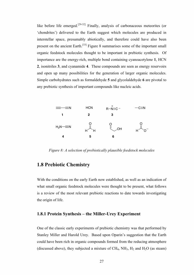
present on the ancient Earth.[33] Figure 8 summarises some of the important small
organic feedstock molecules thought to be important in prebiotic synthesis. Of
importance are the energy-rich, multiple bond containing cyanoacetylene 1, HCN
2, isonitriles 3, and cyanamide 4. These compounds are seen as energy reservoirs
and open up many possibilities for the generation of larger organic molecules.
Simple carbohydrates such as formaldehyde 5 and glycolaldehyde 6 are pivotal to
any prebiotic synthesis of important compounds like nucleic acids.
Figure 8: A selection of prebiotically plausible feedstock molecules
1.8 Prebiotic Chemistry
With the conditions on the early Earth now established, as well as an indication of
what small organic feedstock molecules were thought to be present, what follows
is a review of the most relevant prebiotic reactions to date towards investigating
the origin of life.
1.8.1 Protein Synthesis – the Miller-Urey Experiment
One of the classic early experiments of prebiotic chemistry was that performed by
Stanley Miller and Harold Urey. Based upon Oparin’s suggestion that the Earth
could have been rich in organic compounds formed from the reducing atmosphere
(discussed above), they subjected a mixture of CH4, NH3, H2 and H2O (as steam)
O
HH
O
OH
OOH
HCNN R N C C N
NH2N
1 2 3
4 5 6
28
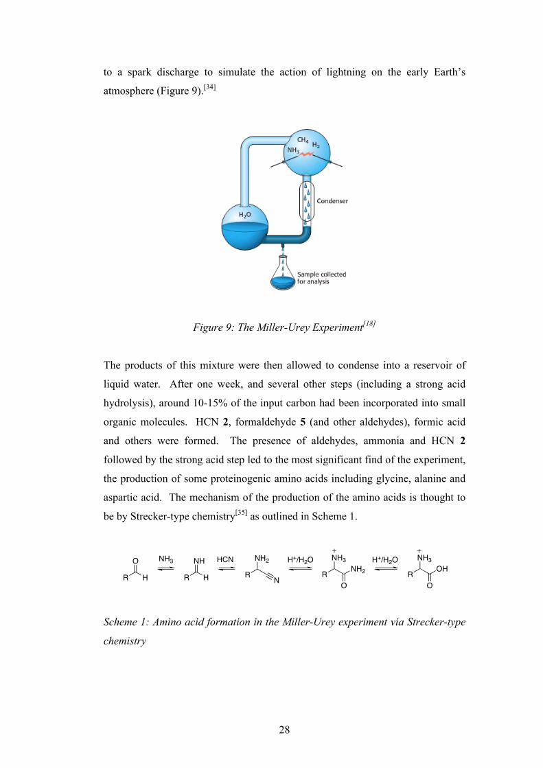
to a spark discharge to simulate the action of lightning on the early Earth’s
atmosphere (Figure 9).[34]
Figure 9: The Miller-Urey Experiment[18]
The products of this mixture were then allowed to condense into a reservoir of
liquid water. After one week, and several other steps (including a strong acid
hydrolysis), around 10-15% of the input carbon had been incorporated into small
organic molecules. HCN 2, formaldehyde 5 (and other aldehydes), formic acid
and others were formed. The presence of aldehydes, ammonia and HCN 2
followed by the strong acid step led to the most significant find of the experiment,
the production of some proteinogenic amino acids including glycine, alanine and
aspartic acid. The mechanism of the production of the amino acids is thought to
be by Strecker-type chemistry[35] as outlined in Scheme 1.
Scheme 1: Amino acid formation in the Miller-Urey experiment via Strecker-type
chemistry
evolutionary time was ribose incorporated to form nucleic acids as we know them today. Despite these uncertainties, an assortment of prebiotic molecules did arise in some fashion, and from this assortment those with properties favorable for the processes that we now associate with life began to interact and to form more complicated compounds. The processes through which modern organisms synthesize molecular building blocks will be discussed in Chapters 24, 25, and 26.
I. The Molecular Design of Life 2. Biochemical Evolution 2.1. Key Organic Molecules Are Used by Living Systems
Figure 2.1. The Urey-Miller Experiment. An electric discharge (simulating lightning) passed through an atmosphere of CH4, NH3, H2O, and H2 leads to the generation of key organic compounds such as amino acids. I. The Molecular Design of Life 2. Biochemical Evolution 2.1. Key Organic Molecules Are Used by Living Systems
Figure 2.2. Products of Prebiotic Synthesis. Amino acids produced in the Urey-Miller experiment. I. The Molecular Design of Life 2. Biochemical Evolution 2.1. Key Organic Molecules Are Used by Living Systems
Figure 2.3. Prebiotic Synthesis of a Nucleic Acid Component. Adenine can be generated by the condensation of HCN. O
HR
NH2
R N
NH
HR
NH3 HCN NH3
RO
NH2NH3
RO
OHH+/H2O H+/H2O
29
With the addition of H2S to the reaction mixture, it was found that 13 of the 20
proteinogenic amino acids could be formed, albeit in very small yield. Despite
the shortcomings of the experiment, such as the prebiotic plausibility of the strong
acid step and the low yields, it was the first experiment of its type to demonstrate
that molecules of biological importance could be made under simulated prebiotic
conditions.
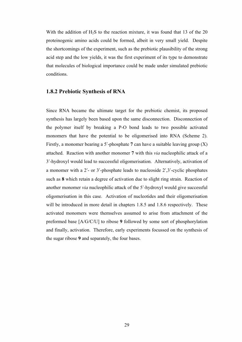
1.8.2 Prebiotic Synthesis of RNA
Since RNA became the ultimate target for the prebiotic chemist, its proposed
synthesis has largely been based upon the same disconnection. Disconnection of
the polymer itself by breaking a P-O bond leads to two possible activated
monomers that have the potential to be oligomerised into RNA (Scheme 2).
Firstly, a monomer bearing a 5ʹ′-phosphate 7 can have a suitable leaving group (X)
attached. Reaction with another monomer 7 with this via nucleophilic attack of a
3ʹ′-hydroxyl would lead to successful oligomerisation. Alternatively, activation of
a monomer with a 2ʹ′- or 3ʹ′-phosphate leads to nucleoside 2ʹ′,3ʹ′-cyclic phosphates
such as 8 which retain a degree of activation due to slight ring strain. Reaction of
another monomer via nucleophilic attack of the 5ʹ′-hydroxyl would give successful
oligomerisation in this case. Activation of nucleotides and their oligomerisation
will be introduced in more detail in chapters 1.8.5 and 1.8.6 respectively. These
activated monomers were themselves assumed to arise from attachment of the
preformed base [A/G/C/U] to ribose 9 followed by some sort of phosphorylation
and finally, activation. Therefore, early experiments focussed on the synthesis of
the sugar ribose 9 and separately, the four bases.
30
Scheme 2: Traditional disconnection of RNA
1.8.2.1 Sugar Synthesis and the Formose Reaction
It had been a long-held belief that for the RNA world to be plausible, a viable
synthesis of its constituent sugar, ribose 9, must be possible under prebiotic
conditions. Although not concerning prebiotic chemistry at all in its original
conception, the formose reaction[36] has long been held as the most viable
prebiotic synthesis of sugars. In its classic form, as performed by Butlerow in
1861, formaldehyde 5 is polymerised under basic conditions in the presence of
calcium hydroxide. His ‘sweet tasting’ product consisted of a highly complex
mixture of tetroses, pentoses, hexoses and more, formed through cycles of aldol,
retro-aldol reactions and tautomerisations. The first slow step is thought to be the
metal ion assisted formation of a formaldehyde anion, which is then able to add to
another formaldehyde 5 to form glycolaldehyde 6. Glycolaldehyde 6, unlike
formaldehyde 5, is readily enolisable and the reaction then proceeds at a much
higher rate (Scheme 3).
O
O OH
BOPO O
O
O OH
BOPO O
O
O OH
BO
PO O
OHO
OPO
OO
or
OO
HO OH
POO X
O
HO OH
OHHO
PO OH
HO O
RNA Activated monomers
7
8
9
B
B
A/G/C/U
+
+
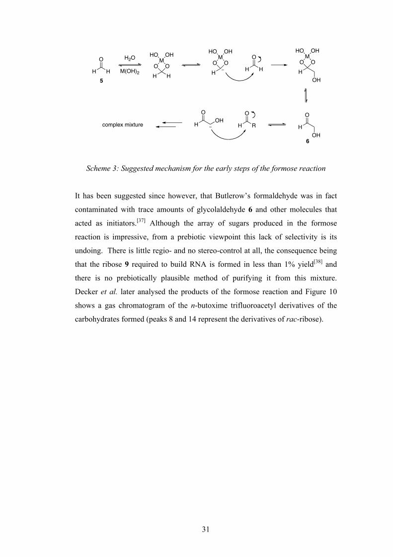
31
Scheme 3: Suggested mechanism for the early steps of the formose reaction
It has been suggested since however, that Butlerow’s formaldehyde was in fact
contaminated with trace amounts of glycolaldehyde 6 and other molecules that
acted as initiators.[37] Although the array of sugars produced in the formose
reaction is impressive, from a prebiotic viewpoint this lack of selectivity is its
undoing. There is little regio- and no stereo-control at all, the consequence being
that the ribose 9 required to build RNA is formed in less than 1% yield[38] and
there is no prebiotically plausible method of purifying it from this mixture.
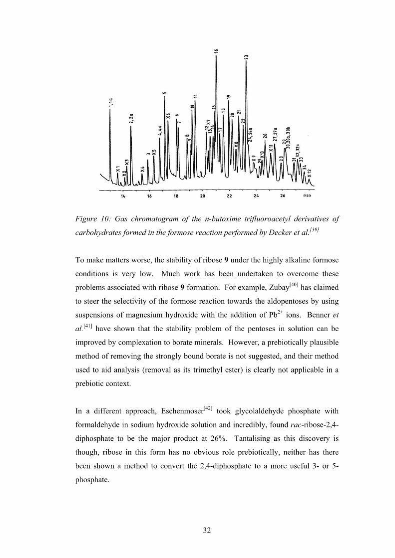
Decker et al. later analysed the products of the formose reaction and Figure 10
shows a gas chromatogram of the n-butoxime trifluoroacetyl derivatives of the
carbohydrates formed (peaks 8 and 14 represent the derivatives of rac-ribose).
O
HH
H2O
M(OH)2 HHO
MO
OHHO
HO
MO
OHHOO
HH HO
MO
OHHO
OH
O
HOH
O
H OHO
H Rcomplex mixture
5
6
32
Figure 10: Gas chromatogram of the n-butoxime trifluoroacetyl derivatives of
carbohydrates formed in the formose reaction performed by Decker et al.[39]
To make matters worse, the stability of ribose 9 under the highly alkaline formose
conditions is very low. Much work has been undertaken to overcome these
problems associated with ribose 9 formation. For example, Zubay[40] has claimed
to steer the selectivity of the formose reaction towards the aldopentoses by using
suspensions of magnesium hydroxide with the addition of Pb2+ ions. Benner et
al.[41] have shown that the stability problem of the pentoses in solution can be
improved by complexation to borate minerals. However, a prebiotically plausible
method of removing the strongly bound borate is not suggested, and their method
used to aid analysis (removal as its trimethyl ester) is clearly not applicable in a
prebiotic context.
In a different approach, Eschenmoser[42] took glycolaldehyde phosphate with
formaldehyde in sodium hydroxide solution and incredibly, found rac-ribose-2,4-
diphosphate to be the major product at 26%. Tantalising as this discovery is
though, ribose in this form has no obvious role prebiotically, neither has there
been shown a method to convert the 2,4-diphosphate to a more useful 3- or 5-
phosphate.
BIOIDS. X.
14 16 18 20 22 24 26 min
Fig_ 1. Gas chromatogram of n-butoxime trifluoroacetyl derivatives of carbohydrates arising in the con- densation of formaldehyde. Temperatures: column, IWC for 2 mitt, then increased from 100 to ISO’C at YC/min, final temperature IgO’C; injection and detector, ZSO’C. Gas flow-rates: nitrogen carrier gas, 2 ml/min; hydrogen. 20 ml!min; air. 200 mi/min_ Sample volume: 1 JL Splitting ratio: 1:12_ Peak identities: see TabIe I(A)_
10 20 30 LO -~ m in
Fig. 2. Autocatalytic consumption of 0.156 mol/i formaldehyde at 4OO’C in the presence of 0.0% mol/l cakitun acetate and 0.087 moI/l NaOH, started by addition of 0.55 mmol/l glycolaldehyde. x = Sample shown in Fig. 1 and Table I.
33
1.8.2.2 Purine Synthesis
Some of the most promising prebiotic chemistry undertaken has concerned the
formation of the purine bases found in nucleic acids. Building on the fact that
HCN 2 was a major product of the Miller-Urey experiment, Oró demonstrated, in
1960, that adenine 12 could be synthesised from this important prebiotic
feedstock molecule.[43] He found that by heating an aqueous solution of
ammonium cyanide at 70°C for several days, followed by acid hydrolysis, adenine
12 could be formed, albeit in a small yield of ≈0.5%. Lowe et al. later confirmed
the presence of other products in the reaction mixture, including guanine 16, and
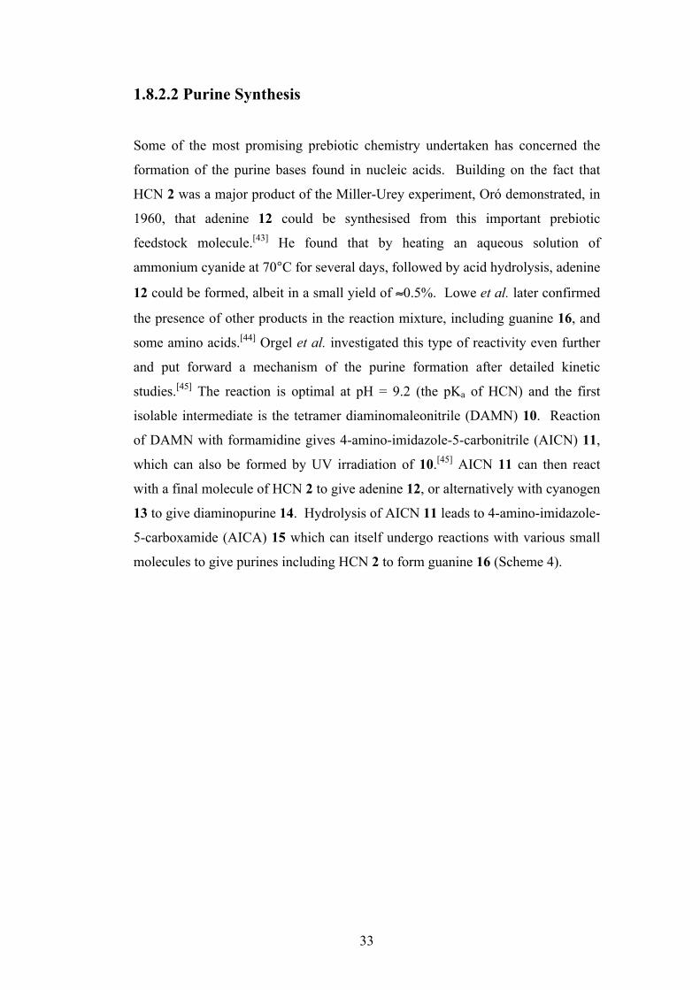
some amino acids.[44] Orgel et al. investigated this type of reactivity even further
and put forward a mechanism of the purine formation after detailed kinetic
studies.[45] The reaction is optimal at pH = 9.2 (the pKa of HCN) and the first
isolable intermediate is the tetramer diaminomaleonitrile (DAMN) 10. Reaction
of DAMN with formamidine gives 4-amino-imidazole-5-carbonitrile (AICN) 11,
which can also be formed by UV irradiation of 10.[45] AICN 11 can then react
with a final molecule of HCN 2 to give adenine 12, or alternatively with cyanogen
13 to give diaminopurine 14. Hydrolysis of AICN 11 leads to 4-amino-imidazole-
5-carboxamide (AICA) 15 which can itself undergo reactions with various small
molecules to give purines including HCN 2 to form guanine 16 (Scheme 4).
34
Scheme 4: Prebiotic syntheses of the purines, including adenine 12 and guanine
16
The yields of the purines under these conditions are low however, and although
they can be produced in higher yields by increasing the concentration
significantly, this is not regarded as being prebiotically plausible. Schwartz has
improved the synthesis of adenine by HCN 2 by the addition of glycolonitrile (the
cyanohydrin of formaldehyde 5) and/or by performing the reaction in ice.[46]
1.8.2.3 Pyrimidine Synthesis
The prebiotic syntheses of the pyrimidine bases cytosine 17 and uracil 18 start
with the spark-discharge product cyanoacetylene 1. When 1M cyanate is reacted
with 0.1M 1 at 100°C for 24 h, cytosine 17 is produced in 5% yield.[27] This can
then be slowly converted to uracil 18 in neutral aqueous solution (t1/2 = 300 years
at 30°C).[47] In an alternative route, cytosine 17 can be produced in up to 50%
yield by the reaction of cyanoacetaldehyde 19 (the hydration product of
cyanoacetylene 1) with urea 20. This second route however requires extremely
high concentrations of urea 20, and to rationalise this prebiotically one has to
imagine a situation whereby such concentrations might be reached in an
HCNHCN HN
N
H2N
N
N H2N
H2N
N
N
HCNHCN
N
NNH
NNH2
NH
NNH
NO
NH2
N
NNH
NNH2
NH2
NH
NNH
NO
NH
NH
NH
NO
O
N
NH NH2
N
210
1115
121416
H2O
HCNNCCNHCNNCCNNCO
h!
13 13 2
NH
NH2or
N
NH NH2
NH2
O

35
evaporating pool of water.[48] Scheme 5 summarises the two routes to the
pyrimidine bases.
Scheme 5: Prebiotic syntheses of the pyrimidine bases cytosine 17 and uracil 18
1.8.2.4 Synthesis of Nucleosides by Attachment of Sugar to Base
As introduced in chapter 1.8.2, it was long assumed that the prebiotic synthesis of
RNA monomers occurred by the joining of a preformed base to the sugar ribose 9,
and the syntheses of these constituents is discussed above. This approach
however, is plagued by problems. The most immediately apparent is the inability
to obtain ribose 9 pure, and in good yield. It was pointed out in chapter 1.8.2.1
that the formose reaction only produces 9 in tiny yield, and that there is no
plausible method of separating it from the many other carbohydrates formed.
Despite this, there have been numerous attempts to directly attach the preformed
base to ribose 9 towards building nucleosides.
Experimentally, the direct addition of ribose 9 to the purines (adenine 12 and
guanine 16) is very poor, and addition of the pyrimidines (cytosine 17 and uracil
18) hasn’t been shown to work at all. The most successful work to date has been
that undertaken by Orgel.[49, 50] He was able to demonstrate yields of up to 8% of
β-D-inosine by heating D-ribose 9 with hypoxanthine in the presence of
magnesium chloride or seawater salts. In the analogous experiment with adenine
12, the case is even worse. Optimistically, β-D-adenosine 21 is formed in just 3%
NNCO
N C ONH
NCO
O NH
NH2
O
N
NH
NH2
O
NO
O
NH2H2N
N N N
N
NH2
O
NNH
NH
O
O
NCO H2O
-CO2
H2O H2O
1
19
20
17
18
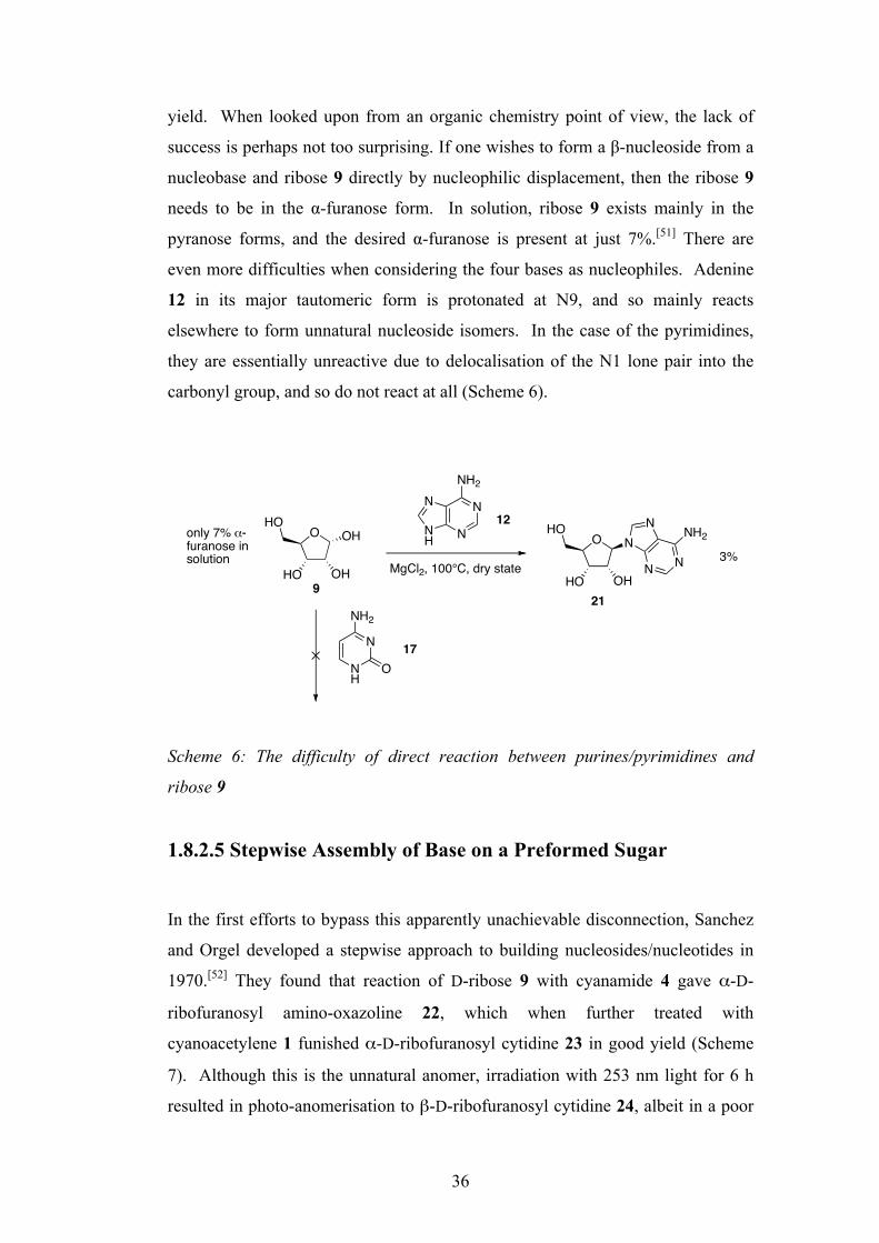
36
yield. When looked upon from an organic chemistry point of view, the lack of
success is perhaps not too surprising. If one wishes to form a β-nucleoside from a
nucleobase and ribose 9 directly by nucleophilic displacement, then the ribose 9
needs to be in the α-furanose form. In solution, ribose 9 exists mainly in the
pyranose forms, and the desired α-furanose is present at just 7%.[51] There are
even more difficulties when considering the four bases as nucleophiles. Adenine
12 in its major tautomeric form is protonated at N9, and so mainly reacts
elsewhere to form unnatural nucleoside isomers. In the case of the pyrimidines,
they are essentially unreactive due to delocalisation of the N1 lone pair into the
carbonyl group, and so do not react at all (Scheme 6).
Scheme 6: The difficulty of direct reaction between purines/pyrimidines and
ribose 9
1.8.2.5 Stepwise Assembly of Base on a Preformed Sugar
In the first efforts to bypass this apparently unachievable disconnection, Sanchez
and Orgel developed a stepwise approach to building nucleosides/nucleotides in
1970.[52] They found that reaction of D-ribose 9 with cyanamide 4 gave α-D-
ribofuranosyl amino-oxazoline 22, which when further treated with
cyanoacetylene 1 funished α-D-ribofuranosyl cytidine 23 in good yield (Scheme
7). Although this is the unnatural anomer, irradiation with 253 nm light for 6 h
resulted in photo-anomerisation to β-D-ribofuranosyl cytidine 24, albeit in a poor
O
HO OH
OHHO
only 7% !-furanose in solution
O
HO OH
HON
N
N N
NH2
MgCl2, 100°C, dry state
21
3%
9
N
NH
NH2
O17
N
NNH
NNH2
12
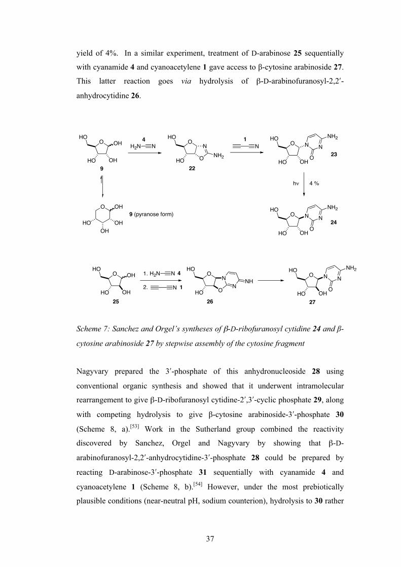
37
yield of 4%. In a similar experiment, treatment of D-arabinose 25 sequentially
with cyanamide 4 and cyanoacetylene 1 gave access to β-cytosine arabinoside 27.
This latter reaction goes via hydrolysis of β-D-arabinofuranosyl-2,2ʹ′-
anhydrocytidine 26.
Scheme 7: Sanchez and Orgel’s syntheses of β-D-ribofuranosyl cytidine 24 and β-
cytosine arabinoside 27 by stepwise assembly of the cytosine fragment
Nagyvary prepared the 3ʹ′-phosphate of this anhydronucleoside 28 using
conventional organic synthesis and showed that it underwent intramolecular
rearrangement to give β-D-ribofuranosyl cytidine-2ʹ′,3ʹ′-cyclic phosphate 29, along
with competing hydrolysis to give β-cytosine arabinoside-3ʹ′-phosphate 30
(Scheme 8, a).[53] Work in the Sutherland group combined the reactivity
discovered by Sanchez, Orgel and Nagyvary by showing that β-D-
arabinofuranosyl-2,2ʹ′-anhydrocytidine-3ʹ′-phosphate 28 could be prepared by
reacting D-arabinose-3ʹ′-phosphate 31 sequentially with cyanamide 4 and
cyanoacetylene 1 (Scheme 8, b).[54] However, under the most prebiotically
plausible conditions (near-neutral pH, sodium counterion), hydrolysis to 30 rather
O
HO OH
OHHO
H2N NO
HO
HO
O
NNH2
N O
HO OH
HON N
O
NH2
h!
O
HO OH
HON N
O
NH2
4 %
O OH
OHOH
HO
O
HO
OHHO
OH
H2N N
N
1.
2.
O
HO
HON N
O
NH2
OH
O
HO
HO
O
NN
NH
9 (pyranose form)
9 22
23
24
25 26 27
4 1
4
1

38
than intramolecular rearrangement to 29 was the dominant pathway, resulting in a
overall yield of just 3.5% of β-D-ribofuranosyl cytidine-2ʹ′,3ʹ′-cyclic phosphate 29
from D-arabinose-3ʹ′-phosphate 31.
Scheme 8: a) Production of β-D-ribofuranosyl cytidine-2ʹ′,3ʹ′-cyclic phosphate 29
by Nagyvary and, b) stepwise formation of β-D-arabinofuranosyl-2,2ʹ′-
anhydrocytidine-3ʹ′-phosphate 28 by the Sutherland group
This method, although avoiding the need for direct attachment of sugar to base,
still has failings: the need to start from pure, D-arabinose-3-phosphate 31 of which
there is no plausible prebiotic synthesis, and the low yield of the desired β-D-
ribofuranosyl cytidine-2ʹ′,3ʹ′-cyclic phosphate 29. However, this different method
of construction offered optimism for several reasons: the unfavourable furanose-
pyranose equilibrium problem is overcome due to the fact that the reaction to
form the amino-oxazoline is selective for the furanose form, the stability of the
amino-oxazolines is far greater than that of their corresponding free sugars,[55] and
unlike the formose reaction and the nucleobase assembly chemistry, this reactivity
displays a measure of selectivity.
Despite offering huge optimism for the formation of RNA monomers, the amino-
oxazolines still had to be prepared from preformed sugars (or sugar-phosphates),
O
O
HO
O
NN
NH2
POHO
O
OHO
N N
O
NH2
OPO
O O
O
OPO
HOO
N
OH
N
O
NH2HO
O
O OH
OHHO
H2N N
O
O
HO
O
NNH2 N
PO OHO
POHO
O
O
O
HO
O
NN
NH2
POHO
O
a)
b)
28 29 30
1
28
4
31
39
which then undergo exposure to nitrogen containing compounds. Indeed, this
requirement for pre-formed sugars, and the need to separate nitrogenous and
oxygenous chemistry were two major obstacles that stood in the way of any
plausible RNA-first theory, and led to theories that perhaps RNA was not the first
genetic material.
1.8.3 Alternative Genetic systems
Due to the difficulty associated with the prebiotic construction of RNA and/or its
monomers, and the scarcity of experimental success, many went on to conclude
that the first genetic system could not have been based upon RNA. Various
alternative (constitutionally ‘simpler’) genetic systems have been proposed to
have arisen first that are capable of Watson-Crick base pairing, both with other
strands of the same polymer and also with RNA. In this way, once the alternative
genetic system was established, it is proposed that the transition to the more
complex RNA-based world could be made, whilst retaining the information stored
in the order of the bases of the polymer.
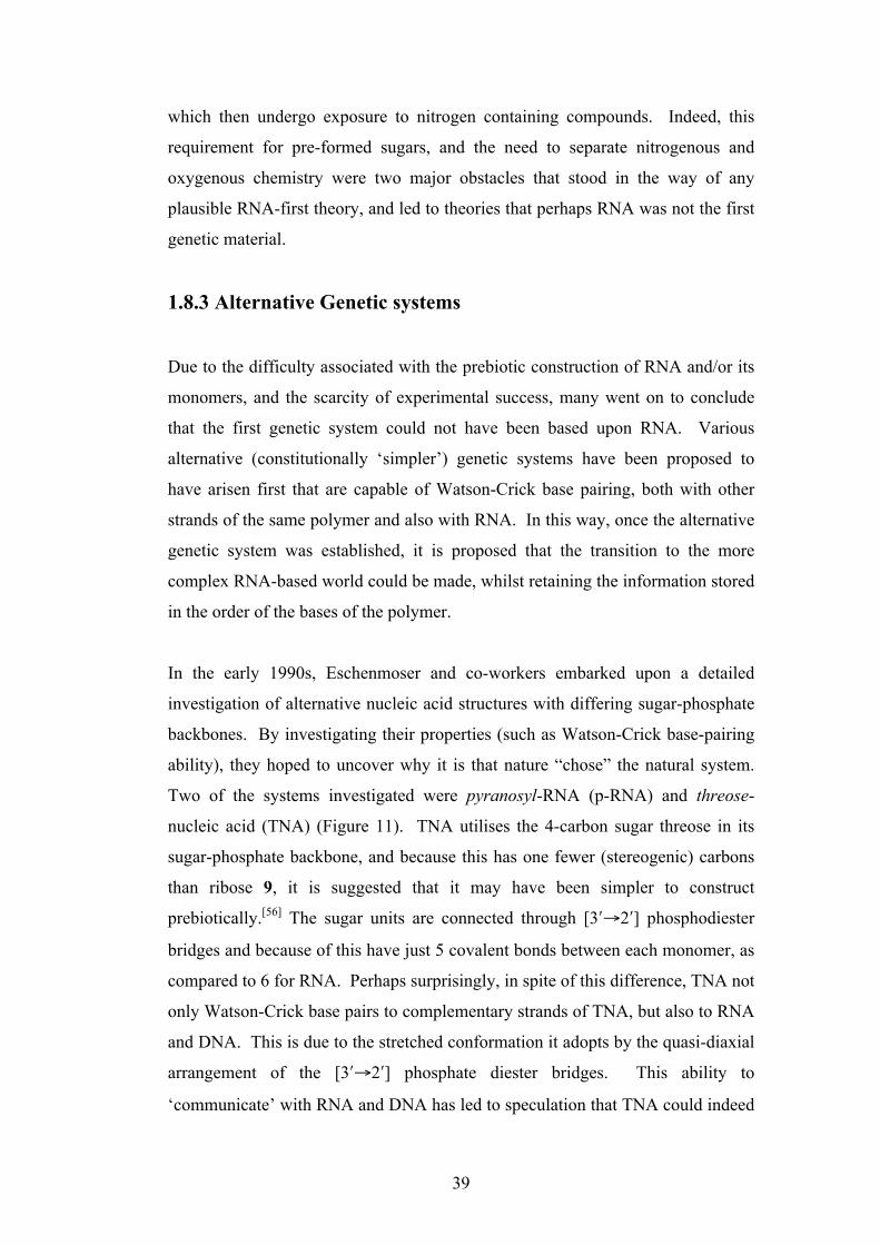
In the early 1990s, Eschenmoser and co-workers embarked upon a detailed
investigation of alternative nucleic acid structures with differing sugar-phosphate
backbones. By investigating their properties (such as Watson-Crick base-pairing
ability), they hoped to uncover why it is that nature “chose” the natural system.
Two of the systems investigated were pyranosyl-RNA (p-RNA) and threose-
nucleic acid (TNA) (Figure 11). TNA utilises the 4-carbon sugar threose in its
sugar-phosphate backbone, and because this has one fewer (stereogenic) carbons
than ribose 9, it is suggested that it may have been simpler to construct
prebiotically.[56] The sugar units are connected through [3ʹ′→2ʹ′] phosphodiester
bridges and because of this have just 5 covalent bonds between each monomer, as
compared to 6 for RNA. Perhaps surprisingly, in spite of this difference, TNA not
only Watson-Crick base pairs to complementary strands of TNA, but also to RNA
and DNA. This is due to the stretched conformation it adopts by the quasi-diaxial
arrangement of the [3ʹ′→2ʹ′] phosphate diester bridges. This ability to
‘communicate’ with RNA and DNA has led to speculation that TNA could indeed
40
have arisen first, followed by effective ‘genetic takeover’ without informational
loss.[57]
p-RNA is an isomer of RNA where the ribose sugar adopts the 6-membered
pyranosyl form rather than 5-membered furanosyl, and these are linked through
[4ʹ′ →2ʹ′] phosphodiester bridges (Figure 11).[58] This was initially seen as a viable
prebiotic genetic system as Eschenmoser had previously shown the selective
formation of ribose-2,4-diphosphate through reaction of glycolaldehyde
phosphate with formaldehyde (see chapter 1.8.2.1).[42]
Figure 11: Threose nucleic acid (TNA) and pyranosyl-RNA (p-RNA)
The Watson-Crick base pairing between complimentary strands of p-RNA is
stronger than that found in RNA and DNA, suggesting that RNA wasn’t ‘selected’
by nature purely for its strength of inter-strand interactions alone.[59] However, the
inability of p-RNA to base pair to RNA or DNA effectively rules it out as an
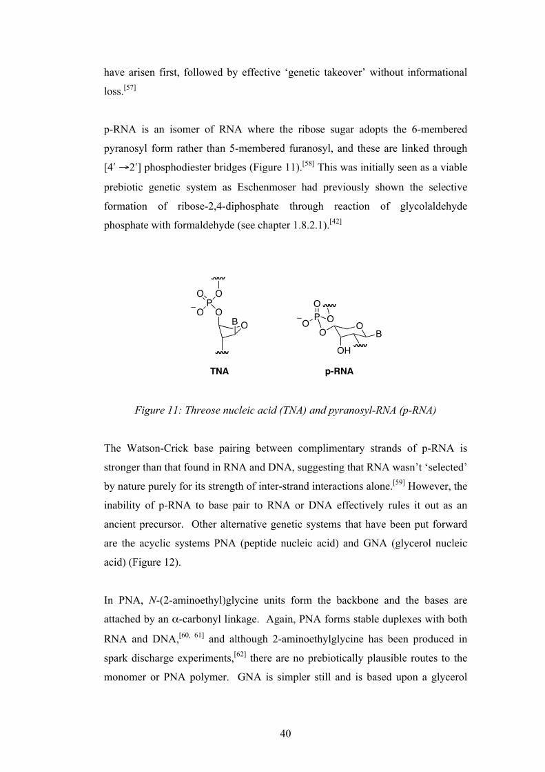
ancient precursor. Other alternative genetic systems that have been put forward
are the acyclic systems PNA (peptide nucleic acid) and GNA (glycerol nucleic
acid) (Figure 12).
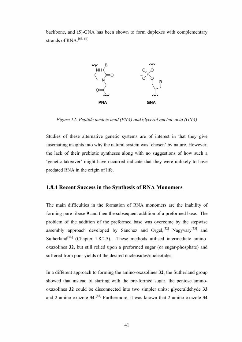
In PNA, N-(2-aminoethyl)glycine units form the backbone and the bases are
attached by an α-carbonyl linkage. Again, PNA forms stable duplexes with both
RNA and DNA,[60, 61] and although 2-aminoethylglycine has been produced in
spark discharge experiments,[62] there are no prebiotically plausible routes to the
monomer or PNA polymer. GNA is simpler still and is based upon a glycerol
OO
B
PO
O
O
TNA
OBO
OH
PO
O O
p-RNA
41
backbone, and (S)-GNA has been shown to form duplexes with complementary
strands of RNA.[63, 64]
Figure 12: Peptide nucleic acid (PNA) and glycerol nucleic acid (GNA)
Studies of these alternative genetic systems are of interest in that they give
fascinating insights into why the natural system was ‘chosen’ by nature. However,
the lack of their prebiotic syntheses along with no suggestions of how such a
‘genetic takeover’ might have occurred indicate that they were unlikely to have
predated RNA in the origin of life.
1.8.4 Recent Success in the Synthesis of RNA Monomers
The main difficulties in the formation of RNA monomers are the inability of
forming pure ribose 9 and then the subsequent addition of a preformed base. The
problem of the addition of the preformed base was overcome by the stepwise
assembly approach developed by Sanchez and Orgel,[52] Nagyvary[53] and
Sutherland[54] (Chapter 1.8.2.5). These methods utilised intermediate amino-
oxazolines 32, but still relied upon a preformed sugar (or sugar-phosphate) and
suffered from poor yields of the desired nucleosides/nucleotides.
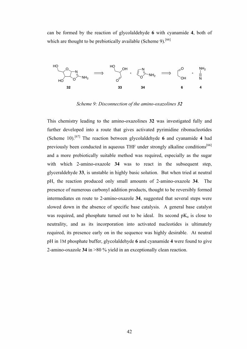
In a different approach to forming the amino-oxazolines 32, the Sutherland group
showed that instead of starting with the pre-formed sugar, the pentose amino-
oxazolines 32 could be disconnected into two simpler units: glyceraldehyde 33
and 2-amino-oxazole 34.[65] Furthermore, it was known that 2-amino-oxazole 34
BO
PO
O O
O
NO
BNH
PNA GNA
42
can be formed by the reaction of glycolaldehyde 6 with cyanamide 4, both of
which are thought to be prebiotically available (Scheme 9).[66]
Scheme 9: Disconnection of the amino-oxazolines 32
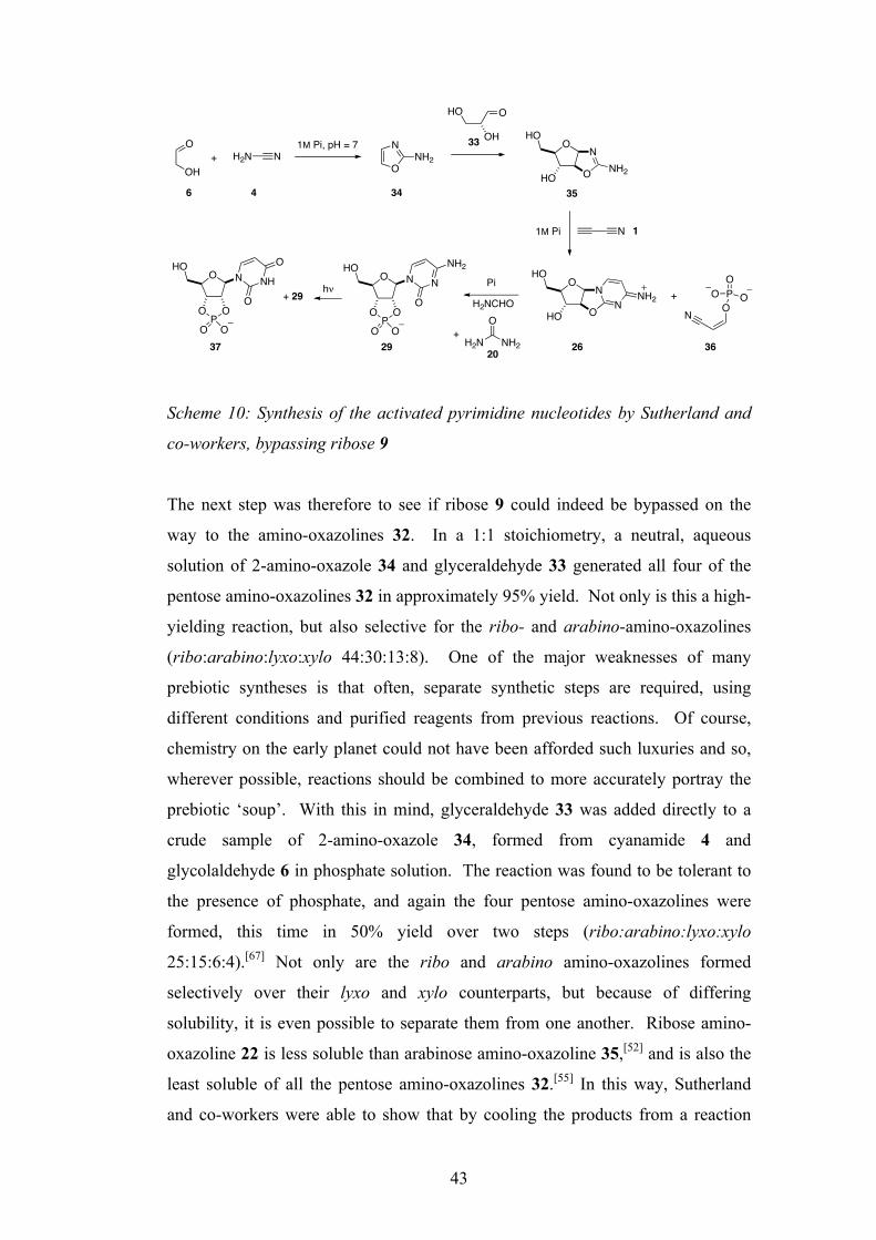
This chemistry leading to the amino-oxazolines 32 was investigated fully and
further developed into a route that gives activated pyrimidine ribonucleotides
(Scheme 10).[67] The reaction between glycolaldehyde 6 and cyanamide 4 had
previously been conducted in aqueous THF under strongly alkaline conditions[66]
and a more prebiotically suitable method was required, especially as the sugar
with which 2-amino-oxazole 34 was to react in the subsequent step,
glyceraldehyde 33, is unstable in highly basic solution. But when tried at neutral
pH, the reaction produced only small amounts of 2-amino-oxazole 34. The
presence of numerous carbonyl addition products, thought to be reversibly formed
intermediates en route to 2-amino-oxazole 34, suggested that several steps were
slowed down in the absence of specific base catalysis. A general base catalyst
was required, and phosphate turned out to be ideal. Its second pKa is close to
neutrality, and as its incorporation into activated nucleotides is ultimately
required, its presence early on in the sequence was highly desirable. At neutral
pH in 1M phosphate buffer, glycolaldehyde 6 and cyanamide 4 were found to give
2-amino-oxazole 34 in >80 % yield in an exceptionally clean reaction.
OHO
HO O
NNH2 O
NNH2
HOOH
O N
NH2O
OH
32 33 34 6 4
43
Scheme 10: Synthesis of the activated pyrimidine nucleotides by Sutherland and
co-workers, bypassing ribose 9
The next step was therefore to see if ribose 9 could indeed be bypassed on the
way to the amino-oxazolines 32. In a 1:1 stoichiometry, a neutral, aqueous
solution of 2-amino-oxazole 34 and glyceraldehyde 33 generated all four of the
pentose amino-oxazolines 32 in approximately 95% yield. Not only is this a high-
yielding reaction, but also selective for the ribo- and arabino-amino-oxazolines
(ribo:arabino:lyxo:xylo 44:30:13:8). One of the major weaknesses of many
prebiotic syntheses is that often, separate synthetic steps are required, using
different conditions and purified reagents from previous reactions. Of course,
chemistry on the early planet could not have been afforded such luxuries and so,
wherever possible, reactions should be combined to more accurately portray the
prebiotic ‘soup’. With this in mind, glyceraldehyde 33 was added directly to a
crude sample of 2-amino-oxazole 34, formed from cyanamide 4 and
glycolaldehyde 6 in phosphate solution. The reaction was found to be tolerant to
the presence of phosphate, and again the four pentose amino-oxazolines were
formed, this time in 50% yield over two steps (ribo:arabino:lyxo:xylo
25:15:6:4).[67] Not only are the ribo and arabino amino-oxazolines formed
selectively over their lyxo and xylo counterparts, but because of differing
solubility, it is even possible to separate them from one another. Ribose amino-
oxazoline 22 is less soluble than arabinose amino-oxazoline 35,[52] and is also the
least soluble of all the pentose amino-oxazolines 32.[55] In this way, Sutherland
and co-workers were able to show that by cooling the products from a reaction
O
OP
O
HO
O O
N N
O
NH2
O
OHNH2N NH2
N
O
HO
OH
O
O
O
N
HO
HO
NH2
N
O
O
N
HO
HO
NNH2
O
OP
O
HO
O O
N NH
O
O
+1M Pi, pH = 7
6 4 34
33
35
11M Pi
H2NCHO
Pi+ 29 h!
37 29 26
PO
O OON
+
36+
O
NH2H2N20
44
between 2-amino-oxazole 34 and glyceraldehyde 33, crystals of pure ribose
amino-oxazoline 22 were formed, and after filtration, the major species in solution
was now the key intermediate, arabinose amino-oxazoline 35.
It had previously been shown by Sanchez and Orgel that treatment of arabinose
amino-oxazoline 35 with cyanoacetylene 1 in an unbuffered reaction furnished β-
arabinocytidine 27, but in a relatively low yield of 58%.[52] Through the careful
study of isolated products, it was revealed the cause of the low yield was two-
fold. Firstly, a rise in pH causes hydrolysis of the anhydronucleoside
intermediate, and secondly, hydroxyl groups undergo reaction with excess
cyanoacetylene 1. To combat this rise in pH a buffer was needed and, as before,
inorganic phosphate was chosen. At pH = 6.5 in the presence of phosphate, there
was little evidence of anhydronucleoside hydrolysis. Using phosphate in this way
also had the added bonus of not simply acting as a pH buffer, but also as a
chemical buffer, reacting with excess cyanoacetylene 1 to form cyanovinyl
phosphate 36, leaving the hydroxyl groups of the anhydronucleoside untouched.
In this way, the arabinose-anhydronucleoside 26 was formed extremely cleanly,
and in 92% yield.
With the arabinose-anhydronucleoside 26 now available through an efficient,
prebiotically plausible route, a subsequent phosphorylation-rearrangement step
was required to convert it to the activated ribonucleotide 29. Prebiotic
phosphorylations of nucleosides have been achieved by heating either in the dry
state with urea 20[68] or in formamide solution.[69] As well as inorganic phosphate,
the Sutherland group were able to utilise pyrophosphate as the phosphorylating
agent, as it is known to be produced in a reaction between inorganic phosphate
and cyanovinyl phosphate 36[70], itself formed in the previous step. Furthermore,
the urea 20 required is formed as a by-product in the reaction that generates 2-
amino-oxazole 34 from glycolaldehyde 6 and cyanamide 4, if the latter is present
in excess. Thus, by heating the arabinose-anhydronucleoside 26 with 0.5
equivalents of pyrophosphate in urea containing ammonium salts the major
product was, remarkably, the activated ribonucleotide β-ribocytidine-2ʹ′,3ʹ′-cyclic
phosphate 29. Alternatively, by heating the anhydronucleoside 26 with inorganic

45
phosphate in formamide solution, an even greater conversion to the activated
ribonucleotide 29 was achieved. Its formation is thought to be via initial
phosphorylation of the 3ʹ′-hydroxyl group of the anhydronucleoside 26, followed
by rearrangement through intramolecular nucleophilic substitution. This is
somewhat surprising as the favoured site of phosphorylation is the secondary 3ʹ′-
hydroxyl rather than the primary 5ʹ′-hydroxyl group of 26. Secondary hydroxyl
groups typically bear more steric hindrance than their primary counterparts, but in
this case investigation of the crystal structure revealed that due to the
conformation of the anhydronucleoside 26, the 3ʹ′-hydroxyl group was indeed
more exposed to phosphorylation than the 5ʹ′-hydroxyl group in what is an
interesting case of predisposition, lacking the requirement for protecting groups
which might be expected to be used in a traditional organic synthesis.
The final step in the synthesis was the partial conversion of 29 to the
corresponding uracil nucleotide, β-ribouridine-2ʹ′,3ʹ′-cyclic phosphate 37. This
was achieved by exposure to UV irradiation for 3 days at near neutral pH. As
well as the partial conversion to 37, this irradiation also served to destroy
nucleotide by-products formed from the previous step that could possibly interfere
with subsequent oligomerisation steps.
This route to the activated pyrimidine ribonucleotides as their 2ʹ′,3ʹ′-cyclic
phosphates 29 and 37 was the first to overcome the problems that had plagued
RNA-first theories for decades: the synthesis of pure ribose and the need to attach
a preformed base. The prebiotically plausible production of these activated
nucleotides now makes the oligomerisation of these monomers into RNA a
realistic challenge.
1.8.5 Nucleotide Activation
As mentioned previously, nucleoside-2ʹ′,3ʹ′-cyclic phosphates 8 are considered to
be good candidates for the synthesis of RNA via monomer oligomerisation.[71-73]
This idea has recently been given strength in light of the prebiotic synthesis of
cytidine- and uridine-2ʹ′,3ʹ′-cyclic phosphate 29 and 37 demonstrated by the

46
Sutherland group.[67] This oligomerisation of nucleoside-2ʹ′,3ʹ′-phosphates 8 faces
several difficulties however. Templated oligomerisation of nucleoside-2ʹ′,3ʹ′-
cyclic phosphates tends to give exclusively unnatural [2ʹ′→5ʹ′] linkages[73] whilst
with non-templated oligomerisation there is usually a mixture of [2ʹ′→5ʹ′] and
[3ʹ′→5ʹ′] linkages.[72] Finally, if such oligomerisations are to be carried out in
aqueous solution, it is inevitable that nucleoside-2ʹ′,3ʹ′-cyclic phosphates will
undergo competing hydrolysis to a mixture of nucleoside-2ʹ′- and 3ʹ′-phosphates 39
and 38 in addition to any oligomerisation. Even if oligomerisations are carried
out in the dry state, any exposure of the cyclic phosphate to water before this step
(e.g. the steps leading to its formation) will also lead to hydrolysis. This problem
can be at least partly overcome if a (continual) repair mechanism is in operation
that converts the mixture of nucleoside-2ʹ′- and 3ʹ′-phosphates (39 and 38) back to
the cyclic material 8 (Scheme 11).
Scheme 11: Hydrolysis of nucleoside-2ʹ′,3ʹ′-cyclic phosphates 8 and the need for
their regeneration for oligomerisation to RNA
Orgel and Lohrmann investigated a range of prebiotically plausible activating
agents for the conversion of uridine-2ʹ′,(3ʹ′)-phosphate to uridine-2ʹ′,3ʹ′-cyclic
phosphate 38.[74] Amongst the activating agents used were cyanoformamide,
cyanamide 4 and cyanate. The most successful of these was cyanamide 4, with a
73% conversion to the 2ʹ′,3ʹ′-cyclic phosphate at pH = 5.0 and at 65°C. The
O B
OP
O
HO
OO
RNA
O BHO
O OHP OOOH
O BHO
HO OP OOOH
+hydrolysis oligomerisation
repair by re-activation
38
39
8
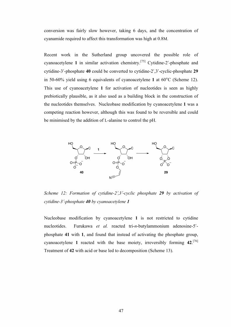
47
conversion was fairly slow however, taking 6 days, and the concentration of
cyanamide required to affect this transformation was high at 0.8M.
Recent work in the Sutherland group uncovered the possible role of
cyanoacetylene 1 in similar activation chemistry.[75] Cytidine-2ʹ′-phosphate and
cytidine-3ʹ′-phosphate 40 could be converted to cytidine-2ʹ′,3ʹ′-cyclic-phosphate 29
in 50-60% yield using 6 equivalents of cyanoacetylene 1 at 60°C (Scheme 12).
This use of cyanoacetylene 1 for activation of nucleotides is seen as highly
prebiotically plausible, as it also used as a building block in the construction of
the nucleotides themselves. Nucleobase modification by cyanoacetylene 1 was a
competing reaction however, although this was found to be reversible and could
be minimised by the addition of L-alanine to control the pH.
Scheme 12: Formation of cytidine-2ʹ′,3ʹ′-cyclic phosphate 29 by activation of
cytidine-3ʹ′-phosphate 40 by cyanoacetylene 1
Nucleobase modification by cyanoacetylene 1 is not restricted to cytidine
nucleotides. Furukawa et al. reacted tri-n-butylammonium adenosine-5ʹ′-
phosphate 41 with 1, and found that instead of activating the phosphate group,
cyanoacetylene 1 reacted with the base moiety, irreversibly forming 42.[76]
Treatment of 42 with acid or base led to decomposition (Scheme 13).
OHO
O OH
C
POO
O
OHO
C
OPO
O O
OHO
O OH
C
POO
O
N
1
40 29
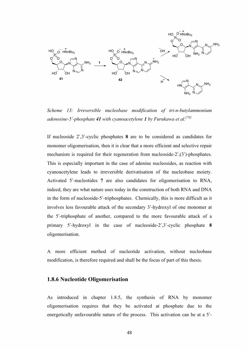
48
Scheme 13: Irreversible nucleobase modification of tri-n-butylammonium
adenosine-5ʹ′-phosphate 41 with cyanoacetylene 1 by Furukawa et al.[76]
If nucleoside 2ʹ′,3ʹ′-cyclic phosphates 8 are to be considered as candidates for
monomer oligomerisation, then it is clear that a more efficient and selective repair
mechanism is required for their regeneration from nucleoside-2ʹ′,(3ʹ′)-phosphates.
This is especially important in the case of adenine nucleosides, as reaction with
cyanoacetylene leads to irreversible derivatisation of the nucleobase moiety.
Activated 5ʹ′-nucleotides 7 are also candidates for oligomerisation to RNA,
indeed, they are what nature uses today in the construction of both RNA and DNA
in the form of nucleoside-5ʹ′-triphosphates. Chemically, this is more difficult as it
involves less favourable attack of the secondary 3ʹ′-hydroxyl of one monomer at
the 5ʹ′-triphosphate of another, compared to the more favourable attack of a
primary 5ʹ′-hydroxyl in the case of nucleoside-2ʹ′,3ʹ′-cyclic phosphate 8
oligomerisation.
A more efficient method of nucleotide activation, without nucleobase
modification, is therefore required and shall be the focus of part of this thesis.
1.8.6 Nucleotide Oligomerisation
As introduced in chapter 1.8.5, the synthesis of RNA by monomer
oligomerisation requires that they be activated at phosphate due to the
energetically unfavourable nature of the process. This activation can be at a 5ʹ′-
O N
HO OH
O N
N N
NH2 O N
HO OH
O N
N N
N NH2
HNnBu3P
HO
O
O
O N
HO OH
O N
NH2 N
N NH2
1
HNN
NH2 N
N NH241 42
OH
H
PO
OHO HNnBu3
P OHO O HNnBu3
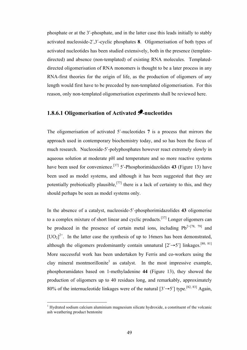
49
phosphate or at the 3ʹ′-phosphate, and in the latter case this leads initially to stably
activated nucleoside-2ʹ′,3ʹ′-cyclic phosphates 8. Oligomerisation of both types of
activated nucleotides has been studied extensively, both in the presence (template-
directed) and absence (non-templated) of existing RNA molecules. Templated-
directed oligomerisation of RNA monomers is thought to be a later process in any
RNA-first theories for the origin of life, as the production of oligomers of any
length would first have to be preceded by non-templated oligomerisation. For this
reason, only non-templated oligomerisation experiments shall be reviewed here.
1.8.6.1 Oligomerisation of Activated 5ʹ′-nucleotides
The oligomerisation of activated 5ʹ′-nucleotides 7 is a process that mirrors the
approach used in contemporary biochemistry today, and so has been the focus of
much research. Nucleoside-5ʹ′-polyphosphates however react extremely slowly in
aqueous solution at moderate pH and temperature and so more reactive systems
have been used for convenience.[37] 5ʹ′-Phosphorimidazolides 43 (Figure 13) have
been used as model systems, and although it has been suggested that they are
potentially prebiotically plausible,[77] there is a lack of certainty to this, and they
should perhaps be seen as model systems only.
In the absence of a catalyst, nucleoside-5ʹ′-phosphorimidazolides 43 oligomerise
to a complex mixture of short linear and cyclic products.[37] Longer oligomers can
be produced in the presence of certain metal ions, including Pb2+[78, 79] and
[UO2]2+. In the latter case the synthesis of up to 16mers has been demonstrated,
although the oligomers predominantly contain unnatural [2ʹ′→5ʹ′] linkages.[80, 81]
More successful work has been undertaken by Ferris and co-workers using the
clay mineral montmorillonite1 as catalyst. In the most impressive example,
phosphoramidates based on 1-methyladenine 44 (Figure 13), they showed the
production of oligomers up to 40 residues long, and remarkably, approximately
80% of the internucleotide linkages were of the natural [3ʹ′→5ʹ′] type.[82, 83] Again,
1 Hydrated sodium calcium aluminium magnesium silicate hydroxide, a constituent of the volcanic ash weathering product bentonite

50
the use of 1-methyladenine nucleotides 44 should be seen as a model system
rather than a strictly prebiotic reagent.
Figure 13: 5ʹ′-Phosphorimidazolides 43 and 1- methyladenine phosphoramidates
44 as used in non-templated oligomerisation experiments
1.8.6.2 Oligomerisation of Nucleoside-2ʹ′ ,3ʹ′-Cyclic Phosphates
Non-templated oligomerisation in aqueous solution is extremely inefficient, and
so Orgel and co-workers in the 1970s concentrated on reactions in the dry state
using various catalysts. When adenosine-2ʹ′,3ʹ′-cyclic phosphate was evaporated
from solution in the presence of simple catalysts (e.g. ethylene diamine and
ethanolamine 109) at high pH and then maintained in the dry state at moderate
temperatures (25-85°C), oligonucleotide formation was observed.[72] Yields of
oligomer up to 68% were found under very dry conditions, and under more
prebiotically relevant conditions yields of up to 25% were obtained. Most of this
material consisted of nucleotide dimers, but material up to hexamer (albeit in
much reduced yield) was also produced. The most promising result of these
studies was that the natural [3ʹ′→5ʹ′] linkages dominated over the unnatural
[2ʹ′→5ʹ′] linkages. It is worth noting that this is in marked contrast to the template-
directed synthesis of nucleotide monomers that almost exclusively leads to
[2ʹ′→5ʹ′] linkages.[84] It is clear that the first life forms based upon RNA could not
have been produced from these oligomerisation reactions alone, as the chain
lengths produced are far too short. However, even the production of short
oligonucleotides, especially trimers, is of huge importance to alternative theories
O
HO OH
BOP
ON
O
NR
O
HO OH
BOP
ON
ON
NN
H2N
43 R = H/CH3 44
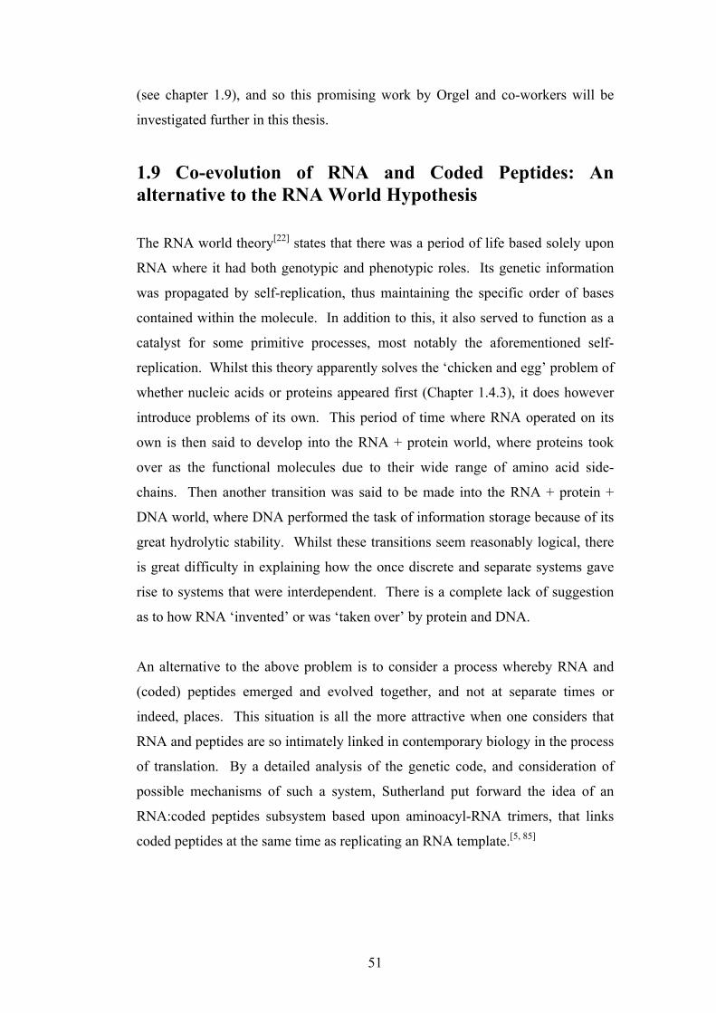
51
(see chapter 1.9), and so this promising work by Orgel and co-workers will be
investigated further in this thesis.
1.9 Co-evolution of RNA and Coded Peptides: An alternative to the RNA World Hypothesis The RNA world theory[22] states that there was a period of life based solely upon
RNA where it had both genotypic and phenotypic roles. Its genetic information
was propagated by self-replication, thus maintaining the specific order of bases
contained within the molecule. In addition to this, it also served to function as a
catalyst for some primitive processes, most notably the aforementioned self-
replication. Whilst this theory apparently solves the ‘chicken and egg’ problem of
whether nucleic acids or proteins appeared first (Chapter 1.4.3), it does however
introduce problems of its own. This period of time where RNA operated on its
own is then said to develop into the RNA + protein world, where proteins took
over as the functional molecules due to their wide range of amino acid side-
chains. Then another transition was said to be made into the RNA + protein +
DNA world, where DNA performed the task of information storage because of its
great hydrolytic stability. Whilst these transitions seem reasonably logical, there
is great difficulty in explaining how the once discrete and separate systems gave
rise to systems that were interdependent. There is a complete lack of suggestion
as to how RNA ‘invented’ or was ‘taken over’ by protein and DNA.
An alternative to the above problem is to consider a process whereby RNA and
(coded) peptides emerged and evolved together, and not at separate times or
indeed, places. This situation is all the more attractive when one considers that
RNA and peptides are so intimately linked in contemporary biology in the process
of translation. By a detailed analysis of the genetic code, and consideration of
possible mechanisms of such a system, Sutherland put forward the idea of an
RNA:coded peptides subsystem based upon aminoacyl-RNA trimers, that links
coded peptides at the same time as replicating an RNA template.[5, 85]

52
Several features of the genetic code are worth noting, and these were pointed out
by Crick in 1968:[16]
• The genetic code is read in triplets
• A triplet code with four bases had the capacity to code for over 60 amino
acids but only 20 are used
• The codons are not assigned randomly to the 20 amino acids
• XYU and XYC always code for the same amino acid
• XYA and XYG usually code for the same amino acid
• XYN (N = any base) codes for the same amino acid in half the cases
• There is often a relationship between the second base of a codon and the
chemical nature of the amino acid side chain it codes for
• Structurally similar amino acids are often coded for by codons with a
single change of base
• The code is (essentially) universal
In order to explain the assignment of the genetic code, there have been several
theories put forward. In the ‘frozen accident’ hypothesis it is suggested that the
initial codon assignments were entirely random, and became fixed in the last
common ancestor.[16] Since the genetic code is now known not to be strictly
universal, this theory seems unlikely, and it also does not explain several patterns
(see above). The ‘historical theory’ reasons that amino acids were assigned
codons in a gradual process in the order that they became available through
biosynthesis.[86] The last major theory is the ‘stereochemical theory’.[15, 16] Here it
is stated that initially there was a direct chemical interaction between the bases of
the RNA codon and the specific side chains of the amino acids. Based upon the
observations laid out above and building on the stereochemical and historical
theories, Sutherland went on to suggest an ancient genetic code that was simpler
than the one in use today.[5]
The aromatic amino acids have long and complex biosynthetic pathways, as do
His, Lys and Met. These are thought to have been late additions to the code, and
were assigned codons only after the enzymes required for their production
evolved. With these amino acids taken away, a pattern begins to emerge. With
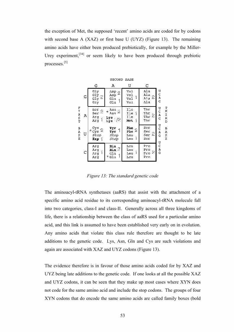
53
the exception of Met, the supposed ‘recent’ amino acids are coded for by codons
with second base A (XAZ) or first base U (UYZ) (Figure 13). The remaining
amino acids have either been produced prebiotically, for example by the Miller-
Urey experiment,[34] or seem likely to have been produced through prebiotic
processes.[5]
Figure 13: The standard genetic code
The aminoacyl-tRNA synthetases (aaRS) that assist with the attachment of a
specific amino acid residue to its corresponding aminoacyl-tRNA molecule fall
into two categories, class-I and class-II. Generally across all three kingdoms of
life, there is a relationship between the class of aaRS used for a particular amino
acid, and this link is assumed to have been established very early on in evolution.
Any amino acids that violate this class rule therefore are thought to be late
additions to the genetic code. Lys, Asn, Gln and Cys are such violations and
again are associated with XAZ and UYZ codons (Figure 13).
The evidence therefore is in favour of those amino acids coded for by XAZ and
UYZ being late additions to the genetic code. If one looks at all the possible XAZ
and UYZ codons, it can be seen that they make up most cases where XYN does
not code for the same amino acid and include the stop codons. The groups of four
XYN codons that do encode the same amino acids are called family boxes (bold
The order of assignment of amino acids to codons has also been considered in thecontext of prebiotic amino acid availabilty. Since translation must have predated anextensive enzyme-mediated metabolism, it is thought that a restricted set of prebioti-cally available amino acids was originally used in translation [22]. Depletion of theseprebiotically available amino acids by incorporation into (coded) peptides would haveprovided a strong driving force for the development of biosynthetic pathways to them.Low catalytic efficiency and restricted scope would most likely have resulted in thepathways being recruited retroacquisitively by catalysis of underlying, predisposedchemistry [28]. According to this hypothesis, the first amino acids must have beenprebiotically available, and other amino acids could not be used until they had becomeavailable for the first time by biosynthesis. Biosynthesis of these later amino acidswould have taken place when a more advanced enzymological repertoire was available,would not have been driven by environmental depletion as discussed above, andconsequently need not (necessarily) have been acquired retroacquisitively [29]. In
Fig. 1. Chemical Analysis of the Genetic Code. In using the genetic code to guide retrosynthetic disconnectionsof RNA :coded peptides, the etiology of the code must be considered. Some of the amino acids have (andrequire) long and complex biosyntheses and appear late additions to the code (bold). An analysis of prebioticavailability suggests that certain amino acids could not have been assigned to the code at the outset(underlined). The allocation of the aminoacyl-tRNA synthetases to either one of two classes (outline) isviolated in one case, and there are certain charging discrepancies (asterisks) which indicate recent assignments.Stop codons (italics) which would otherwise severely limit the length of (random sequence) RNA translationproducts are thought to be late assignments. Finally, parsimony suggests that those amino acids in family boxes(framed in bold) and only coded by the first two bases of the codon are the earliest assignments. When the codeis viewed according to these (chemical) criteria, a distinct pattern emerges with XAZ and UYZ codons
appearing as late assignments.
CHEMISTRY & BIODIVERSITY ± Vol. 1 (2004)210
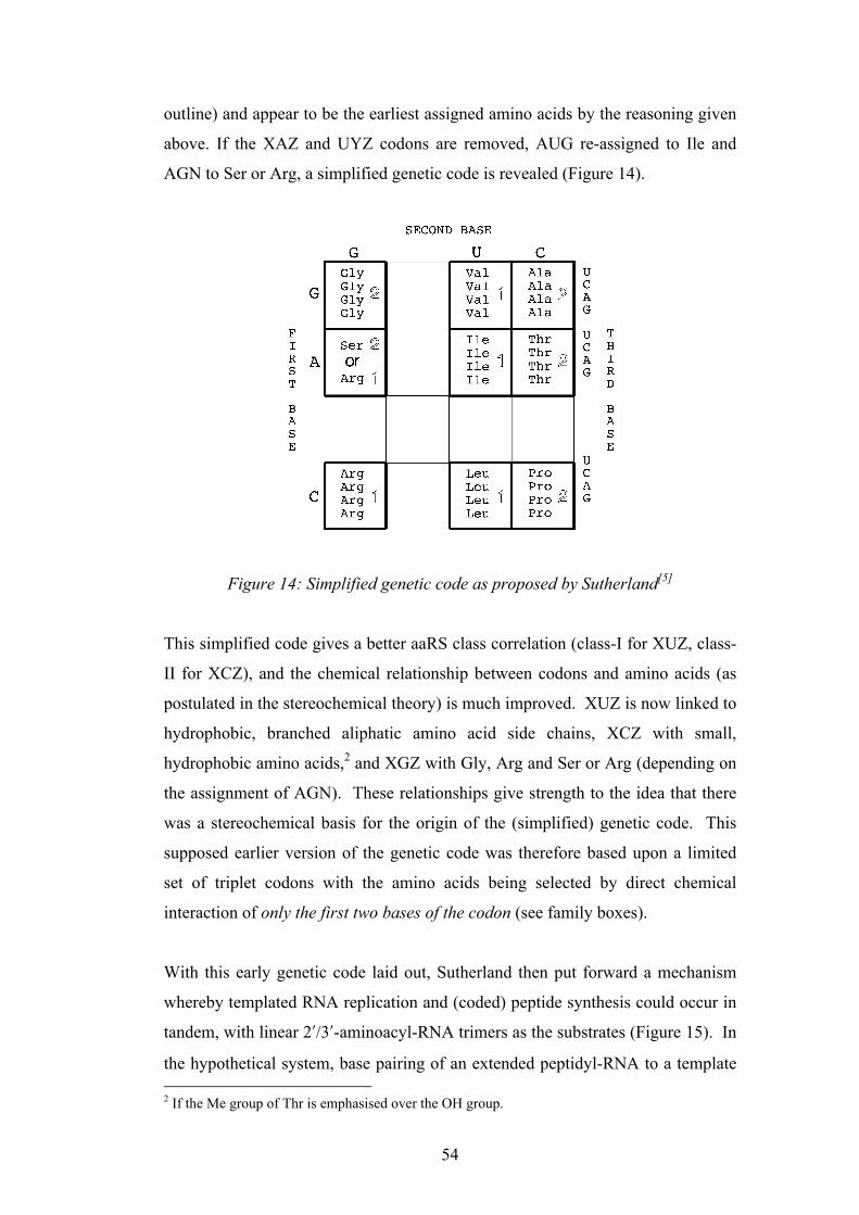
54
outline) and appear to be the earliest assigned amino acids by the reasoning given
above. If the XAZ and UYZ codons are removed, AUG re-assigned to Ile and
AGN to Ser or Arg, a simplified genetic code is revealed (Figure 14).
Figure 14: Simplified genetic code as proposed by Sutherland[5]
This simplified code gives a better aaRS class correlation (class-I for XUZ, class-
II for XCZ), and the chemical relationship between codons and amino acids (as
postulated in the stereochemical theory) is much improved. XUZ is now linked to
hydrophobic, branched aliphatic amino acid side chains, XCZ with small,
hydrophobic amino acids,2 and XGZ with Gly, Arg and Ser or Arg (depending on
the assignment of AGN). These relationships give strength to the idea that there
was a stereochemical basis for the origin of the (simplified) genetic code. This
supposed earlier version of the genetic code was therefore based upon a limited
set of triplet codons with the amino acids being selected by direct chemical
interaction of only the first two bases of the codon (see family boxes).
With this early genetic code laid out, Sutherland then put forward a mechanism
whereby templated RNA replication and (coded) peptide synthesis could occur in
tandem, with linear 2ʹ′/3ʹ′-aminoacyl-RNA trimers as the substrates (Figure 15). In
the hypothetical system, base pairing of an extended peptidyl-RNA to a template 2 If the Me group of Thr is emphasised over the OH group.
unassigned to codons. The improved chemical relationship between codons (oranticodons) and amino acids is particularly striking. XUZ is now associated withhydrophobic, branched aliphatic amino acid side chains. XCZ is associated with smallhydrophobic amino acids (if emphasis is placed on the Me group rather than the OHgroup of Thr) and XGZ is associated with Gly, Arg and, depending on the assignmentof AGN, Ser. Additionally, a potential association with the first base of the codonemerges. Within the codon sets XGZ, XUZ and XCZ, GYZ codes for the smallestamino acid, and for the codon sets XUZ and XCZ, AYZ codes for !-branched aminoacids, whilst CYZ codes for !-unbranched amino acids of comparable size. Thisimproved chemical relationship between codons (or anticodons) and amino acidsstrongly implies a chemical (stereochemical) basis for the origin of the codingrelationship.
According to this (retrosynthetic) analysis, the early genetic code was initially basedon a limited set of triplet codons with the amino acid being specified by direct chemicalcoding by two bases of the codon/anticodon only (family boxes). Subsequent evolutionprogressively sampled other codons resulting in their gradual recruitment andassignment to −old× or −new× amino acids. Such a model benefits from the advantagesimplicit in the Osawa!Jukes [38] and Schultz!Yarus [39] mechanisms for codonreassignments in later evolution but does not require initial loss and subsequentreassignment of codons merely a late assignment. Crucial to such a model is a
Fig. 2. Postulated Early, Compositionally-Restricted Chemical Genetic Code.Removal of XAZ and UYZ codonassignments, reassignment of AUG to Ile and AGN to Ser or Arg leaves a simplified code and an improved
chemical relationship between the amino acid side chains and the bases of the codons (or anticodons).
CHEMISTRY & BIODIVERSITY ± Vol. 1 (2004) 213
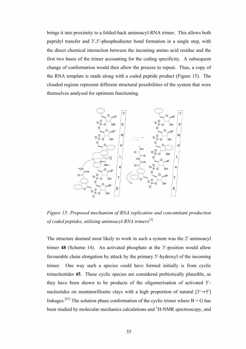
55
brings it into proximity to a folded-back aminoacyl-RNA trimer. This allows both
peptidyl transfer and 3ʹ′,5ʹ′-phosphodiester bond formation in a single step, with
the direct chemical interaction between the incoming amino acid residue and the
first two bases of the trimer accounting for the coding specificity. A subsequent
change of conformation would then allow the process to repeat. Thus, a copy of
the RNA template is made along with a coded peptide product (Figure 15). The
clouded regions represent different structural possibilities of the system that were
themselves analysed for optimum functioning.
Figure 15: Proposed mechanism of RNA replication and concomitant production
of coded peptides, utilising aminoacyl-RNA trimers[5]
The structure deemed most likely to work in such a system was the 2ʹ′-aminoacyl
trimer 48 (Scheme 14). An activated phosphate at the 3ʹ′-position would allow
favourable chain elongation by attack by the primary 5ʹ′-hydroxyl of the incoming
trimer. One way such a species could have formed initially is from cyclic
trinucleotides 45. These cyclic species are considered prebiotically plausible, as
they have been shown to be products of the oligomerisation of activated 5ʹ′-
nucleotides on montmorillonite clays with a high proportion of natural [3ʹ′→5ʹ′]
linkages.[87] The solution phase conformation of the cyclic trimer where B = G has
been studied by molecular mechanics calculations and 1H-NMR spectroscopy, and
Intramolecular contact between the amino acid side chain and the first two bases of thetrimer in a −folded-back× conformation was proposed to account for stereochemicalcoding. It was postulated that the coding might arise during synthesis of the trimers ormight result from the greater stability of correctly aminoacylated trimers towardshydrolysis. According to the scheme, RNA replication with aminoacyl-RNA trimerswould produce a coded peptide product and a copy of the template.
Aminoacyl-RNA Trimer Options. The retrosynthetic analysis of RNA :codedpeptides thus far points to two stages of synthesis involving assembly and oligomerisa-tion of aminoacyl-RNA trimers. The intermediacy of trimers extends retrosynthesisoptions because there exists the possibility that one of the residues of the trimer mightbe abasic or bear a modified base (Scheme 5). Such modifications need not preventtemplate association because two bases alone might provide sufficient binding. In thesecases, the base would have to be added or the modification removed either duringoligomerisation or afterwards.
There are a daunting number of possible trimer structures and oligomerisationchemistries so our strategy has been to select what are thought to be the most likelypossibilities (Scheme 6) and then to investigate them by preliminary experiments. Any
Scheme 5. Hypothetical Linked-RNA Replication and (Coded) Peptide Synthesis. Base pairing to a templatebrings an extended peptidyl-RNA and a folded-back aminoacyl-RNA trimer into proximity, allowing peptidyltransfer and 3!,5!-phosphodiester bond formation. Subsequent change of conformation to the extended stateallows continuation of the process. Coding is postulated to arise through interaction of the amino acid side chainand the first two bases of the trimer (R :B1, B2). A number of possible trimer structures and transfer chemistries
are consistent with this general scheme (clouds).
CHEMISTRY & BIODIVERSITY ± Vol. 1 (2004) 215
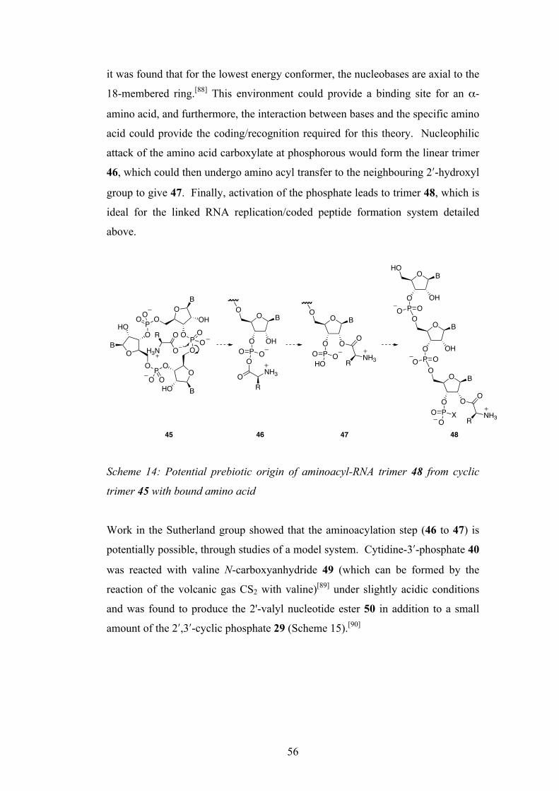
56
it was found that for the lowest energy conformer, the nucleobases are axial to the
18-membered ring.[88] This environment could provide a binding site for an α-
amino acid, and furthermore, the interaction between bases and the specific amino
acid could provide the coding/recognition required for this theory. Nucleophilic
attack of the amino acid carboxylate at phosphorous would form the linear trimer
46, which could then undergo amino acyl transfer to the neighbouring 2ʹ′-hydroxyl
group to give 47. Finally, activation of the phosphate leads to trimer 48, which is
ideal for the linked RNA replication/coded peptide formation system detailed
above.
Scheme 14: Potential prebiotic origin of aminoacyl-RNA trimer 48 from cyclic
trimer 45 with bound amino acid
Work in the Sutherland group showed that the aminoacylation step (46 to 47) is
potentially possible, through studies of a model system. Cytidine-3ʹ′-phosphate 40
was reacted with valine N-carboxyanhydride 49 (which can be formed by the
reaction of the volcanic gas CS2 with valine)[89] under slightly acidic conditions
and was found to produce the 2'-valyl nucleotide ester 50 in addition to a small
amount of the 2ʹ′,3ʹ′-cyclic phosphate 29 (Scheme 15).[90]
PO
O
O
OP
O P O
O
O
O
HOOH
HO
B
B
B
OO
OO
OO
O
O
R
H3N
OO
O OH
B
POO
O
OR
NH3
OO
O O
B
POHO
O
O
NH3R
O BO
O B
O OH
O
P OO
O
O OH
HO
P OO
O
B
POO
X
OO
NH3R
45 47 4846

57
Scheme 15: Aminoacylation of cytidine-3ʹ′-phosphate 40 with valine N-
carboxyanhydride 49
The (coded) aminoacylation of an RNA trimer is therefore a valuable target for
the further development of the RNA:coded peptides theory, and this shall be
investigated in this thesis.
1.10 Compartmentalisation
This thesis so far has concentrated on the genetic aspects of life’s origin, in
questioning how nucleic acids and also (coded) peptides may have arisen. Whilst
these ideas may be at the very core of the problem, there are also other types of
molecule that must not be dismissed if we are to gain a real understanding of how
the most primitive of all life forms, the LCA, emerged on Earth.
The role of amphiphilic compounds in the origin of life is one of huge importance.
Without compartmentalisation, life itself could not be distinguished from an
otherwise disparate array of molecular interactions. If one wants to consider the
concept of the LCA, then this primitive life form must have had some sort of
boundary structure, almost certainly a lipid bilayer. Without a membrane, any
self-replicating molecules (such as nucleic acids) or products of metabolism
would simply diffuse away and be lost by the ‘organism’. It is apparent that some
sort of concentrating mechanism is needed to overcome the high dilution of an
early global ocean, and allow life to evolve.[91] It is therefore of interest to
O C
O OH
HO
P OO
O
NHO
O
O
O C
O OH
HO
POOO
O
NH3
O C
O O
HO
POOO O
NH3
O CHO
OPO
O O
CO2
+
+
40
49
50
29
+
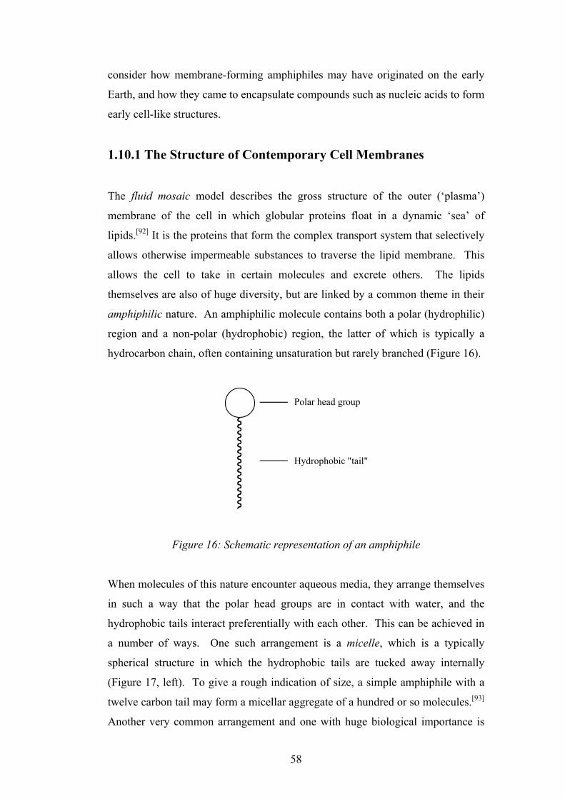
58
consider how membrane-forming amphiphiles may have originated on the early
Earth, and how they came to encapsulate compounds such as nucleic acids to form
early cell-like structures.
1.10.1 The Structure of Contemporary Cell Membranes
The fluid mosaic model describes the gross structure of the outer (‘plasma’)
membrane of the cell in which globular proteins float in a dynamic ‘sea’ of
lipids.[92] It is the proteins that form the complex transport system that selectively
allows otherwise impermeable substances to traverse the lipid membrane. This
allows the cell to take in certain molecules and excrete others. The lipids
themselves are also of huge diversity, but are linked by a common theme in their
amphiphilic nature. An amphiphilic molecule contains both a polar (hydrophilic)
region and a non-polar (hydrophobic) region, the latter of which is typically a
hydrocarbon chain, often containing unsaturation but rarely branched (Figure 16).
Figure 16: Schematic representation of an amphiphile
When molecules of this nature encounter aqueous media, they arrange themselves
in such a way that the polar head groups are in contact with water, and the
hydrophobic tails interact preferentially with each other. This can be achieved in
a number of ways. One such arrangement is a micelle, which is a typically
spherical structure in which the hydrophobic tails are tucked away internally
(Figure 17, left). To give a rough indication of size, a simple amphiphile with a
twelve carbon tail may form a micellar aggregate of a hundred or so molecules.[93]
Another very common arrangement and one with huge biological importance is
Polar head group
Hydrophobic "tail"
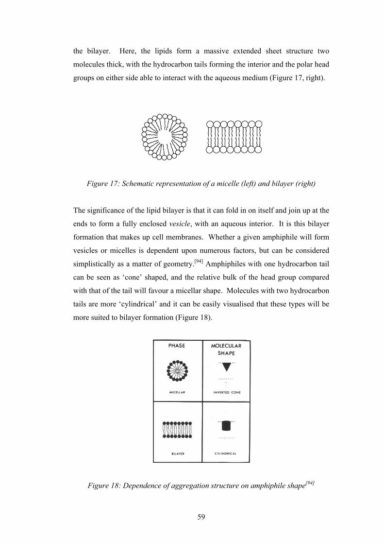
59
the bilayer. Here, the lipids form a massive extended sheet structure two
molecules thick, with the hydrocarbon tails forming the interior and the polar head
groups on either side able to interact with the aqueous medium (Figure 17, right).
Figure 17: Schematic representation of a micelle (left) and bilayer (right)
The significance of the lipid bilayer is that it can fold in on itself and join up at the
ends to form a fully enclosed vesicle, with an aqueous interior. It is this bilayer
formation that makes up cell membranes. Whether a given amphiphile will form
vesicles or micelles is dependent upon numerous factors, but can be considered
simplistically as a matter of geometry.[94] Amphiphiles with one hydrocarbon tail
can be seen as ‘cone’ shaped, and the relative bulk of the head group compared
with that of the tail will favour a micellar shape. Molecules with two hydrocarbon
tails are more ‘cylindrical’ and it can be easily visualised that these types will be
more suited to bilayer formation (Figure 18).
Figure 18: Dependence of aggregation structure on amphiphile shape[94]

60
The variety of lipids used to form membranes in biology is enormous, but there
are essentially three common types: phospholipids, glycolipids and cholesterol.[18]
Of these, the phospholipids are the most common, and are found in all biological
membranes. A typical phospholipid, phosphatidylcholine, is shown in Figure 19.
Figure 19: Phosphatidylcholine
Phosphatidylcholine illustrates the common features of a phosphoglyceride: a
glycerol backbone is attached to two fatty acid chains and a phosphorylated
quaternary ethanolamine. If one considers the possibilities of variation, firstly at
the hydrocarbon tails (length, saturation, branching, asymmetry), and also of the
identity of the phosphorylated alcohol, the vast scope for diversity can be
appreciated for just one subclass of membranous lipid.
1.10.2 Amphiphiles on the Prebiotic Earth
Several clues as to which amphiphilic molecules were available on the prebiotic
Earth lie in the content of carbonaceous meteorites (see chapter 1.7). Deamer and
Pashley extracted lipid-like compounds from the Murchison CM2 chondrite,3 and
although the chemical composition of these lipids was not fully determined, they
were shown to assemble into vesicle-type structures.[95] Naraoka et al. went a
stage further and identified various lipid-like molecules present in three CM2
3 A meteorite that fell in Murchison, Australia on the 28th September 1969. Amongst other compounds, the discovery of various amino acids sparked much scientific interest.
O
O O PO
OO
NO
O
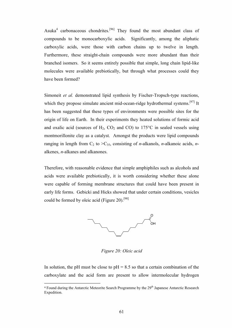
61
Asuka4 carbonaceous chondrites.[96] They found the most abundant class of
compounds to be monocarboxylic acids. Significantly, among the aliphatic
carboxylic acids, were those with carbon chains up to twelve in length.
Furthermore, these straight-chain compounds were more abundant than their
branched isomers. So it seems entirely possible that simple, long chain lipid-like
molecules were available prebiotically, but through what processes could they
have been formed?
Simoneit et al. demonstrated lipid synthesis by Fischer-Tropsch-type reactions,
which they propose simulate ancient mid-ocean-ridge hydrothermal systems.[97] It
has been suggested that these types of environments were possible sites for the
origin of life on Earth. In their experiments they heated solutions of formic acid
and oxalic acid (sources of H2, CO2 and CO) to 175°C in sealed vessels using
montmorillonite clay as a catalyst. Amongst the products were lipid compounds
ranging in length from C2 to >C35, consisting of n-alkanols, n-alkanoic acids, n-
alkenes, n-alkanes and alkanones.
Therefore, with reasonable evidence that simple amphiphiles such as alcohols and
acids were available prebiotically, it is worth considering whether these alone
were capable of forming membrane structures that could have been present in
early life forms. Gebicki and Hicks showed that under certain conditions, vesicles
could be formed by oleic acid (Figure 20).[98]
Figure 20: Oleic acid
In solution, the pH must be close to pH = 8.5 so that a certain combination of the
carboxylate and the acid form are present to allow intermolecular hydrogen
4 Found during the Antarctic Meteorite Search Programme by the 29th Japanese Antarctic Research Expedition.
O
OH

62
bonding. If all molecules are present as the acid, then droplets are produced, and
if present entirely as the anion, only micelles are formed. In fact, this requirement
for a specific pH value in order to form vesicles is a feature common to all long-
chain carboxylic acids. Studies into the role of amphiphiles in the origin of life
have focussed almost entirely on carboxylic acids of some sort. Walde et al.
showed that vesicles composed of caprylic acid (octanoic acid) and oleic acid
could undergo ‘autopoietic’ self-reproduction - an increase of their population due
to a reaction which takes place within the vesicles themselves.[99] In this case, the
reaction was the alkaline hydrolysis of the corresponding anhydrides of the acids,
and the subsequent release of the acid/carboxylate allowed the growth and
eventual division of the vesicles. This fascinating work was some of the first
towards building a ‘protocell’ - a synthetic system that models what the very first
cell-type structures may have been.
Others have since gone further towards the goal of the protocell, by taking a
simple self-replicating vesicle system and adding to it things such as nucleic acids
and even ribozymes. These studies offer tantalising insights into what the LCA
could have been composed of at the origin of life. Szostak et al. are making
efforts towards a model protocell system based on the encapsulation of self-
replicating nucleic acids in self-replicating membrane vesicles.[100] They took a
mixture of myristoleic acid (tetradecanoic acid) and its glycerol monoester to
construct vesicles that encapsulated a hammerhead ribozyme. The ribozyme was
able to self-cleave under conditions where the vesicles also self-replicated. The
use of the glycerol monoester was vital for stabilisation, as the Mg2+ cations
needed to activate the ribozyme for self-cleavage causes fatty acid vesicles to
precipitate. This is a general problem for fatty acid vesicles, and other divalent
metal cations that were almost certainly present in the early oceans (such as Ca2+)
cause the same problem, even at low concentrations.[101]
Although the various studies based upon carboxylic acid vesicles have certainly
given insights into how a primitive cell may have been constructed, their
sensitivity to ionic environment perhaps questions their prebiotic relevance. This
need for a specific pH, and the required addition of glycerol esters to overcome
the disruptive influence of divalent metal cations has even led to some suggesting
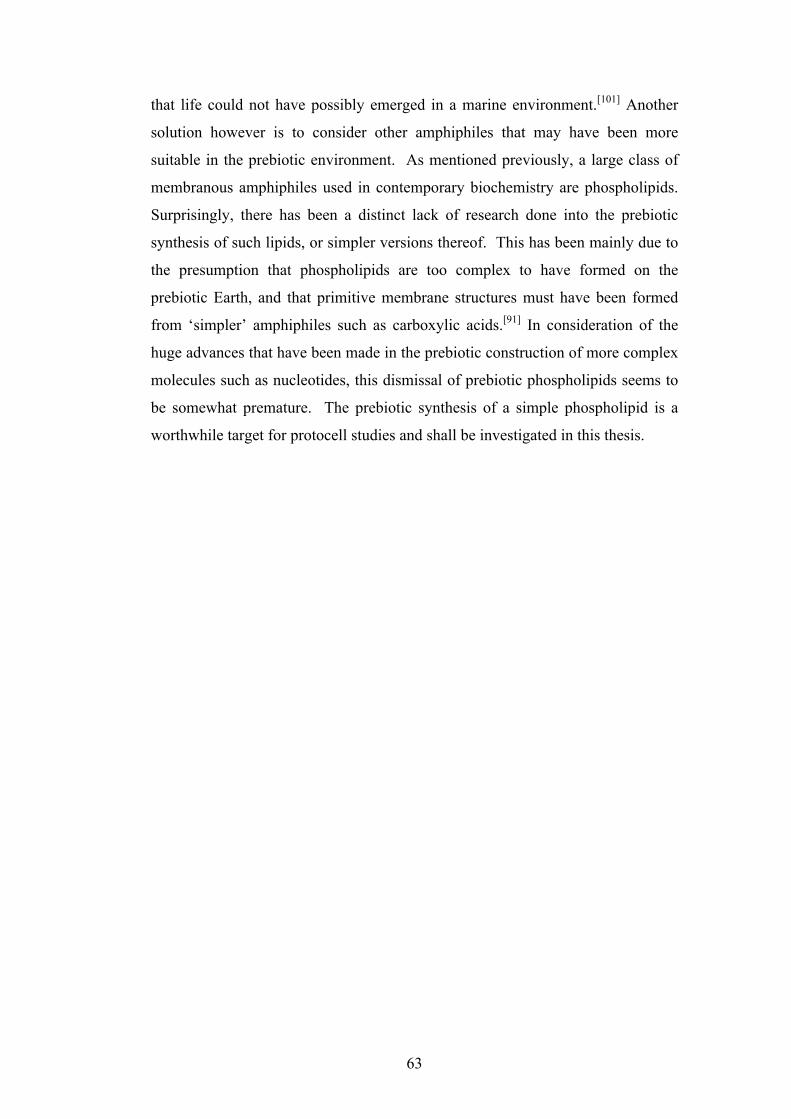
63
that life could not have possibly emerged in a marine environment.[101] Another
solution however is to consider other amphiphiles that may have been more
suitable in the prebiotic environment. As mentioned previously, a large class of
membranous amphiphiles used in contemporary biochemistry are phospholipids.
Surprisingly, there has been a distinct lack of research done into the prebiotic
synthesis of such lipids, or simpler versions thereof. This has been mainly due to
the presumption that phospholipids are too complex to have formed on the
prebiotic Earth, and that primitive membrane structures must have been formed
from ‘simpler’ amphiphiles such as carboxylic acids.[91] In consideration of the
huge advances that have been made in the prebiotic construction of more complex
molecules such as nucleotides, this dismissal of prebiotic phospholipids seems to
be somewhat premature. The prebiotic synthesis of a simple phospholipid is a
worthwhile target for protocell studies and shall be investigated in this thesis.
64
1.11 Project Aims
• Recent breakthroughs in the Sutherland group have demonstrated the
prebiotic synthesis of activated pyrimidine ribonucleotides in the form of
2ʹ′,3ʹ′-cyclic phosphates. This gives strength to the idea that these species
should be considered as candidates for oligomerisation in the formation of
RNA, which is central to any ‘RNA-first’ theory for the origin of life.
Hydrolysis of these monomers to the respective 2ʹ′- and 3ʹ′-
monophosphates is inevitable in water however, and so a means of re-
activation to the cyclic phosphates is investigated. The activation
chemistry outlined above is investigated as part of a multicomponent
reaction so that in addition to the nucleotide activation, important prebiotic
side products are formed that would support the emergence of molecules
such as peptides alongside RNA at the origin of life.
• The theory of the co-emergence of RNA and coded peptides put forward
by Sutherland requires aminoacyl-RNA trimers to be formed prebiotically,
and this is investigated also as part of a multicomponent reaction.
• The dry-state oligomerisation of ribonucleotide-2ʹ′,3ʹ′-cyclic phosphates by
Orgel and co-workers has been shown to be promising towards the
formation of short RNA molecules. One of the most effective catalysts for
this process is ethanolamine, and this oligomerisation mechanism is
probed using 1D/2D NMR spectroscopy to identify key intermediates.
• The compartmentalisation of important prebiotic molecules is thought to
be essential for the origin of life. This was most likely to have been
achieved by vesicles formed by primitive amphiphiles, and so a
predisposed phosphorylation reaction is investigated to produce simple
phospholipids that have the potential to form bilayer vesicles.
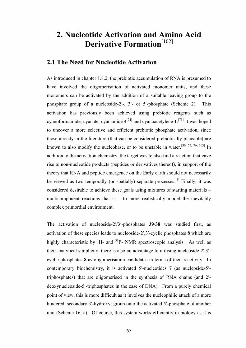
65
2. Nucleotide Activation and Amino Acid Derivative Formation[102]
2.1 The Need for Nucleotide Activation As introduced in chapter 1.8.2, the prebiotic accumulation of RNA is presumed to
have involved the oligomerisation of activated monomer units, and these
monomers can be activated by the addition of a suitable leaving group to the
phosphate group of a nucleoside-2ʹ′-, 3ʹ′- or 5ʹ′-phosphate (Scheme 2). This
activation has previously been achieved using prebiotic reagents such as
cyanoformamide, cyanate, cyanamide 4[74] and cyanoacetylene 1.[75] It was hoped
to uncover a more selective and efficient prebiotic phosphate activation, since
those already in the literature (that can be considered prebiotically plausible) are
known to also modify the nucleobase, or to be unstable in water.[30, 75, 76, 103] In
addition to the activation chemistry, the target was to also find a reaction that gave
rise to non-nucleotide products (peptides or derivatives thereof), in support of the
theory that RNA and peptide emergence on the Early earth should not necessarily
be viewed as two temporally (or spatially) separate processes.[5] Finally, it was
considered desirable to achieve these goals using mixtures of starting materials –
multicomponent reactions that is – to more realistically model the inevitably
complex primordial environment.
The activation of nucleoside-2ʹ′/3ʹ′-phosphates 39/38 was studied first, as
activation of these species leads to nucleoside-2ʹ′,3ʹ′-cyclic phosphates 8 which are
highly characteristic by 1H- and 31P- NMR spectroscopic analysis. As well as
their analytical simplicity, there is also an advantage to utilising nucleoside-2ʹ′,3ʹ′-
cyclic phosphates 8 as oligomerisation candidates in terms of their reactivity. In
contemporary biochemistry, it is activated 5ʹ′-nucleotides 7 (as nucleoside-5ʹ′-
triphosphates) that are oligomerised in the synthesis of RNA chains (and 2ʹ′-
deoxynucleoside-5ʹ′-triphosphates in the case of DNA). From a purely chemical
point of view, this is more difficult as it involves the nucleophilic attack of a more
hindered, secondary 3ʹ′-hydroxyl group onto the activated 5ʹ′-phosphate of another
unit (Scheme 16, a). Of course, this system works efficiently in biology as it is
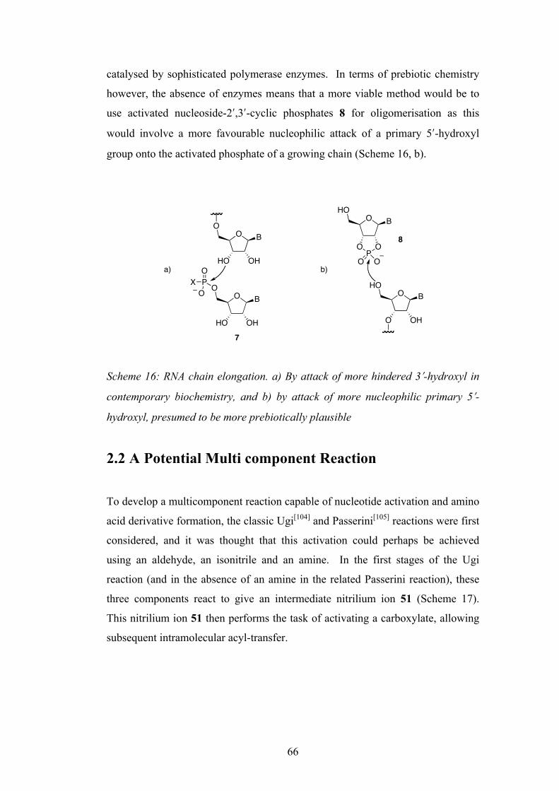
66
catalysed by sophisticated polymerase enzymes. In terms of prebiotic chemistry
however, the absence of enzymes means that a more viable method would be to
use activated nucleoside-2ʹ′,3ʹ′-cyclic phosphates 8 for oligomerisation as this
would involve a more favourable nucleophilic attack of a primary 5ʹ′-hydroxyl
group onto the activated phosphate of a growing chain (Scheme 16, b).
Scheme 16: RNA chain elongation. a) By attack of more hindered 3ʹ′-hydroxyl in
contemporary biochemistry, and b) by attack of more nucleophilic primary 5ʹ′-
hydroxyl, presumed to be more prebiotically plausible
2.2 A Potential Multi component Reaction
To develop a multicomponent reaction capable of nucleotide activation and amino
acid derivative formation, the classic Ugi[104] and Passerini[105] reactions were first
considered, and it was thought that this activation could perhaps be achieved
using an aldehyde, an isonitrile and an amine. In the first stages of the Ugi
reaction (and in the absence of an amine in the related Passerini reaction), these
three components react to give an intermediate nitrilium ion 51 (Scheme 17).
This nitrilium ion 51 then performs the task of activating a carboxylate, allowing
subsequent intramolecular acyl-transfer.
O BO
HO OH
O BHO
OPO
O O
O BO
HO OH
PO
XO O B
HO
O OH
a) b)
7
8
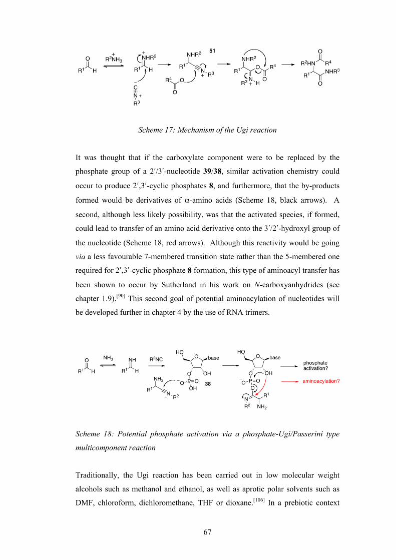
67
Scheme 17: Mechanism of the Ugi reaction
It was thought that if the carboxylate component were to be replaced by the
phosphate group of a 2ʹ′/3ʹ′-nucleotide 39/38, similar activation chemistry could
occur to produce 2ʹ′,3ʹ′-cyclic phosphates 8, and furthermore, that the by-products
formed would be derivatives of α-amino acids (Scheme 18, black arrows). A
second, although less likely possibility, was that the activated species, if formed,
could lead to transfer of an amino acid derivative onto the 3ʹ′/2ʹ′-hydroxyl group of
the nucleotide (Scheme 18, red arrows). Although this reactivity would be going
via a less favourable 7-membered transition state rather than the 5-membered one
required for 2ʹ′,3ʹ′-cyclic phosphate 8 formation, this type of aminoacyl transfer has
been shown to occur by Sutherland in his work on N-carboxyanhydrides (see
chapter 1.9).[90] This second goal of potential aminoacylation of nucleotides will
be developed further in chapter 4 by the use of RNA trimers.
Scheme 18: Potential phosphate activation via a phosphate-Ugi/Passerini type
multicomponent reaction
Traditionally, the Ugi reaction has been carried out in low molecular weight
alcohols such as methanol and ethanol, as well as aprotic polar solvents such as
DMF, chloroform, dichloromethane, THF or dioxane.[106] In a prebiotic context
O
HR1
CNR3
NHR2
HR1R2NH3
NHR2
R1 N R3
O
R4 O
NHR2
R1N
O R4
OR3
R2HN
R1 NHR3
O
O
R4
51
H
O
HR1
NH3 NH
HR1
R2NC
NH2
R1 N R2
O
O OH
baseHO
P OOH
O
phosphateactivation?
O
O OH
baseHO
P OOO
NR2
R1
NH2
aminoacylation?38

68
such a multicomponent reaction would have to be feasible in aqueous solution,
and recent literature suggests that some Passerini and Ugi reactions are not only
possible, but actually accelerated in water.[105] Encouraged by this, a detailed
study of a potentially prebiotic four-component reaction in water was undertaken.
The aldehyde chosen was iso-butyraldehyde 52, in the hope of forming
derivatives, or achieving aminoacylation of the proteinogenic amino acid valine if
the chemistry was successful. It was decided to use tert-butylisonitrile 53 simply
for its ease of handling, as its odour is substantially less pungent than some other
isonitriles.5 Finally, the amine used would be ammonia (as ammonium chloride
for ease of handling), again so that the potential by-products formed would be
derivatives of an α-amino acid.
2.3 Reaction of Nucleoside-2ʹ′ /3ʹ′-Phosphates with an Isonitrile, Aldehyde and Ammonia
Preliminary experiments were first carried out to determine the optimum
conditions for the multicomponent reaction, and it turned out that a slightly acidic
pH/pD was necessary (presumably because of the need for the aldehyde to be at
least partially protonated) and an excess of isonitrile, aldehyde and ammonia gave
the best results. Thus, addition of four equivalents each of iso-butyraldehyde 52
and tert-butylisonitrile 53 to a solution of β-D-adenosine-3ʹ′-phosphate 54 (100
mM) and NH4Cl (1M) at pH = 6 resulted in a heterogeneous reaction mixture that
was stirred at 40°C overnight. Initial 1H-NMR analysis showed that a variety of
products had been produced, and a means of separating these was sought to aid
identification (Scheme 19). The aqueous reaction mixture was first lyophilised to
remove any of the unreacted volatiles, followed by re-suspension in water and
adjustment to pH = 6. Initial extraction with dichloromethane followed by flash
column chromatography isolated the hydroxy amide 55 and its iso-butyrate ester
56. Re-adjustment of the aqueous phase to pH = 11.5 and a further organic
extraction revealed amino acid derivative 57 and a more mobile minor
unidentified product. This unidentified product could possibly be dimeric
5 The unpleasant smell of isonitriles is well documented, and Ugi even went so far as to state that “It is true that many potential workers in this field have been turned away by the odor”.
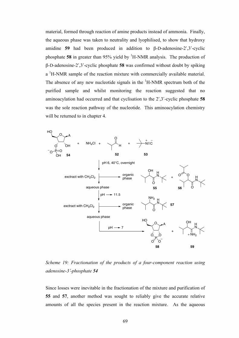
69
material, formed through reaction of amine products instead of ammonia. Finally,
the aqueous phase was taken to neutrality and lyophilised, to show that hydroxy
amidine 59 had been produced in addition to β-D-adenosine-2ʹ′,3ʹ′-cyclic
phosphate 58 in greater than 95% yield by 1H-NMR analysis. The production of
β-D-adenosine-2ʹ′,3ʹ′-cyclic phosphate 58 was confirmed without doubt by spiking
a 1H-NMR sample of the reaction mixture with commercially available material.
The absence of any new nucleotide signals in the 1H-NMR spectrum both of the
purified sample and whilst monitoring the reaction suggested that no
aminoacylation had occurred and that cyclisation to the 2ʹ′,3ʹ′-cyclic phosphate 58
was the sole reaction pathway of the nucleotide. This aminoacylation chemistry
will be returned to in chapter 4.
Scheme 19: Fractionation of the products of a four-component reaction using
adenosine-3ʹ′-phosphate 54
Since losses were inevitable in the fractionation of the mixture and purification of
55 and 57, another method was sought to reliably give the accurate relative
amounts of all the species present in the reaction mixture. As the aqueous
O
O OH
AHO
P OOH
O
NH4ClO
H N C+ ++
pH 6, 40°C, overnight
organicphaseexctract with CH2Cl2
aqueous phase
pH 11.5
aqueous phase
exctract with CH2Cl2 organicphase
pH 7
OH
O
HN + O
O
HN
O
NH2
O
HN
O AHO
OP
O
O O
+
OH
NH2
HN
54 52 53
55 56
57
58 59
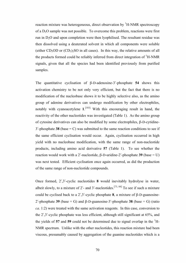
70
reaction mixture was heterogeneous, direct observation by 1H-NMR spectroscopy
of a D2O sample was not possible. To overcome this problem, reactions were first
run in D2O and upon completion were then lyophilised. The resultant residue was
then dissolved using a deuterated solvent in which all components were soluble
(either CD3OD or (CD3)2SO in all cases). In this way, the relative amounts of all
the products formed could be reliably inferred from direct integration of 1H-NMR
signals, given that all the species had been identified previously from purified
samples.
The quantitative cyclisation of β-D-adenosine-3ʹ′-phosphate 54 shows this
activation chemistry to be not only very efficient, but the fact that there is no
modification of the nucleobase shows it to be highly selective also, as the amino
group of adenine derivatives can undergo modification by other electrophiles,
notably with cyanoacetylene 1.[103] With this encouraging result in hand, the
reactivity of the other nucleotides was investigated (Table 1). As the amino group
of cytosine derivatives can also be modified by some electrophiles, β-D-cytidine-
3ʹ′-phosphate 38 (base = C) was submitted to the same reaction conditions to see if
the same efficient cyclisation would occur. Again, cyclisation occurred in high
yield with no nucleobase modification, with the same range of non-nucleotide
products, including amino acid derivative 57 (Table 1). To see whether the
reaction would work with a 2ʹ′-nucleotide, β-D-uridine-2ʹ′-phosphate 39 (base = U)
was next tested. Efficient cyclisation once again occurred, as did the production
of the same range of non-nucleotide compounds.
Once formed, 2ʹ′,3ʹ′-cyclic nucleotides 8 would inevitably hydrolyse in water,
albeit slowly, to a mixture of 2ʹ′- and 3ʹ′-nucleotides.[73, 84] To see if such a mixture
could be cyclised back to a 2ʹ′,3ʹ′-cyclic phosphate 8, a mixture of β-D-guanosine-
2ʹ′-phosphate 39 (base = G) and β-D-guanosine-3ʹ′-phosphate 38 (base = G) (ratio
ca. 1:2) were treated with the same activation reagents. In this case, conversion to
the 2ʹ′,3ʹ′-cyclic phosphate was less efficient, although still significant at 65%, and
the yields of 57 and 59 could not be determined due to signal overlap in the 1H-
NMR spectrum. Unlike with the other nucleotides, this reaction mixture had been
viscous, presumably caused by aggregation of the guanine nucleotides which is a
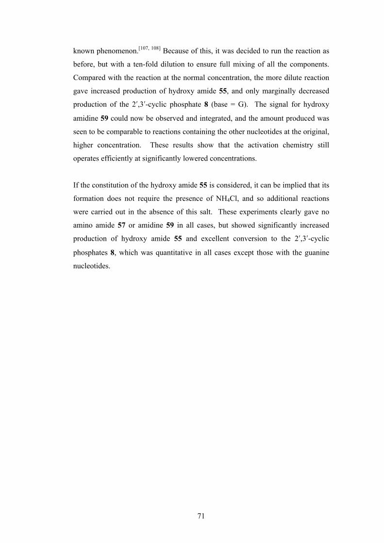
71
known phenomenon.[107, 108] Because of this, it was decided to run the reaction as
before, but with a ten-fold dilution to ensure full mixing of all the components.
Compared with the reaction at the normal concentration, the more dilute reaction
gave increased production of hydroxy amide 55, and only marginally decreased
production of the 2ʹ′,3ʹ′-cyclic phosphate 8 (base = G). The signal for hydroxy
amidine 59 could now be observed and integrated, and the amount produced was
seen to be comparable to reactions containing the other nucleotides at the original,
higher concentration. These results show that the activation chemistry still
operates efficiently at significantly lowered concentrations.
If the constitution of the hydroxy amide 55 is considered, it can be implied that its
formation does not require the presence of NH4Cl, and so additional reactions
were carried out in the absence of this salt. These experiments clearly gave no
amino amide 57 or amidine 59 in all cases, but showed significantly increased
production of hydroxy amide 55 and excellent conversion to the 2ʹ′,3ʹ′-cyclic
phosphates 8, which was quantitative in all cases except those with the guanine
nucleotides.

72
Nucleotide ± NH4Cl Relative amounts [%][a]
(100 mM) (1M) 8 55 57 59
38 (base = A) + 100 100 50 90
38 (base = C) + 90 162 41 81
39 (base = U) + 84 117 36 79
39 + 38 (base = G)[b] + 65 135 n.d.[c] n.d.[c]
39 + 38 (base = G)[d] +[e] 60 200 n.d.[c] 82
38 (base = A) - 100 160 - -
38 (base = C) - 100 201 - -
39 (base = U) - 100 167 - -
39 + 38 (base = G) - 70 188 - -
[a] Based on starting nucleotide; for 55, 57 and 59 this leads to relative amounts higher than 100% in some cases (because four equivalents of both 52 and 53 were used), but allows direct comparison of the relative amounts of the nucleotide products. [b] Ratio of 39 (base = G) and 38 (base = G) ca. 1:2. [c] Relative amount could not be determined due to signal overlap. [d] 10 mM. [e] 100 mM NH4Cl. Table 1: The effect of nucleotide structure on the relative amount of 8 and other products
Figure 21 shows a selected region of the CD3OD 1H-NMR spectrum of the
reaction of cytidine 3ʹ′-phosphate 38 (base = C) with tert-butylisonitrile 53 and
iso-butyraldehyde 52 in the presence of NH4Cl. It shows the signals for the α-
protons of the hydroxy amide 55, amino amide 57 and hydroxy amidine 59 as
well as signals for H-C(2ʹ′), H-C(4ʹ′) and H2C(5ʹ′) of the 2ʹ′,3ʹ′-cyclic phosphate 8
(base = C). The signal for H-C(3ʹ′) of the 2ʹ′,3ʹ′-cyclic phosphate is obscured by the
HOD peak.
O
O OH
BHO
P OOHO
O
HO O
BHO
PO OOH
O BHO
OPO
O O
OH
O
HN
NH2
O
HN
OH
NH2
HN
55 57 5983938

73
Figure 21: 1H-NMR (CD3OD) spectrum of the products of the reaction of 8 (base
= C) with 52, 53 and NH4Cl
2.4 Activation Using Only an Isonitrile
Mizuno and Kobayashi have shown that isonitriles alone are capable of phosphate
activation in pyridine.[109] They demonstrated that a mixture of uridine-2ʹ′(3ʹ′)-
phosphate could be converted to the 2ʹ′,3ʹ′-cyclic phosphate in 90% yield by
treatment with cyclohexyl isonitrile. In light of this, a control experiment was
conducted in the absence of the aldehyde and NH4Cl to see if similar activation
with just the isonitrile could occur in aqueous solution. A 1:2 mixture of cytidine-
2ʹ′-phosphate 39 (base = C) and cytidine 3ʹ′-phosphate 38 (base = C) was reacted in
D2O with 53 at pD = 6 overnight (Figure 22). In this experiment only very slow
cyclisation of the nucleotides occurred, with only a 44% conversion to the 2ʹ′,3ʹ′-
cyclic phosphate 8 (base = C), compared to the corresponding multicomponent
reactions that went to 90% and 100% conversion (with and without NH4Cl
respectively). This shows that, in aqueous solution at least, the phosphate
activation chemistry is greatly accelerated when conducted as part of the
multicomponent reaction in the presence of an aldehyde, either with or without
NH4Cl. This can be rationalised through the increased electrophilicity of the
nitrilium ion 61 over the (protonated) isonitrile alone.
5.0 4.9 4.8 4.7 4.6 4.5 4.4 4.3 4.2 4.1 4.0 3.9 3.8 3.7 3.6 3.5
! (1H) / ppm
8 (base = C) H-C(2')
HOD
8 (base = C) H-C(4')
8 (base = C) H2C(5')
59 H"
55 H"
57 H"

74
Figure 22: Selected region of the 1H-NMR spectrum of the reaction of 39 (base =
C)/ 40 (base = C) with 53 showing only partial conversion to the 2ʹ′,3ʹ′-cyclic
phosphate 8 (base = C)
2.5 Proposed Mechanism
Although the mechanism of the reactions have not been studied in detail in this
work, the constitution of the various products and comparison with the known Ugi
and related Passerini reactions[106] give clues as to how they might be formed.
Reversible reaction of ammonia with iso-butyraldehyde 52 gives the iminium ion
60, and reaction of this with tert-butylisonitrile 53 forms the amino nitrilium ion
61 (X = NH2). Alternatively (or exclusively in the absence of NH4Cl), direct
reaction of the aldehyde 52 with the isonitrile 53 would give the hydroxy nitrilium
ion 61 (X = OH). Reaction of either nitrilium ions with a 2ʹ′- or 3ʹ′-nucleotide
39/38 would lead to the imidoyl phosphate 62, and the attack of the 2ʹ′-hydroxyl of
62 at phosphorous generates simultaneously the 2ʹ′,3ʹ′-cyclic phosphate 8, and
hydroxy amide 55 or amino amide 57. Hydroxy amidine 59 is thought to result
from competing attack of ammonia on the nitrilium ion 61 (X = OH), and attack
by water on these nitrilium ions also occurs, which explains why the combined
yields of hydroxy amide 55 and amino amide 57 are 1.5-2.7 times higher than the
yield of the 2ʹ′,3ʹ′-cyclic phosphates 8. Consistent with this, the products 55, 57
and 59 were formed in similar yields in the absence of nucleotides, but the
reaction proceeded more slowly.
5.1 5.0 4.9 4.8 4.7 4.6 4.5 4.4 4.3 4.2 4.1 4.0
! (1H) / ppm
8 (base = C) H-C(2')
8 (base = C) H-C(3')
39 (base = C) H-C(2')
38 (base = C) H-C(3')
38 (base = C) H-C(2')
39 (base = C) H-C(3')
39 (base = C) H-C(4')
38 (base = C) H-C(4')
8 (base = C) H-C(4')
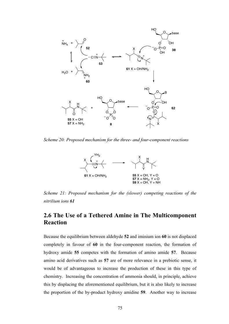
75
Scheme 20: Proposed mechanism for the three- and four-component reactions
Scheme 21: Proposed mechanism for the (slower) competing reactions of the
nitrilium ions 61
2.6 The Use of a Tethered Amine in The Multicomponent Reaction
Because the equilibrium between aldehyde 52 and iminium ion 60 is not displaced
completely in favour of 60 in the four-component reaction, the formation of
hydroxy amide 55 competes with the formation of amino amide 57. Because
amino acid derivatives such as 57 are of more relevance in a prebiotic sense, it
would be of advantageous to increase the production of these in this type of
chemistry. Increasing the concentration of ammonia should, in principle, achieve
this by displacing the aforementioned equilibrium, but it is also likely to increase
the proportion of the by-product hydroxy amidine 59. Another way to increase
ONH4 +
NH2H2O +
C N N
X
O
O OH
baseHO
P OOOH
O
O OH
BHO
P OOO
NX
O baseHO
OP
O
O O
X
O
HN +
61 X = OH/NH2
55 X = OH57 X = NH2
52
60
53
38
62
8
NX
YH2 X
Y
HN
61 X = OH/NH2 55 X = OH, Y = O57 X = NH2, Y = O59 X = OH, Y = NH
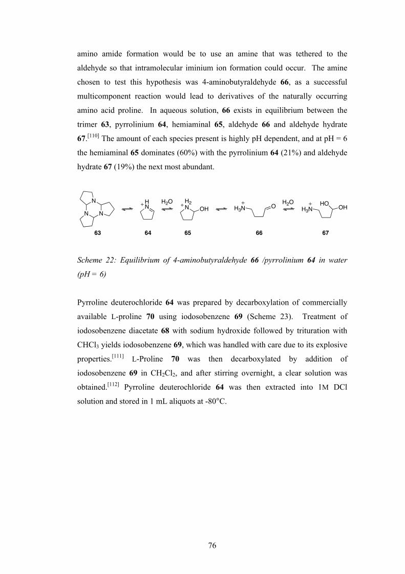
76
amino amide formation would be to use an amine that was tethered to the
aldehyde so that intramolecular iminium ion formation could occur. The amine
chosen to test this hypothesis was 4-aminobutyraldehyde 66, as a successful
multicomponent reaction would lead to derivatives of the naturally occurring
amino acid proline. In aqueous solution, 66 exists in equilibrium between the
trimer 63, pyrrolinium 64, hemiaminal 65, aldehyde 66 and aldehyde hydrate
67.[110] The amount of each species present is highly pH dependent, and at pH = 6
the hemiaminal 65 dominates (60%) with the pyrrolinium 64 (21%) and aldehyde
hydrate 67 (19%) the next most abundant.
Scheme 22: Equilibrium of 4-aminobutyraldehyde 66 /pyrrolinium 64 in water
(pH = 6)
Pyrroline deuterochloride 64 was prepared by decarboxylation of commercially
available L-proline 70 using iodosobenzene 69 (Scheme 23). Treatment of
iodosobenzene diacetate 68 with sodium hydroxide followed by trituration with
CHCl3 yields iodosobenzene 69, which was handled with care due to its explosive
properties.[111] L-Proline 70 was then decarboxylated by addition of
iodosobenzene 69 in CH2Cl2, and after stirring overnight, a clear solution was
obtained.[112] Pyrroline deuterochloride 64 was then extracted into 1M DCl
solution and stored in 1 mL aliquots at -80°C.
N N
N HN
H2N OH OH3N H3N OHHOH2O H2O
63 64 65 66 67
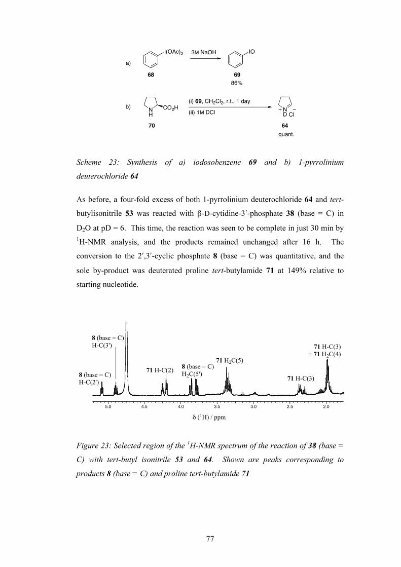
77
Scheme 23: Synthesis of a) iodosobenzene 69 and b) 1-pyrrolinium
deuterochloride 64
As before, a four-fold excess of both 1-pyrrolinium deuterochloride 64 and tert-
butylisonitrile 53 was reacted with β-D-cytidine-3ʹ′-phosphate 38 (base = C) in
D2O at pD = 6. This time, the reaction was seen to be complete in just 30 min by 1H-NMR analysis, and the products remained unchanged after 16 h. The
conversion to the 2ʹ′,3ʹ′-cyclic phosphate 8 (base = C) was quantitative, and the
sole by-product was deuterated proline tert-butylamide 71 at 149% relative to
starting nucleotide.
Figure 23: Selected region of the 1H-NMR spectrum of the reaction of 38 (base =
C) with tert-butyl isonitrile 53 and 64. Shown are peaks corresponding to
products 8 (base = C) and proline tert-butylamide 71
I(OAc)2 3M NaOH IO
68 69
NH
CO2H(i) 69, CH2Cl2, r.t., 1 day
(ii) 1M DCl ND Cl
a)
b)
86%
70 64quant.
5.0 4.5 4.0 3.5 3.0 2.5 2.0
! (1H) / ppm
8 (base = C) H-C(3')
8 (base = C) H-C(2')
71 H-C(2) 8 (base = C) H2C(5')
71 H2C(5)
71 H-C(3)
71 H-C(3)+ 71 H2C(4)
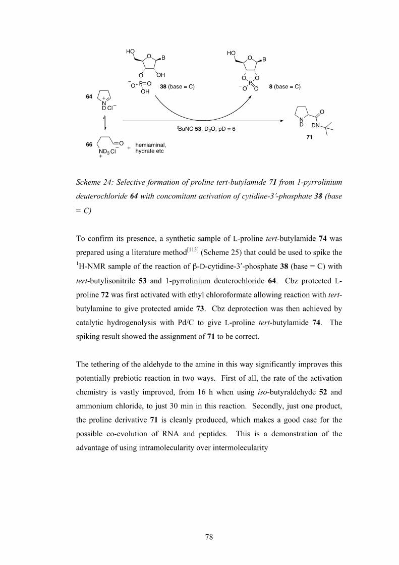
78
Scheme 24: Selective formation of proline tert-butylamide 71 from 1-pyrrolinium
deuterochloride 64 with concomitant activation of cytidine-3ʹ′-phosphate 38 (base
= C)
To confirm its presence, a synthetic sample of L-proline tert-butylamide 74 was
prepared using a literature method[113] (Scheme 25) that could be used to spike the 1H-NMR sample of the reaction of β-D-cytidine-3ʹ′-phosphate 38 (base = C) with
tert-butylisonitrile 53 and 1-pyrrolinium deuterochloride 64. Cbz protected L-
proline 72 was first activated with ethyl chloroformate allowing reaction with tert-
butylamine to give protected amide 73. Cbz deprotection was then achieved by
catalytic hydrogenolysis with Pd/C to give L-proline tert-butylamide 74. The
spiking result showed the assignment of 71 to be correct.
The tethering of the aldehyde to the amine in this way significantly improves this
potentially prebiotic reaction in two ways. First of all, the rate of the activation
chemistry is vastly improved, from 16 h when using iso-butyraldehyde 52 and
ammonium chloride, to just 30 min in this reaction. Secondly, just one product,
the proline derivative 71 is cleanly produced, which makes a good case for the
possible co-evolution of RNA and peptides. This is a demonstration of the
advantage of using intramolecularity over intermolecularity
ND Cl
ND
O
DN
ClND3
O hemiaminal, hydrate etc
O
O OH
HO
P OOOH
OHO
OP
O
OO
tBuNC 53, D2O, pD = 6
64
6671
8 (base = C)38 (base = C)
B B

79
Scheme 25: Conventional synthesis of L-proline tert-butylamide 74 for sample
spiking
2.7 Activation of Nucleoside-5ʹ′-Phosphates
Having investigated the activation chemistry of 2ʹ′- and 3ʹ′-nucleotides 39/38 , it
was now decided to consider the isomeric 5ʹ′-nucleotides such as 76. Activation
of 5ʹ′-nucleotides does not lead to nucleoside 3ʹ′,5ʹ′-cyclic phosphates and so is not
indicated by stable nucleotide products. However, formation of by-products
hydroxy amide 55, amino amide 57 and hydroxy amidine 59 more rapidly than in
the minus nucleotide control would indicate that these were being formed via
transient activation to the imidoyl phosphates, followed by hydrolysis. This was
investigated by adding four equivalents of tert-butylisonitrile 53 and iso-
butyraldehyde 52 to a solution of β-D-cytidine-5ʹ′-phosphate 76 at pH = 6 in the
presence and absence of NH4Cl. In the presence of NH4Cl all three by-products
were formed (relative amounts based on nucleotide: hydroxy amide 55, 233%;
amino amide 57, 30%; hydroxy amidine 59, 31%), and in its absence only
hydroxy amide 55 was observed (370%).
These reactions were significantly faster than the minus-nucleotide control and
suggest that transient activation to the imidoyl phosphate 75 had taken place. The
fact that this species is not observed by 1H-NMR means that it is quickly
hydrolysed and so such species could only be an intermediate in a templated
oligomerisation of RNA in aqueous solution so that the 3ʹ′-hydroxyl group of a
growing chain could have a high enough effective molarity relative to water and
therefore would be able to compete as a nucleophile. Therefore, although the
reaction involving a 5ʹ′-nucleotide is not as attractive for a prebiotic scenario as the
reaction involving 2ʹ′/3ʹ′-nucleotides (that produces stably activated 2ʹ′,3ʹ′-cyclic
NCbz
O
OHNCbz
O
HNNH
O
HN
(i) EtOCOCl, Et3N, THF, 0 °C (ii) tBuNH2, 0 ! 70°C Pd/C, H2, MeOH
78%72 73 74
quant.
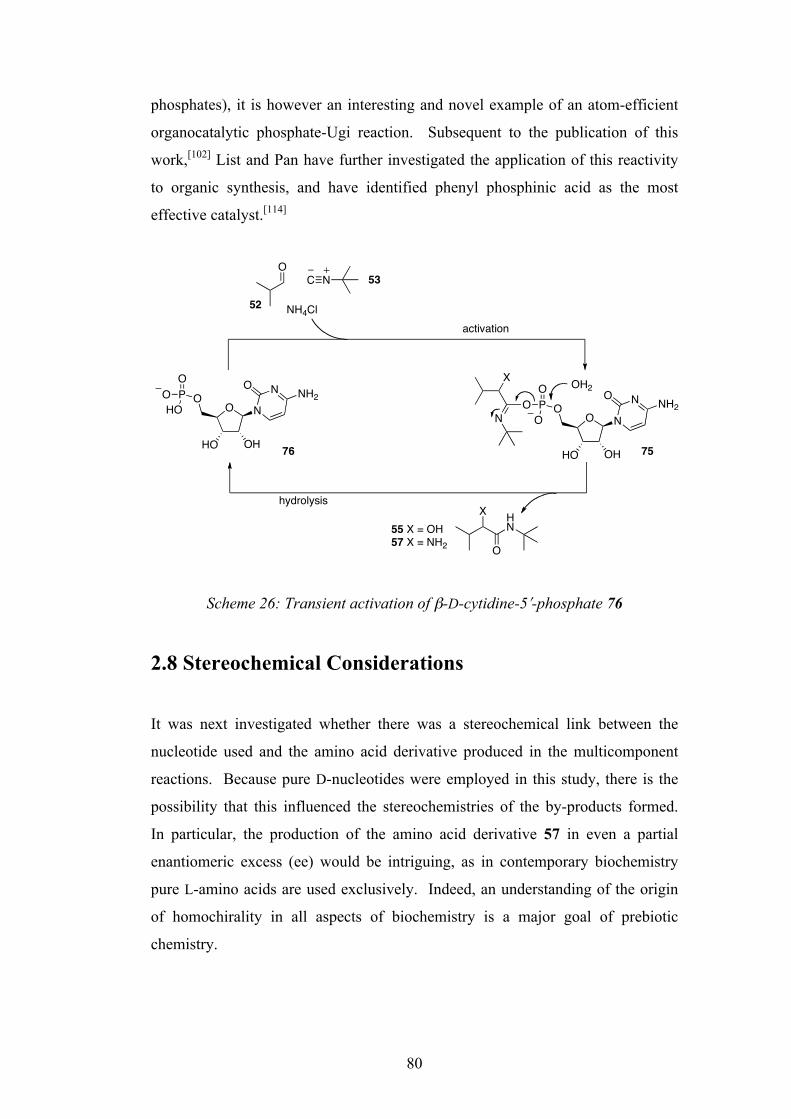
80
phosphates), it is however an interesting and novel example of an atom-efficient
organocatalytic phosphate-Ugi reaction. Subsequent to the publication of this
work,[102] List and Pan have further investigated the application of this reactivity
to organic synthesis, and have identified phenyl phosphinic acid as the most
effective catalyst.[114]
Scheme 26: Transient activation of β-D-cytidine-5ʹ′-phosphate 76
2.8 Stereochemical Considerations
It was next investigated whether there was a stereochemical link between the
nucleotide used and the amino acid derivative produced in the multicomponent
reactions. Because pure D-nucleotides were employed in this study, there is the
possibility that this influenced the stereochemistries of the by-products formed.
In particular, the production of the amino acid derivative 57 in even a partial
enantiomeric excess (ee) would be intriguing, as in contemporary biochemistry
pure L-amino acids are used exclusively. Indeed, an understanding of the origin
of homochirality in all aspects of biochemistry is a major goal of prebiotic
chemistry.
O
HO OH
ON
NONH2P
OOHO O
HO OH
ON
NONH2P
OO
O
OC N
X
N
NH4Cl
X HN
O
OH2
55 X = OH57 X = NH2
activation
hydrolysis
52
53
7576

81
To test this possibility, the valine-tert-butylamide 57 produced from the reaction
of β-D-adenosine-3ʹ′-phosphate 54 with tert-butylisonitrile 53 and iso-
butyraldehyde 52 in the presence of NH4Cl (Scheme 19) was analysed by chiral
gas chromatography (GC). For comparison, a sample of D-valine-tert-butylamide
79 was synthesised according to a literature procedure.[115] Boc-D-valine 77 was
converted to the protected tert-butyl amide 78 using DCC, which was then
deprotected with trifluoroacetic acid to give D-valine-tert-butylamide 79 (Scheme
27). The disappointing yields were not optimised as a sufficient amount was
prepared for characterisation and the GC study.
Scheme 27: Synthesis of D-valine-tert-butylamide 79 for chiral GC analysis of 57
produced from the reaction of β-D-adenosine-3ʹ′-phosphate 54 with tert-
butylisonitrile 53 and iso-butyraldehyde 52 in the presence of NH4Cl (Scheme 19)
The D-isomer was found to elute at ~63.9 min, and the L-isomer at ~65.3 min
(Figure 24). Integration of these peaks for the gas chromatogram for valine-tert-
butylamide 57 produced by the multi-component reaction indicated an L-ee of 0.8
%. This low value can be taken to indicate that, within experimental error, the
amino amide was essentially racemic or close to racemic. Whilst this result at
first may seem of no relevance to the origin of homochirality, it has been recently
demonstrated by Blackmond et al. that small enantiomeric excesses of amino
acids can be greatly amplified by processes that can be considered prebiotic.[116]
NH O
Boc
(i) DCC, CH2Cl2, 0°C
OH(ii) tBuNH2, O°C ! r.t.
NH O
BocHN H2N
O
HN
CF3CO2H, CH2Cl2
79787738% 29%

82
Figure 24: Gas chromatogram of a sample of 57 from the reaction of 54, 52, 53,
and NH4Cl
L-57
D-57

83
3. Prebiotic Synthesis of Small Metabolites[117] 3.1 Using a Tethered Phosphate in the Multicomponent Reaction To further test the scope of the phosphate Passerini and Ugi type reactions, the
possibility of using intramolecularity to increase the efficiency was next
investigated. If the phosphate group were to be attached to the aldehyde, the
reaction of the isonitrile with the aldehyde could be followed by fast,
intramolecular addition of the phosphate to the newly formed nitrilium ion. The
aldehyde chosen was glycolaldehyde phosphate 81, which has been shown by
Eschenmoser and co-workers to be prebiotically available by the phosphorylation
of glycolaldehyde 6 with amidotriphosphate 80 in aqueous solution.[118]
Scheme 28: Formation of glycolaldehyde phosphate 81 by phosphorylation of
glycolaldehyde 6 with amidotriphosphate 80
Glycolaldehyde phosphate 81 was reacted overnight at r.t. with methyl isonitrile
82 in 1:1 stoichiometry in D2O at pD = 6. The 1H-NMR analysis showed that a
single product had been cleanly formed in >95% yield (Figure 25) that was
assigned as the glyceric acid amide derivative 83. The quantitative formation of
83 using only one equivalent of the isonitrile 82 (as opposed to four in the
previous multicomponent reactions) shows that rapid intramolecular phosphate
addition rather than hydrolysis occurs in nitrilium intermediate 84 to give 85.
Hydrolysis of 85 then leads to the product 83 (Scheme 29). It was found that if an
excess of isonitrile 82 was used, a small amount of conversion to the glyceric acid
amide-2,3-cyclic phosphate occurred. The inefficiency of this process compared
to the cyclisation of nucleoside-2ʹ′/3ʹ′-phosphates is presumably due to entropic
OHO P
O
OH2N OPO
OXP O
O
O+
0.25M MgCl2, H2O, pH = 7, r.t.O
OPO
OO
6 8180
Mg2+

84
reasons, as there is free rotation about the C(2)-C(3) bond in 83 in contrast to the
restricted C(2ʹ′)-C(3ʹ′) bond rotation on nucleoside-2ʹ′/3ʹ′-phosphates 39/38.
Scheme 29: Formation of the glyceric amide derivative 83 via efficient
intramolecular addition of phosphate to the nitrilium ion 84
Figure 25: 1H-NMR spectrum showing the sole product 83 of the reaction
between glycolaldehyde phosphate 81 and methyl isonitrile 82
3.2 Three-Component Reaction with Phosphate Transfer
To further expand this type of reactivity, it was hoped that a phosphate-Passerini
reaction could be developed where transphosphorylation could occur onto the
newly formed hydroxyl group, akin to the transfer of the acyl group in the
traditional Passerini reaction. To find the right reagents for this phosphate
OH
OPO
OO
NHO
C N
OH
ON
PO
OO
O
OPO
OO
OP O
OH
NH
O O
H2O
+D2O, pD = 6, r.t., o.n.
81
84 85
8382
4.0 3.94.14.24.3 2.8 2.7 2.6
OH
ONHOP
OO
O
H-C(2) H2C(3) MeNH
! (1") / ppm
23
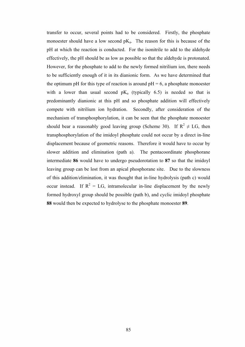
85
transfer to occur, several points had to be considered. Firstly, the phosphate
monoester should have a low second pKa. The reason for this is because of the
pH at which the reaction is conducted. For the isonitrile to add to the aldehyde
effectively, the pH should be as low as possible so that the aldehyde is protonated.
However, for the phosphate to add to the newly formed nitrilium ion, there needs
to be sufficiently enough of it in its dianionic form. As we have determined that
the optimum pH for this type of reaction is around pH = 6, a phosphate monoester
with a lower than usual second pKa (typically 6.5) is needed so that is
predominantly dianionic at this pH and so phosphate addition will effectively
compete with nitrilium ion hydration. Secondly, after consideration of the
mechanism of transphosphorylation, it can be seen that the phosphate monoester
should bear a reasonably good leaving group (Scheme 30). If R2 ≠ LG, then
transphosphorylation of the imidoyl phosphate could not occur by a direct in-line
displacement because of geometric reasons. Therefore it would have to occur by
slower addition and elimination (path a). The pentacoordinate phosphorane
intermediate 86 would have to undergo pseudorotation to 87 so that the imidoyl
leaving group can be lost from an apical phosphorane site. Due to the slowness
of this addition/elimination, it was thought that in-line hydrolysis (path c) would
occur instead. If R2 = LG, intramolecular in-line displacement by the newly
formed hydroxyl group should be possible (path b), and cyclic imidoyl phosphate
88 would then be expected to hydrolyse to the phosphate monoester 89.
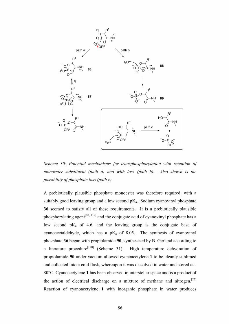
86
Scheme 30: Potential mechanisms for transphosphorylation with retention of
monoester substituent (path a) and with loss (path b). Also shown is the
possibility of phosphate loss (path c)
A prebiotically plausible phosphate monoester was therefore required, with a
suitably good leaving group and a low second pKa. Sodium cyanovinyl phosphate
36 seemed to satisfy all of these requirements. It is a prebiotically plausible
phosphorylating agent[70, 119] and the conjugate acid of cyanovinyl phosphate has a
low second pKa of 4.6, and the leaving group is the conjugate base of
cyanoacetaldehyde, which has a pKa of 8.05. The synthesis of cyanovinyl
phosphate 36 began with propiolamide 90, synthesised by B. Gerland according to
a literature procedure[120] (Scheme 31). High temperature dehydration of
propiolamide 90 under vacuum allowed cyanoacetylene 1 to be cleanly sublimed
and collected into a cold flask, whereupon it was dissolved in water and stored at -
80°C. Cyanoacetylene 1 has been observed in interstellar space and is a product of
the action of electrical discharge on a mixture of methane and nitrogen.[27]
Reaction of cyanoacetylene 1 with inorganic phosphate in water produces
P OO
R1
NH
O
OR2O
P OO
R1
NHOR2O O
NHO
OR1
POR2
O
O
H
OR1
NHO
POOR2
O
OP
O NHO
O
R1H2O
OR1
NHO
POO
O
HOR1
NHOP
OR2
O
OH2O
HOR1
NHO
PO
OOR2
O
+
path a path b
path c
!
86
87
88
89
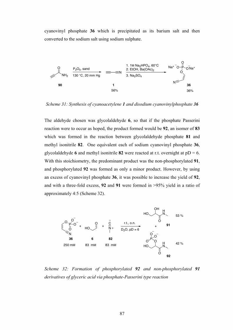
87
cyanovinyl phosphate 36 which is precipitated as its barium salt and then
converted to the sodium salt using sodium sulphate.
Scheme 31: Synthesis of cyanoacetylene 1 and disodium cyanovinylphosphate 36
The aldehyde chosen was glycolaldehyde 6, so that if the phosphate Passerini
reaction were to occur as hoped, the product formed would be 92, an isomer of 83
which was formed in the reaction between glycolaldehyde phosphate 81 and
methyl isonitrile 82. One equivalent each of sodium cyanovinyl phosphate 36,
glycolaldehyde 6 and methyl isonitrile 82 were reacted at r.t. overnight at pD = 6.
With this stoichiometry, the predominant product was the non-phosphorylated 91,
and phosphorylated 92 was formed as only a minor product. However, by using
an excess of cyanovinyl phosphate 36, it was possible to increase the yield of 92,
and with a three-fold excess, 92 and 91 were formed in >95% yield in a ratio of
approximately 4:5 (Scheme 32).
Scheme 32: Formation of phosphorylated 92 and non-phosphorylated 91
derivatives of glyceric acid via phosphate-Passerini type reaction
O
NH2
P2O5, sand
130 °C, 20 mm HgN
1. 1M Na2HPO4, 60°C2. EtOH, Ba(OAc)2
PO
OO
O
N
Na+ Na+
90 1 3656% 36%
3. Na2SO4
O
N
PO
OO
OHO
CN D2O, pD = 6
r.t., o.n.
OHHO
O
HN
OHO
O
HN
PO
O O
+
42 %
53 %
250 mM 83 mM 83 mM
+ +
36 6 82
91
92
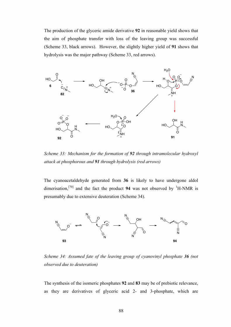
88
The production of the glyceric amide derivative 92 in reasonable yield shows that
the aim of phosphate transfer with loss of the leaving group was successful
(Scheme 33, black arrows). However, the slightly higher yield of 91 shows that
hydrolysis was the major pathway (Scheme 33, red arrows).
Scheme 33: Mechanism for the formation of 92 through intramolecular hydroxyl
attack at phosphorous and 91 through hydrolysis (red arrows)
The cyanoacetaldehyde generated from 36 is likely to have undergone aldol
dimerisation,[70] and the fact the product 94 was not observed by 1H-NMR is
presumably due to extensive deuteration (Scheme 34).
Scheme 34: Assumed fate of the leaving group of cyanovinyl phosphate 36 (not
observed due to deuteration)
The synthesis of the isomeric phosphates 92 and 83 may be of prebiotic relevance,
as they are derivatives of glyceric acid 2- and 3-phosphate, which are
OHO
NC
OHHO
NPO
OO
O
N
NH
OO
HOP
OH OO
N
NH
HO OPO OHOH2O
OHO
O
HN
POO O
H2O
OH
O
HNHO
92 91
6
8236
O
N
ON
ON N
OH
ON
NO
N
93 94
89
intermediates in the glycolysis pathway.[18] Based on these results, it is reasonable
to suggest that biology possibly evolved to use these types of compounds because
of their availability through abiotic syntheses such as the ones demonstrated here.
The new multicomponent reactions uncovered here using phosphates and
isonitriles have demonstrated nucleotide activation, formation of amino acid
derivatives, and production of small molecules that may have been of significance
in early metabolic cycles. The other goal that was set out earlier of
aminoacylation of nucleotides has not been seen however. In the next chapter this
second goal will be investigated further by using RNA trimers.
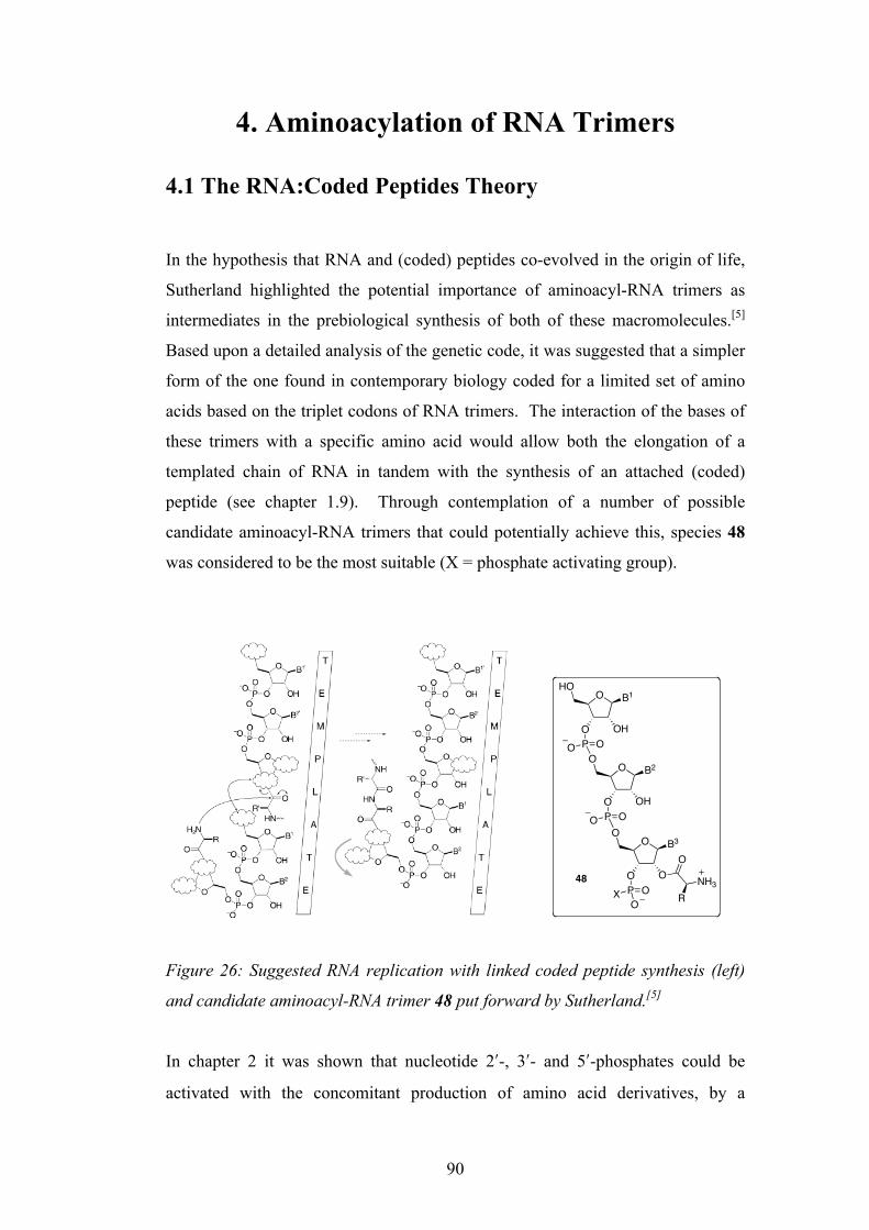
90
4. Aminoacylation of RNA Trimers 4.1 The RNA:Coded Peptides Theory In the hypothesis that RNA and (coded) peptides co-evolved in the origin of life,
Sutherland highlighted the potential importance of aminoacyl-RNA trimers as
intermediates in the prebiological synthesis of both of these macromolecules.[5]
Based upon a detailed analysis of the genetic code, it was suggested that a simpler
form of the one found in contemporary biology coded for a limited set of amino
acids based on the triplet codons of RNA trimers. The interaction of the bases of
these trimers with a specific amino acid would allow both the elongation of a
templated chain of RNA in tandem with the synthesis of an attached (coded)
peptide (see chapter 1.9). Through contemplation of a number of possible
candidate aminoacyl-RNA trimers that could potentially achieve this, species 48
was considered to be the most suitable (X = phosphate activating group).
Figure 26: Suggested RNA replication with linked coded peptide synthesis (left)
and candidate aminoacyl-RNA trimer 48 put forward by Sutherland.[5]
In chapter 2 it was shown that nucleotide 2ʹ′-, 3ʹ′- and 5ʹ′-phosphates could be
activated with the concomitant production of amino acid derivatives, by a
Intramolecular contact between the amino acid side chain and the first two bases of thetrimer in a −folded-back× conformation was proposed to account for stereochemicalcoding. It was postulated that the coding might arise during synthesis of the trimers ormight result from the greater stability of correctly aminoacylated trimers towardshydrolysis. According to the scheme, RNA replication with aminoacyl-RNA trimerswould produce a coded peptide product and a copy of the template.
Aminoacyl-RNA Trimer Options. The retrosynthetic analysis of RNA :codedpeptides thus far points to two stages of synthesis involving assembly and oligomerisa-tion of aminoacyl-RNA trimers. The intermediacy of trimers extends retrosynthesisoptions because there exists the possibility that one of the residues of the trimer mightbe abasic or bear a modified base (Scheme 5). Such modifications need not preventtemplate association because two bases alone might provide sufficient binding. In thesecases, the base would have to be added or the modification removed either duringoligomerisation or afterwards.
There are a daunting number of possible trimer structures and oligomerisationchemistries so our strategy has been to select what are thought to be the most likelypossibilities (Scheme 6) and then to investigate them by preliminary experiments. Any
Scheme 5. Hypothetical Linked-RNA Replication and (Coded) Peptide Synthesis. Base pairing to a templatebrings an extended peptidyl-RNA and a folded-back aminoacyl-RNA trimer into proximity, allowing peptidyltransfer and 3!,5!-phosphodiester bond formation. Subsequent change of conformation to the extended stateallows continuation of the process. Coding is postulated to arise through interaction of the amino acid side chainand the first two bases of the trimer (R :B1, B2). A number of possible trimer structures and transfer chemistries
are consistent with this general scheme (clouds).
CHEMISTRY & BIODIVERSITY ± Vol. 1 (2004) 215
O
O O
O
P OXO
O
O OH
O
P OO
O B1
O OH
HO
P OO
B2
B3
O
NH3R
48

91
multicomponent reaction akin to the Ugi and Passerini reactions. It was suggested
that using this same chemistry, it might be possible to transfer an amino acid
derivative to the 2ʹ′- or 3ʹ′-hydroxyl group of the nucleotide (Scheme 18). In the
case of nucleotide monomers, no such aminoacylation was observed. In the case
of 2ʹ′- and 3ʹ′-nucleotides 39/38, these were cyclised to the 2ʹ′,3ʹ′-cyclic phosphates
8, and the 5ʹ′-nucleotides were transiently activated giving no directly observed
intermediates. With both cases, a range of amino acid derivatives was produced
as by-products.
In consideration of this multicomponent reaction, and the candidate aminoacyl-
RNA trimer put forward by Sutherland, it was hoped that aminoacylation might
be achieved, at least in part, if a nucleotide trimer were used instead of a
monomer. In this way, perhaps the conformational folding, and interaction of the
bases of the trimer could assist with allowing the amino acid derivative to transfer
to the nearby hydroxyl group. In keeping with the notion that this primitive
aminoacylation relied upon a direct, specific interaction of the bases of the trimer
with a particular amino acid derivative to be transferred, therefore the triplet code
and the amino acid should be matched. Earlier in chapter 2.6, it was shown that
an amide derivative of proline 71 could be produced by the reaction of
pyrrolinium 64, an isonitrile 53 and either a 3ʹ′- or 5ʹ′nucleotide, 38 or 76
respectively. In the modern genetic code (and the simplified one put forward by
Sutherland),[5] proline is coded for by the triplet codon CCC. Therefore is was
hoped that by using an RNA trimer of CCC in the multi-component reaction, that
the direct interaction between the bases and amino acid that could have
underpinned the primitive genetic code would assist in transferring the amino acid
derivative to the terminus of the trimer. In this way, it may be possible to form an
aminoacylated RNA trimer that could have been an ancient precursor of what
nature now uses, tRNA.
4.2 Synthesis of RNA Trimer with Terminal 3ʹ′-Phosphate
It was decided to first synthesise a CCC RNA trimer with a phosphate group
attached to the 3ʹ′-terminal hydroxyl. In this way, successful transfer of the amino
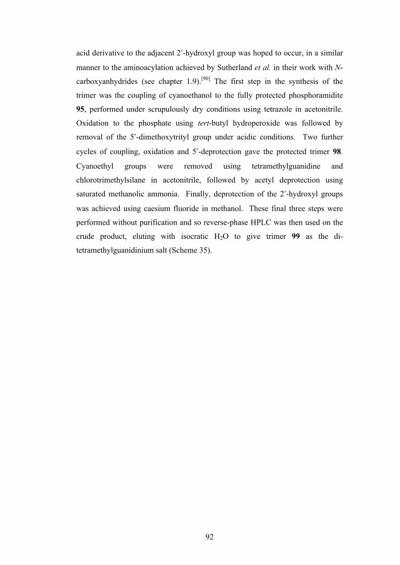
92
acid derivative to the adjacent 2ʹ′-hydroxyl group was hoped to occur, in a similar
manner to the aminoacylation achieved by Sutherland et al. in their work with N-
carboxyanhydrides (see chapter 1.9).[90] The first step in the synthesis of the
trimer was the coupling of cyanoethanol to the fully protected phosphoramidite
95, performed under scrupulously dry conditions using tetrazole in acetonitrile.
Oxidation to the phosphate using tert-butyl hydroperoxide was followed by
removal of the 5ʹ′-dimethoxytrityl group under acidic conditions. Two further
cycles of coupling, oxidation and 5ʹ′-deprotection gave the protected trimer 98.
Cyanoethyl groups were removed using tetramethylguanidine and
chlorotrimethylsilane in acetonitrile, followed by acetyl deprotection using
saturated methanolic ammonia. Finally, deprotection of the 2ʹ′-hydroxyl groups
was achieved using caesium fluoride in methanol. These final three steps were
performed without purification and so reverse-phase HPLC was then used on the
crude product, eluting with isocratic H2O to give trimer 99 as the di-
tetramethylguanidinium salt (Scheme 35).
93
(i) HO(CH2)2CN, tetrazole, MeCN, 3Å molecular sieves, (ii) tBuOOH, (iii) DCA, CH2Cl2 (85%
over 3 steps), (iv) 95, tetrazole, MeCN, 3Å molecular sieves, (v) tBuOOH, (vi) DCA, CH2Cl2
(87% over 3 steps), (vii) 95, tetrazole, MeCN, 3Å molecular sieves, (viii) tBuOOH, (ix) DCA,
CH2Cl2 (63% over 3 steps), (x) TMG, TMSCl, MeCN, (xi) NH3/MeOH, (xii) CsF, MeOH (55%
over 3 steps).
Scheme 35: Synthesis of CCC RNA trimer 99 with terminal 3ʹ′-phosphate group
4.3 Multicomponent Reaction of RNA Trimer With Terminal 3ʹ′-Phosphate
As with the nucleotide monomers, the CCC-3ʹ′-phosphate trimer 99 was reacted
with four equivalents each of 1-pyrrolinium 64 and methyl isonitrile 82 in D2O at
O N
O OTBDMS
NDMTrO
ONHAc
PN
O
N
O N
O OTBDMS
NO
ONHAc
P OOO
N
N
O N
O OTBDMS
NHO
ONHAc
P OON
O N
O OTBDMS
NO
ONHAc
P OOO
N
N
O N
O OTBDMS
NO
ONHAc
P OON
O N
O OTBDMS
NHO
ONHAc
P OON
O N
O OH
NO
ONH2
P OOOH
O N
O OH
NO
ONH2
P OO
O N
O OH
NHO
ONH2
P OO
NH2
N(CH3)2(H3C)2N
(i) ! (vi)
(vii) ! (ix)
(x) ! (xii)Cs
2
95
9899
97

94
pD = 6. The only new nucleotide signals observed by 1H-NMR spectroscopy
were due to the trimer-2ʹ′,3ʹ′-cyclic phosphate 100, with the characteristic H-C(2ʹ′)
and H-C(3ʹ′) signals observed at ∼5.0 ppm. In the aminoacylation of cytidine-3ʹ′-
phosphate 40 with Val-NCA by Sutherland et al. (Scheme 15), the product of
successful acyl transfer 50 had a characteristic triplet at 5.4-5.5 ppm in the 1H-
NMR spectrum corresponding to H-C(2ʹ′). The absence of any similar signals
observed in this reaction suggests that transfer of the amino acid derivative onto
the 2ʹ′-hydroxyl had not occurred. After 3 hours, the 2ʹ′,3ʹ′-cyclic phosphate 100
was produced quantitatively, and the proline amide derivative 101 in 206% (based
on starting trimer).
Scheme 36: Reaction of the 3ʹ′-phosphate CCC trimer 99 with 1-pyrrolinium 64
and methyl isonitrile 82
4.4 Synthesis of RNA Trimer with Terminal 5ʹ′-Phosphate
It was next attempted to show transfer of the amino acid derivative onto the 2ʹ′- or
3ʹ′-hydroxyl group of an RNA CCC trimer bearing the phosphate group at the
terminal 5ʹ′-position. It was hoped that the trimer could undergo conformational
folding such that the activated terminal 5ʹ′-position would be close in space to the
O N
O
ONO
NH2
P O
O N
O
ONO
NH2
P
O N
O
HONO
NH2
P OOH
OH
OH
O
O
OHO
O
ND , MeNC 82
D2O, pD = 6
O N
O
ONO
NH2
P O
O NO
NONH2
O N
O
HONO
NH2
P OOH
OH
O
O
OP
O
O O
ND
O
DN+
99 100
101
quant.
206% (based on 99)
64
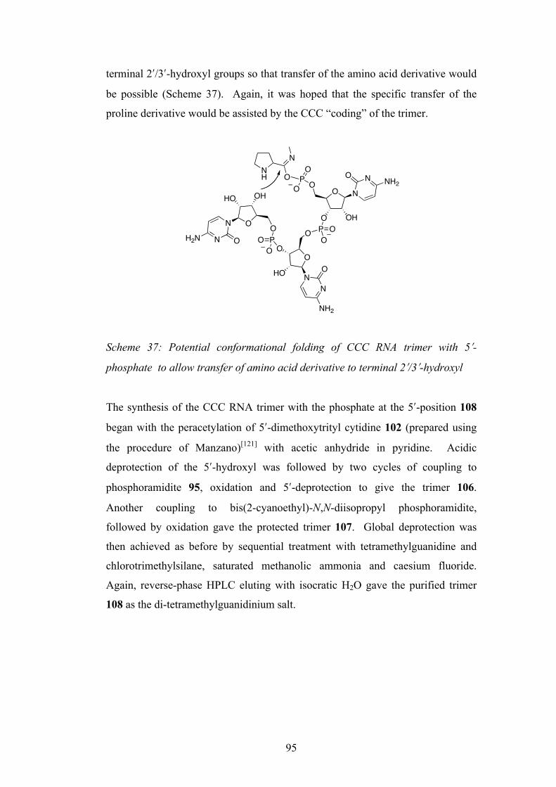
95
terminal 2ʹ′/3ʹ′-hydroxyl groups so that transfer of the amino acid derivative would
be possible (Scheme 37). Again, it was hoped that the specific transfer of the
proline derivative would be assisted by the CCC “coding” of the trimer.
Scheme 37: Potential conformational folding of CCC RNA trimer with 5ʹ′-
phosphate to allow transfer of amino acid derivative to terminal 2ʹ′/3ʹ′-hydroxyl
The synthesis of the CCC RNA trimer with the phosphate at the 5ʹ′-position 108
began with the peracetylation of 5ʹ′-dimethoxytrityl cytidine 102 (prepared using
the procedure of Manzano)[121] with acetic anhydride in pyridine. Acidic
deprotection of the 5ʹ′-hydroxyl was followed by two cycles of coupling to
phosphoramidite 95, oxidation and 5ʹ′-deprotection to give the trimer 106.
Another coupling to bis(2-cyanoethyl)-N,N-diisopropyl phosphoramidite,
followed by oxidation gave the protected trimer 107. Global deprotection was
then achieved as before by sequential treatment with tetramethylguanidine and
chlorotrimethylsilane, saturated methanolic ammonia and caesium fluoride.
Again, reverse-phase HPLC eluting with isocratic H2O gave the purified trimer
108 as the di-tetramethylguanidinium salt.
O N
O OH
NONH2O
O
N
O
HO
N
O
NH2
POON
OHHO
N OH2N PO
NH
N
O PO
O
OOO
O
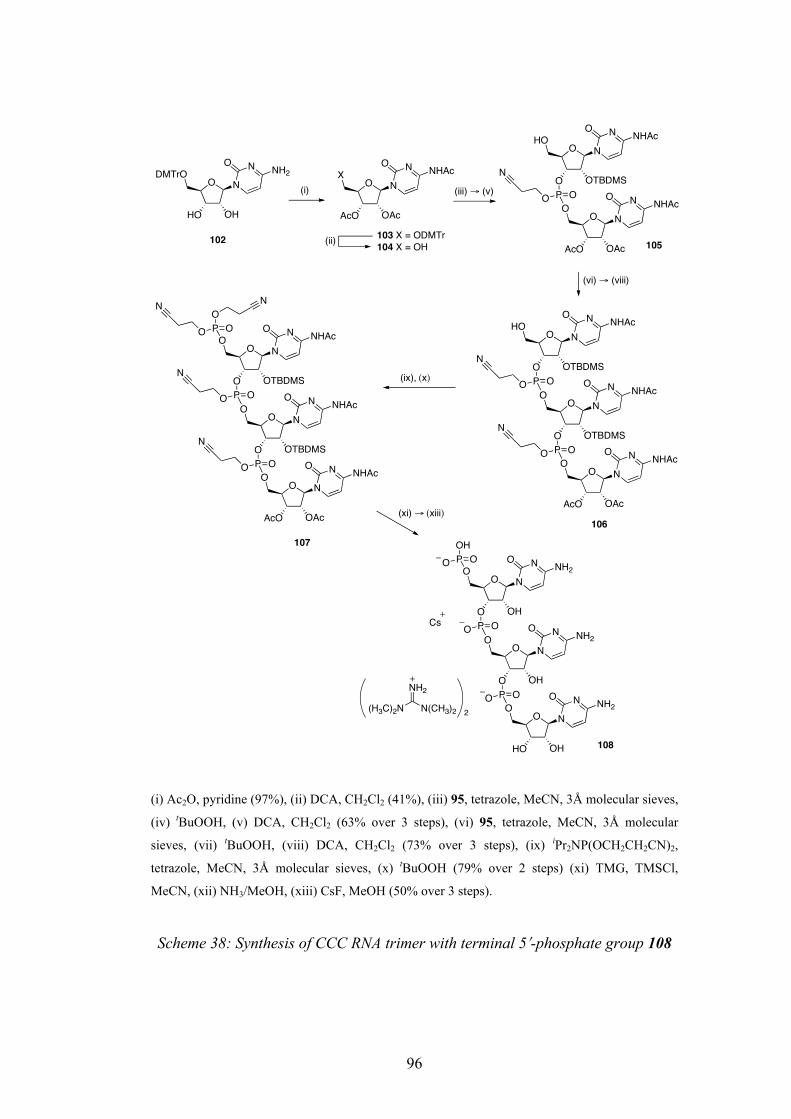
96
(i) Ac2O, pyridine (97%), (ii) DCA, CH2Cl2 (41%), (iii) 95, tetrazole, MeCN, 3Å molecular sieves,
(iv) tBuOOH, (v) DCA, CH2Cl2 (63% over 3 steps), (vi) 95, tetrazole, MeCN, 3Å molecular
sieves, (vii) tBuOOH, (viii) DCA, CH2Cl2 (73% over 3 steps), (ix) iPr2NP(OCH2CH2CN)2,
tetrazole, MeCN, 3Å molecular sieves, (x) tBuOOH (79% over 2 steps) (xi) TMG, TMSCl,
MeCN, (xii) NH3/MeOH, (xiii) CsF, MeOH (50% over 3 steps).
Scheme 38: Synthesis of CCC RNA trimer with terminal 5ʹ′-phosphate group 108
O N
HO OH
NDMTrO
ONH2
O N
AcO OAc
NX
ONHAc
O N
AcO OAc
NO
ONHAc
O N
O OTBDMS
NHO
ONHAc
P OO
N
O N
AcO OAc
NO
ONHAc
O N
O OTBDMS
NO
ONHAc
P OO
N
O N
O OTBDMS
NHO
ONHAc
P OO
N
O N
AcO OAc
NO
ONHAc
O N
O OTBDMS
NO
ONHAc
P OO
N
O N
O OTBDMS
NO
ONHAc
P OO
N
P OO
O
N N
O N
HO OH
NO
ONH2
O N
O OH
NO
ONH2
P OO
O N
O OH
NO
ONH2
P OO
P OOH
O
NH2
(H3C)2N N(CH3)2
103 X = ODMTr104 X = OH
(i)
(ii)
(iii) ! (v)
(vi) ! (viii)
(ix), (x)
(xi) ! (xiii)
102
106
107
2
Cs
108
105

97
4.5 Multicomponent Reaction of RNA Trimer with Terminal 5ʹ′-Phosphate
The trimer was reacted in the same way as before, with 4 equivalents each of 1-
pyrrolinium 64 and methyl isonitrile 82 in D2O at pD = 6. Again, there were no
signals present in the 1H-NMR spectrum to suggest transfer of the amino acid
derivative to the 2ʹ′- or 3ʹ′-hydroxyl group. Proline methyl amide 101 was
produced in 190% yield (based on the starting trimer), and the fact that the signals
due to the trimer were unchanged suggests that this species had been transiently
activated, much in the same way as in the previous multicomponent reactions with
a 5ʹ′-nucleotide monomer.
In conclusion, the phosphate Ugi-type multicomponent reaction that has been
developed has not been shown to successfully cause transfer of amino acid
derivatives onto nucleotide monomers or trimers. However, the prebiotically
plausible activation of monomer and trimer nucleoside-2ʹ′/3ʹ′-phosphates should be
seen as highly significant as the 2ʹ′,3ʹ′-cyclic phosphates formed (whether
monomers or trimers) are candidates for prebiotic oligomerisation reactions,
which shall be investigated in the next chapter.
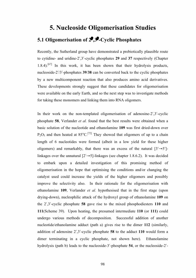
98
5. Nucleoside Oligomerisation Studies 5.1 Oligomerisation of 2ʹ′ ,3ʹ′-Cyclic Phosphates Recently, the Sutherland group have demonstrated a prebiotically plausible route
to cytidine- and uridine-2ʹ′,3ʹ′-cyclic phosphates 29 and 37 respectively (Chapter
1.8.4).[67] In this work, it has been shown that their hydrolysis products,
nucleoside-2ʹ′/3ʹ′-phosphates 39/38 can be converted back to the cyclic phosphates
by a new multicomponent reaction that also produces amino acid derivatives.
These developments strongly suggest that these candidates for oligomerisation
were available on the early Earth, and so the next step was to investigate methods
for taking these monomers and linking them into RNA oligomers.
In their work on the non-templated oligomerisation of adenosine-2ʹ′,3ʹ′-cyclic
phosphate 58, Verlander et al. found that the best results were obtained when a
basic solution of the nucleotide and ethanolamine 109 was first dried-down over
P2O5 and then heated at 85°C.[72] They showed that oligomers of up to a chain
length of 6 nucleotides were formed (albeit in a low yield for these higher
oligomers) and remarkably, that there was an excess of the natural [3ʹ′→5ʹ′]-
linkages over the unnatural [2ʹ′→5]-linkages (see chapter 1.8.6.2). It was decided
to embark upon a detailed investigation of this promising method of
oligomerisation in the hope that optimising the conditions and/or changing the
catalyst used could increase the yields of the higher oligomers and possibly
improve the selectivity also. In their rationale for the oligomerisation with
ethanolamine 109, Verlander et al. hypothesised that in the first stage (upon
drying-down), nucleophilic attack of the hydroxyl group of ethanolamine 109 on
the 2ʹ′,3ʹ′-cyclic phosphate 58 gave rise to the mixed phosphodiesters 110 and
111(Scheme 39). Upon heating, the presumed intermediate 110 (or 111) could
undergo various methods of decomposition. Successful addition of another
nucleotide/ethanolamine adduct (path a) gives rise to the dimer 112 (similarly,
addition of adenosine 2ʹ′,3ʹ′-cyclic phosphate 58 to the adduct 110 would form a
dimer terminating in a cyclic phosphate, not shown here). Ethanolamine
hydrolysis (path b) leads to the nucleoside-3ʹ′-phosphate 54, or the nucleoside-2ʹ′-
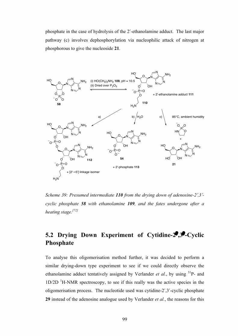
99
phosphate in the case of hydrolysis of the 2ʹ′-ethanolamine adduct. The last major
pathway (c) involves dephosphorylation via nucleophilic attack of nitrogen at
phosphorous to give the nucleoside 21.
Scheme 39: Presumed intermediate 110 from the drying down of adenosine-2ʹ′,3ʹ′-
cyclic phosphate 58 with ethanolamine 109, and the fates undergone after a
heating stage.[72]
5.2 Drying Down Experiment of Cytidine-2ʹ′ ,3ʹ′-Cyclic Phosphate
To analyse this oligomerisation method further, it was decided to perform a
similar drying-down type experiment to see if we could directly observe the
ethanolamine adduct tentatively assigned by Verlander et al., by using 31P- and
1D/2D 1H-NMR spectroscopy, to see if this really was the active species in the
oligomerisation process. The nucleotide used was cytidine-2ʹ′,3ʹ′-cyclic phosphate
29 instead of the adenosine analogue used by Verlander et al., the reasons for this
O N
OP
O
HO N
N N
NH2
O O
O NHO N
N N
NH2
O OHP OOO
H2N
(i) HO(CH2)2NH2 109, pH = 10.5(ii) Dried over P2O5
+ 2'-ethanolamine adduct 111
O NHO N
N N
NH2
O OHP OO
O NO N
N N
NH2
O OHP OOO
H2N
O NHO N
N N
NH2
O OHP OOO
OP
HN
O O
O NHO N
N N
NH2
HO OH
+
H2Oa) b) c) 85°C, ambient humidity
58 110
+ [2'!5'] linkage isomer+ 2'-phosphate 113
21112 54

100
being two-fold. Firstly, its commercial availability at the time and secondly, this
species has recently been shown to be produced (as well as its uridine counterpart)
in a new prebiotically plausible synthesis developed by the Sutherland group.[67]
Accordingly, a pH = 10.5 solution of cytidine-2ʹ′,3ʹ′-phosphate 29 and a five-fold
excess of ethanolamine 109 was rapidly taken down to dryness on a high-vacuum
rotary evaporator (Scheme 40), followed by dissolving in D2O for NMR analysis.
Scheme 40: Drying-down experiment of cytidine-2ʹ′,3ʹ′-cyclic phosphate 29 in the
presence of ethanolamine 109, with the three major products formed shown
The 1H-NMR spectrum (Figure 27) revealed that there were three nucleotide
species present in the reaction mixture. Unreacted (or re-cyclised) cytidine-2ʹ′,3ʹ′-
cyclic phosphate 29 was still present at 31% as can be seen by the distinctive
signals for H-C(2ʹ′) and H-C(3ʹ′) at 5.75 and 4.35 ppm respectively, both with
phosphorous couplings (JH,P = 6.4 Hz). The most abundant species at 46% was
tentatively assigned as the 3ʹ′-ethanolamine adduct 114, due to an apparent triplet
at 4.35 ppm (H-C(2ʹ′)) and an apparent triplet of doublets at 4.49 ppm (H-C(3ʹ′))
showing a pronounced phosphorous coupling (JH,P = 8.1 Hz). The least abundant
species was assumed to be the 2ʹ′-ethanolamine adduct 115, showing a double
doublet at 4.27 ppm for H-C(3ʹ′) and an apparent triplet of doublets for H-C(2ʹ′)
with phosphorous coupling (JH,P = 8.1 Hz). Finally, a new signal at 3.93 ppm
with phosphorous coupling appeared to be due to the CH2OP protons of both
ethanolamine adducts, compared to the more upfield triplet (3.63 ppm) due to the
corresponding CH2OH signals of free ethanolamine. The 31P-NMR spectrum
showed a characteristic signal at -20.2 ppm for the 2ʹ′,3ʹ′-cyclic phosphate 29, and
further signals at -0.27 and -0.19 ppm for the 3ʹ′- and 2ʹ′-ethanolamine adduct
O NHO
N
O
NH2
OP
O
O O
O NHO
N
O
NH2
O OHP OOOH2N
O NHO
N
O
NH2
HO OP OOOH2N
+ + 29 31%(i) 109, pH = 10.5
(ii) Dried in vacuo
114 46% 115 23%29
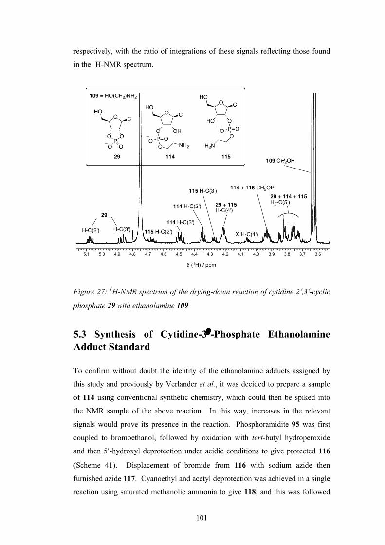
101
respectively, with the ratio of integrations of these signals reflecting those found
in the 1H-NMR spectrum.
Figure 27: 1H-NMR spectrum of the drying-down reaction of cytidine 2ʹ′,3ʹ′-cyclic
phosphate 29 with ethanolamine 109
5.3 Synthesis of Cytidine-3ʹ′-Phosphate Ethanolamine Adduct Standard
To confirm without doubt the identity of the ethanolamine adducts assigned by
this study and previously by Verlander et al., it was decided to prepare a sample
of 114 using conventional synthetic chemistry, which could then be spiked into
the NMR sample of the above reaction. In this way, increases in the relevant
signals would prove its presence in the reaction. Phosphoramidite 95 was first
coupled to bromoethanol, followed by oxidation with tert-butyl hydroperoxide
and then 5ʹ′-hydroxyl deprotection under acidic conditions to give protected 116
(Scheme 41). Displacement of bromide from 116 with sodium azide then
furnished azide 117. Cyanoethyl and acetyl deprotection was achieved in a single
reaction using saturated methanolic ammonia to give 118, and this was followed
5.1 5.0 4.9 4.8 4.7 4.6 4.5 4.4 4.3 4.2 4.1 4.0 3.9 3.8 3.7 3.6
! (1H) / ppm
29
H-C(2') H-C(3') 115 H-C(2')114 H-C(3')
114 H-C(2')
115 H-C(3')
X H-C(4')
29 + 115H-C(4')
114 + 115 CH2OP29 + 114 + 115 H2-C(5')
109 CH2OH
O CHO
OP
O
O O
O CHO
O OHP OOO NH2
O CHO
HO OP OOO
H2N
109 = HO(CH2)NH2
29 114 115
102
by 2ʹ′-hydroxyl deprotection using caesium fluoride. Reduction of azide 119 was
first attempted with tris(2-carboxyethyl)phosphine which proved unsuccessful,
and so it was decided to employ a catalytic hydrogenolysis instead using Pd/C in
H2O. This method was also not without its problems, however. It was found that
although the azide was successfully reduced to amine 114, the resultant
compound had a tendency to undergo partial conversion to the 2ʹ′,3ʹ′-cyclic
phosphate 29 the longer it stayed under the reaction conditions, presumably due to
general acid/base catalysis of the amine. After several carefully timed repeats, a
reaction time of precisely 1 h 15 min was found to give the optimal results with a
ratio of the desired compound 114 to the 2ʹ′,3ʹ′-cyclic phosphate 29 of 6.3:1. The
presence of the 2ʹ′,3ʹ′-cyclic phosphate 29 was not deemed a serious problem, as
this species was already in the reaction mixture of the drying down experiment
(Scheme 40) and moreover, would serve as an additional positive identification.
The crude mixture of 114 and 29 was not purified using reverse-phase HPLC as it
was feared this would cause further reversion to the 2ʹ′,3ʹ′-cyclic phosphate 29 and
in any case, the crude sample prepared was satisfactorily clean enough in the 1H-
NMR dimension for the sole purpose of sample spiking.
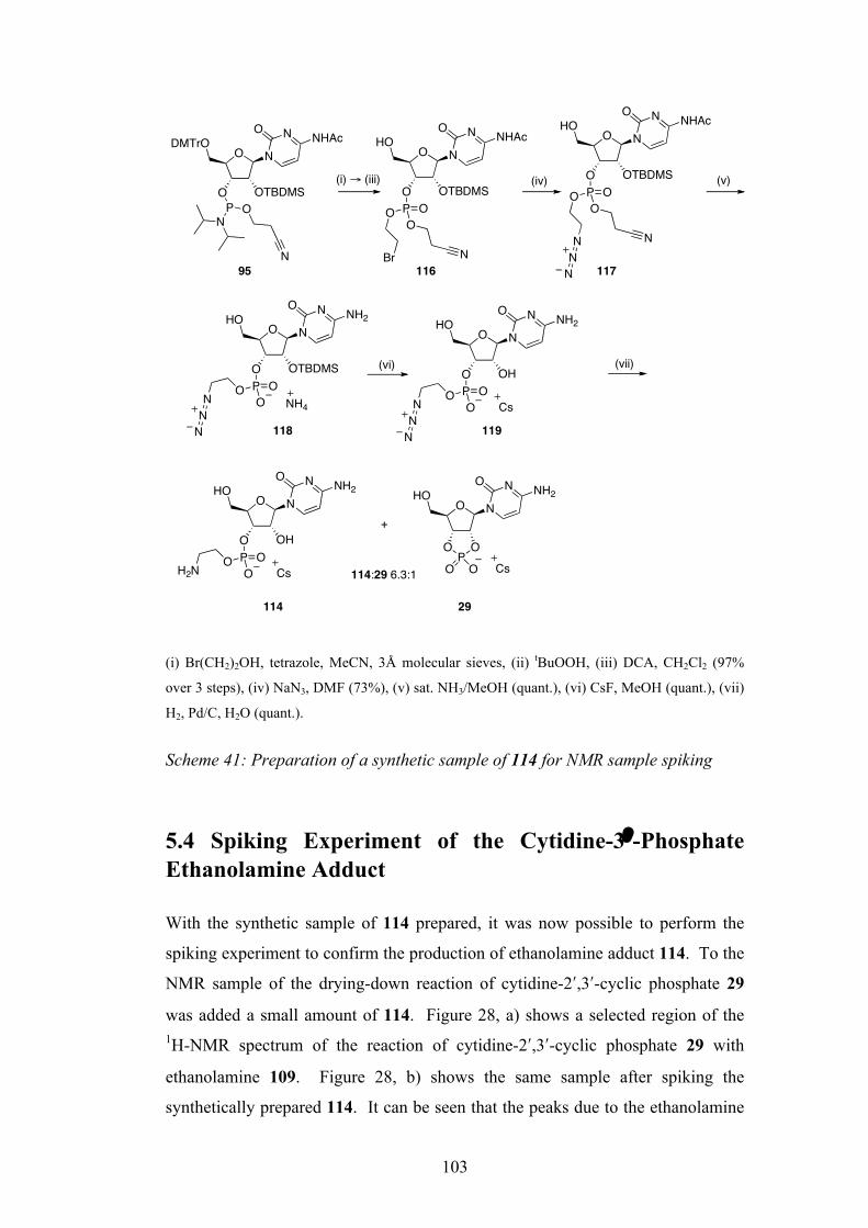
103
(i) Br(CH2)2OH, tetrazole, MeCN, 3Å molecular sieves, (ii) tBuOOH, (iii) DCA, CH2Cl2 (97%
over 3 steps), (iv) NaN3, DMF (73%), (v) sat. NH3/MeOH (quant.), (vi) CsF, MeOH (quant.), (vii)
H2, Pd/C, H2O (quant.). Scheme 41: Preparation of a synthetic sample of 114 for NMR sample spiking
5.4 Spiking Experiment of the Cytidine-3ʹ′-Phosphate Ethanolamine Adduct
With the synthetic sample of 114 prepared, it was now possible to perform the
spiking experiment to confirm the production of ethanolamine adduct 114. To the
NMR sample of the drying-down reaction of cytidine-2ʹ′,3ʹ′-cyclic phosphate 29
was added a small amount of 114. Figure 28, a) shows a selected region of the 1H-NMR spectrum of the reaction of cytidine-2ʹ′,3ʹ′-cyclic phosphate 29 with
ethanolamine 109. Figure 28, b) shows the same sample after spiking the
synthetically prepared 114. It can be seen that the peaks due to the ethanolamine
O N
O OTBDMS
NHO
ONHAc
P OOO
N
O N
O OTBDMS
NDMTrO
ONHAc
PN
O
N
O N
O OTBDMS
NHO
ONHAc
P OO
N
O N
O OTBDMS
NHO
ONH2
P OOON
NN
NH4
O N
O OH
NHO
ONH2
P OOOH2N Cs
O N
NHO
ONH2
OP
O
O O Cs
(i) ! (iii) (iv)
O N
O OH
NHO
ONH2
P OOON
NN
Cs
(vi)
(v)
+
114:29 6.3:1
(vii)
114 29
Br
O
NN
N95 116 117
118 119
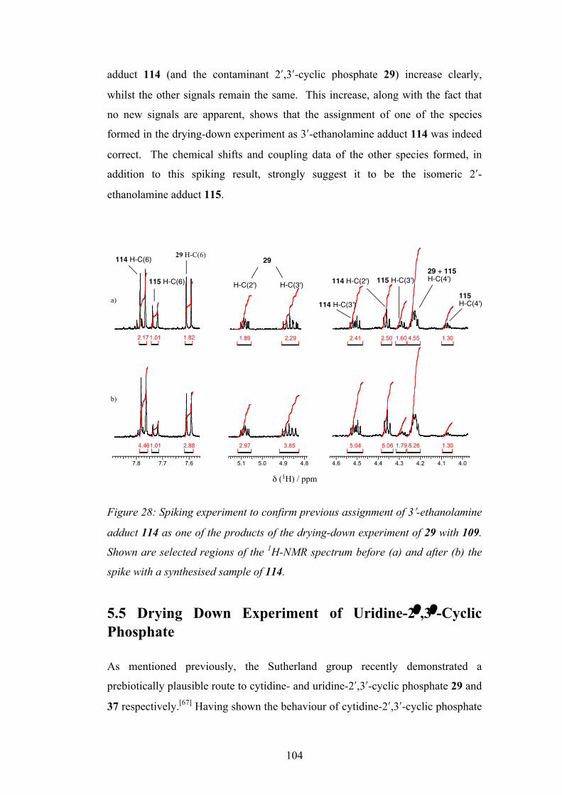
104
adduct 114 (and the contaminant 2ʹ′,3ʹ′-cyclic phosphate 29) increase clearly,
whilst the other signals remain the same. This increase, along with the fact that
no new signals are apparent, shows that the assignment of one of the species
formed in the drying-down experiment as 3ʹ′-ethanolamine adduct 114 was indeed
correct. The chemical shifts and coupling data of the other species formed, in
addition to this spiking result, strongly suggest it to be the isomeric 2ʹ′-
ethanolamine adduct 115.
Figure 28: Spiking experiment to confirm previous assignment of 3ʹ′-ethanolamine
adduct 114 as one of the products of the drying-down experiment of 29 with 109.
Shown are selected regions of the 1H-NMR spectrum before (a) and after (b) the
spike with a synthesised sample of 114.
5.5 Drying Down Experiment of Uridine-2ʹ′ ,3ʹ′-Cyclic Phosphate
As mentioned previously, the Sutherland group recently demonstrated a
prebiotically plausible route to cytidine- and uridine-2ʹ′,3ʹ′-cyclic phosphate 29 and
37 respectively.[67] Having shown the behaviour of cytidine-2ʹ′,3ʹ′-cyclic phosphate
1.821.012.17
7.8 7.7 7.6
2.881.014.46
1.304.551.602.502.412.291.89
5.1 5.0 4.9 4.8 4.6 4.5 4.4 4.3 4.2 4.1 4.0
1.308.261.795.065.043.852.97
! (1H) / ppm
a)
b)
29
H-C(2') H-C(3')
114 H-C(6) 29 H-C(6)
115 H-C(6)
114 H-C(3')
114 H-C(2') 115 H-C(3')29 + 115H-C(4')
115H-C(4')
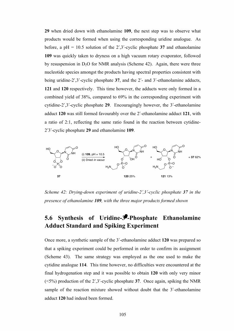
105
29 when dried down with ethanolamine 109, the next step was to observe what
products would be formed when using the corresponding uridine analogue. As
before, a pH = 10.5 solution of the 2ʹ′,3ʹ′-cyclic phosphate 37 and ethanolamine
109 was quickly taken to dryness on a high vacuum rotary evaporator, followed
by resuspension in D2O for NMR analysis (Scheme 42). Again, there were three
nucleotide species amongst the products having spectral properties consistent with
being uridine-2ʹ′,3ʹ′-cyclic phosphate 37, and the 2ʹ′- and 3ʹ′-ethanolamine adducts,
121 and 120 respectively. This time however, the adducts were only formed in a
combined yield of 38%, compared to 69% in the corresponding experiment with
cytidine-2ʹ′,3ʹ′-cyclic phosphate 29. Encouragingly however, the 3ʹ′-ethanolamine
adduct 120 was still formed favourably over the 2ʹ′-ethanolamine adduct 121, with
a ratio of 2:1, reflecting the same ratio found in the reaction between cytidine-
2ʹ′3ʹ′-cyclic phosphate 29 and ethanolamine 109.
Scheme 42: Drying-down experiment of uridine-2ʹ′,3ʹ′-cyclic phosphate 37 in the
presence of ethanolamine 109, with the three major products formed shown
5.6 Synthesis of Uridine-3ʹ′-Phosphate Ethanolamine Adduct Standard and Spiking Experiment
Once more, a synthetic sample of the 3ʹ′-ethanolamine adduct 120 was prepared so
that a spiking experiment could be performed in order to confirm its assignment
(Scheme 43). The same strategy was employed as the one used to make the
cytidine analogue 114. This time however, no difficulties were encountered at the
final hydrogenation step and it was possible to obtain 120 with only very minor
(<5%) production of the 2ʹ′,3ʹ′-cyclic phosphate 37. Once again, spiking the NMR
sample of the reaction mixture showed without doubt that the 3ʹ′-ethanolamine
adduct 120 had indeed been formed.
O NHO
NH
OO
PO
O O
O NHO
NH
O
O
O OHP OOOH2N
O NHO
NH
O
O
HO OP OOOH2N
+ + 37 62%(i) 109, pH = 10.5
(ii) Dried in vacuo
120 25% 121 13%37
O
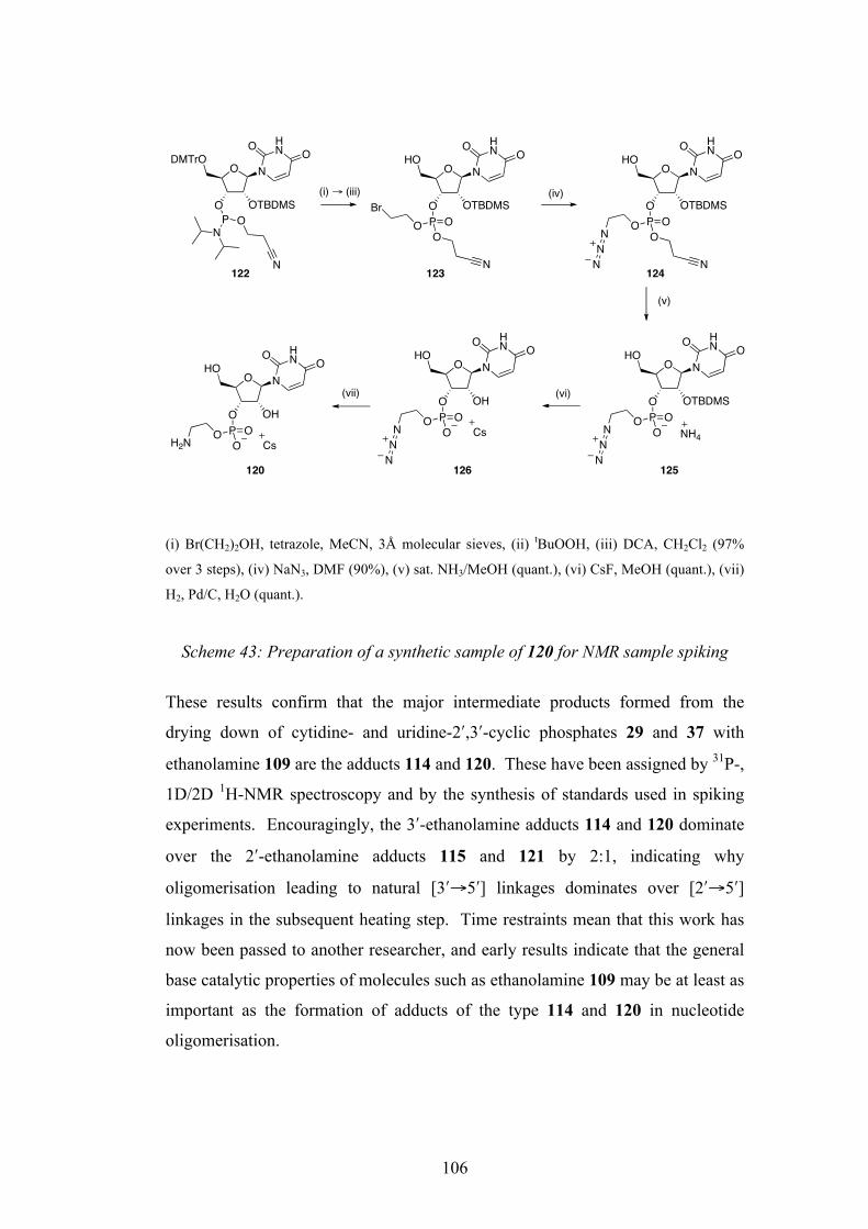
106
(i) Br(CH2)2OH, tetrazole, MeCN, 3Å molecular sieves, (ii) tBuOOH, (iii) DCA, CH2Cl2 (97%
over 3 steps), (iv) NaN3, DMF (90%), (v) sat. NH3/MeOH (quant.), (vi) CsF, MeOH (quant.), (vii)
H2, Pd/C, H2O (quant.).
Scheme 43: Preparation of a synthetic sample of 120 for NMR sample spiking
These results confirm that the major intermediate products formed from the
drying down of cytidine- and uridine-2ʹ′,3ʹ′-cyclic phosphates 29 and 37 with
ethanolamine 109 are the adducts 114 and 120. These have been assigned by 31P-,
1D/2D 1H-NMR spectroscopy and by the synthesis of standards used in spiking
experiments. Encouragingly, the 3ʹ′-ethanolamine adducts 114 and 120 dominate
over the 2ʹ′-ethanolamine adducts 115 and 121 by 2:1, indicating why
oligomerisation leading to natural [3ʹ′→5ʹ′] linkages dominates over [2ʹ′→5ʹ′]
linkages in the subsequent heating step. Time restraints mean that this work has
now been passed to another researcher, and early results indicate that the general
base catalytic properties of molecules such as ethanolamine 109 may be at least as
important as the formation of adducts of the type 114 and 120 in nucleotide
oligomerisation.
O N
O OTBDMS
HN
HOO
P OOO
Br
N
O N
O OTBDMS
HN
DMTrOO
O
PN
O
N
O N
O OTBDMS
HN
HOO
O
P OOO
N
NN
N
O N
O OTBDMS
HN
HOO
O
P OOON
NN
NH4
O N
O OH
HN
HOO
O
P OOOH2N Cs
(i) ! (iii) (iv)
O N
O OH
HN
HOO
O
P OOON
NN
Cs
(vi)
(v)
(vii)
O
120
122 123 124
125126
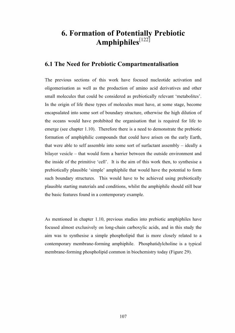
107
6. Formation of Potentially Prebiotic Amphiphiles[122]
6.1 The Need for Prebiotic Compartmentalisation The previous sections of this work have focused nucleotide activation and
oligomerisation as well as the production of amino acid derivatives and other
small molecules that could be considered as prebiotically relevant ‘metabolites’.
In the origin of life these types of molecules must have, at some stage, become
encapsulated into some sort of boundary structure, otherwise the high dilution of
the oceans would have prohibited the organisation that is required for life to
emerge (see chapter 1.10). Therefore there is a need to demonstrate the prebiotic
formation of amphiphilic compounds that could have arisen on the early Earth,
that were able to self assemble into some sort of surfactant assembly – ideally a
bilayer vesicle – that would form a barrier between the outside environment and
the inside of the primitive ‘cell’. It is the aim of this work then, to synthesise a
prebiotically plausible ‘simple’ amphiphile that would have the potential to form
such boundary structures. This would have to be achieved using prebiotically
plausible starting materials and conditions, whilst the amphiphile should still bear
the basic features found in a contemporary example.
As mentioned in chapter 1.10, previous studies into prebiotic amphiphiles have
focused almost exclusively on long-chain carboxylic acids, and in this study the
aim was to synthesise a simple phospholipid that is more closely related to a
contemporary membrane-forming amphiphile. Phosphatidylcholine is a typical
membrane-forming phospholipid common in biochemistry today (Figure 29).
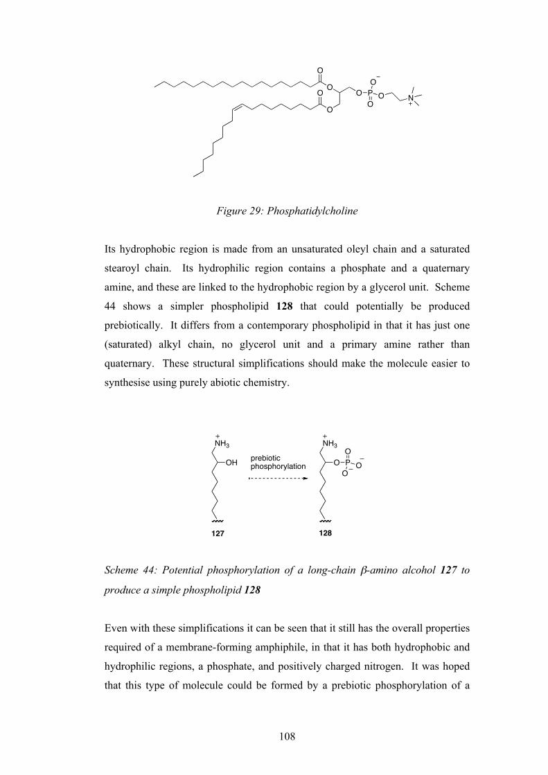
108
Figure 29: Phosphatidylcholine
Its hydrophobic region is made from an unsaturated oleyl chain and a saturated
stearoyl chain. Its hydrophilic region contains a phosphate and a quaternary
amine, and these are linked to the hydrophobic region by a glycerol unit. Scheme
44 shows a simpler phospholipid 128 that could potentially be produced
prebiotically. It differs from a contemporary phospholipid in that it has just one
(saturated) alkyl chain, no glycerol unit and a primary amine rather than
quaternary. These structural simplifications should make the molecule easier to
synthesise using purely abiotic chemistry.
Scheme 44: Potential phosphorylation of a long-chain β-amino alcohol 127 to
produce a simple phospholipid 128
Even with these simplifications it can be seen that it still has the overall properties
required of a membrane-forming amphiphile, in that it has both hydrophobic and
hydrophilic regions, a phosphate, and positively charged nitrogen. It was hoped
that this type of molecule could be formed by a prebiotic phosphorylation of a
O
O O PO
OO
NO
O
OH
NH3
O
NH3
prebiotic phosphorylation
127 128
PO
OO

109
long chain β-amino alcohol 127 (Scheme 44) in a predisposed reaction.
Furthermore, if it was possible to partially phosphorylate the long chain β-amino
alcohol 127, then mixtures of the overall negatively charged product 128 and the
positively charged β-amino alcohol 127, could have the potential to form what are
known as ‘catanionic vesicles’ – vesicles made from oppositely charged single-
chained amphiphiles.[123, 124] Kaler et al. showed that spontaneous, single-walled
vesicles can be formed from aqueous mixtures of single-tailed cationic and
anionic surfactants.[124] This vesicle formation results from anion-cation surfactant
pairs that then act as double-tailed zwitterionic surfactants.
The prebiotic availability of long chain β-amino alcohols, whilst not investigated
in this work, can be reasonably assumed as they are the potential reduction
products of cyanohydrins 130, which themselves could arise from the reaction of
HCN 2 and aldehydes 129 (Scheme 45). Long-chain aldehydes 129 are found in
the oxygenate fraction of Fischer-Tropsch reaction products[125], and Fischer-
Tropsch type processes have been shown to occur under aqueous, prebiotic
conditions (discussed in chapter 1.10.2).[97]
Scheme 45: Rationale for the prebiotic availability of long-chain β-amino
alcohols 127
6.2 A Potentially Predisposed Phosphorylation
The inspiration of a potentially predisposed phosphorylation of β-amino alcohols
came from work by Quimby and Flautt[126] and separately, Eschenmoser and co-
NH2
OHOH
N
HCN 2 [H]
129 130 127
O
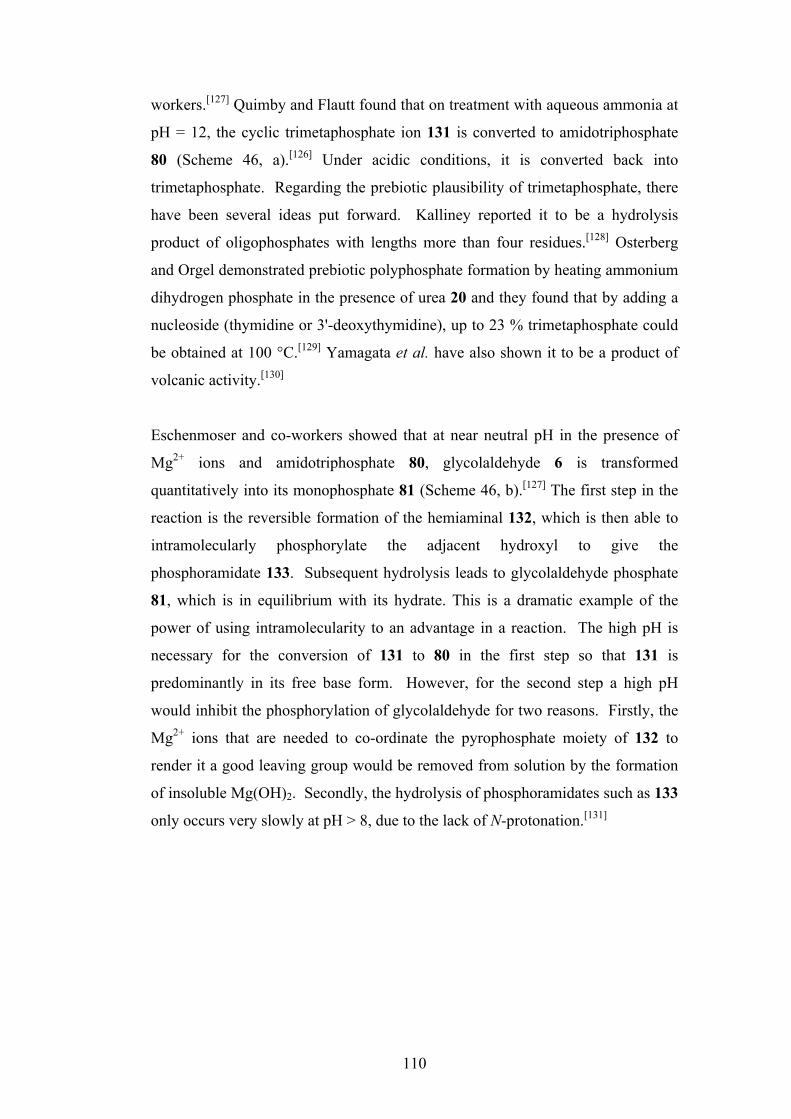
110
workers.[127] Quimby and Flautt found that on treatment with aqueous ammonia at
pH = 12, the cyclic trimetaphosphate ion 131 is converted to amidotriphosphate
80 (Scheme 46, a).[126] Under acidic conditions, it is converted back into
trimetaphosphate. Regarding the prebiotic plausibility of trimetaphosphate, there
have been several ideas put forward. Kalliney reported it to be a hydrolysis
product of oligophosphates with lengths more than four residues.[128] Osterberg
and Orgel demonstrated prebiotic polyphosphate formation by heating ammonium
dihydrogen phosphate in the presence of urea 20 and they found that by adding a
nucleoside (thymidine or 3'-deoxythymidine), up to 23 % trimetaphosphate could
be obtained at 100 °C.[129] Yamagata et al. have also shown it to be a product of
volcanic activity.[130]
Eschenmoser and co-workers showed that at near neutral pH in the presence of
Mg2+ ions and amidotriphosphate 80, glycolaldehyde 6 is transformed
quantitatively into its monophosphate 81 (Scheme 46, b).[127] The first step in the
reaction is the reversible formation of the hemiaminal 132, which is then able to
intramolecularly phosphorylate the adjacent hydroxyl to give the
phosphoramidate 133. Subsequent hydrolysis leads to glycolaldehyde phosphate
81, which is in equilibrium with its hydrate. This is a dramatic example of the
power of using intramolecularity to an advantage in a reaction. The high pH is
necessary for the conversion of 131 to 80 in the first step so that 131 is
predominantly in its free base form. However, for the second step a high pH
would inhibit the phosphorylation of glycolaldehyde for two reasons. Firstly, the
Mg2+ ions that are needed to co-ordinate the pyrophosphate moiety of 132 to
render it a good leaving group would be removed from solution by the formation
of insoluble Mg(OH)2. Secondly, the hydrolysis of phosphoramidates such as 133
only occurs very slowly at pH > 8, due to the lack of N-protonation.[131]

111
Scheme 46: a) Formation of amidotriphosphate 80 from trimetaphosphate 131
and ammonia.[126] b) Eschenmoser’s phosphorylation of glycolaldehyde 6 using
amidotriphosphate[127]
In the case of β-amino alcohols, it was hoped to use trimetaphosphate 131 in a
similar intramolecular phosphorylation reaction, but at one pH, in a single
reaction and without the need for Mg2+ ions. Furthermore, it was anticipated that
altering the length of the alkyl chain could control the key steps. Tsuhako et
al.[132] showed that ethanolamines are capable of reacting with trimetaphosphate
131, and so at a pH value high enough so that 134 is at least partially available as
its free base form, reaction with 131 should give the N-triphosphate 135. If R =
short alkyl chain, this species would not be expected to undergo intramolecular
attack of the OH group to give phosphoramidate 136 due to the poor leaving
group ability of uncoordinated/unprotonated pyrophosphate. However if R is
sufficiently long enough, 135 is likely to be incorporated into some sort of
surfactant assembly, and this would be expected to alter its reactivity. This
incorporation would cause the unfavourable proximity of multiple negative
charges on the pyrophosphate moiety leading to charge repulsion, and so
presumably increase its protonation level and therefore also its leaving group
ability. If phosphoramidate 136 was then formed, then it too would be expected
OP O P
OPO O
OO
OO
NH3, pH = 12
H2NP
O
OOPO
O OPO
OO
O OHHN
PO
OOPO
O OPO
OO
HO
HO
OP
HN
O O O
O
P OO
OOH
HO
HO
Mg2+, pH = 7OH
O
HO
P OO
OHO
Mg2+
80
a)
b)
131 80
H+
6 132 133
81
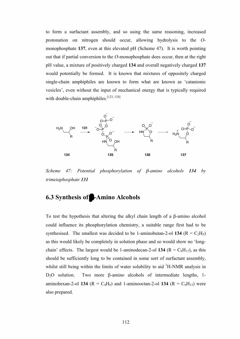
112
to form a surfactant assembly, and so using the same reasoning, increased
protonation on nitrogen should occur, allowing hydrolysis to the O-
monophosphate 137, even at this elevated pH (Scheme 47). It is worth pointing
out that if partial conversion to the O-monophosphate does occur, then at the right
pH value, a mixture of positively charged 134 and overall negatively charged 137
would potentially be formed. It is known that mixtures of oppositely charged
single-chain amphiphiles are known to form what are known as ‘catanionic
vesicles’, even without the input of mechanical energy that is typically required
with double-chain amphiphiles.[123, 124]
Scheme 47: Potential phosphorylation of β-amino alcohols 134 by
trimetaphosphate 131
6.3 Synthesis of β-Amino Alcohols
To test the hypothesis that altering the alkyl chain length of a β-amino alcohol
could influence its phosphorylation chemistry, a suitable range first had to be
synthesised. The smallest was decided to be 1-aminobutan-2-ol 134 (R = C2H5)
as this would likely be completely in solution phase and so would show no ‘long-
chain’ effects. The largest would be 1-aminodecan-2-ol 134 (R = C8H17), as this
should be sufficiently long to be contained in some sort of surfactant assembly,
whilst still being within the limits of water solubility to aid 1H-NMR analysis in
D2O solution. Two more β-amino alcohols of intermediate lengths, 1-
aminohexan-2-ol 134 (R = C4H9) and 1-aminooctan-2-ol 134 (R = C6H13) were
also prepared.
H2N OH
RHN OH
R
PO O
O
POO O
POO
O
R
OP
HN
O O
H3N O
R
POOO131
134 135 136 137

113
Terminal epoxides 138 were ring-opened using sodium azide to give the
corresponding β-azido alcohols 139, as characterised by a distinctive absorbance
at 2100 cm-1 in the IR spectrum. These were then catalytically hydrogenolysed to
give the range of β-amino alcohols 134, confirmed by the loss of the azide
absorbance at 2100 cm-1 in the IR spectrum (Scheme 48).
Scheme 48: Synthesis of a range of β-amino alcohols 134. R = C2H5, C4H9, C6H13
and C8H17
6.4 Reaction of β-Amino Alcohols with Trimetaphosphate
Preliminary experiments showed that reaction of 134 with trimetaphosphate 131
was insignificant at pD < 10. This is presumably due to the fact that the amine
group of 134 is mainly protonated at this pH and so unable to act as a nucleophile.
At pD = 10 however, 134 was found to react with 2.5 equivalents of 131 over a
number of days, the reactions being monitored by 1H- and 31P-NMR spectroscopy.
For the two shortest-chain compounds 134 (R = C2H5) and 134 (R = C4H9), they
were partially converted (19% in both cases) to a new species which had spectral
properties consistent with being the N-triphosphate 135. In the 1H-NMR
spectrum of the reaction between 134 (R = C4H9) and 131, signals for H-C(2) and
H2-C(1) of the new species were shifted upfield relative to those of the starting
material 134 (R = C4H9) (Figure 30).
O
R
NaN3
H2O/EtOH
R
HO NN
NH2, Pd/C
EtOH R
HO NH2
R = C2H5 (35%)R = C4H9 (59%)R = C6H13 (69 %)R = C8H17 (62%)
138 139 134
R = C2H5 (94%)R = C4H9 (97%)R = C6H13 (94 %)R = C8H17 (98%)

114
Figure 30: 1H-NMR spectrum of the reaction of 134 (R = C4H9) with sodium
trimetaphosphate 131
The 1H-coupled 31P-NMR spectrum showed a doublet of apparent of triplets (δ =
0.0 ppm, JP,P = 19.8 Hz, JH,P = 8.5 Hz, JH,P = 7.9 Hz for 135 (R = C4H9)) for Pα
which collapsed to a doublet (JP,P = 19.8 Hz) in the 1H-decoupled spectrum
(Figure 31). Also consistent with an N-triphosphate 135 was a doublet (δ = -5.0
ppm, JP,P = 19.2 Hz) for Pγ and an apparent triplet (-20.4 ppm, JP,P = 21.3, JP,P =
19.2 Hz) for Pβ (Figure 31).
3.8 3.7 3.6 3.5 3.4 3.3 3.2 3.1 3.0 2.9 2.8 2.7
135 (R = C4H9)H2C(1)
134 (R = C4H9)H2C(1)
134 (R = C4H9)H-C(2)
135 (R = C4H9)H-C(2)
! (1") / ppm
NH2HO
R
NHHO
R
P OO
O
P OOO
P OO
O
134 R = C4H9 135 R = C4H9

115
Figure 31: 31P-NMR analysis of the products from the reaction of 134 (R = C4H9)
with 131. Selected regions of the 1H-decoupled spectrum (bottom) and 1H-
coupled (top) are shown
In the case of the longest-chain compound (R = C8H17), 134 was partially
converted in significant yield (40% after 6 days, 55% after 6 weeks) to a single
species that had NMR spectroscopic data consistent with the O-monophosphate
137. A key factor in the assignment of the O-monophosphate 137 was the
observation of a doublet signal in the 1H-coupled 31P-NMR spectrum at δ = 4.0
(JH,P = 8.5 Hz) which collapsed to a singlet in the 1H-decoupled spectrum (Figure
32).
-4 -6 -20 -220
1H-coupled
1H-decoupled
P! P" PPi
131
P#
135 (R = C4H9)
$ (31P) / ppm
NHHO
R
P OO
O
P OOO
P OO
O
135 R = C4H9

116
Figure 32: 31P-NMR analysis of the reaction of 134 (R = C8H17) with 131.
Selected regions of the 1H-decoupled spectrum (bottom) and 1H-coupled spectrum
(top) are shown
6.5 Synthesis of Standard and Spiking Experiment
To verify this assignment of the O-monophosphate, a sample of 137 (R = C8H17)
was made using conventional synthesis which could then be used to spike the 1H-
NMR sample of the reaction products of 134 (R = C8H17) and 131. The
previously prepared azidoalcohol 139 (R = C8H17) served as a protected amine
and was phosphitylated using di-tert-butyl diisopropyl phosphoramidite, followed
by oxidation to give protected phosphate 140. Staudinger-type reduction of the
azide was then achieved using polymer-bound triphenylphosphine to reveal amine
141. Finally, the phosphate protection was removed under acidic conditions to
give 137 (R = C8H17) as its hydrochloride salt (Scheme 49).
5 4 3 2 1 0 -1
5 4 3 2
! (31P) / ppm
137 (R = C8H17)
Pi
1H-coupled
1H-decoupled
NH2O
R
137 R = C8H17
P OO
O
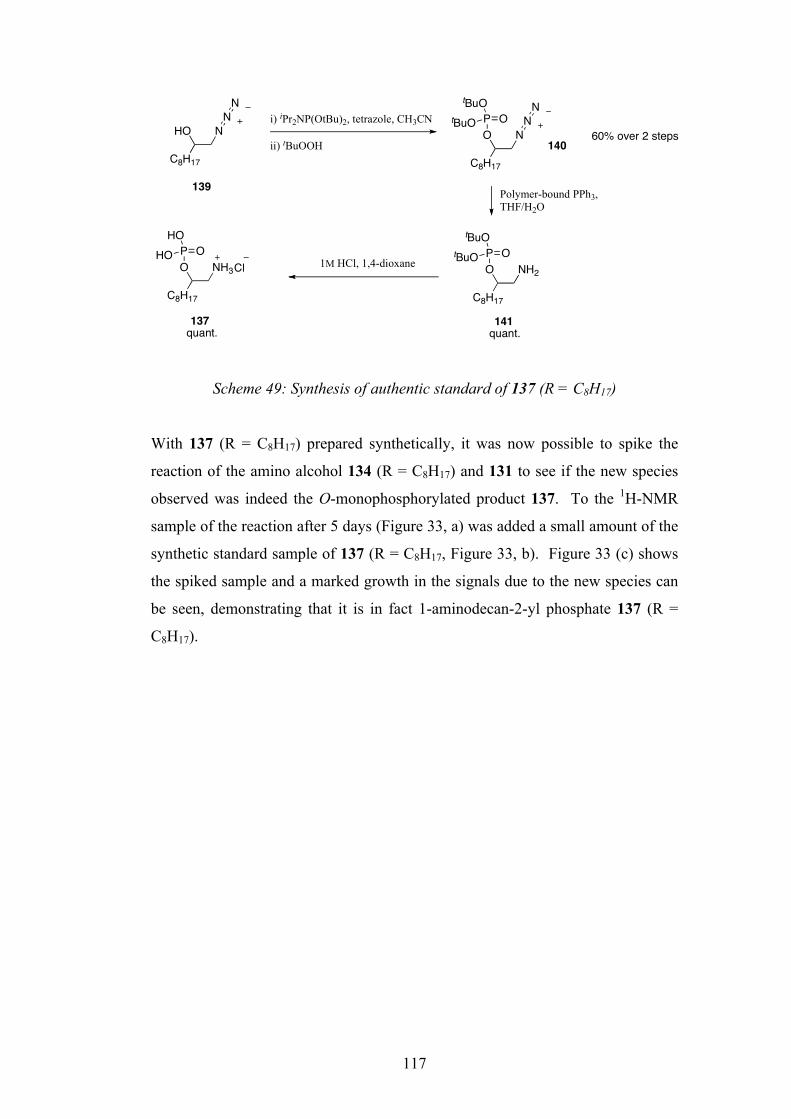
117
Scheme 49: Synthesis of authentic standard of 137 (R = C8H17)
With 137 (R = C8H17) prepared synthetically, it was now possible to spike the
reaction of the amino alcohol 134 (R = C8H17) and 131 to see if the new species
observed was indeed the O-monophosphorylated product 137. To the 1H-NMR
sample of the reaction after 5 days (Figure 33, a) was added a small amount of the
synthetic standard sample of 137 (R = C8H17, Figure 33, b). Figure 33 (c) shows
the spiked sample and a marked growth in the signals due to the new species can
be seen, demonstrating that it is in fact 1-aminodecan-2-yl phosphate 137 (R =
C8H17).
C8H17
NHON
N
C8H17
NON
NP OtBuO
tBuO
C8H17
NH2OP OtBuO
tBuO
C8H17
NH3OP OHO
HO
Cl 1M HCl, 1,4-dioxane
i) iPr2NP(OtBu)2, tetrazole, CH3CN
ii) tBuOOH
Polymer-bound PPh3, THF/H2O
139
140
141137
60% over 2 steps
quant.quant.
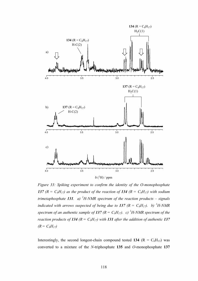
118
Figure 33: Spiking experiment to confirm the identity of the O-monophosphate
137 (R = C8H17) as the product of the reaction of 134 (R = C8H17) with sodium
trimetaphosphate 131. a) 1H-NMR spectrum of the reaction products – signals
indicated with arrows suspected of being due to 137 (R = C8H17). b) 1H-NMR
spectrum of an authentic sample of 137 (R = C8H17). c) 1H-NMR spectrum of the
reaction products of 134 (R = C8H17) with 131 after the addition of authentic 137
(R = C8H17)
Interestingly, the second longest-chain compound tested 134 (R = C6H13) was
converted to a mixture of the N-triphosphate 135 and O-monophosphate 137
4.0 3.5 3.0 2.5
4.0 3.5 3.0 2.5
4.0 3.5 3.0 2.5
a)
b)
c)
134 (R = C8H17)H2C(1)
134 (R = C8H17)H-C(2)
137 (R = C8H17)H2C(1)
137 (R = C8H17)H-C(2)
! (1H) / ppm
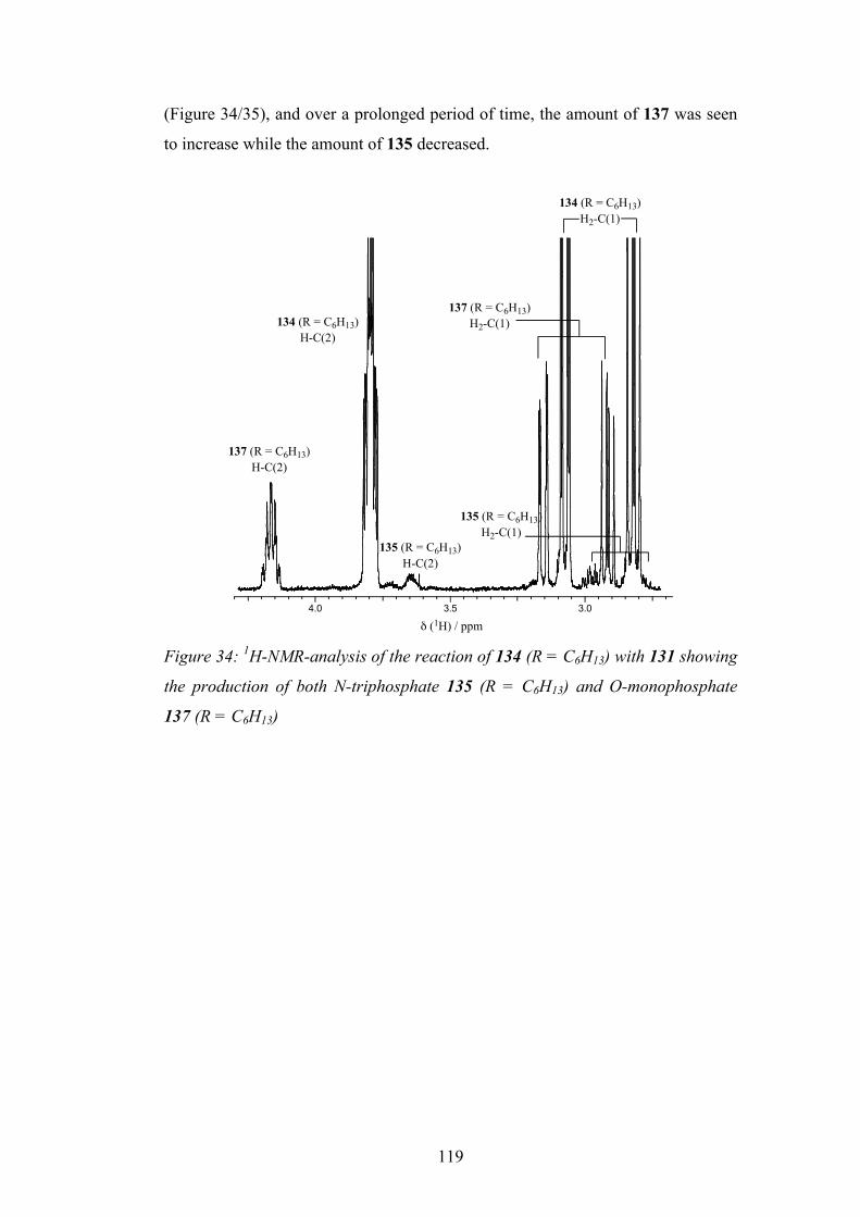
119
(Figure 34/35), and over a prolonged period of time, the amount of 137 was seen
to increase while the amount of 135 decreased.
Figure 34: 1H-NMR-analysis of the reaction of 134 (R = C6H13) with 131 showing
the production of both N-triphosphate 135 (R = C6H13) and O-monophosphate
137 (R = C6H13)
4.0 3.5 3.0
! (1H) / ppm
137 (R = C6H13)H-C(2)
134 (R = C6H13)H-C(2)
137 (R = C6H13)H2-C(1)
135 (R = C6H13)H-C(2)
135 (R = C6H13)H2-C(1)
134 (R = C6H13)H2-C(1)
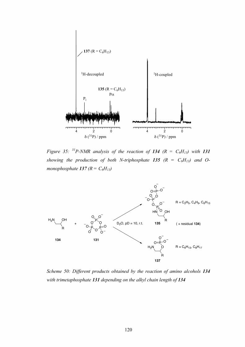
120
Figure 35: 31P-NMR analysis of the reaction of 134 (R = C6H13) with 131
showing the production of both N-triphosphate 135 (R = C6H13) and O-
monophosphate 137 (R = C6H13)
Scheme 50: Different products obtained by the reaction of amino alcohols 134
with trimetaphosphate 131 depending on the alkyl chain length of 134
4 2 0 4 2 0
! (31P) / ppm ! (31P) / ppm
135 (R = C6H13)P"
Pi
137 (R = C6H13)
1H-decoupled 1H-coupled
H2N OH
ROP O P
OPO O
OO
OO
HN OH
R
PO O
O
POO O
POO
O
H2N O
R
POO
O
+ D2O, pD = 10, r.t.
R = C2H5, C4H9, C6H13
R = C6H13, C8H17
( + residual 134)
134 131
135
137
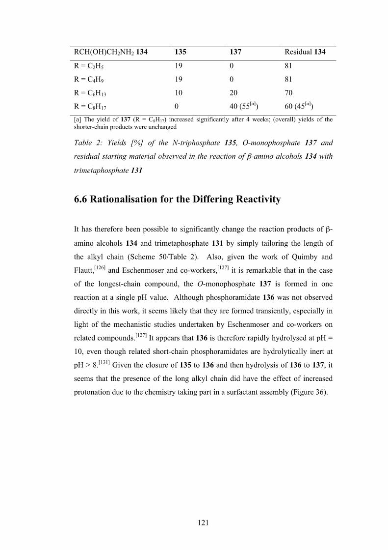
121
RCH(OH)CH2NH2 134 135 137 Residual 134
R = C2H5 19 0 81
R = C4H9 19 0 81
R = C6H13 10 20 70
R = C8H17 0 40 (55[a]) 60 (45[a]) [a] The yield of 137 (R = C8H17) increased significantly after 4 weeks; (overall) yields of the shorter-chain products were unchanged Table 2: Yields [%] of the N-triphosphate 135, O-monophosphate 137 and
residual starting material observed in the reaction of β-amino alcohols 134 with
trimetaphosphate 131
6.6 Rationalisation for the Differing Reactivity
It has therefore been possible to significantly change the reaction products of β-
amino alcohols 134 and trimetaphosphate 131 by simply tailoring the length of
the alkyl chain (Scheme 50/Table 2). Also, given the work of Quimby and
Flautt,[126] and Eschenmoser and co-workers,[127] it is remarkable that in the case
of the longest-chain compound, the O-monophosphate 137 is formed in one
reaction at a single pH value. Although phosphoramidate 136 was not observed
directly in this work, it seems likely that they are formed transiently, especially in
light of the mechanistic studies undertaken by Eschenmoser and co-workers on
related compounds.[127] It appears that 136 is therefore rapidly hydrolysed at pH =
10, even though related short-chain phosphoramidates are hydrolytically inert at
pH > 8.[131] Given the closure of 135 to 136 and then hydrolysis of 136 to 137, it
seems that the presence of the long alkyl chain did have the effect of increased
protonation due to the chemistry taking part in a surfactant assembly (Figure 36).
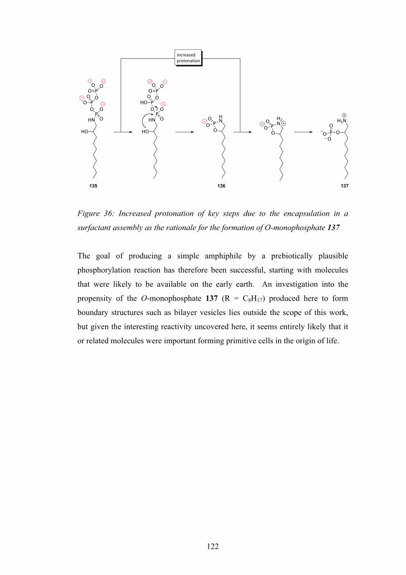
122
Figure 36: Increased protonation of key steps due to the encapsulation in a
surfactant assembly as the rationale for the formation of O-monophosphate 137
The goal of producing a simple amphiphile by a prebiotically plausible
phosphorylation reaction has therefore been successful, starting with molecules
that were likely to be available on the early earth. An investigation into the
propensity of the O-monophosphate 137 (R = C8H17) produced here to form
boundary structures such as bilayer vesicles lies outside the scope of this work,
but given the interesting reactivity uncovered here, it seems entirely likely that it
or related molecules were important forming primitive cells in the origin of life.
HNPO
OOPO
O OPOOO
HO
HNPO
OOPHO
O OPOOO
HO
HNP
O
OO
H3N
OPO
OO
H2NP
O
OO
!"#$%&'%()*$+,+"&,!+"
135 136 137
123
7. Conclusions
• A new multicomponent reaction was developed, involving a nucleotide,
aldehyde, isonitrile and ammonia. In the case of nucleotide-2ʹ′(3ʹ′)-
phosphates, these were stably activated in excellent yield to the
corresponding 2ʹ′,3ʹ′-cyclic phosphates which are considered candidates for
the oligomerisation to RNA. With nucleoside-5ʹ′-phosphates, these were
transiently activated and can be considered as candidates for templated
oligomerisation. In both cases, a variety of potentially important prebiotic
side products were formed. This gives support to the idea that RNA could
have emerged not in isolation, but alongside peptides and other small
molecules important for primitive metabolism.
• The activation chemistry was developed further and used on
glycolaldehyde and glycolaldehyde phosphate. These prebiotically
available molecules were cleanly converted to glyceric acid derivatives.
Glyceric acid 2- and 3-phosphate are intermediates in the glycolysis
pathway in contemporary biology, and simple prebiotic syntheses such as
these suggest that biology evolved to use such compounds in metabolic
cycles as they became available abiotically.
• The goal of forming an aminoacyl-RNA trimer using the multicomponent
activation chemistry proved unsuccessful in this work. This suggests that
another method must be found to form these species, to give support to the
RNA:coded peptides theory put forward by Sutherland.
• The dry state oligomerisation of nucleoside-2ʹ′,3ʹ′-cyclic phosphates using
catalytic ethanolamine was investigated, and key intermediate adducts
were identified. The preference for the natural [3ʹ′→5ʹ′] linkages of the
oligomers found by Orgel and co-workers was rationalised by the 2:1 ratio
of the 3ʹ′-ethanolamine adducts to the 2ʹ′-ethanolamine adducts formed in
the drying down experiments. This work now needs further investigation
to identify other possible catalysts and conditions to efficiently form short
124
RNA oligomers from prebiotically plausible nucleoside-2ʹ′,3ʹ′-cyclic
phosphates.
• A long-chain amino alcohol was phosphorylated partially to its O-
monophosphate using the trimetaphosphate ion in a single reaction and at
one pH value. Mixtures of the overall negatively charged product and
positively charged starting material have the potential to form catanionic
vesicles which could have been of importance in the compartmentalisation
of an early genetic system. It was found that when using the
corresponding short-chain amino alcohols, conversion to the N-
triphosphates occurred instead. This striking difference in reactivity based
upon differing alkyl chain-length is apparently due to perturbed pKa values
of the species depending on whether they are incorporated into a surfactant
assembly (long-chain), or simply reacting in the solution phase (short-
chain). The determination of the vesicle-forming properties of such
phospholipids lies beyond the scope of this work but should be seen as a
worthwhile goal, as these species have the potential to be superior to
previously investigated prebiotic amphiphiles such as carboxylic acids.
125
8. Experimental
8.1 General
Reagents and solvents purchased from Acros, Fluka, Lancaster, Sigma-Aldrich,
Chemgenes and Synthon. Acetonitrile, pyridine, CH2Cl2, triethylamine, and
toluene were distilled over calcium hydride before use. Tetrahydrofuran (THF)
was distilled from sodium metal using benzophenone as an indicator. All
reactions requiring an inert atmosphere were carried out using apparatus oven
dried at 120°C and cooled under a nitrogen atmosphere. Brine refers to a
saturated solution of aqueous sodium chloride. DCl was prepared by the addition
of oxalylchloride to D2O and NaOD was prepared by dissolving sodium metal in
D2O.
Silica gel flash chromatography was carried out using Merck 9385 silica gel 60
(230-400 mesh) and alumina flash column chromatography was carried out with
Brockmann grade specified (prepared from commercial grade I alumina shaken
with the required volume of water, allowed to cool to room temperature and stand
for >10 h). Thin layer chromatography (TLC) was carried out using plates pre-
coated with Merck silica gel (60-254 mesh).
Reverse phase high performance liquid chromatography (RP-HPLC) was carried
out on a Gilson HPLC system equipped with Gilson 306 pumps, a variable
wavelength detector, an 806 manometric module and a model 231 Biosample
injector. A rainin Dyanamax C18 Microsorb column (21.1 mm × 250 mm) was
used at a flow rate of 15 mL min-1 with UV detection at 255 nm. HPLC
instrument control, data collection and analysis were performed on a PC equipped
with Microsoft Windows 95, and Unipoint v1.65 software.
Melting points (m.p.) were measured on a Sanyo Gallenkamp variable heater.
Values have been quoted to the nearest degree, and are uncorrected.
126
Ultraviolet spectra were recorded using a Hewlett Packard 8452A diode-array
spectrometer. Wavelengths are accurate to ±4 nm and extinction coefficients (e)
are accurate to ±10%.
Infrared spectra were recorded as solvent-cast films on sodium chloride plates in
specified solvent (film) or as neat sample pressed between sodium chloride plates
on an ATI Mattson Genesis series FT-IR spectrometer. Alternatively solid IR
(solid) were recorded on a Bruker Equinox 55/ Bruker FRA 106/5 with coherent
500 mW laser as Attenuated Total Reflectance spectra with ‘golden gate’
attachment with resolution of 2 cm-1. Absorption maxima are quoted in
wavenumbers (cm-1).
Proton Nuclear Magnetic Resonance (1H-NMR) spectra were recorded on a
Varian INOVA 300 ‘Athos’ 300 MHz spectrometer with autosampler, a Varian
INOVA 400 MHz spectrometer and a Bruker AMX 500 MHz spectrometer with
autosampler, operating at ambient probe temperature, using an internal deuterium
lock. Carbon Nuclear Magnetic Resonance (13C NMR) spectra were recorded on
a Varian Inova 300 ‘Athos’ 75 MHz spectrometer and a Bruker 400 INOVA 100
MHz spectrometer. Phosphorus Nuclear Magnetic Resonance (31P NMR) and
Proton decoupled Phosphorus Nuclear Magnetic Resonance (31P-NMR) spectra
were recorded on a 162 MHz spectrometer Bruker 400 INOVA. All chemical
shifts are quoted in parts per million (ppm). They are reported as multiplicity,
coupling constant (J measured in Hertz (Hz)), integration (xN, where x = number
of proton per molecule and N = nucleus irradiated), and assignment. The signal
splittings are recorded as singlet (s), broad singlet (br. s), doublet (d), doublet of
doublets (dd), double double doublet (ddd), triplet (t), doublet of triplets (dt),
triplet of doublets (td), quartet (q), doublet of quartets (dq), triplet of quartets (tq),
quintet (quin.), sextet (sex.), heptet (h) and multiplet (m). The notation (abx)
refers to a methylene spin system coupled to an adjacent proton.
Mass spectrometry (MS) and High Resonance Mass Spectrometry (HRMS) were
carried out on a VG Trio 2000 instrument, Thermofinnigan MAT/95XP
spectrometer or a Kratos Concept IS instrument. The modes of ionisation were
electron impact (EI) and chemical ionisation (CI), electrospray ionisation (ESI) or
127
atmospheric pressure ionisation (APCI). ESI-MS ionisation is denoted (neg.) or
(pos.) for anion and cation detection respectively.
Purified water was used from a Milli-Q system.
Solvents with boiling points less than 80 °C were removed on a Büchi Rotavapor
T114 connected to a house vacuum. High boiling solvents were evaporated at
<0.1 Torr on a Büchi RE111 connected to an Edwards Speedivac 2 mechanical
vacuum pump. Frozen aqueous solutions were lyophilised using an Edwards RV5
mechanical vacuum pump in conjunction with an E-C Modulyo airtight freezer.
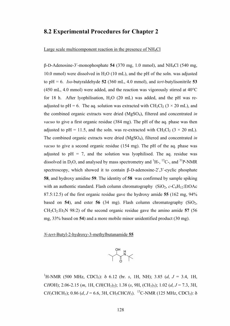
128
8.2 Experimental Procedures for Chapter 2
Large scale multicomponent reaction in the presence of NH4Cl
β-D-Adenosine-3ʹ′-monophosphate 54 (370 mg, 1.0 mmol), and NH4Cl (540 mg,
10.0 mmol) were dissolved in H2O (10 mL), and the pH of the soln. was adjusted
to pH = 6. Iso-butyraldehyde 52 (360 mL, 4.0 mmol), and tert-butylisonitrile 53
(450 mL, 4.0 mmol) were added, and the reaction was vigorously stirred at 40°C
for 18 h. After lyophilisation, H2O (20 mL) was added, and the pH was re-
adjusted to pH = 6. The aq. solution was extracted with CH2Cl2 (3 × 20 mL), and
the combined organic extracts were dried (MgSO4), filtered and concentrated in
vacuo to give a first organic residue (384 mg). The pH of the aq. phase was then
adjusted to pH = 11.5, and the soln. was re-extracted with CH2Cl2 (3 × 20 mL).
The combined organic extracts were dried (MgSO4), filtered and concentrated in
vacuo to give a second organic residue (154 mg). The pH of the aq. phase was
adjusted to pH = 7, and the solution was lyophilised. The aq. residue was
dissolved in D2O, and analysed by mass spectrometry and 1H-, 13C-, and 31P-NMR
spectroscopy, which showed it to contain β-D-adenosine-2ʹ′,3ʹ′-cyclic phosphate
58, and hydroxy amidine 59. The identity of 58 was confirmed by sample spiking
with an authentic standard. Flash column chromatography (SiO2, c-C6H12:EtOAc
87.5:12.5) of the first organic residue gave the hydroxy amide 55 (162 mg, 94%
based on 54), and ester 56 (34 mg). Flash column chromatography (SiO2,
CH2Cl2:Et3N 98:2) of the second organic residue gave the amino amide 57 (56
mg, 33% based on 54) and a more mobile minor unidentified product (30 mg).
N-tert-Butyl-2-hydroxy-3-methylbutanamide 55
1H-NMR (500 MHz, CDCl3): δ 6.12 (br. s, 1H, NH); 3.85 (d, J = 3.4, 1H,
CHOH); 2.06-2.15 (m, 1H, CH(CH3)2); 1.38 (s, 9H, (CH3)3); 1.02 (d, J = 7.3, 3H,
CH3CHCH3); 0.86 (d, J = 6.6, 3H, CH3CHCH3). 13C-NMR (125 MHz, CDCl3): δ
OH HN
O

129
172.6 (CO); 76.0 (CHOH); 51.0 (C(CH3)3); 31.9 (CH(CH3)2); 28.7 (C(CH3)3);
19.2 (CHCH3); 15.3 (CHCH3). ESI-MS (pos., H2O): 174 (100%, [M + H]+). HR-
ESI-MS (pos.): 174.1494 (C9H20NO2+; calc. 174.1489).[133]
1-(tert-Butylcarbamoyl)-2-methylpropyl isobutyrate 56
1H-NMR (300 MHz, CDCl3): δ 5.76 (br. s, 1H, NH); 4.98 (d, J = 4.1, 1H, CHO);
2.60-2.74 (m, 1H, (CH3)2CHC=O); 2.24-2.40 (m, 1H, (CH3)2CHCHO); 1.35 (s,
9H, (CH3)3); 1.24 (d, J = 7.0, 6H, (CH3)2CHC=O); 0.94 (d, J = 6.9, 6H,
(CH3)2CHCO). 13C-NMR (75 MHz, CDCl3): δ 175.5; 168.5; 77.7; 51.2; 34.1;
30.5; 28.7; 19.0; 18.8; 16.7. HR-ESI-MS (pos.): 244.1916 (C13H26NO3+; calc.
244.1907). IR (CHCl3): νmax/cm-1 1680 (CONH), 1738 (COO).
N-tert-Butyl-2-amino-3-methylbutanamide 57
1H-NMR (500 MHz, CDCl3): δ 7.10 (br. s, 1H, NH); 3.07 (d, J = 3.8, 1H,
CHNH2); 2.20-2.29 (m, 1H, CH(CH3)2); 1.33 (s, 9H, (CH3)3); 0.94 (d, J = 6.9, 3H,
CH3CH); 0.79 (d, J = 6.6, 3H, CHCH3). 13C-NMR (125 MHz, CDCl3): δ 172.5
(CO); 59.4 (CHNH2); 49.4 (C(CH3)3); 29.8 (CH(CH3)2); 27.7 (C(CH3)3); 18.7
(CHCH3); 14.9 (CHCH3). ESI-MS (pos., H2O): 173 (100%, [M + H]+). HR-ESI-
MS (pos.): 173.1644 (C9H21N2O+; calc. 173.1648).
O HN
O
O
NH2 HN
O
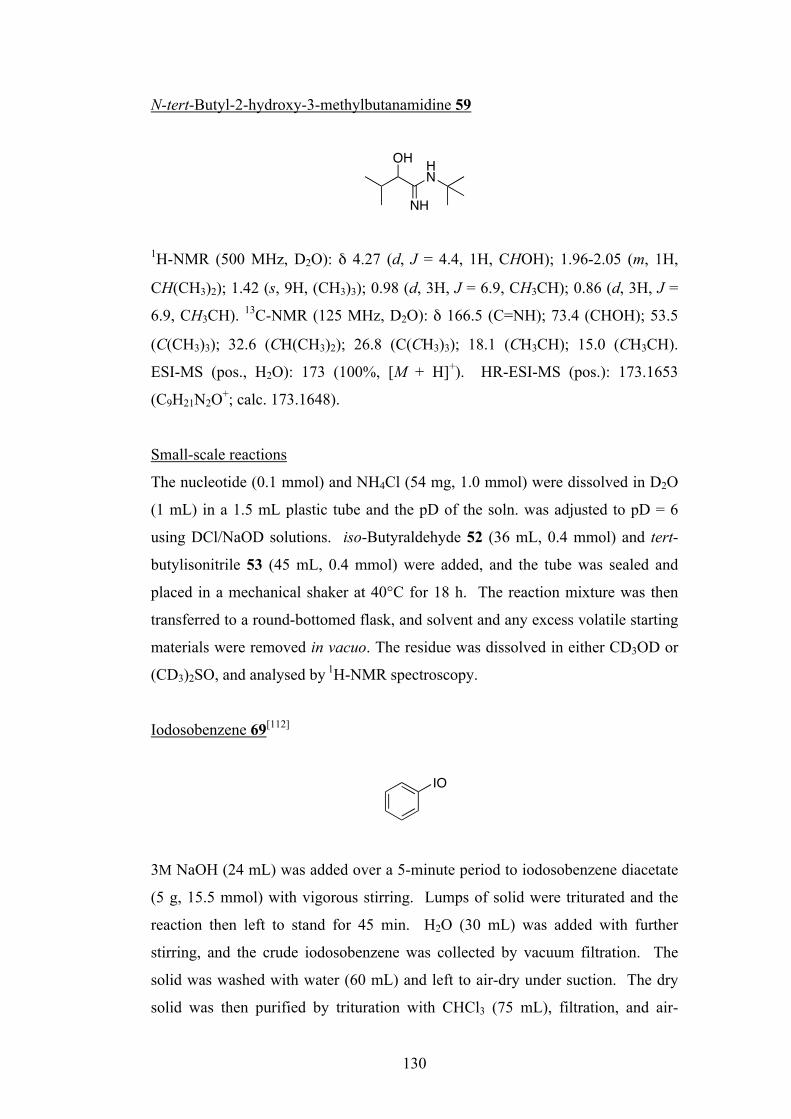
130
N-tert-Butyl-2-hydroxy-3-methylbutanamidine 59
1H-NMR (500 MHz, D2O): δ 4.27 (d, J = 4.4, 1H, CHOH); 1.96-2.05 (m, 1H,
CH(CH3)2); 1.42 (s, 9H, (CH3)3); 0.98 (d, 3H, J = 6.9, CH3CH); 0.86 (d, 3H, J =
6.9, CH3CH). 13C-NMR (125 MHz, D2O): δ 166.5 (C=NH); 73.4 (CHOH); 53.5
(C(CH3)3); 32.6 (CH(CH3)2); 26.8 (C(CH3)3); 18.1 (CH3CH); 15.0 (CH3CH).
ESI-MS (pos., H2O): 173 (100%, [M + H]+). HR-ESI-MS (pos.): 173.1653
(C9H21N2O+; calc. 173.1648).
Small-scale reactions
The nucleotide (0.1 mmol) and NH4Cl (54 mg, 1.0 mmol) were dissolved in D2O
(1 mL) in a 1.5 mL plastic tube and the pD of the soln. was adjusted to pD = 6
using DCl/NaOD solutions. iso-Butyraldehyde 52 (36 mL, 0.4 mmol) and tert-
butylisonitrile 53 (45 mL, 0.4 mmol) were added, and the tube was sealed and
placed in a mechanical shaker at 40°C for 18 h. The reaction mixture was then
transferred to a round-bottomed flask, and solvent and any excess volatile starting
materials were removed in vacuo. The residue was dissolved in either CD3OD or
(CD3)2SO, and analysed by 1H-NMR spectroscopy.
Iodosobenzene 69[112]
3M NaOH (24 mL) was added over a 5-minute period to iodosobenzene diacetate
(5 g, 15.5 mmol) with vigorous stirring. Lumps of solid were triturated and the
reaction then left to stand for 45 min. H2O (30 mL) was added with further
stirring, and the crude iodosobenzene was collected by vacuum filtration. The
solid was washed with water (60 mL) and left to air-dry under suction. The dry
solid was then purified by trituration with CHCl3 (75 mL), filtration, and air-
OH HN
NH
IO
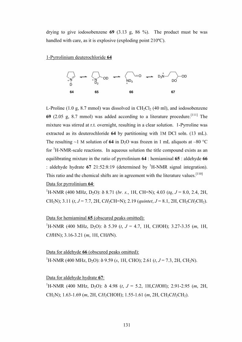
131
drying to give iodosobenzene 69 (3.13 g, 86 %). The product must be was
handled with care, as it is explosive (exploding point 210ºC).
1-Pyrrolinium deuterochloride 64
L-Proline (1.0 g, 8.7 mmol) was dissolved in CH2Cl2 (40 ml), and iodosobenzene
69 (2.05 g, 8.7 mmol) was added according to a literature procedure.[111] The
mixture was stirred at r.t. overnight, resulting in a clear solution. 1-Pyrroline was
extracted as its deuterochloride 64 by partitioning with 1M DCl soln. (13 mL).
The resulting ~1 M solution of 64 in D2O was frozen in 1 mL aliquots at –80 °C
for 1H-NMR-scale reactions. In aqueous solution the title compound exists as an
equilibrating mixture in the ratio of pyrrolinium 64 : hemiaminal 65 : aldehyde 66
: aldehyde hydrate 67 21:52:8:19 (determined by 1H-NMR signal integration).
This ratio and the chemical shifts are in agreement with the literature values.[110]
Data for pyrrolinium 64: 1H-NMR (400 MHz, D2O): δ 8.71 (br. s., 1H, CH=N); 4.03 (tq, J = 8.0, 2.4, 2H,
CH2N); 3.11 (t, J = 7.7, 2H, CH2CH=N); 2.19 (quintet, J = 8.1, 2H, CH2CH2CH2).
Data for hemiaminal 65 (obscured peaks omitted): 1H-NMR (400 MHz, D2O): δ 5.39 (t, J = 4.7, 1H, CHOH); 3.27-3.35 (m, 1H,
CHHN); 3.16-3.21 (m, 1H, CHHN).
Data for aldehyde 66 (obscured peaks omitted): 1H-NMR (400 MHz, D2O): δ 9.59 (s, 1H, CHO); 2.61 (t, J = 7.3, 2H, CH2N).
Data for aldehyde hydrate 67: 1H-NMR (400 MHz, D2O): δ 4.98 (t, J = 5.2, 1H,CHOH); 2.91-2.95 (m, 2H,
CH2N); 1.63-1.69 (m, 2H, CH2CHOH); 1.55-1.61 (m, 2H, CH2CH2CH2).
ND
ND2
OD O D3NDO
ODND3
64 65 66 67

132
Reaction of cytidine-3ʹ′-phosphate 38 (B = C) with tert-butylisonitrile 53 and 1-
pyrrolinium deuterochloride 64
Solutions of 1-pyrrolinium deuterochloride 64 in D2O (1M, 400 µL, 0.4 mmol)
and β-D-cytidine-3ʹ′-phosphate 38 (B = C) in D2O (0.167 M, 600 µL, 0.1 mmol)
were mixed in a 1.5 mL plastic tube and the resulting solution was adjusted to pD
= 6. Tert-butylisonitrile 53 (45 µL, 0.4 mmol) was added, and the tube was then
sealed and shaken for 5 min. Half of the reaction mixture was immediately taken
for 1H-NMR analysis, and the other half was placed in a mechanical shaker at
40°C for 18 h prior to 1H-NMR analysis. The presence of proline tert-butylamide
71 in the samples was confirmed by spiking with an authentic sample of L-proline
tert-butylamide 74 prepared from L-Cbz-proline 72 according to a literature
method.[113]
N-Cbz-L-proline-tert-butylamide 73[113]
N-Cbz-L-proline 72 (500 mg, 2.0 mmol) and triethylamine (278 µL, 2.0 mmol)
were dissolved in THF (8 mL) and the solution cooled to 0°C. Ethyl
chloroformate (190 µL, 2.0 mmol) was added drop-wise (15 min), and the
solution stirred for a further 30 min. tert-Butylamine (210 µL, 2.0 mmol) was
then added drop-wise (15 min.). The resulting solution was stirred at 0°C for 1h,
at r.t. for 16 h, and finally heated under reflux for 3 h. After cooling to r.t., the
solution was diluted with EtOAc, filtered and concentrated under vacuum to give
the crude product. This was then purified by flash column chromatography
(eluting with c-C6H12:EtOAc 2:1) to give N-Cbz-L-proline tert-butylamide 73 as a
white crystalline product (472 mg, 78 %). M.p. 82-85°C. 1H-NMR (400 MHz,
CDCl3): δ 7.30-7.38 (m, 5H, C6H5); 5.16-5.22 (m, 2H, CH2O); 4.21 (m, 1H, CH);
3.51 (m, 2H, CH2N); 1.86-2.26 (m, 4H, CH2CH2CH); 1.28 (s, 9H, (CH3)3). 13C-
NMR (125 MHz, CDCl3): δ 127.77-128.48 (C5H5); 67.19 (CH2O); 60.04 (CH);
50.95 (C(CH3)3); 46.54 (CH2N); 28.54 ((CH3)3); 26.93 (CH2CH); 22.58
N
OO
O
HN
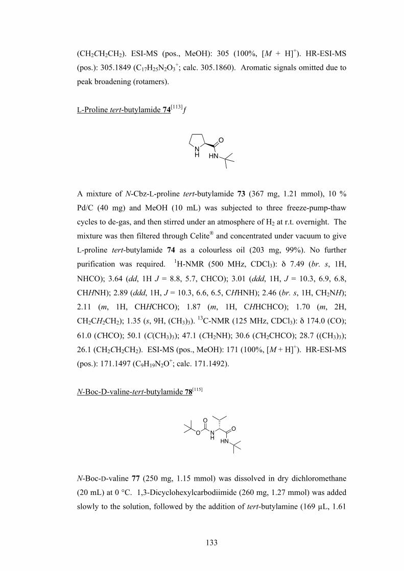
133
(CH2CH2CH2). ESI-MS (pos., MeOH): 305 (100%, [M + H]+). HR-ESI-MS
(pos.): 305.1849 (C17H25N2O3+; calc. 305.1860). Aromatic signals omitted due to
peak broadening (rotamers).
L-Proline tert-butylamide 74[113]ƒ
A mixture of N-Cbz-L-proline tert-butylamide 73 (367 mg, 1.21 mmol), 10 %
Pd/C (40 mg) and MeOH (10 mL) was subjected to three freeze-pump-thaw
cycles to de-gas, and then stirred under an atmosphere of H2 at r.t. overnight. The
mixture was then filtered through Celite® and concentrated under vacuum to give
L-proline tert-butylamide 74 as a colourless oil (203 mg, 99%). No further
purification was required. 1H-NMR (500 MHz, CDCl3): δ 7.49 (br. s, 1H,
NHCO); 3.64 (dd, 1H J = 8.8, 5.7, CHCO); 3.01 (ddd, 1H, J = 10.3, 6.9, 6.8,
CHHNH); 2.89 (ddd, 1H, J = 10.3, 6.6, 6.5, CHHNH); 2.46 (br. s, 1H, CH2NH);
2.11 (m, 1H, CHHCHCO); 1.87 (m, 1H, CHHCHCO); 1.70 (m, 2H,
CH2CH2CH2); 1.35 (s, 9H, (CH3)3). 13C-NMR (125 MHz, CDCl3): δ 174.0 (CO);
61.0 (CHCO); 50.1 (C(CH3)3); 47.1 (CH2NH); 30.6 (CH2CHCO); 28.7 ((CH3)3);
26.1 (CH2CH2CH2). ESI-MS (pos., MeOH): 171 (100%, [M + H]+). HR-ESI-MS
(pos.): 171.1497 (C9H19N2O+; calc. 171.1492).
N-Boc-D-valine-tert-butylamide 78[115]
N-Boc-D-valine 77 (250 mg, 1.15 mmol) was dissolved in dry dichloromethane
(20 mL) at 0 °C. 1,3-Dicyclohexylcarbodiimide (260 mg, 1.27 mmol) was added
slowly to the solution, followed by the addition of tert-butylamine (169 µL, 1.61
NH
O
HN
O
NH
O O
HN
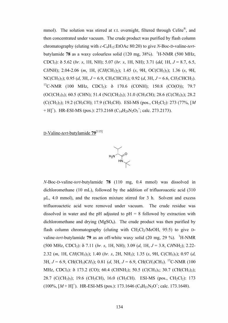
134
mmol). The solution was stirred at r.t. overnight, filtered through Celite®, and
then concentrated under vacuum. The crude product was purified by flash column
chromatography (eluting with c-C6H12:EtOAc 80:20) to give N-Boc-D-valine-tert-
butylamide 78 as a waxy colourless solid (120 mg, 38%). 1H-NMR (500 MHz,
CDCl3): δ 5.62 (br. s, 1H, NH); 5.07 (br. s, 1H, NH); 3.71 (dd, 1H, J = 8.7, 6.5,
CHNH); 2.04-2.06 (m, 1H, (CH(CH3)2); 1.45 (s, 9H, OC(CH3)3); 1.36 (s, 9H,
NC(CH3)3); 0.95 (d, 3H, J = 6.9, CH3CHCH3); 0.92 (d, 3H, J = 6.6, CH3CHCH3). 13C-NMR (100 MHz, CDCl3): δ 170.6 (CONH); 150.8 (CO(O)); 79.7
(OC(CH3)3); 60.5 (CHN); 51.4 (NC(CH3)3); 31.0 (CH3CH); 28.6 (C(CH3)3); 28.2
(C(CH3)3); 19.2 (CH3CH); 17.9 (CH3CH). ESI-MS (pos., CH2Cl2): 273 (77%, [M
+ H]+). HR-ESI-MS (pos.): 273.2168 (C14H29N2O3+; calc. 273.2173).
D-Valine-tert-butylamide 79[115]
N-Boc-D-valine-tert-butylamide 78 (110 mg, 0.4 mmol) was dissolved in
dichloromethane (10 mL), followed by the addition of trifluoroacetic acid (310
µL, 4.0 mmol), and the reaction mixture stirred for 3 h. Solvent and excess
trifluoroactetic acid were removed under vacuum. The crude residue was
dissolved in water and the pH adjusted to pH = 8 followed by extraction with
dichloromethane and drying (MgSO4). The crude product was then purified by
flash column chromatography (eluting with CH2Cl2/MeOH, 95:5) to give D-
valine-tert-butylamide 79 as an off-white waxy solid (20 mg, 29 %). 1H-NMR
(500 MHz, CDCl3): δ 7.11 (br. s, 1H, NH); 3.09 (d, 1H, J = 3.8, CHNH2); 2.22-
2.32 (m, 1H, CH(CH3)2); 1.40 (br. s, 2H, NH2); 1.35 (s, 9H, C(CH3)3); 0.97 (d,
3H, J = 6.9, CH(CH3)CH3); 0.81 (d, 3H, J = 6.9, CH(CH3)CH3). 13C-NMR (100
MHz, CDCl3): δ 173.2 (CO); 60.4 (CHNH2); 50.5 (C(CH3)3; 30.7 (CH(CH3)2);
28.7 (C(CH3)3); 19.6 (CH3CH), 16.0 (CH3CH). ESI-MS (pos., CH2Cl2): 173
(100%, [M + H]+). HR-ESI-MS (pos.): 173.1646 (C9H21N2O+; calc. 173.1648).
HNH2N
O

135
Chiral GC analysis of 71
Samples of L-valine-tert-butylamide (Bachem), and synthetically prepared D-
valine-tert-butylamide 79, were analysed by chiral GC (Varian CP7503 with
flame ionisation detection; column: 25 m x 32 mm chiral-dexCB; helium carrier
gas). The D-isomer was found to elute at ~63.9 min, and the L-isomer at ~65.3
min. Manual integration of these peaks for a gas chromatogram of 57 isolated
from the large-scale four-component reaction of β-D-adenosine-3ʹ′-phosphate 54,
52, 53, and NH4Cl indicated an L-ee of 0.8%. Given the errors inherent in chiral
GC analysis, and the fact that the sample of 57 had been fractionated, this low
value can be taken to indicate that 57 formed in the reaction was either racemic, or
close to racemic.

136
8.3 Experimental Procedures for Chapter 3
Reaction of glycolaldehyde phosphate 81 with methyl isonitrile 82
Glycolaldehyde phosphate disodium salt6 81 (55 mg, 0.3 mmol) was dissolved in
D2O (3 mL) and the pD was adjusted to pD = 6 using NaOD-DCl solution.
Methyl isonitrile 82 (18 µL, 0.3 mmol) was then added and the solution stirred at
r.t. overnight. 83 was formed in >95% yield (by 1H-NMR signal integration).
2-(Methylcarbamoyl)-2-hydroxyethyl phosphate sodium salt 83
1H-NMR (500 MHz, D2O): δ 4.25 (app. t, J = 4.1, 3.8, 1H, CHOH); 4.02 (ddd, J
= 11.0, 6.3, 2.8, 1H, CHHOP); 3.96 (ddd, J = 11.4, 7.3, 5.0, 1H, CHHOP); 2.73
(s, 3H, CH3). 13C-NMR (75 MHz, D2O): δ 174.3 (CONH); 71.6 (CHOH); 66.3
(CH2OP); 25.6 (CH3). 31P-NMR (81 MHz, D2O): δ 1.32 (t, J = 6.4).
Cyanoacetylene 1
Propiolamide 90 (2 g, 29.0 mmol, prepared by B. Gerland according to a literature
procedure[120]), oven dried chromatography sand (14 g) and P2O5 (6.35 g, 44.7
mmol) were crushed together in a mortar and pestle and the mixture was dry
distilled under reduced pressure (20 mm Hg) at 130 °C for 1 h. Cyanoacetylene 1
(829 mg, 56 %) was collected at -78 °C as a white solid which was immediately
dissolved in H2O to make a 1M solution which was either used immediately or
stored at -80 °C. 1H-NMR (500 MHz, CDCl3): δ 2.52 (s, 1H). 13C-NMR (100
MHz, D2O): δ 105.1 (CN); 76.2 (t, J = 40.7, CCN); 55.9 (t, J = 8.0, DCC).
6 Donated by S. Pitsch (ETH, Zürich).
OH HN
O
OPO
OHONa
N
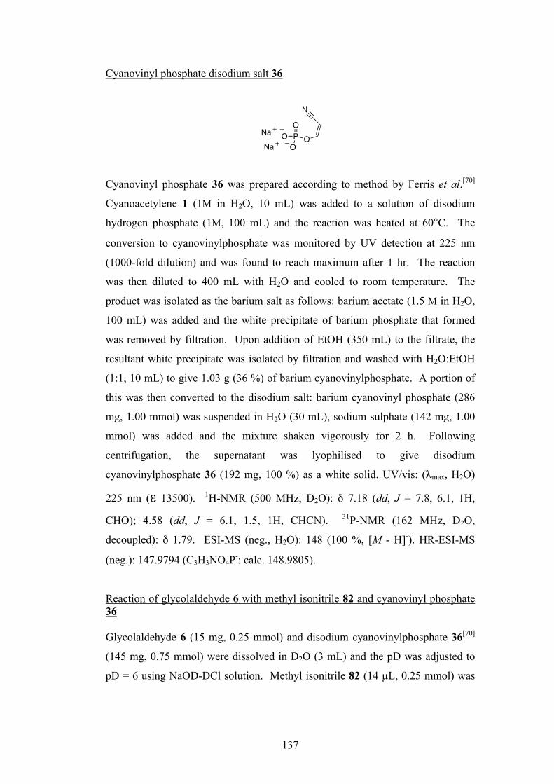
137
Cyanovinyl phosphate disodium salt 36
Cyanovinyl phosphate 36 was prepared according to method by Ferris et al.[70]
Cyanoacetylene 1 (1M in H2O, 10 mL) was added to a solution of disodium
hydrogen phosphate (1M, 100 mL) and the reaction was heated at 60°C. The
conversion to cyanovinylphosphate was monitored by UV detection at 225 nm
(1000-fold dilution) and was found to reach maximum after 1 hr. The reaction
was then diluted to 400 mL with H2O and cooled to room temperature. The
product was isolated as the barium salt as follows: barium acetate (1.5 M in H2O,
100 mL) was added and the white precipitate of barium phosphate that formed
was removed by filtration. Upon addition of EtOH (350 mL) to the filtrate, the
resultant white precipitate was isolated by filtration and washed with H2O:EtOH
(1:1, 10 mL) to give 1.03 g (36 %) of barium cyanovinylphosphate. A portion of
this was then converted to the disodium salt: barium cyanovinyl phosphate (286
mg, 1.00 mmol) was suspended in H2O (30 mL), sodium sulphate (142 mg, 1.00
mmol) was added and the mixture shaken vigorously for 2 h. Following
centrifugation, the supernatant was lyophilised to give disodium
cyanovinylphosphate 36 (192 mg, 100 %) as a white solid. UV/vis: (λmax, H2O)
225 nm (ε 13500). 1H-NMR (500 MHz, D2O): δ 7.18 (dd, J = 7.8, 6.1, 1H,
CHO); 4.58 (dd, J = 6.1, 1.5, 1H, CHCN). 31P-NMR (162 MHz, D2O,
decoupled): δ 1.79. ESI-MS (neg., H2O): 148 (100 %, [M - H]-). HR-ESI-MS
(neg.): 147.9794 (C3H3NO4P-; calc. 148.9805).
Reaction of glycolaldehyde 6 with methyl isonitrile 82 and cyanovinyl phosphate 36 Glycolaldehyde 6 (15 mg, 0.25 mmol) and disodium cyanovinylphosphate 36[70]
(145 mg, 0.75 mmol) were dissolved in D2O (3 mL) and the pD was adjusted to
pD = 6 using NaOD-DCl solution. Methyl isonitrile 82 (14 µL, 0.25 mmol) was
PO
OO
O
N
Na
Na

138
then added and the solution stirred at r.t. overnight. 91 and 92 were formed in ca.
95% yield in a 4:5 ratio (by 1H-NMR signal integration).
2,3-Di-hydroxy-N-methyl-propanamide 91
1H-NMR (500 MHz, D2O): δ 4.16 (dd, J = 4.6, 3.6, 1H, CHOH); 3.76 (dd, J =
12.0, 3.47, 1H, CHHOH); 3.72 (dd, J = 11.7, 4.9, 1H, CHHOH); 2.72 (s, 3H,
CH3). 13C-NMR (75 MHz, D2O): δ 160.2 (CONH); 72.3 (CHOH); 63.3
(CH2OH); 25.6 (CH3). ESI-MS (pos., H2O): 142 (100 %, [M + Na]+).
1-(Methylcarbamoyl)-2-hydroxyethyl phosphate sodium salt 92
1H-NMR (500 MHz, D2O): δ 4.44 (app. dt, J = 9.6, 3.2, 2.8, 1H, CHOP); 3.85
(dd, J = 12.3, 2.8, 1H, CHHOH); 3.81 (dd, J = 12.0, 3.2, 1H, CHHOH); 2.74 (s,
3H, CH3). 13C-NMR (75 MHz, D2O): δ 160.2 (CONH); 75.2 (CHOP); 63.3
(CH2OH); 25.7 (CH3). 31P-NMR (81 MHz, D2O): δ 2.14 (d, J = 8.9). ESI-MS
(neg., H2O): 198 (87 %, [M]-). HR-ESI-MS (neg.): 198.0181 (C4H9NO6P-; calc.
198.0173).
HO OH
ONH
HO O
ONH
POOH
O Na
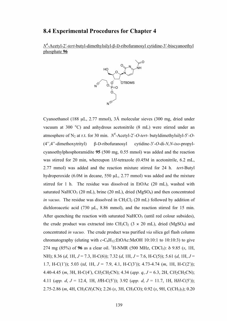
139
8.4 Experimental Procedures for Chapter 4 N4-Acetyl-2ʹ′-tert-butyl-dimethylsilyl-β-D-ribofuranosyl cytidine-3ʹ′-biscyanoethyl phosphate 96
Cyanoethanol (188 µL, 2.77 mmol), 3Å molecular sieves (300 mg, dried under
vacuum at 300 °C) and anhydrous acetonitrile (8 mL) were stirred under an
atmosphere of N2 at r.t. for 30 min. N4-Acetyl-2ʹ′-O-tert- butyldimethylsilyl-5ʹ′-O-
(4ʹ′ʹ′,4ʹ′ʹ′-dimethoxytrityl) β-D-ribofuranosyl cytidine-3ʹ′-O-di-N,N-iso-propyl-
cyanoethylphosphoramidite 95 (500 mg, 0.55 mmol) was added and the reaction
was stirred for 20 min, whereupon 1H-tetrazole (0.45M in acetonitrile, 6.2 mL,
2.77 mmol) was added and the reaction mixture stirred for 24 h. tert-Butyl
hydroperoxide (6.0M in decane, 550 µL, 2.77 mmol) was added and the mixture
stirred for 1 h. The residue was dissolved in EtOAc (20 mL), washed with
saturated NaHCO3 (20 mL), brine (20 mL), dried (MgSO4) and then concentrated
in vacuo. The residue was dissolved in CH2Cl2 (20 mL) followed by addition of
dichloroacetic acid (730 µL, 8.86 mmol), and the reaction stirred for 15 min.
After quenching the reaction with saturated NaHCO3 (until red colour subsides),
the crude product was extracted into CH2Cl2 (3 × 20 mL), dried (MgSO4) and
concentrated in vacuo. The crude product was purified via silica gel flash column
chromatography (eluting with c-C6H12:EtOAc:MeOH 10:10:1 to 10:10:3) to give
274 mg (85%) of 96 as a clear oil. 1H-NMR (500 MHz, CDCl3): δ 9.85 (s, 1H,
NH); 8.36 (d, 1H, J = 7.3, H-C(6)); 7.32 (d, 1H, J = 7.6, H-C(5)); 5.61 (d, 1H, J =
1.7, H-C(1ʹ′)); 5.03 (td, 1H, J = 7.9, 4.1, H-C(3ʹ′)); 4.73-4.74 (m, 1H, H-C(2ʹ′));
4.40-4.45 (m, 3H, H-C(4ʹ′), CH2CH2CN); 4.34 (app. q, J = 6.3, 2H, CH2CH2CN);
4.11 (app. d, J = 12.4, 1H, HH-C(5ʹ′)); 3.92 (app. d, J = 11.7, 1H, HH-C(5ʹ′));
2.75-2.86 (m, 4H, CH2CH2CN); 2.26 (s, 3H, CH3CO); 0.92 (s, 9H, C(CH3)3); 0.20
O N
O OTBDMS
HONO
NH
O
P OON
O
N
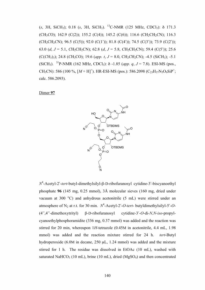
140
(s, 3H, SiCH3); 0.18 (s, 3H, SiCH3). 13C-NMR (125 MHz, CDCl3): δ 171.3
(CH3CO); 162.9 (C(2)); 155.2 (C(4)); 145.2 (C(6)); 116.6 (CH2CH2CN); 116.3
(CH2CH2CN); 96.5 (C(5)); 92.0 (C(1ʹ′)); 81.8 (C(4ʹ′)); 74.5 (C(3ʹ′)); 73.9 (C(2ʹ′));
63.0 (d, J = 5.1, CH2CH2CN); 62.8 (d, J = 5.8, CH2CH2CN); 59.4 (C(5ʹ′)); 25.6
(C(CH3)3); 24.8 (CH3CO); 19.6 (app. t, J = 8.0, CH2CH2CN); -4.5 (SiCH3); -5.1
(SiCH3). 31P-NMR (162 MHz, CDCl3): δ -1.85 (app. q, J = 7.8). ESI-MS (pos.,
CH3CN): 586 (100 %, [M + H]+). HR-ESI-MS (pos.): 586.2098 (C23H37N5O9SiP+;
calc. 586.2093).
Dimer 97
N4-Acetyl-2ʹ′-tert-butyl-dimethylsilyl-β-D-ribofuranosyl cytidine-3ʹ′-biscyanoethyl
phosphate 96 (145 mg, 0.25 mmol), 3Å molecular sieves (160 mg, dried under
vacuum at 300 °C) and anhydrous acetonitrile (5 mL) were stirred under an
atmosphere of N2 at r.t. for 30 min. N4-Acetyl-2ʹ′-O-tert- butyldimethylsilyl-5ʹ′-O-
(4ʹ′ʹ′,4ʹ′ʹ′-dimethoxytrityl) β-D-ribofuranosyl cytidine-3ʹ′-O-di-N,N-iso-propyl-
cyanoethylphosphoramidite (336 mg, 0.37 mmol) was added and the reaction was
stirred for 20 min, whereupon 1H-tetrazole (0.45M in acetonitrile, 4.4 mL, 1.98
mmol) was added and the reaction mixture stirred for 24 h. tert-Butyl
hydroperoxide (6.0M in decane, 250 µL, 1.24 mmol) was added and the mixture
stirred for 1 h. The residue was dissolved in EtOAc (10 mL), washed with
saturated NaHCO3 (10 mL), brine (10 mL), dried (MgSO4) and then concentrated
O N
O OTBDMS
HONO
NH
O
P OON
O N
O OTBDMS
ONO
NH
O
P OOO
N
N
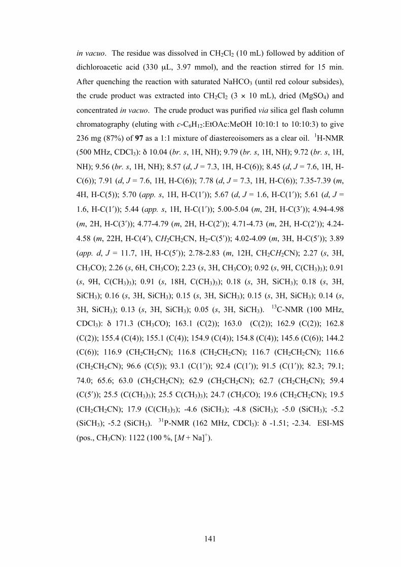
141
in vacuo. The residue was dissolved in CH2Cl2 (10 mL) followed by addition of
dichloroacetic acid (330 µL, 3.97 mmol), and the reaction stirred for 15 min.
After quenching the reaction with saturated NaHCO3 (until red colour subsides),
the crude product was extracted into CH2Cl2 (3 × 10 mL), dried (MgSO4) and
concentrated in vacuo. The crude product was purified via silica gel flash column
chromatography (eluting with c-C6H12:EtOAc:MeOH 10:10:1 to 10:10:3) to give
236 mg (87%) of 97 as a 1:1 mixture of diastereoisomers as a clear oil. 1H-NMR
(500 MHz, CDCl3): δ 10.04 (br. s, 1H, NH); 9.79 (br. s, 1H, NH); 9.72 (br. s, 1H,
NH); 9.56 (br. s, 1H, NH); 8.57 (d, J = 7.3, 1H, H-C(6)); 8.45 (d, J = 7.6, 1H, H-
C(6)); 7.91 (d, J = 7.6, 1H, H-C(6)); 7.78 (d, J = 7.3, 1H, H-C(6)); 7.35-7.39 (m,
4H, H-C(5)); 5.70 (app. s, 1H, H-C(1ʹ′)); 5.67 (d, J = 1.6, H-C(1ʹ′)); 5.61 (d, J =
1.6, H-C(1ʹ′)); 5.44 (app. s, 1H, H-C(1ʹ′)); 5.00-5.04 (m, 2H, H-C(3ʹ′)); 4.94-4.98
(m, 2H, H-C(3ʹ′)); 4.77-4.79 (m, 2H, H-C(2ʹ′)); 4.71-4.73 (m, 2H, H-C(2ʹ′)); 4.24-
4.58 (m, 22H, H-C(4ʹ′), CH2CH2CN, H2-C(5ʹ′)); 4.02-4.09 (m, 3H, H-C(5ʹ′)); 3.89
(app. d, J = 11.7, 1H, H-C(5ʹ′)); 2.78-2.83 (m, 12H, CH2CH2CN); 2.27 (s, 3H,
CH3CO); 2.26 (s, 6H, CH3CO); 2.23 (s, 3H, CH3CO); 0.92 (s, 9H, C(CH3)3); 0.91
(s, 9H, C(CH3)3); 0.91 (s, 18H, C(CH3)3); 0.18 (s, 3H, SiCH3); 0.18 (s, 3H,
SiCH3); 0.16 (s, 3H, SiCH3); 0.15 (s, 3H, SiCH3); 0.15 (s, 3H, SiCH3); 0.14 (s,
3H, SiCH3); 0.13 (s, 3H, SiCH3); 0.05 (s, 3H, SiCH3). 13C-NMR (100 MHz,
CDCl3): δ 171.3 (CH3CO); 163.1 (C(2)); 163.0 (C(2)); 162.9 (C(2)); 162.8
(C(2)); 155.4 (C(4)); 155.1 (C(4)); 154.9 (C(4)); 154.8 (C(4)); 145.6 (C(6)); 144.2
(C(6)); 116.9 (CH2CH2CN); 116.8 (CH2CH2CN); 116.7 (CH2CH2CN); 116.6
(CH2CH2CN); 96.6 (C(5)); 93.1 (C(1ʹ′)); 92.4 (C(1ʹ′)); 91.5 (C(1ʹ′)); 82.3; 79.1;
74.0; 65.6; 63.0 (CH2CH2CN); 62.9 (CH2CH2CN); 62.7 (CH2CH2CN); 59.4
(C(5ʹ′)); 25.5 (C(CH3)3); 25.5 C(CH3)3); 24.7 (CH3CO); 19.6 (CH2CH2CN); 19.5
(CH2CH2CN); 17.9 (C(CH3)3); -4.6 (SiCH3); -4.8 (SiCH3); -5.0 (SiCH3); -5.2
(SiCH3); -5.2 (SiCH3). 31P-NMR (162 MHz, CDCl3): δ -1.51; -2.34. ESI-MS
(pos., CH3CN): 1122 (100 %, [M + Na]+).
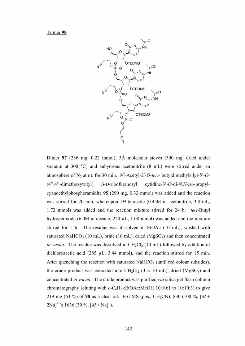
142
Trimer 98
Dimer 97 (236 mg, 0.22 mmol), 3Å molecular sieves (300 mg, dried under
vacuum at 300 °C) and anhydrous acetonitrile (8 mL) were stirred under an
atmosphere of N2 at r.t. for 30 min. N4-Acetyl-2ʹ′-O-tert- butyldimethylsilyl-5ʹ′-O-
(4ʹ′ʹ′,4ʹ′ʹ′-dimethoxytrityl) β-D-ribofuranosyl cytidine-3ʹ′-O-di-N,N-iso-propyl-
cyanoethylphosphoramidite 95 (290 mg, 0.32 mmol) was added and the reaction
was stirred for 20 min, whereupon 1H-tetrazole (0.45M in acetonitrile, 3.8 mL,
1.72 mmol) was added and the reaction mixture stirred for 24 h. tert-Butyl
hydroperoxide (6.0M in decane, 220 µL, 1.08 mmol) was added and the mixture
stirred for 1 h. The residue was dissolved in EtOAc (10 mL), washed with
saturated NaHCO3 (10 mL), brine (10 mL), dried (MgSO4) and then concentrated
in vacuo. The residue was dissolved in CH2Cl2 (10 mL) followed by addition of
dichloroacetic acid (285 µL, 3.44 mmol), and the reaction stirred for 15 min.
After quenching the reaction with saturated NaHCO3 (until red colour subsides),
the crude product was extracted into CH2Cl2 (3 × 10 mL), dried (MgSO4) and
concentrated in vacuo. The crude product was purified via silica gel flash column
chromatography (eluting with c-C6H12:EtOAc:MeOH 10:10:1 to 10:10:3) to give
219 mg (63 %) of 98 as a clear oil. ESI-MS (pos., CH3CN): 830 (100 %, [M +
2Na]2+); 1636 (30 %, [M + Na]+).
O N
O OTBDMS
ONO
NH
O
P OON
O N
O OTBDMS
ONO
NH
O
P OOO
N
N
O N
O OTBDMS
HONO
NH
O
P OON
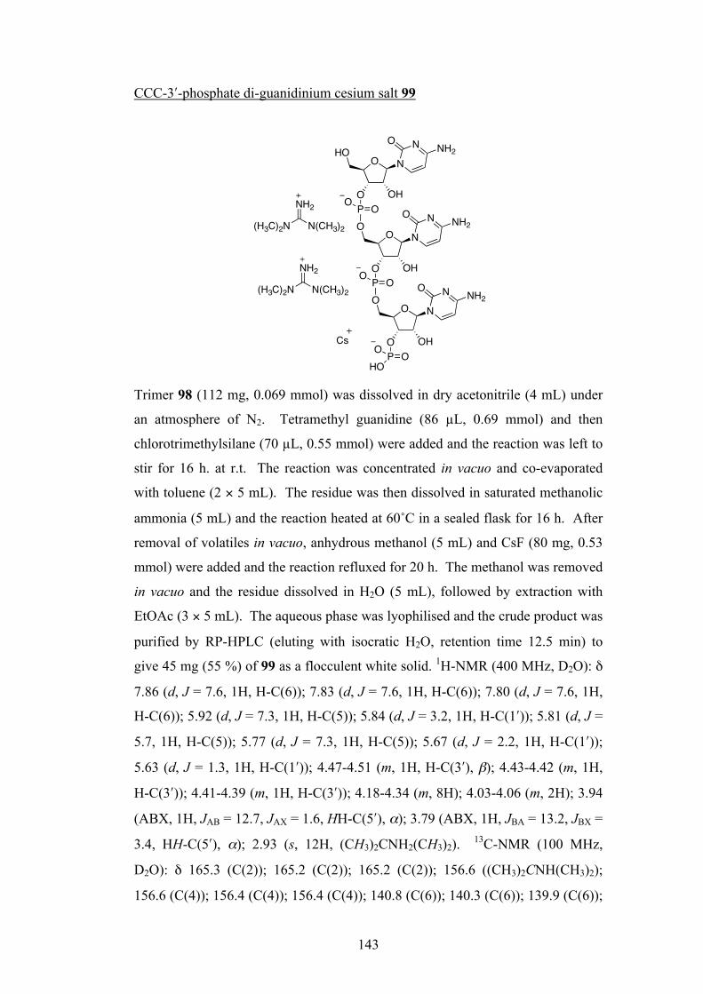
143
CCC-3ʹ′-phosphate di-guanidinium cesium salt 99
Trimer 98 (112 mg, 0.069 mmol) was dissolved in dry acetonitrile (4 mL) under
an atmosphere of N2. Tetramethyl guanidine (86 µL, 0.69 mmol) and then
chlorotrimethylsilane (70 µL, 0.55 mmol) were added and the reaction was left to
stir for 16 h. at r.t. The reaction was concentrated in vacuo and co-evaporated
with toluene (2 × 5 mL). The residue was then dissolved in saturated methanolic
ammonia (5 mL) and the reaction heated at 60˚C in a sealed flask for 16 h. After
removal of volatiles in vacuo, anhydrous methanol (5 mL) and CsF (80 mg, 0.53
mmol) were added and the reaction refluxed for 20 h. The methanol was removed
in vacuo and the residue dissolved in H2O (5 mL), followed by extraction with
EtOAc (3 × 5 mL). The aqueous phase was lyophilised and the crude product was
purified by RP-HPLC (eluting with isocratic H2O, retention time 12.5 min) to
give 45 mg (55 %) of 99 as a flocculent white solid. 1H-NMR (400 MHz, D2O): δ
7.86 (d, J = 7.6, 1H, H-C(6)); 7.83 (d, J = 7.6, 1H, H-C(6)); 7.80 (d, J = 7.6, 1H,
H-C(6)); 5.92 (d, J = 7.3, 1H, H-C(5)); 5.84 (d, J = 3.2, 1H, H-C(1ʹ′)); 5.81 (d, J =
5.7, 1H, H-C(5)); 5.77 (d, J = 7.3, 1H, H-C(5)); 5.67 (d, J = 2.2, 1H, H-C(1ʹ′));
5.63 (d, J = 1.3, 1H, H-C(1ʹ′)); 4.47-4.51 (m, 1H, H-C(3ʹ′), β); 4.43-4.42 (m, 1H,
H-C(3ʹ′)); 4.41-4.39 (m, 1H, H-C(3ʹ′)); 4.18-4.34 (m, 8H); 4.03-4.06 (m, 2H); 3.94
(ABX, 1H, JAB = 12.7, JAX = 1.6, HH-C(5ʹ′), α); 3.79 (ABX, 1H, JBA = 13.2, JBX =
3.4, HH-C(5ʹ′), α); 2.93 (s, 12H, (CH3)2CNH2(CH3)2). 13C-NMR (100 MHz,
D2O): δ 165.3 (C(2)); 165.2 (C(2)); 165.2 (C(2)); 156.6 ((CH3)2CNH(CH3)2);
156.6 (C(4)); 156.4 (C(4)); 156.4 (C(4)); 140.8 (C(6)); 140.3 (C(6)); 139.9 (C(6));
O N
O
ONO
NH2
P OO
O N
O
ONO
NH2
P OHOO
O N
O
HONO
NH2
P OOOH
OH
OH
NH2
N(CH3)2(H3C)2N
NH2
N(CH3)2(H3C)2N
Cs
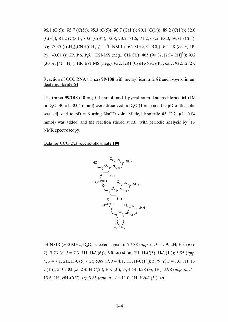
144
96.1 (C(5)); 95.7 (C(5)); 95.3 (C(5)); 90.7 (C(1ʹ′)); 90.1 (C(1ʹ′)); 89.2 (C(1ʹ′)); 82.0
(C(3ʹ′)); 81.2 (C(3ʹ′)); 80.6 (C(3ʹ′)); 73.8; 73.2; 71.6; 71.2; 63.5; 63.0; 59.31 (C(5ʹ′),
α); 37.35 ((CH3)2CNH(CH3)2). 31P-NMR (162 MHz, CDCl3): δ 1.48 (br. s, 1P,
Pγ); -0.01 (s, 2P, Pα, Pβ). ESI-MS (neg., CH2Cl2): 465 (90 %, [M – 2H]2-); 932
(30 %, [M – H]-). HR-ESI-MS (neg.): 932.1284 (C27H37N9O22P3-; calc. 932.1272).
Reaction of CCC RNA trimers 99/108 with methyl isonitrile 82 and 1-pyrrolinium deuterochloride 64
The trimer 99/108 (10 mg, 0.1 mmol) and 1-pyrrolinium deuterochloride 64 (1M
in D2O, 40 µL, 0.04 mmol) were dissolved in D2O (1 mL) and the pD of the soln.
was adjusted to pD = 6 using NaOD soln. Methyl isonitrile 82 (2.2 µL, 0.04
mmol) was added, and the reaction stirred at r.t., with periodic analysis by 1H-
NMR spectroscopy.
Data for CCC-2ʹ′,3ʹ′-cyclic-phosphate 100
1H-NMR (500 MHz, D2O, selected signals): δ 7.88 (app. t., J = 7.9, 2H, H-C(6) ×
2); 7.73 (d, J = 7.3, 1H, H-C(6)); 6.01-6.04 (m, 2H, H-C(5), H-C(1ʹ′)); 5.95 (app.
t., J = 7.1, 2H, H-C(5) × 2); 5.89 (d, J = 4.1, 1H, H-C(1ʹ′)); 5.79 (d, J = 1.6, 1H, H-
C(1ʹ′)); 5.0-5.02 (m, 2H, H-C(2ʹ′), H-C(3ʹ′), γ); 4.54-4.58 (m, 1H); 3.98 (app. d., J =
13.6, 1H, HH-C(5ʹ′), α); 3.85 (app. d., J = 11.0, 1H, HH-C(5ʹ′), α).
O N
NO
ONH2
O N
O OH
NO
ONH2
P OO
O N
O OH
NHO
ONH2
P OO
OPO
O O
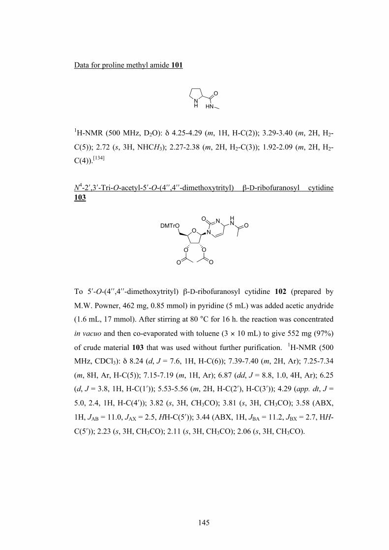
145
Data for proline methyl amide 101
1H-NMR (500 MHz, D2O): δ 4.25-4.29 (m, 1H, H-C(2)); 3.29-3.40 (m, 2H, H2-
C(5)); 2.72 (s, 3H, NHCH3); 2.27-2.38 (m, 2H, H2-C(3)); 1.92-2.09 (m, 2H, H2-
C(4)).[134]
N4-2ʹ′,3ʹ′-Tri-O-acetyl-5ʹ′-O-(4ʹ′ʹ′,4ʹ′ʹ′-dimethoxytrityl) β-D-ribofuranosyl cytidine 103
To 5ʹ′-O-(4ʹ′ʹ′,4ʹ′ʹ′-dimethoxytrityl) β-D-ribofuranosyl cytidine 102 (prepared by
M.W. Powner, 462 mg, 0.85 mmol) in pyridine (5 mL) was added acetic anydride
(1.6 mL, 17 mmol). After stirring at 80 °C for 16 h. the reaction was concentrated
in vacuo and then co-evaporated with toluene (3 × 10 mL) to give 552 mg (97%)
of crude material 103 that was used without further purification. 1H-NMR (500
MHz, CDCl3): δ 8.24 (d, J = 7.6, 1H, H-C(6)); 7.39-7.40 (m, 2H, Ar); 7.25-7.34
(m, 8H, Ar, H-C(5)); 7.15-7.19 (m, 1H, Ar); 6.87 (dd, J = 8.8, 1.0, 4H, Ar); 6.25
(d, J = 3.8, 1H, H-C(1ʹ′)); 5.53-5.56 (m, 2H, H-C(2ʹ′), H-C(3ʹ′)); 4.29 (app. dt, J =
5.0, 2.4, 1H, H-C(4ʹ′)); 3.82 (s, 3H, CH3CO); 3.81 (s, 3H, CH3CO); 3.58 (ABX,
1H, JAB = 11.0, JAX = 2.5, HH-C(5ʹ′)); 3.44 (ABX, 1H, JBA = 11.2, JBX = 2.7, HH-
C(5ʹ′)); 2.23 (s, 3H, CH3CO); 2.11 (s, 3H, CH3CO); 2.06 (s, 3H, CH3CO).
NH HN
O
O N
O O
OO
NO HN ODMTrO

146
N4-2ʹ′,3ʹ′-Tri-O-acetyl-β-D-ribofuranosyl cytidine 104
N4-2ʹ′,3ʹ′-Tri-O-acetyl-5ʹ′-O-(4ʹ′ʹ′,4ʹ′ʹ′-dimethoxytrityl) β-D-ribofuranosyl cytidine
103 (552 mg, 0.82 mmol) was dissolved in CH2Cl2 (10 mL). Dichloroacetic acid
(1.12 mL, 13.6 mmol) was added and the reaction stirred at r.t for 15 min
whereupon CH2Cl2 was removed in vacuo. The residue was dissolved in EtOAc
(10 mL), washed with saturated NaHCO3 then partitioned between H2O, and the
organic phase combined with four further extracts which were dried (MgSO4) and
concentrated in vacuo. The crude material was purified by flash column
chromatography (eluting with c-C6H12:EtOAc:MeOH 15:25:2) to give 126 mg (41
%) of 104 as a yellow solid. M.p. 149-153 °C. 1H-NMR (300 MHz, CD3OD): δ
8.45 (d, J = 7.6, 1H, H-C(6)); 7.45 (d, J = 7.6, 1H, H-C(5)); 6.13 (d, J = 4.8, 1H,
H-C(1ʹ′)); 5.49 (m, 1H, H-C(2ʹ′)); 5.42 (m, 1H, H-C(3ʹ′)); 4.28 (dt, J = 4.7, 2.5, 1H,
H-C(4ʹ′)); 3.90 (ABX, 1H, JAB = 12.3, JAX = 2.5, HH-C(5ʹ′)); 3.78 (ABX, 1H, JBA
= 12.5, JBX = 2.6, HH-C(5ʹ′)); 2.18 (s, 3H, CH3CO); 2.10 (s, 3H, CH3CO); 2.07 (s,
3H, CH3CO). 13C-NMR (75 MHz, CD3OD): δ 173.1 (CH3CO); 171.6 (CH3CO);
171.3 (CH3CO); 168.8 (C(2)); 164.7 (C(4)); 146.6 (C(6)); 98.5 (C(5)); 90.2
(C(1ʹ′)); 85.0 (C(4ʹ′)); 75.7 (C(2ʹ′)); 72.1 (C(3ʹ′)); 61.9 (C(5ʹ′)); 24.7 (CH3CO); 20.6
(CH3CO); 20.4 (CH3CO). ESI-MS (neg., CH3OH): 368 (100 %, [M - H]-).[135]
O N
O O
OO
NO HN OHO
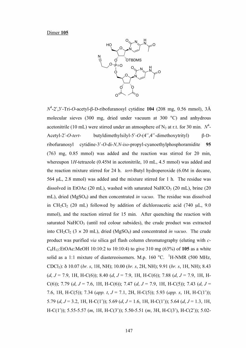
147
Dimer 105
N4-2ʹ′,3ʹ′-Tri-O-acetyl-β-D-ribofuranosyl cytidine 104 (208 mg, 0.56 mmol), 3Å
molecular sieves (300 mg, dried under vacuum at 300 °C) and anhydrous
acetonitrile (10 mL) were stirred under an atmosphere of N2 at r.t. for 30 min. N4-
Acetyl-2ʹ′-O-tert- butyldimethylsilyl-5ʹ′-O-(4ʹ′ʹ′,4ʹ′ʹ′-dimethoxytrityl) β-D-
ribofuranosyl cytidine-3ʹ′-O-di-N,N-iso-propyl-cyanoethylphosphoramidite 95
(763 mg, 0.85 mmol) was added and the reaction was stirred for 20 min,
whereupon 1H-tetrazole (0.45M in acetonitrile, 10 mL, 4.5 mmol) was added and
the reaction mixture stirred for 24 h. tert-Butyl hydroperoxide (6.0M in decane,
564 µL, 2.8 mmol) was added and the mixture stirred for 1 h. The residue was
dissolved in EtOAc (20 mL), washed with saturated NaHCO3 (20 mL), brine (20
mL), dried (MgSO4) and then concentrated in vacuo. The residue was dissolved
in CH2Cl2 (20 mL) followed by addition of dichloroacetic acid (740 µL, 9.0
mmol), and the reaction stirred for 15 min. After quenching the reaction with
saturated NaHCO3 (until red colour subsides), the crude product was extracted
into CH2Cl2 (3 × 20 mL), dried (MgSO4) and concentrated in vacuo. The crude
product was purified via silica gel flash column chromatography (eluting with c-
C6H12:EtOAc:MeOH 10:10:2 to 10:10:4) to give 310 mg (63%) of 105 as a white
solid as a 1:1 mixture of diastereoisomers. M.p. 160 °C. 1H-NMR (500 MHz,
CDCl3): δ 10.07 (br. s, 1H, NH); 10.00 (br. s, 2H, NH); 9.91 (br. s, 1H, NH); 8.43
(d, J = 7.9, 1H, H-C(6)); 8.40 (d, J = 7.9, 1H, H-C(6)); 7.88 (d, J = 7.9, 1H, H-
C(6)); 7.79 (d, J = 7.6, 1H, H-C(6)); 7.47 (d, J = 7.9, 1H, H-C(5)); 7.43 (d, J =
7.6, 1H, H-C(5)); 7.34 (app. t, J = 7.1, 2H, H-C(5)); 5.93 (app. s, 1H, H-C(1ʹ′));
5.79 (d, J = 3.2, 1H, H-C(1ʹ′)); 5.69 (d, J = 1.6, 1H, H-C(1ʹ′)); 5.64 (d, J = 1.3, 1H,
H-C(1ʹ′)); 5.55-5.57 (m, 1H, H-C(3ʹ′)); 5.50-5.51 (m, 3H, H-C(3ʹ′), H-C(2ʹ′)); 5.02-
O
OO
ON
NO HN
O
O
HON
NO HN
P OOOTBDMS
OO
O
O
N
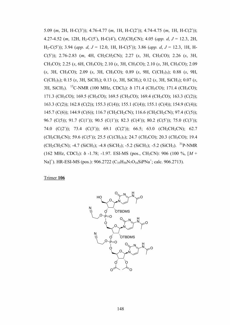
148
5.09 (m, 2H, H-C(3ʹ′)); 4.76-4.77 (m, 1H, H-C(2ʹ′)); 4.74-4.75 (m, 1H, H-C(2ʹ′));
4.27-4.52 (m, 12H, H2-C(5ʹ′), H-C(4ʹ′), CH2CH2CN); 4.05 (app. d, J = 12.3, 2H,
H2-C(5ʹ′)); 3.94 (app. d, J = 12.0, 1H, H-C(5ʹ′)); 3.86 (app. d, J = 12.3, 1H, H-
C(5ʹ′)); 2.76-2.83 (m, 4H, CH2CH2CN); 2.27 (s, 3H, CH3CO); 2.26 (s, 3H,
CH3CO); 2.25 (s, 6H, CH3CO); 2.10 (s, 3H, CH3CO); 2.10 (s, 3H, CH3CO); 2.09
(s, 3H, CH3CO); 2.09 (s, 3H, CH3CO); 0.89 (s, 9H, C(CH3)3); 0.88 (s, 9H,
C(CH3)3); 0.15 (s, 3H, SiCH3); 0.13 (s, 3H, SiCH3); 0.12 (s, 3H, SiCH3); 0.07 (s,
3H, SiCH3). 13C-NMR (100 MHz, CDCl3): δ 171.4 (CH3CO); 171.4 (CH3CO);
171.3 (CH3CO); 169.5 (CH3CO); 169.5 (CH3CO); 169.4 (CH3CO); 163.3 (C(2));
163.3 (C(2)); 162.8 (C(2)); 155.3 (C(4)); 155.1 (C(4)); 155.1 (C(4)); 154.9 (C(4));
145.7 (C(6)); 144.9 (C(6)); 116.7 (CH2CH2CN); 116.6 (CH2CH2CN); 97.4 (C(5));
96.7 (C(5)); 91.7 (C(1ʹ′)); 90.5 (C(1ʹ′)); 82.3 (C(4ʹ′)); 80.2 (C(5ʹ′)); 75.0 (C(3ʹ′));
74.0 (C(2ʹ′)); 73.4 (C(3ʹ′)); 69.1 (C(2ʹ′)); 66.5; 63.0 (CH2CH2CN); 62.7
(CH2CH2CN); 59.6 (C(5ʹ′)); 25.5 (C(CH3)3); 24.7 (CH3CO); 20.3 (CH3CO); 19.4
(CH2CH2CN); -4.7 (SiCH3); -4.8 (SiCH3); -5.2 (SiCH3); -5.2 (SiCH3). 31P-NMR
(162 MHz, CDCl3): δ -1.78; -1.97. ESI-MS (pos., CH3CN): 906 (100 %, [M +
Na]+). HR-ESI-MS (pos.): 906.2722 (C35H50N7O16SiPNa+; calc. 906.2713).
Trimer 106
O
OO
ON
NO HN
O
O
ON
NO HN
P OO
O
O
HON
NO HN
P OOOTBDMS
OTBDMS
OO
O
O
O
N
N
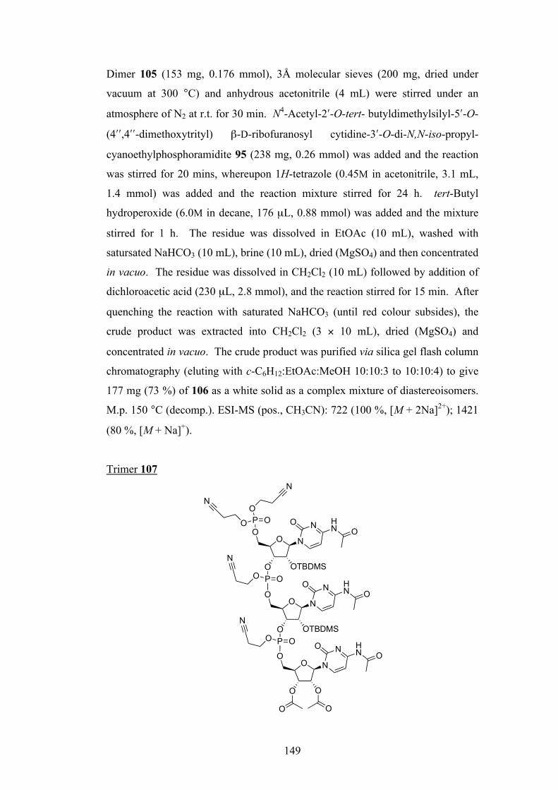
149
Dimer 105 (153 mg, 0.176 mmol), 3Å molecular sieves (200 mg, dried under
vacuum at 300 °C) and anhydrous acetonitrile (4 mL) were stirred under an
atmosphere of N2 at r.t. for 30 min. N4-Acetyl-2ʹ′-O-tert- butyldimethylsilyl-5ʹ′-O-
(4ʹ′ʹ′,4ʹ′ʹ′-dimethoxytrityl) β-D-ribofuranosyl cytidine-3ʹ′-O-di-N,N-iso-propyl-
cyanoethylphosphoramidite 95 (238 mg, 0.26 mmol) was added and the reaction
was stirred for 20 mins, whereupon 1H-tetrazole (0.45M in acetonitrile, 3.1 mL,
1.4 mmol) was added and the reaction mixture stirred for 24 h. tert-Butyl
hydroperoxide (6.0M in decane, 176 µL, 0.88 mmol) was added and the mixture
stirred for 1 h. The residue was dissolved in EtOAc (10 mL), washed with
satursated NaHCO3 (10 mL), brine (10 mL), dried (MgSO4) and then concentrated
in vacuo. The residue was dissolved in CH2Cl2 (10 mL) followed by addition of
dichloroacetic acid (230 µL, 2.8 mmol), and the reaction stirred for 15 min. After
quenching the reaction with saturated NaHCO3 (until red colour subsides), the
crude product was extracted into CH2Cl2 (3 × 10 mL), dried (MgSO4) and
concentrated in vacuo. The crude product was purified via silica gel flash column
chromatography (eluting with c-C6H12:EtOAc:MeOH 10:10:3 to 10:10:4) to give
177 mg (73 %) of 106 as a white solid as a complex mixture of diastereoisomers.
M.p. 150 °C (decomp.). ESI-MS (pos., CH3CN): 722 (100 %, [M + 2Na]2+); 1421
(80 %, [M + Na]+).
Trimer 107
O
OO
ON
NO HN
O
O
ON
NO HN
P OO
O
O
ON
NO HN
P OOOTBDMS
OTBDMS
OO
O
O
O
N
N
P OO
O
N
N

150
Trimer 106 (169 mg, 0.12 mmol), 3Å molecular sieves (150 mg, dried under
vacuum at 300 °C) and anhydrous acetonitrile (4 mL) were stirred under an
atmosphere of N2 at r.t. for 30 mins. Bis(2-cyanoethyl)-N,N-
diisopropylphosphoramidite (48 µL, 0.18 mmol) was added and the reaction was
stirred for 20 min, whereupon 1H-tetrazole (0.45M in acetonitrile, 2.1 mL, 0.96
mmol) was added and the reaction mixture stirred for 24 h. tert-Butyl
hydroperoxide (6.0M in decane, 120 µL, 0.6 mmol) was added and the mixture
stirred for 1 h. The residue was dissolved in EtOAc (10 mL), washed with
saturated NaHCO3 (10 mL), brine (10 mL), dried (MgSO4) and then concentrated
in vacuo. The crude product was purified via silica gel flash column
chromatography (eluting with c-C6H12:EtOAc:MeOH 10:10:3 to 10:10:4.5) to
give 151 mg (79 %) of 107 as a white solid as a complex mixture of
diastereoisomers. ESI-MS (pos., CH3CN): 815 (100 %, [M + 2Na]2+); 1607 (80
%, [M + Na]+).
CCC-5ʹ′-phosphate di-guanidinium cesium salt 108
Trimer 107 (153 mg, 0.097 mmol) was dissolved in dry acetonitrile (5.5 mL)
under an atmosphere of N2. Tetramethyl guanidine (121 µL, 0.97 mmol) and then
chlorotrimethylsilane (98 µL, 0.78 mmol) were added and the reaction was left to
stir for 16 h at r.t. The reaction was concentrated in vacuo and co-evaporated with
O
OHHO
ON
NONH2
O
O
ON
NONH2
P OO
O
O
ON
NONH2
P OO
P OOH
O
OH
OH
NH2
N(CH3)2(H3C)2N
NH2
N(CH3)2(H3C)2N
Cs
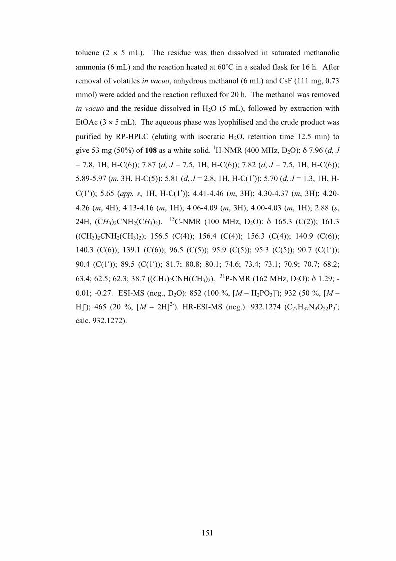
151
toluene (2 × 5 mL). The residue was then dissolved in saturated methanolic
ammonia (6 mL) and the reaction heated at 60˚C in a sealed flask for 16 h. After
removal of volatiles in vacuo, anhydrous methanol (6 mL) and CsF (111 mg, 0.73
mmol) were added and the reaction refluxed for 20 h. The methanol was removed
in vacuo and the residue dissolved in H2O (5 mL), followed by extraction with
EtOAc (3 × 5 mL). The aqueous phase was lyophilised and the crude product was
purified by RP-HPLC (eluting with isocratic H2O, retention time 12.5 min) to
give 53 mg (50%) of 108 as a white solid. 1H-NMR (400 MHz, D2O): δ 7.96 (d, J
= 7.8, 1H, H-C(6)); 7.87 (d, J = 7.5, 1H, H-C(6)); 7.82 (d, J = 7.5, 1H, H-C(6));
5.89-5.97 (m, 3H, H-C(5)); 5.81 (d, J = 2.8, 1H, H-C(1ʹ′)); 5.70 (d, J = 1.3, 1H, H-
C(1ʹ′)); 5.65 (app. s, 1H, H-C(1ʹ′)); 4.41-4.46 (m, 3H); 4.30-4.37 (m, 3H); 4.20-
4.26 (m, 4H); 4.13-4.16 (m, 1H); 4.06-4.09 (m, 3H); 4.00-4.03 (m, 1H); 2.88 (s,
24H, (CH3)2CNH2(CH3)2). 13C-NMR (100 MHz, D2O): δ 165.3 (C(2)); 161.3
((CH3)2CNH2(CH3)2); 156.5 (C(4)); 156.4 (C(4)); 156.3 (C(4)); 140.9 (C(6));
140.3 (C(6)); 139.1 (C(6)); 96.5 (C(5)); 95.9 (C(5)); 95.3 (C(5)); 90.7 (C(1ʹ′));
90.4 (C(1ʹ′)); 89.5 (C(1ʹ′)); 81.7; 80.8; 80.1; 74.6; 73.4; 73.1; 70.9; 70.7; 68.2;
63.4; 62.5; 62.3; 38.7 ((CH3)2CNH(CH3)2). 31P-NMR (162 MHz, D2O): δ 1.29; -
0.01; -0.27. ESI-MS (neg., D2O): 852 (100 %, [M – H2PO3]-); 932 (50 %, [M –
H]-); 465 (20 %, [M – 2H]2-). HR-ESI-MS (neg.): 932.1274 (C27H37N9O22P3-;
calc. 932.1272).
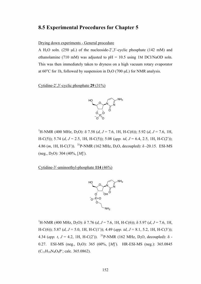
152
8.5 Experimental Procedures for Chapter 5
Drying down experiments - General procedure
A H2O soln. (250 µL) of the nucleoside-2ʹ′,3ʹ′-cyclic phosphate (142 mM) and
ethanolamine (710 mM) was adjusted to pH = 10.5 using 1M DCl/NaOD soln.
This was then immediately taken to dryness on a high vacuum rotary evaporator
at 60°C for 1h, followed by suspension in D2O (700 µL) for NMR analysis.
Cytidine-2ʹ′,3ʹ′-cyclic phosphate 29 (31%)
1H-NMR (400 MHz, D2O): δ 7.58 (d, J = 7.6, 1H, H-C(6)); 5.92 (d, J = 7.6, 1H,
H-C(5)); 5.74 (d, J = 2.5, 1H, H-C(5)); 5.08 (app. td, J = 6.4, 2.5, 1H, H-C(2ʹ′));
4.86 (m, 1H, H-C(3ʹ′)). 31P-NMR (162 MHz, D2O, decoupled): δ -20.15. ESI-MS
(neg., D2O): 304 (40%, [M]-).
Cytidine-3ʹ′-aminoethyl-phosphate 114 (46%)
1H-NMR (400 MHz, D2O): δ 7.76 (d, J = 7.6, 1H, H-C(6)); δ 5.97 (d, J = 7.6, 1H,
H-C(6)); 5.87 (d, J = 5.0, 1H, H-C(1ʹ′)); 4.49 (app. td, J = 8.1, 5.2, 1H, H-C(3ʹ′));
4.34 (app. t, J = 4.2, 1H, H-C(2ʹ′)). 31P-NMR (162 MHz, D2O, decoupled): δ -
0.27. ESI-MS (neg., D2O): 365 (60%, [M]-). HR-ESI-MS (neg.): 365.0845
(C11H18N4O8P-; calc. 365.0862).
O N
OPO
HON
O
O O
NH2
O NHO
N
OO OHP OOO
NH2
NH2
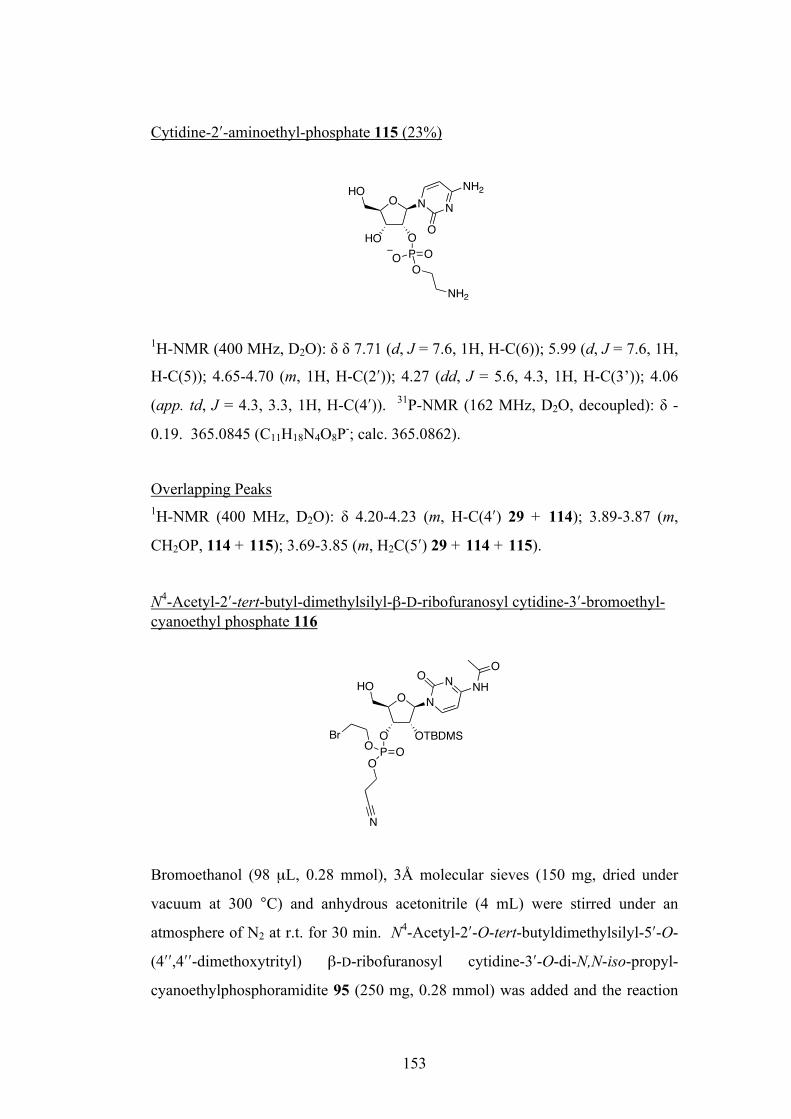
153
Cytidine-2ʹ′-aminoethyl-phosphate 115 (23%)
1H-NMR (400 MHz, D2O): δ δ 7.71 (d, J = 7.6, 1H, H-C(6)); 5.99 (d, J = 7.6, 1H,
H-C(5)); 4.65-4.70 (m, 1H, H-C(2ʹ′)); 4.27 (dd, J = 5.6, 4.3, 1H, H-C(3’)); 4.06
(app. td, J = 4.3, 3.3, 1H, H-C(4ʹ′)). 31P-NMR (162 MHz, D2O, decoupled): δ -
0.19. 365.0845 (C11H18N4O8P-; calc. 365.0862).
Overlapping Peaks 1H-NMR (400 MHz, D2O): δ 4.20-4.23 (m, H-C(4ʹ′) 29 + 114); 3.89-3.87 (m,
CH2OP, 114 + 115); 3.69-3.85 (m, H2C(5ʹ′) 29 + 114 + 115).
N4-Acetyl-2ʹ′-tert-butyl-dimethylsilyl-β-D-ribofuranosyl cytidine-3ʹ′-bromoethyl-cyanoethyl phosphate 116
Bromoethanol (98 µL, 0.28 mmol), 3Å molecular sieves (150 mg, dried under
vacuum at 300 °C) and anhydrous acetonitrile (4 mL) were stirred under an
atmosphere of N2 at r.t. for 30 min. N4-Acetyl-2ʹ′-O-tert-butyldimethylsilyl-5ʹ′-O-
(4ʹ′ʹ′,4ʹ′ʹ′-dimethoxytrityl) β-D-ribofuranosyl cytidine-3ʹ′-O-di-N,N-iso-propyl-
cyanoethylphosphoramidite 95 (250 mg, 0.28 mmol) was added and the reaction
O NHO
N
OHO O
P OO
O
NH2
NH2
O
O OTBDMS
HON
NONH
P OOO
Br
N
O

154
was stirred for 20 mins, whereupon 1H-tetrazole (0.45M in acetonitrile, 4 mL,
1.39 mmol) was added and the reaction mixture stirred for 24 h. tert-Butyl
hydroperoxide (6.0M in decane, 277 µL, 1.39 mmol) was added and the mixture
stirred for 1 h. The mixture was then filtered through Celite®, and solvents were
removed in vacuo. The residue was dissolved in EtOAc (10 mL), washed with
saturated NaHCO3 (10 mL), brine (10 mL), dried (MgSO4) and then concentrated
in vacuo. The residue was dissolved in CH2Cl2 (10 mL) followed by addition of
dichloroacetic acid (365 µL, 4.43 mmol), and the reaction stirred for 15 mins.
After quenching the reaction with saturated NaHCO3, the crude product was
extracted into CH2Cl2 (3 × 10 mL), dried (MgSO4) and concentrated in vacuo.
The crude product was purified via silica gel flash column chromatography
(eluting with c-C6H12:EtOAc:MeOH 10:10:1 to 10:10:3) to give 172 mg (97%) of
116 as a ~1:1 mixture of diastereoisomers as a white solid. Rf: 0.39 (c-
C6H12:EtOAc:MeOH 10:10:3). M.p 82-85°C. 1H-NMR (400 MHz, CDCl3): δ
9.66 (br. s, 1H, NH, a); 9.57 (br. s, 1H, NH, b); 8.34 (d, J = 7.6, 1H, H-C(6), a);
8.30 (d, J = 7.6, 1H, H-C(6), b); 7.38 (d, J = 7.3, 1H, H-C(5), b); 7.34 (d, J = 7.3,
1H, H-C(5), a); 5.62 (d, J = 2.5, 1H, H-C(1ʹ′), b); 5.89 (d, J = 2.3, 1H, H-C(1ʹ′),
b); 4.93-5.01 (m, 2H, H-C(3ʹ′), b + a); 4.74-4.77 (m, 2H, H-C(2ʹ′), b + a); 4.38-
4.48 (m, 8H, CH2CH2CN a, CH2CH2Br b + a, H-C(4ʹ′) b + a); 4.33 (dt, J = 7.7,
6.1, 2H, CH2CH2CN, b); 4.03-4.13 (m, 2H, HH-C(5ʹ′), b + a); 3.92-3.96 (m, 2H,
HH-C(5ʹ′), b + a); 3.54-3.59 (m, 4H, CH2CH2Br, b + a); 2.75-2.85 (m, 4H,
CH2CH2CN, b + a); 2.27 (s, 3H, CH3CO, b); 2.26 (s, 3H, CH3CO, a); 0.92 (s, 9H,
C(CH3)3, a); 0.92 (s, 9H, C(CH3)3, b); 0.20 (s, 3H, SiCH3, a); 0.18 (s, 3H, SiCH3,
b); 0.18 (s, 3H, SiCH3, a); 0.15 (s, 3H, SiCH3, b). 13C-NMR (100 MHz, CDCl3):
δ 171.4 (CH3CO), 171.4 (CH3CO); 162.9 (C(2)), 162.8 (C(2)); 155.3 (C(4)),
155.1 (C(4)); 145.4 (C(6)), 145.2 (C(6)); 116.5 (CH2CH2CN), 116.3
(CH2CH2CN); 96.6 (C(5)), 96.5 (C(5)); 91.9 (C(1ʹ′)), 91.7 (C(1ʹ′)); 82.2 (C(4ʹ′)),
81.9 (C(4ʹ′)); 74.5 (C(3ʹ′)), 74.4 (C(3ʹ′)); 74.1 (C(2ʹ′)), 74.0 (C(2ʹ′)); 67.8 (d, J = 5.5,
CH2CH2Br); 67.6 (d, J = 5.5, CH2CH2Br); 62.8 (d, J = 5.5, CH2CH2CN); 62.5 (d,
J = 5.5, CH2CH2CN); 59.6 (C(5ʹ′)), 59.3 (C(5ʹ′)); 29.7 (d, J = 7.4, CH2CH2Br);
29.2 (d, J = 8.3, CH2CH2Br); 25.5 (C(CH3)3); 24.8, 24.8 (CH3CO); 19.5 (app. t, J
= 7.8, CH2CH2CN); -4.5 (SiCH3); -4.6 (SiCH3); -5.2 (SiCH3). 31P-NMR (162
MHz, CDCl3): δ -1.72 (app. spt., J = 7.8). ESI-MS (pos., CH2Cl2): 661 (100 %,
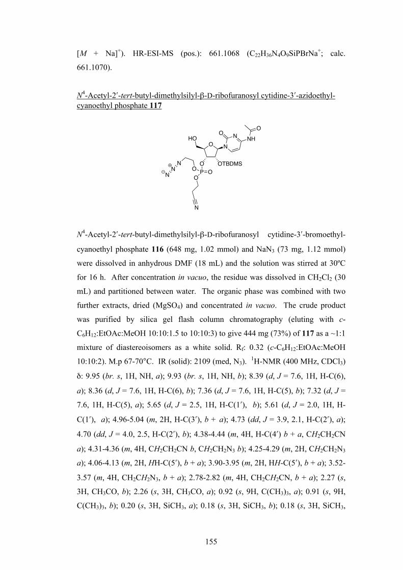
155
[M + Na]+). HR-ESI-MS (pos.): 661.1068 (C22H36N4O9SiPBrNa+; calc.
661.1070).
N4-Acetyl-2ʹ′-tert-butyl-dimethylsilyl-β-D-ribofuranosyl cytidine-3ʹ′-azidoethyl-cyanoethyl phosphate 117
N4-Acetyl-2ʹ′-tert-butyl-dimethylsilyl-β-D-ribofuranosyl cytidine-3ʹ′-bromoethyl-
cyanoethyl phosphate 116 (648 mg, 1.02 mmol) and NaN3 (73 mg, 1.12 mmol)
were dissolved in anhydrous DMF (18 mL) and the solution was stirred at 30ºC
for 16 h. After concentration in vacuo, the residue was dissolved in CH2Cl2 (30
mL) and partitioned between water. The organic phase was combined with two
further extracts, dried (MgSO4) and concentrated in vacuo. The crude product
was purified by silica gel flash column chromatography (eluting with c-
C6H12:EtOAc:MeOH 10:10:1.5 to 10:10:3) to give 444 mg (73%) of 117 as a ~1:1
mixture of diastereoisomers as a white solid. Rf: 0.32 (c-C6H12:EtOAc:MeOH
10:10:2). M.p 67-70°C. IR (solid): 2109 (med, N3). 1H-NMR (400 MHz, CDCl3)
δ: 9.95 (br. s, 1H, NH, a); 9.93 (br. s, 1H, NH, b); 8.39 (d, J = 7.6, 1H, H-C(6),
a); 8.36 (d, J = 7.6, 1H, H-C(6), b); 7.36 (d, J = 7.6, 1H, H-C(5), b); 7.32 (d, J =
7.6, 1H, H-C(5), a); 5.65 (d, J = 2.5, 1H, H-C(1ʹ′), b); 5.61 (d, J = 2.0, 1H, H-
C(1ʹ′), a); 4.96-5.04 (m, 2H, H-C(3ʹ′), b + a); 4.73 (dd, J = 3.9, 2.1, H-C(2ʹ′), a);
4.70 (dd, J = 4.0, 2.5, H-C(2ʹ′), b); 4.38-4.44 (m, 4H, H-C(4ʹ′) b + a, CH2CH2CN
a); 4.31-4.36 (m, 4H, CH2CH2CN b, CH2CH2N3 b); 4.25-4.29 (m, 2H, CH2CH2N3
a); 4.06-4.13 (m, 2H, HH-C(5ʹ′), b + a); 3.90-3.95 (m, 2H, HH-C(5ʹ′), b + a); 3.52-
3.57 (m, 4H, CH2CH2N3, b + a); 2.78-2.82 (m, 4H, CH2CH2CN, b + a); 2.27 (s,
3H, CH3CO, b); 2.26 (s, 3H, CH3CO, a); 0.92 (s, 9H, C(CH3)3, a); 0.91 (s, 9H,
C(CH3)3, b); 0.20 (s, 3H, SiCH3, a); 0.18 (s, 3H, SiCH3, b); 0.18 (s, 3H, SiCH3,
O
O OTBDMS
HON
NONH
P OOO
N
O
NN
N
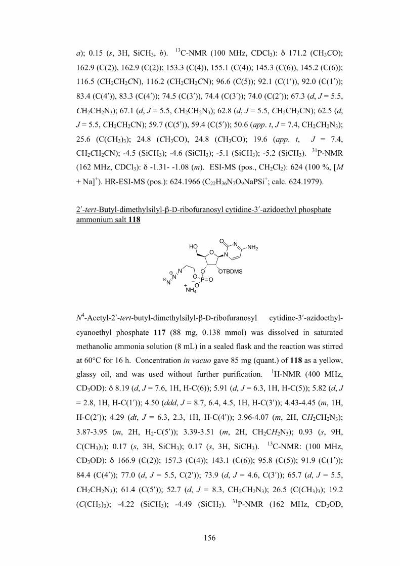
156
a); 0.15 (s, 3H, SiCH3, b). 13C-NMR (100 MHz, CDCl3): δ 171.2 (CH3CO);
162.9 (C(2)), 162.9 (C(2)); 153.3 (C(4)), 155.1 (C(4)); 145.3 (C(6)), 145.2 (C(6));
116.5 (CH2CH2CN), 116.2 (CH2CH2CN); 96.6 (C(5)); 92.1 (C(1ʹ′)), 92.0 (C(1ʹ′));
83.4 (C(4ʹ′)), 83.3 (C(4ʹ′)); 74.5 (C(3ʹ′)), 74.4 (C(3ʹ′)); 74.0 (C(2ʹ′)); 67.3 (d, J = 5.5,
CH2CH2N3); 67.1 (d, J = 5.5, CH2CH2N3); 62.8 (d, J = 5.5, CH2CH2CN); 62.5 (d,
J = 5.5, CH2CH2CN); 59.7 (C(5ʹ′)), 59.4 (C(5ʹ′)); 50.6 (app. t, J = 7.4, CH2CH2N3);
25.6 (C(CH3)3); 24.8 (CH3CO), 24.8 (CH3CO); 19.6 (app. t, J = 7.4,
CH2CH2CN); -4.5 (SiCH3); -4.6 (SiCH3); -5.1 (SiCH3); -5.2 (SiCH3). 31P-NMR
(162 MHz, CDCl3): δ -1.31- -1.08 (m). ESI-MS (pos., CH2Cl2): 624 (100 %, [M
+ Na]+). HR-ESI-MS (pos.): 624.1966 (C22H36N7O9NaPSi+; calc. 624.1979).
2ʹ′-tert-Butyl-dimethylsilyl-β-D-ribofuranosyl cytidine-3ʹ′-azidoethyl phosphate ammonium salt 118
N4-Acetyl-2ʹ′-tert-butyl-dimethylsilyl-β-D-ribofuranosyl cytidine-3ʹ′-azidoethyl-
cyanoethyl phosphate 117 (88 mg, 0.138 mmol) was dissolved in saturated
methanolic ammonia solution (8 mL) in a sealed flask and the reaction was stirred
at 60°C for 16 h. Concentration in vacuo gave 85 mg (quant.) of 118 as a yellow,
glassy oil, and was used without further purification. 1H-NMR (400 MHz,
CD3OD): δ 8.19 (d, J = 7.6, 1H, H-C(6)); 5.91 (d, J = 6.3, 1H, H-C(5)); 5.82 (d, J
= 2.8, 1H, H-C(1ʹ′)); 4.50 (ddd, J = 8.7, 6.4, 4.5, 1H, H-C(3ʹ′)); 4.43-4.45 (m, 1H,
H-C(2ʹ′)); 4.29 (dt, J = 6.3, 2.3, 1H, H-C(4ʹ′)); 3.96-4.07 (m, 2H, CH2CH2N3);
3.87-3.95 (m, 2H, H2-C(5ʹ′)); 3.39-3.51 (m, 2H, CH2CH2N3); 0.93 (s, 9H,
C(CH3)3); 0.17 (s, 3H, SiCH3); 0.17 (s, 3H, SiCH3). 13C-NMR: (100 MHz,
CD3OD): δ 166.9 (C(2)); 157.3 (C(4)); 143.1 (C(6)); 95.8 (C(5)); 91.9 (C(1ʹ′));
84.4 (C(4ʹ′)); 77.0 (d, J = 5.5, C(2ʹ′)); 73.9 (d, J = 4.6, C(3ʹ′)); 65.7 (d, J = 5.5,
CH2CH2N3); 61.4 (C(5ʹ′)); 52.7 (d, J = 8.3, CH2CH2N3); 26.5 (C(CH3)3); 19.2
(C(CH3)3); -4.22 (SiCH3); -4.49 (SiCH3). 31P-NMR (162 MHz, CD3OD,
O
O OTBDMS
HON
NONH2
P OOO
NN
NNH4
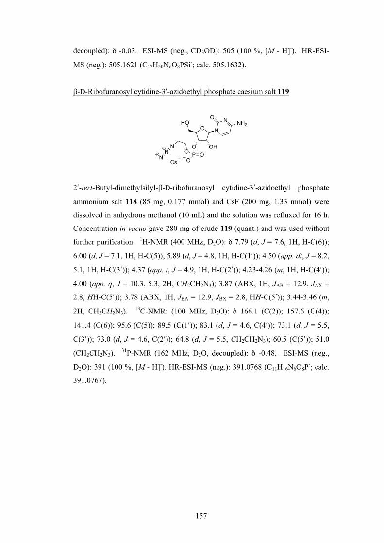
157
decoupled): δ -0.03. ESI-MS (neg., CD3OD): 505 (100 %, [M - H]-). HR-ESI-
MS (neg.): 505.1621 (C17H30N6O8PSi-; calc. 505.1632).
β-D-Ribofuranosyl cytidine-3ʹ′-azidoethyl phosphate caesium salt 119
2ʹ′-tert-Butyl-dimethylsilyl-β-D-ribofuranosyl cytidine-3ʹ′-azidoethyl phosphate
ammonium salt 118 (85 mg, 0.177 mmol) and CsF (200 mg, 1.33 mmol) were
dissolved in anhydrous methanol (10 mL) and the solution was refluxed for 16 h.
Concentration in vacuo gave 280 mg of crude 119 (quant.) and was used without
further purification. 1H-NMR (400 MHz, D2O): δ 7.79 (d, J = 7.6, 1H, H-C(6));
6.00 (d, J = 7.1, 1H, H-C(5)); 5.89 (d, J = 4.8, 1H, H-C(1ʹ′)); 4.50 (app. dt, J = 8.2,
5.1, 1H, H-C(3ʹ′)); 4.37 (app. t, J = 4.9, 1H, H-C(2ʹ′)); 4.23-4.26 (m, 1H, H-C(4ʹ′));
4.00 (app. q, J = 10.3, 5.3, 2H, CH2CH2N3); 3.87 (ABX, 1H, JAB = 12.9, JAX =
2.8, HH-C(5ʹ′)); 3.78 (ABX, 1H, JBA = 12.9, JBX = 2.8, HH-C(5ʹ′)); 3.44-3.46 (m,
2H, CH2CH2N3). 13C-NMR: (100 MHz, D2O): δ 166.1 (C(2)); 157.6 (C(4));
141.4 (C(6)); 95.6 (C(5)); 89.5 (C(1ʹ′)); 83.1 (d, J = 4.6, C(4ʹ′)); 73.1 (d, J = 5.5,
C(3ʹ′)); 73.0 (d, J = 4.6, C(2ʹ′)); 64.8 (d, J = 5.5, CH2CH2N3); 60.5 (C(5ʹ′)); 51.0
(CH2CH2N3). 31P-NMR (162 MHz, D2O, decoupled): δ -0.48. ESI-MS (neg.,
D2O): 391 (100 %, [M - H]-). HR-ESI-MS (neg.): 391.0768 (C11H16N6O8P-; calc.
391.0767).
O
O
HON
NONH2
P OOO
NN
N
OH
Cs
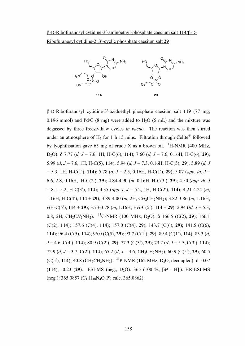
158
β-D-Ribofuranosyl cytidine-3ʹ′-aminoethyl-phosphate caesium salt 114/β-D-
Ribofuranosyl cytidine-2ʹ′,3ʹ′-cyclic phosphate caesium salt 29
β-D-Ribofuranosyl cytidine-3ʹ′-azidoethyl phosphate caesium salt 119 (77 mg,
0.196 mmol) and Pd/C (8 mg) were added to H2O (5 mL) and the mixture was
degassed by three freeze-thaw cycles in vacuo. The reaction was then stirred
under an atmosphere of H2 for 1 h 15 mins. Filtration through Celite® followed
by lyophilisation gave 65 mg of crude X as a brown oil. 1H-NMR (400 MHz,
D2O): δ 7.77 (d, J = 7.6, 1H, H-C(6), 114); 7.60 (d, J = 7.6, 0.16H, H-C(6), 29);
5.99 (d, J = 7.6, 1H, H-C(5), 114); 5.94 (d, J = 7.3, 0.16H, H-C(5), 29); 5.89 (d, J
= 5.3, 1H, H-C(1ʹ′), 114); 5.78 (d, J = 2.5, 0.16H, H-C(1ʹ′), 29); 5.07 (app. td, J =
6.6, 2.8, 0.16H, H-C(2ʹ′), 29); 4.84-4.90 (m, 0.16H, H-C(3ʹ′), 29); 4.50 (app. dt, J
= 8.1, 5.2, H-C(3ʹ′), 114); 4.35 (app. t, J = 5.2, 1H, H-C(2ʹ′), 114); 4.21-4.24 (m,
1.16H, H-C(4ʹ′), 114 + 29); 3.89-4.00 (m, 2H, CH2CH2NH2); 3.82-3.86 (m, 1.16H,
HH-C(5ʹ′), 114 + 29); 3.73-3.78 (m, 1.16H, HH-C(5ʹ′), 114 + 29); 2.94 (td, J = 5.3,
0.8, 2H, CH2CH2NH2). 13C-NMR (100 MHz, D2O): δ 166.5 (C(2), 29); 166.1
(C(2), 114); 157.6 (C(4), 114); 157.0 (C(4), 29); 143.7 (C(6), 29); 141.5 (C(6),
114); 96.4 (C(5), 114); 96.0 (C(5), 29); 93.7 (C(1ʹ′), 29); 89.4 (C(1ʹ′), 114); 83.3 (d,
J = 4.6, C(4ʹ′), 114); 80.9 (C(2ʹ′), 29); 77.3 (C(3ʹ′), 29); 73.2 (d, J = 5.5, C(3ʹ′), 114);
72.9 (d, J = 3.7, C(2ʹ′), 114); 65.2 (d, J = 4.6, CH2CH2NH2); 60.9 (C(5ʹ′), 29); 60.5
(C(5ʹ′), 114); 40.8 (CH2CH2NH2). 31P-NMR (162 MHz, D2O, decoupled): δ -0.07
(114); -0.23 (29). ESI-MS (neg., D2O): 365 (100 %, [M - H]-). HR-ESI-MS
(neg.): 365.0857 (C11H18N4O8P-; calc. 365.0862).
O
O
HON
NONH2
P OOO
H2N OH
OHO
N
NONH2
OPO
OOCs Cs
114 29
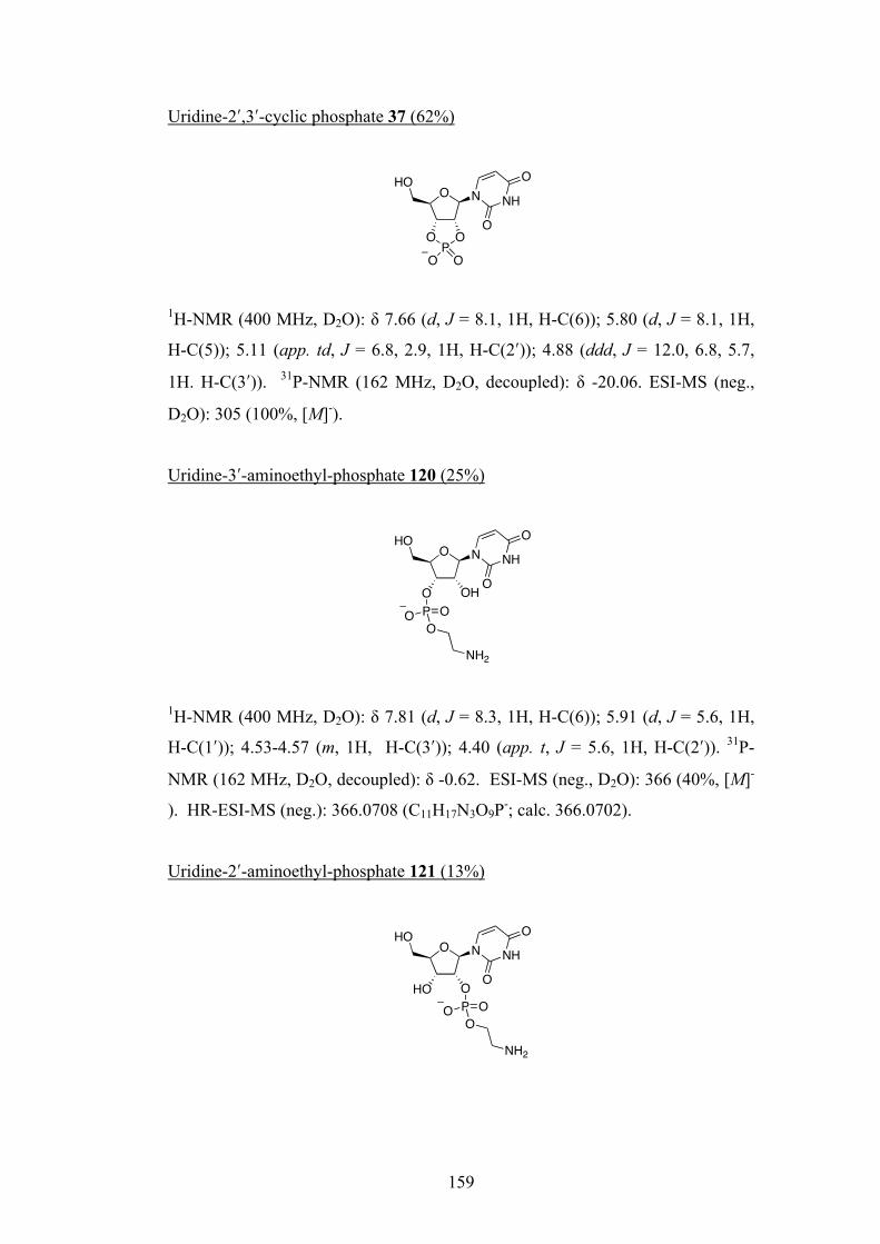
159
Uridine-2ʹ′,3ʹ′-cyclic phosphate 37 (62%)
1H-NMR (400 MHz, D2O): δ 7.66 (d, J = 8.1, 1H, H-C(6)); 5.80 (d, J = 8.1, 1H,
H-C(5)); 5.11 (app. td, J = 6.8, 2.9, 1H, H-C(2ʹ′)); 4.88 (ddd, J = 12.0, 6.8, 5.7,
1H. H-C(3ʹ′)). 31P-NMR (162 MHz, D2O, decoupled): δ -20.06. ESI-MS (neg.,
D2O): 305 (100%, [M]-).
Uridine-3ʹ′-aminoethyl-phosphate 120 (25%)
1H-NMR (400 MHz, D2O): δ 7.81 (d, J = 8.3, 1H, H-C(6)); 5.91 (d, J = 5.6, 1H,
H-C(1ʹ′)); 4.53-4.57 (m, 1H, H-C(3ʹ′)); 4.40 (app. t, J = 5.6, 1H, H-C(2ʹ′)). 31P-
NMR (162 MHz, D2O, decoupled): δ -0.62. ESI-MS (neg., D2O): 366 (40%, [M]-
). HR-ESI-MS (neg.): 366.0708 (C11H17N3O9P-; calc. 366.0702).
Uridine-2ʹ′-aminoethyl-phosphate 121 (13%)
O N
OPO
HONH
O
O
O O
O NHO
NH
O
O
O OHP OOO
NH2
O NHO
NH
O
O
HO OP OO
O
NH2
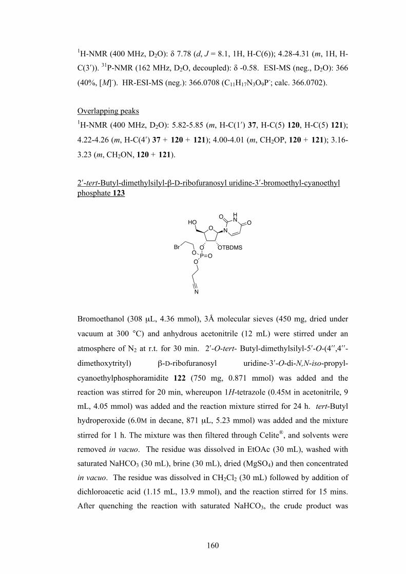
160
1H-NMR (400 MHz, D2O): δ 7.78 (d, J = 8.1, 1H, H-C(6)); 4.28-4.31 (m, 1H, H-
C(3ʹ′)). 31P-NMR (162 MHz, D2O, decoupled): δ -0.58. ESI-MS (neg., D2O): 366
(40%, [M]-). HR-ESI-MS (neg.): 366.0708 (C11H17N3O9P-; calc. 366.0702).
Overlapping peaks 1H-NMR (400 MHz, D2O): 5.82-5.85 (m, H-C(1ʹ′) 37, H-C(5) 120, H-C(5) 121);
4.22-4.26 (m, H-C(4ʹ′) 37 + 120 + 121); 4.00-4.01 (m, CH2OP, 120 + 121); 3.16-
3.23 (m, CH2ON, 120 + 121).
2ʹ′-tert-Butyl-dimethylsilyl-β-D-ribofuranosyl uridine-3ʹ′-bromoethyl-cyanoethyl phosphate 123
Bromoethanol (308 µL, 4.36 mmol), 3Å molecular sieves (450 mg, dried under
vacuum at 300 °C) and anhydrous acetonitrile (12 mL) were stirred under an
atmosphere of N2 at r.t. for 30 min. 2ʹ′-O-tert- Butyl-dimethylsilyl-5ʹ′-O-(4ʹ′ʹ′,4ʹ′ʹ′-
dimethoxytrityl) β-D-ribofuranosyl uridine-3ʹ′-O-di-N,N-iso-propyl-
cyanoethylphosphoramidite 122 (750 mg, 0.871 mmol) was added and the
reaction was stirred for 20 min, whereupon 1H-tetrazole (0.45M in acetonitrile, 9
mL, 4.05 mmol) was added and the reaction mixture stirred for 24 h. tert-Butyl
hydroperoxide (6.0M in decane, 871 µL, 5.23 mmol) was added and the mixture
stirred for 1 h. The mixture was then filtered through Celite®, and solvents were
removed in vacuo. The residue was dissolved in EtOAc (30 mL), washed with
saturated NaHCO3 (30 mL), brine (30 mL), dried (MgSO4) and then concentrated
in vacuo. The residue was dissolved in CH2Cl2 (30 mL) followed by addition of
dichloroacetic acid (1.15 mL, 13.9 mmol), and the reaction stirred for 15 mins.
After quenching the reaction with saturated NaHCO3, the crude product was
O
O OTBDMS
HON
HNO
P OOO
Br
N
O

161
extracted into CH2Cl2 (3 × 30 mL), dried (MgSO4) and concentrated in vacuo.
The crude product was purified via silica gel flash column chromatography
(eluting with c-C6H12:EtOAc:MeOH 10:10:1 to 10:10:2) to give 407 mg (97%) of
123 as a ~1:1 mixture of diastereoisomers as a white solid. Rf : 0.56 (c-
C6H12:EtOAc:MeOH 10:10:3). 1H-NMR (400 MHz, CD3OD): δ 8.05 (d, J = 8.1,
1H, H-C(6), a); 8.05 (d, J = 8.1, 1H, H-C(6), b); 5.99 (d, J = 2.3, 1H, H-C(1ʹ′), a);
5.98 (d, J = 2.0, 1H, H-C(1ʹ′), b); 5.75 (d, J = 8.1, 2H, H-C(5), b + a); 4.83-4.86
(m, 2H, H-C(3ʹ′), b + a); 4.56 (ddd, J = 6.3, 4.5, 1.8, 2H, H-C(2ʹ′), b + a); 4.42-4.49
(m, 4H, CH2CH2Br, b + a); 4.32-4.39 (m, 6H, CH2CH2CN, b + a, H-C(4ʹ′), b + a);
3.84 (m, 4H, H2-C(5ʹ′), b + a); 3.66-3.69 (m, 4H, CH2CH2Br, b + a); 2.92-2.95 (m,
4H, CH2CH2CN, b + a); 0.90 (s, 18H, C(CH3)3, b + a); 0.13 (s, 6H, SiCH3, b + a);
0.09 (s, 6H, SiCH3, b + a). 13C-NMR (100 MHz, CD3OD): δ 165.9 (C(2)); 152.5
(C(4)); 142.2 (C(6)); 118.5 (CH2CH2CN); 103.4 (C(5)); 89.2 (C(1ʹ′)), 89.1 (C(1ʹ′));
85.1 (C(4ʹ′)); 79.1 (C(3ʹ′)); 75.8 (C(3ʹ′)); 69.5 (d, J = 5.5, CH2CH2Br); 69.4 (d, J =
5.5, CH2CH2Br); 64.7 (d, J = 5.5, CH2CH2CN); 64.6 (d, J = 5.5, CH2CH2CN);
62.0 (C(5ʹ′)), 62.0 (C(5ʹ′)); 31.1 (d, J = 8.3, CH2CH2Br); 30.9 (d, J = 7.4,
CH2CH2Br); 26.3 (C(CH3)3); 20.2 (d, J = 7.4, CH2CH2CN); 20.2 (d, J = 7.4,
CH2CH2CN); 20.2 (d, J = 7.4, CH2CH2CN); 19.1 (C(CH3)3); -4.4 (SiCH3); -4.7
(SiCH3). 31P-NMR (162 MHz, CD3OD, decoupled): δ -2.92, -3.00. ESI-MS
(pos., CD3OD): 620.2 (100 %, [M + Na]+), 622.2 (95 %, [M + Na]+). HR-ESI-MS
(pos.): 620.0786 (C20H33N3O9NaP79BrSi+; calc. 620.0805), 622.0777
(C20H33N3O9NaP81BrSi+; calc. 622.0784).
2ʹ′-tert-Butyl-dimethylsilyl-β-D-ribofuranosyl uridine-3ʹ′-azidoethyl-cyanoethyl phosphate 124
O
O OTBDMS
HON
HNO
P OOO
N
NN
N
O

162
2ʹ′-tert-Butyl-dimethylsilyl-β-D-ribofuranosyl uridine-3ʹ′-bromoethyl-cyanoethyl
phosphate 123 (200 mg, 0.335 mmol) and NaN3 (24 mg, 0.369 mmol) were
dissolved in anhydrous DMF (6 mL) and the solution was stirred at 30ºC for 16 h.
After concentration in vacuo, the residue was dissolved in CH2Cl2 (5 mL) and
partitioned between water. The organic phase was combined with four further
extracts, dried (MgSO4) and concentrated in vacuo to give 169 mg (90%) of 124
as a ~1:1 mixture of diastereoisomers as an off-white solid. No further
purification was necessary. M.p 150-153°C. IR (solid): 2108 (med, N3). 1H-
NMR (400 MHz, CD3OD): δ 8.05 (d, J = 8.1, 2H, H-C(6), b + a); 5.99 (d, J = 6.1,
1H, H-C(1ʹ′), a); 5.99 (d, J = 6.1, 1H, H-C(1ʹ′), b); 5.75 (d, J = 8.1, 2H, H-C(5ʹ′), b
+ a); 4.83-4.86 (m, 2H, H-C(3ʹ′), b + a); 4.54-4.57 (m, 2H, H-C(2ʹ′), b + a); 4.27-
4.38 (m, 10H, H-C(4ʹ′), CH2CH2CN, CH2CH2N3, b + a); 3.79-3.88 (m, 4H, H2-
C(5ʹ′), b + a); 3.58-3.62 (m, 4H, CH2CH2N3, b + a); 2.92-2.95 (m, 4H,
CH2CH2CN, b + a); 0.90 (s, 18H, C(CH3)3, b + a); 0.14 (s, SiCH3, b); 0.13 (s,
SiCH3, a); 0.09 (s, SiCH3, b); 0.09 (s, SiCH3, a). 13C-NMR (100 MHz, CD3OD):
δ 166.0 (C(2)); 152.5 (C(4); 142.2 (C(6)); 118.5 (CH2CH2CN); 103.4 (C(5)); 89.2
(C(1ʹ′)), 89.1 (C(1ʹ′)); 85.2 (C(4ʹ′)), 85.2 (C(4ʹ′)); 79.1 (d, J = 5.5, C(3ʹ′)); 79.1 (d, J
= 5.5, C(3ʹ′)); 75.9 (C(2ʹ′)), 75.8 (C(2ʹ′)); 68.8 (d, J = 5.5, CH2CH2N3); 68.7 (d, J =
5.5, CH2CH2N3); 64.7 (d, J = 5.5, CH2CH2CN); 64.5 (d, J = 5.5, CH2CH2CN);
62.0 (C(5ʹ′)); 62.0 (C(5ʹ′)); 52.1 (d, J = 7.4, CH2CH2N3); 26.3 (C(CH3)3); 20.2 (d, J
= 7.4, CH2CH2CN); 19.1 (C(CH3)3); -4.4 (SiCH3): -4.5 (SiCH3); -4.7 (SiCH3); -
4.8 (SiCH3). 31P-NMR (162 MHz, CD3OD, decoupled): δ -2.72, -2.75. ESI-MS
(pos., CH3OH): 583 (94 %, [M + Na]+). HR-ESI-MS (pos.): 583.1700
(C20H33N6O9NaPSi+; calc. 583.1714).
2ʹ′-tert-Butyl-dimethylsilyl-β-D-ribofuranosyl uridine-3ʹ′-azidoethyl phosphate ammonium salt 125
O
O OTBDMS
HON
HNO
P OOO
NN
N
O
NH4
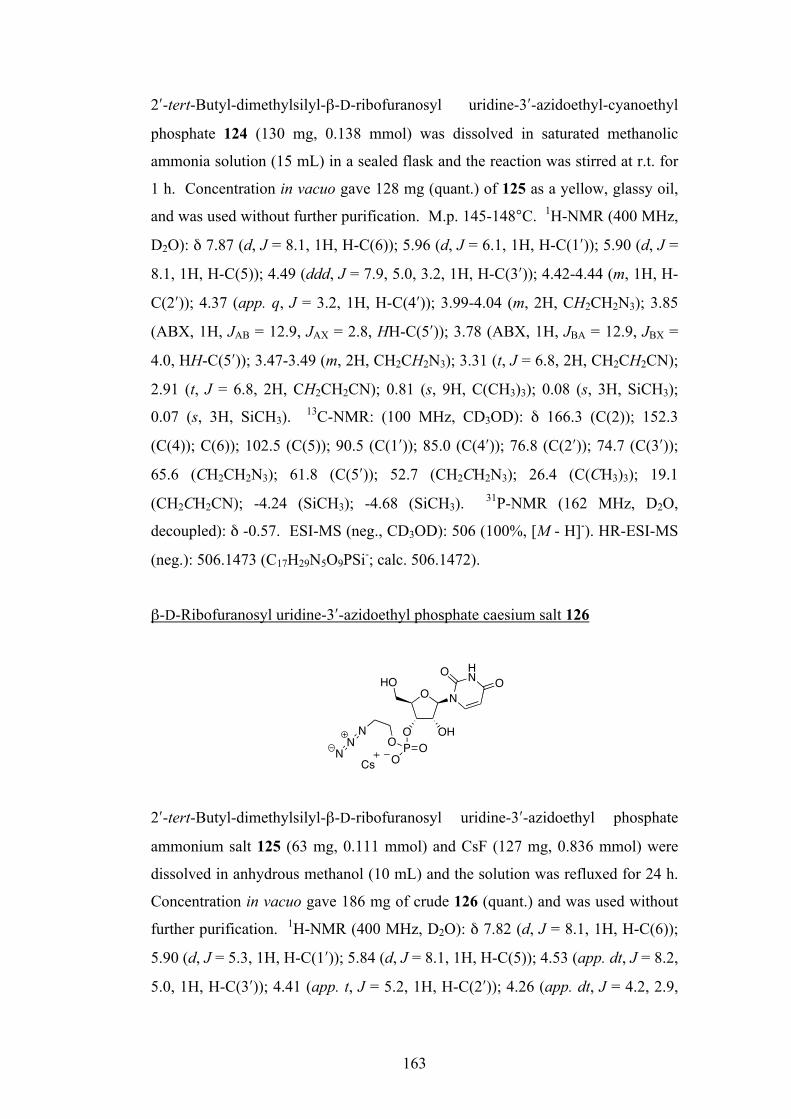
163
2ʹ′-tert-Butyl-dimethylsilyl-β-D-ribofuranosyl uridine-3ʹ′-azidoethyl-cyanoethyl
phosphate 124 (130 mg, 0.138 mmol) was dissolved in saturated methanolic
ammonia solution (15 mL) in a sealed flask and the reaction was stirred at r.t. for
1 h. Concentration in vacuo gave 128 mg (quant.) of 125 as a yellow, glassy oil,
and was used without further purification. M.p. 145-148°C. 1H-NMR (400 MHz,
D2O): δ 7.87 (d, J = 8.1, 1H, H-C(6)); 5.96 (d, J = 6.1, 1H, H-C(1ʹ′)); 5.90 (d, J =
8.1, 1H, H-C(5)); 4.49 (ddd, J = 7.9, 5.0, 3.2, 1H, H-C(3ʹ′)); 4.42-4.44 (m, 1H, H-
C(2ʹ′)); 4.37 (app. q, J = 3.2, 1H, H-C(4ʹ′)); 3.99-4.04 (m, 2H, CH2CH2N3); 3.85
(ABX, 1H, JAB = 12.9, JAX = 2.8, HH-C(5ʹ′)); 3.78 (ABX, 1H, JBA = 12.9, JBX =
4.0, HH-C(5ʹ′)); 3.47-3.49 (m, 2H, CH2CH2N3); 3.31 (t, J = 6.8, 2H, CH2CH2CN);
2.91 (t, J = 6.8, 2H, CH2CH2CN); 0.81 (s, 9H, C(CH3)3); 0.08 (s, 3H, SiCH3);
0.07 (s, 3H, SiCH3). 13C-NMR: (100 MHz, CD3OD): δ 166.3 (C(2)); 152.3
(C(4)); C(6)); 102.5 (C(5)); 90.5 (C(1ʹ′)); 85.0 (C(4ʹ′)); 76.8 (C(2ʹ′)); 74.7 (C(3ʹ′));
65.6 (CH2CH2N3); 61.8 (C(5ʹ′)); 52.7 (CH2CH2N3); 26.4 (C(CH3)3); 19.1
(CH2CH2CN); -4.24 (SiCH3); -4.68 (SiCH3). 31P-NMR (162 MHz, D2O,
decoupled): δ -0.57. ESI-MS (neg., CD3OD): 506 (100%, [M - H]-). HR-ESI-MS
(neg.): 506.1473 (C17H29N5O9PSi-; calc. 506.1472).
β-D-Ribofuranosyl uridine-3ʹ′-azidoethyl phosphate caesium salt 126
2ʹ′-tert-Butyl-dimethylsilyl-β-D-ribofuranosyl uridine-3ʹ′-azidoethyl phosphate
ammonium salt 125 (63 mg, 0.111 mmol) and CsF (127 mg, 0.836 mmol) were
dissolved in anhydrous methanol (10 mL) and the solution was refluxed for 24 h.
Concentration in vacuo gave 186 mg of crude 126 (quant.) and was used without
further purification. 1H-NMR (400 MHz, D2O): δ 7.82 (d, J = 8.1, 1H, H-C(6));
5.90 (d, J = 5.3, 1H, H-C(1ʹ′)); 5.84 (d, J = 8.1, 1H, H-C(5)); 4.53 (app. dt, J = 8.2,
5.0, 1H, H-C(3ʹ′)); 4.41 (app. t, J = 5.2, 1H, H-C(2ʹ′)); 4.26 (app. dt, J = 4.2, 2.9,
O
O
HON
HNO
P OOO
NN
N
OH
O
Cs

164
H-C(4ʹ′); 3.99-4.03 (m, 2H, CH2CH2N3); 3.86 (ABX, 1H, JAB = 12.6, JAX = 2.8,
HH-C(5ʹ′)); 3.77 (ABX, 1H, JBA = 12.9, JBX = 4.0, HH-C(5ʹ′)); 3.44-3.47 (m, 2H,
CH2CH2N3). 13C-NMR: (100 MHz, D2O): δ 165.7 (C(2)); 151.7 (C(4)); 141.5
(C(6)); 102.4 (C(5)); 88.6 (C(1ʹ′)); 83.6 (d, J = 3.7, C(4ʹ′)); 73.2 (d, J = 4.6, C(3ʹ′));
72.7 (d, J = 4.6, C(2ʹ′)); 64.8 (d, J = 5.5, CH2CH2N3); 60.5 (C(5ʹ′)); 50.9 (d, J =
7.4, CH2CH2N3). 31P-NMR: (162 MHz, D2O, decoupled): δ -0.44. ESI-MS (neg.,
D2O): 392 (100 %, [M - H]-). HR-ESI-MS (neg.): 392.0601 (C11H15N5O9P-; calc.
392.0607).
β-D-Ribofuranosyl uridine-3ʹ′-aminoethyl phosphate caesium salt 120
β-D-Ribofuranosyl uridine-3ʹ′-azidoethyl phosphate caesium salt 126 (96 mg,
0.262 mmol) and Pd/C (10 mg) were added to H2O (5 mL) and the mixture was
degassed by three freeze-thaw cycles in vacuo. The reaction was then stirred
under an atmosphere of H2 for 1 h 15 min. Filtration through Celite® followed by
lyophilisation gave 82 mg (quant.) of crude 120 as a brown oil. 1H-NMR (400
MHz, D2O): δ 7.78 (d, J = 8.1, 1H, H-C(6)); 5.90 (d, J = 5.8, 1H, H-C(1ʹ′)); 5.82
(d, J = 8.1, 1H, H-C(5)); 4.54 (ddd, J = 8.1, 5.2, 4.4, 1H, H-C(3ʹ′)); 4.39 (app. t, J
= 5.2, 1H, H-C(2ʹ′)); 4.22-4.25 (m, 1H, H-C(4ʹ′)); 4.05-4.10 (m, 2H, CH2CH2NH2);
3.83 (ABX, 1H, JAB = 12.9, JAX = 2.8, HH-C(5ʹ′)); 3.75 (ABX, 1H, JBA = 12.9, JBX
= 4.0, HH-C(5ʹ′)); 3.17-3.19 (m, 2H, CH2CH2NH2). 13C-NMR (100 MHz, D2O): δ
168.7 (C(2)); 153.2 (C(4)); 141.2 (C(6)); 102.6 (C(5)); 88.5 (C(1ʹ′)); 83.6 (C(4ʹ′));
73.5 (C(3ʹ′)); 72.5 (C(2ʹ′)); 62.8 (d, J = 6.5, CH2CH2NH2); 60.5 (C(5ʹ′)); 39.9
(CH2CH2NH2). 31P-NMR (162 MHz, D2O, decoupled): δ -0.56. ESI-MS (neg.,
D2O): 366 (20 %, [M - H]-). HR-ESI-MS (neg.): 366.0703 (C11H17N3O9P-; calc.
366.0702).
O
O
HON
HNO
P OOO
H2N OH
O
Cs
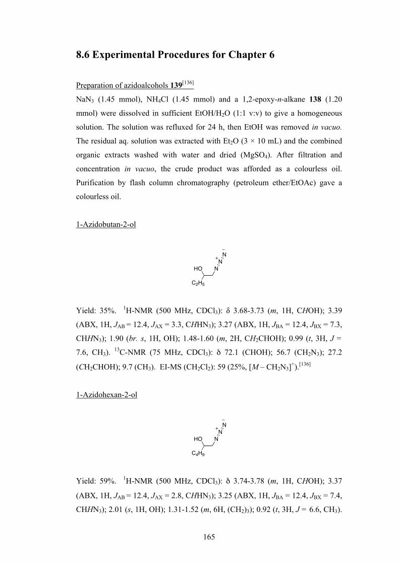
165
8.6 Experimental Procedures for Chapter 6
Preparation of azidoalcohols 139[136]
NaN3 (1.45 mmol), NH4Cl (1.45 mmol) and a 1,2-epoxy-n-alkane 138 (1.20
mmol) were dissolved in sufficient EtOH/H2O (1:1 v:v) to give a homogeneous
solution. The solution was refluxed for 24 h, then EtOH was removed in vacuo.
The residual aq. solution was extracted with Et2O (3 × 10 mL) and the combined
organic extracts washed with water and dried (MgSO4). After filtration and
concentration in vacuo, the crude product was afforded as a colourless oil.
Purification by flash column chromatography (petroleum ether/EtOAc) gave a
colourless oil.
1-Azidobutan-2-ol
Yield: 35%. 1H-NMR (500 MHz, CDCl3): δ 3.68-3.73 (m, 1H, CHOH); 3.39
(ABX, 1H, JAB = 12.4, JAX = 3.3, CHHN3); 3.27 (ABX, 1H, JBA = 12.4, JBX = 7.3,
CHHN3); 1.90 (br. s, 1H, OH); 1.48-1.60 (m, 2H, CH2CHOH); 0.99 (t, 3H, J =
7.6, CH3). 13C-NMR (75 MHz, CDCl3): δ 72.1 (CHOH); 56.7 (CH2N3); 27.2
(CH2CHOH); 9.7 (CH3). EI-MS (CH2Cl2): 59 (25%, [M – CH2N3]+).[136]
1-Azidohexan-2-ol
Yield: 59%. 1H-NMR (500 MHz, CDCl3): δ 3.74-3.78 (m, 1H, CHOH); 3.37
(ABX, 1H, JAB = 12.4, JAX = 2.8, CHHN3); 3.25 (ABX, 1H, JBA = 12.4, JBX = 7.4,
CHHN3); 2.01 (s, 1H, OH); 1.31-1.52 (m, 6H, (CH2)3); 0.92 (t, 3H, J = 6.6, CH3).
C2H5
HO NNN
C4H9
HO NNN
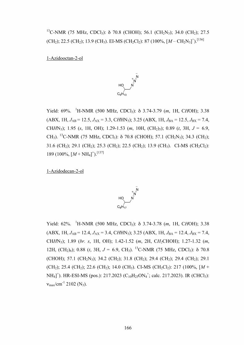
166
13C-NMR (75 MHz, CDCl3): δ 70.8 (CHOH); 56.1 (CH2N3); 34.0 (CH2); 27.5
(CH2); 22.5 (CH2); 13.9 (CH3). EI-MS (CH2Cl2): 87 (100%, [M – CH2N3]+).[136]
1-Azidooctan-2-ol
Yield: 69%. 1H-NMR (500 MHz, CDCl3): δ 3.74-3.79 (m, 1H, CHOH); 3.38
(ABX, 1H, JAB = 12.5, JAX = 3.3, CHHN3); 3.25 (ABX, 1H, JBA = 12.5, JBX = 7.4,
CHHN3); 1.95 (s, 1H, OH); 1.29-1.53 (m, 10H, (CH2)5); 0.89 (t, 3H, J = 6.9,
CH3). 13C-NMR (75 MHz, CDCl3): δ 70.8 (CHOH); 57.1 (CH2N3); 34.3 (CH2);
31.6 (CH2); 29.1 (CH2); 25.3 (CH2); 22.5 (CH2); 13.9 (CH3). CI-MS (CH2Cl2):
189 (100%, [M + NH4]+).[137]
1-Azidodecan-2-ol
Yield: 62%. 1H-NMR (500 MHz, CDCl3): δ 3.74-3.78 (m, 1H, CHOH); 3.38
(ABX, 1H, JAB = 12.4, JAX = 3.4, CHHN3); 3.25 (ABX, 1H, JBA = 12.4, JBX = 7.4,
CHHN3); 1.89 (br. s, 1H, OH); 1.42-1.52 (m, 2H, CH2CHOH); 1.27-1.32 (m,
12H, (CH2)6); 0.88 (t, 3H, J = 6.9, CH3). 13C-NMR (75 MHz, CDCl3): δ 70.8
(CHOH); 57.1 (CH2N3); 34.2 (CH2); 31.8 (CH2); 29.4 (CH2); 29.4 (CH2); 29.1
(CH2); 25.4 (CH2); 22.6 (CH2); 14.0 (CH3). CI-MS (CH2Cl2): 217 (100%, [M +
NH4]+). HR-ESI-MS (pos.): 217.2023 (C10H25ON4+; calc. 217.2023). IR (CHCl3):
νmax/cm-1 2102 (N3).
C6H13
HO NNN
C8H17
HO NNN

167
Preparation of aminoalcohols 134[136]
The azidoalcohol (1.34 mmol) and Pd/C catalyst (0.1 eq. w:w) were placed in a
flask with EtOH (3 mL). The mixture was degassed by 3 freeze-pump-thaw cycles
from an atmosphere of H2. The mixture was then stirred under an atmosphere of
H2 at r.t. for 20 h. After filtration through Celite®, EtOH was removed in vacuo to
give the product as a white solid. No further purification was necessary.
1-Aminobutan-2-ol
Yield: 94%. 1H-NMR (500 MHz, CDCl3): δ 3.40-3.45 (m, 1H, CHOH); 2.78
(ABX, 1H, JAB = 12.7, JAX = 3.0, CHHNH2); 2.52 (ABX, 1H, JBA = 12.7, JBX =
4.4, CHHNH2); 2.54 (s, 1H, OH); 1.39-1.45 (m, 2H, CH2CHOH); 0.92 (t, 3H, J =
7.4, CH3). 13C-NMR (75 MHz, CDCl3): δ 73.2 (CHOH); 46.9 (CH2NH2); 27.6
(CH2CHOH); 9.9 (CH3). APCI-MS (CH3OH): 90 (4%, [M + H]+).[138]
1-Aminohexan-2-ol
Yield: 97%. 1H-NMR (500 MHz, CDCl3): δ 3.47-3.52 (m, 1H, CHOH); 2.80
(ABX, 1H, JAB = 12.7, JAX = 3.2, CHHNH2); 2.51 (ABX, 1H, JBA = 12.7, JBX =
8.4, CHHNH2); 2.19 (s, 1H, OH); 1.28-1.43 (m, 6H, (CH2)3); 0.89 (t, 3H, J = 7.1,
CH3). 13C-NMR (75 MHz, CDCl3): δ 71.9 (CHOH); 47.4 (CH2NH2); 34.4 (CH2);
27.8 (CH2); 22.7 (CH2); 13.9 (CH3). APCI-MS (CH3OH): 118 (12%, [M +
H]+).[139]
1-Aminooctan-2-ol
C2H5
HO NH2
C4H9
HO NH2
C6H13
HO NH2

168
Yield: 94%. 1H-NMR (500 MHz, CDCl3): δ 3.48-3.53 (m, 1H, CHOH); 2.82
(ABX, 1H, JAB = 12.7, JAX = 3.5, CHHNH2); 2.52 (ABX, 1H, JBA = 12.7, JBX =
8.4, CHHNH2); 2.24 (s, 1H, OH); 1.25-1.43 (m, 10H, (CH2)5); 0.87 (t, 3H, J =
6.9, CH3). 13C-NMR (75 MHz, CDCl3): δ 71.9 (CHOH); 47.3 (CH2NH2); 34.8
(CH2); 31.7 (CH2); 29.3 (CH2); 25.6 (CH2); 22.5 (CH2); 14.0 (CH3). APCI-MS
(CH3OH): 146 (31%, [M + H]+).[140]
1-Aminodecan-2-ol
Yield: 98%. 1H-NMR (300 MHz, CD3OD): δ 3.43-3.51 (m, 1H, CHOH); 2.65
(ABX, 1H, JAB = 13.0, JAX = 3.7, CHHNH2); 2.50 (ABX, 1H, JBA = 13.0, JBX =
8.0, CHHNH2); 1.28-1.41 (m, 14H, (CH2)7); 0.87 (t, 3H, J = 6.7, CH3). 13C-NMR
(75 MHz, CD3OD): δ 73.6 (CHOH); 36.0 (CH2); 33.1 (CH2); 30.8 (CH2); 30.7
(CH2); 30.4 (CH2); 26.8 (CH2); 23.7 (CH2); 14.4 (CH3). CI-MS (CH3OH): 174
(100%, [M + H]+). HR-ESI-MS (pos.): 174.1855 (C10H24ON+; calc. 174.1852).
Reaction of aminoalcohols 134 with sodium trimetaphosphate 131
A solution (or suspension) of the aminoalcohol 134 (100 mM) and sodium
trimetaphosphate 131 (250 mM) in D2O was adjusted to pD = 10.0 using DCl
solution and maintained at 26°C. The pD was monitored daily and readjusted to
pD = 10.0 when necessary using NaOD solution 1H-NMR spectra were obtained
daily, after filtration where necessary. 31P-NMR spectra were recorded at the end
of the experiment.
C8H17
HO NH2

169
N-Triphosphates 135 and O-monophosphates 137
N-(2-Hydroxyhexyl)- P-amidotriphosphate 135 (R = C4H9) 31P-NMR (162 MHz, D2O): δ 0.0 (ddd, JP,P = 19.8, JH,P = 8.5, 7.9, Pα); –5.0 (d, J
= 19.2, Pγ); –20.4 (app. t, J = 19.2, 21.3, Pβ).
N-(2-Hydroxyoctyl)-P-amidotriphosphate 135 (R = C6H13) 31P-NMR (162 MHz, D2O): δ 0.0 (ddd, JP,P = 21.3, JH,P = 8.5, Pα); –5.0 (d, J =
21.3, Pγ); –20.5 (t, J = 21.3, Pβ).
1-Aminooctan-2-yl-phosphate 137 (R = C6H13) 31P-NMR (162 MHz, D2O): δ 4.0 (d, J = 8.5).
1-Aminodecan-2-yl-phosphate 137 (R = C8H17) 31P-NMR (162 MHz, D2O): δ 4.0 (d, J = 8.5).
Di-tert-butyl 1-azidodecan-2-yl phosphate 140
1-Azidodecan-2-ol 139 (R = C8H17) (60 mg, 0.3 mmol), 3Å molecular sieves
(dried under vacuum at 300°C overnight, 250 mg) and (t-BuO)2PN(i-Pr)2 (0.19
R
OHHNP OP OPOOO
O
OO
O
O
R
H2NP OO
O
135 137
N
C8H17
ONN
P OO
O

170
mL, 0.6 mmol) were placed in a dry flask under an atmosphere of N2, and dry
CH2Cl2 (5 mL) was added. The mixture was stirred for 25 min, and then 1H-
tetrazole (0.45M in MeCN, 2 ml, 0.9 mmol) was added dropwise. After stirring
o.n., all of the starting material had been consumed (according to analysis by
t.l.c.). tert-Butyl hydroperoxide (70% aq., 0.17 mL, 1.2 mmol) was then added,
and the mixture stirred for a further 1 h. The mixture was then filtered through
Celite®, and solvents were removed in vacuo. The residue was dissolved in
EtOAc (10 mL) and added to H2O. The organic phase was collected and
combined with 2 further EtOAc extracts; these were then washed with brine and
dried (MgSO4). Concentration in vacuo gave the crude product which was
purified by flash column chromatography (activated Brockmann grade III
alumina; c-C6H12/EtOAc 92:8) to give 140 as a colourless oil (70 mg, 60%). 1H-
NMR (300 MHz, CDCl3): δ 4.32-4.41 (m, CH(O-)); 3.55 (ABX, 1H, JAB = 12.9,
JAX = 4.1, CHHN3); 3.34 (ABX, 1H, JBA = 12.9, JBX = 4.5, CHHN3); 1.59-1.75
(m, 2H, CH2CH(O-)); 1.49 (s, 9H, C(CH3)3); 1.48 (s, 9H, C(CH3)3); 1.18-1.37 (m,
12H, (CH2)6); 0.87 (t, 3H, J = 6.7, CH3). 13C-NMR (75 MHz, CDCl3): δ 82.5 (d,
J = 7.5, C(CH3)3); 82.3 (d, J = 7.5, C(CH3)3); 76.1 (d, J = 6.3, CH2CH(O-)); 54.0
(d, J = 4.3, CH2N3); 32.6 (d, J = 5.2, CH2CH(O-)); 31.8 (CH2); 29.8 (d, J = 4.3,
C(CH3)3); 29.4 (CH2); 29.4 (CH2); 29.1 (CH2); 24.9 (CH2); 22.6 (CH2); 14.0
(CH3). 31P-NMR (162 MHz, CDCl3): δ –9.26 (d, J = 6.3). APCI-MS (pos.,
CH2Cl2): 392 (44%, [M + H]+). HR-ESI-MS (pos.): 392.2670 (C18H39O4N3P+;
calc. 392.2673). IR (CHCl3): νmax/cm-1 2104 (N3).
Di-tert-butyl 1-aminodecan-2-yl phosphate 141
Di-tert-butyl 1-azidodecan-2-yl phosphate 140 (50 mg, 0.128 mmol) was
dissolved in THF/H2O (1.5 mL, 95:5 v:v). Polymer-bound PPh3 (107 mg, 0.32
mmol) was added to the solution and the reaction mixture was stirred at 50°C for
NH2
C8H17
OP OO
O
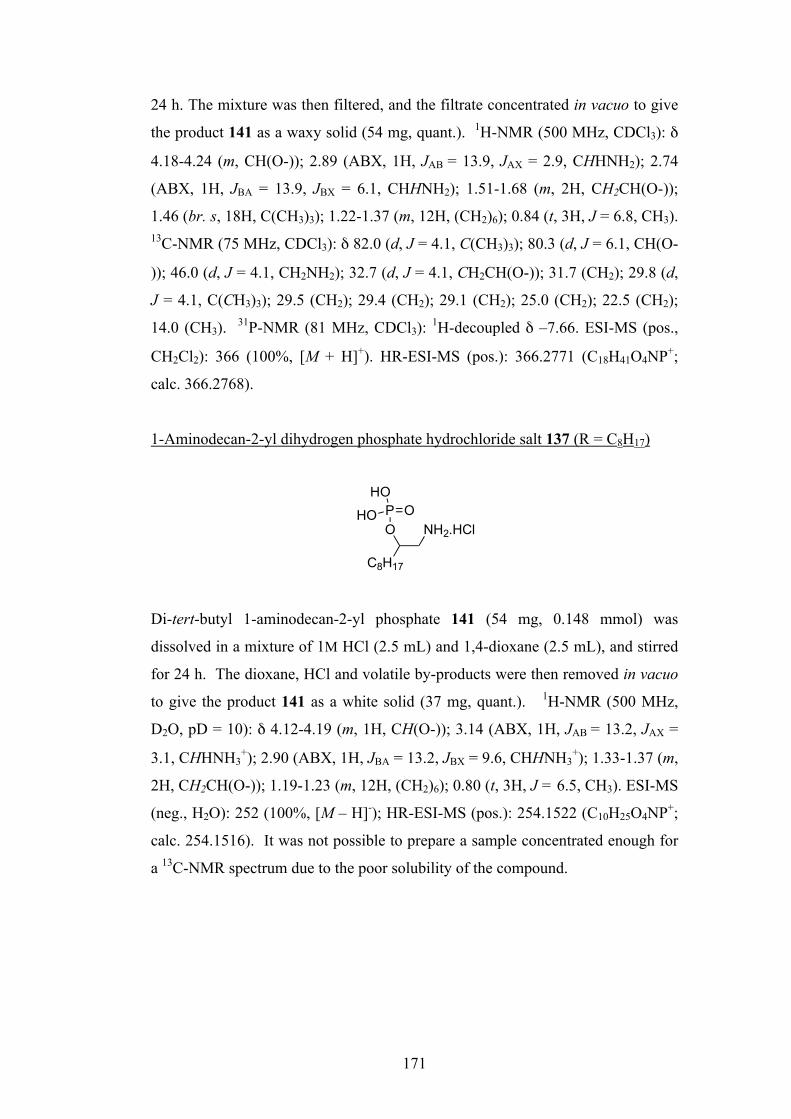
171
24 h. The mixture was then filtered, and the filtrate concentrated in vacuo to give
the product 141 as a waxy solid (54 mg, quant.). 1H-NMR (500 MHz, CDCl3): δ
4.18-4.24 (m, CH(O-)); 2.89 (ABX, 1H, JAB = 13.9, JAX = 2.9, CHHNH2); 2.74
(ABX, 1H, JBA = 13.9, JBX = 6.1, CHHNH2); 1.51-1.68 (m, 2H, CH2CH(O-));
1.46 (br. s, 18H, C(CH3)3); 1.22-1.37 (m, 12H, (CH2)6); 0.84 (t, 3H, J = 6.8, CH3).
13C-NMR (75 MHz, CDCl3): δ 82.0 (d, J = 4.1, C(CH3)3); 80.3 (d, J = 6.1, CH(O-
)); 46.0 (d, J = 4.1, CH2NH2); 32.7 (d, J = 4.1, CH2CH(O-)); 31.7 (CH2); 29.8 (d,
J = 4.1, C(CH3)3); 29.5 (CH2); 29.4 (CH2); 29.1 (CH2); 25.0 (CH2); 22.5 (CH2);
14.0 (CH3). 31P-NMR (81 MHz, CDCl3): 1H-decoupled δ –7.66. ESI-MS (pos.,
CH2Cl2): 366 (100%, [M + H]+). HR-ESI-MS (pos.): 366.2771 (C18H41O4NP+;
calc. 366.2768).
1-Aminodecan-2-yl dihydrogen phosphate hydrochloride salt 137 (R = C8H17)
Di-tert-butyl 1-aminodecan-2-yl phosphate 141 (54 mg, 0.148 mmol) was
dissolved in a mixture of 1M HCl (2.5 mL) and 1,4-dioxane (2.5 mL), and stirred
for 24 h. The dioxane, HCl and volatile by-products were then removed in vacuo
to give the product 141 as a white solid (37 mg, quant.). 1H-NMR (500 MHz,
D2O, pD = 10): δ 4.12-4.19 (m, 1H, CH(O-)); 3.14 (ABX, 1H, JAB = 13.2, JAX =
3.1, CHHNH3+); 2.90 (ABX, 1H, JBA = 13.2, JBX = 9.6, CHHNH3
+); 1.33-1.37 (m,
2H, CH2CH(O-)); 1.19-1.23 (m, 12H, (CH2)6); 0.80 (t, 3H, J = 6.5, CH3). ESI-MS
(neg., H2O): 252 (100%, [M – H]-); HR-ESI-MS (pos.): 254.1522 (C10H25O4NP+;
calc. 254.1516). It was not possible to prepare a sample concentrated enough for
a 13C-NMR spectrum due to the poor solubility of the compound.
NH2.HCl
C8H17
OP O
HO
HO

172
References
[1] P. Ball, Nature 2006, 442, 500.
[2] A. Eschenmoser, Tetrahedron 2007, 63, 12821.
[3] F. Crick, Nature 1970, 227, 561.
[4] J. D. Watson, F. H. C. Crick, Nature 1953, 171, 737.
[5] V. Borsenberger, M. A. Crowe, J. Lehbauer, J. Raftery, M. Helliwell, K.
Bhutia, T. Cox, J. D. Sutherland, Chem. Biodiversity 2004, 1, 203.
[6] C. R. Woese, Proc. Natl. Acad. Sci. U. S. A. 2000, 97, 8392.
[7] C. R. Darwin, On the Origin of Species, John Murray, London, 1859.
[8] G. F. Joyce, in Origins of Life: The Central Concepts (Eds.: D. W.
Deamer, G. R. Fleischaker), Jones and Bartlett, Boston, 1994.
[9] G. Wächtershäuser, Proc. Natl. Acad. Sci. U. S. A. 1990, 87, 200.
[10] A. G. Cairns-Smith, Chem. Eur. J. 2008, 14, 3830.
[11] P. Ball, Elegant Solutions: Ten Beautiful Experiments in Chemistry, Royal
Society of Chemistry, Cambridge, 2005.
[12] http://www.biologycorner.com/resources/DNA-replication.jpg.
[13] http://www.wiley.com/legacy/college/boyer/tRNA/trna_diagram.gif.
[14] http://chsweb.lr.k12.nj.us/Translation%20Notes_files/image007.jpg.
[15] C. Woese, The Genetic Code, the Molecular Basis for Genetic Expression,
Harper and Row, New York, 1967.
[16] F. H. C. Crick, J. Mol. Biol. 1968, 38, 367.
[17] L. E. Orgel, J. Mol. Biol. 1968, 38, 381.
[18] L. Stryer, J. M. Berg, J. L. Tymoczko, Biochemistry, 5th ed., Freeman,
New York, 2002.
[19] K. Kruger, P. J. Grabowski, A. J. Zaug, J. Sands, D. E. Gottschling, T. R.
Cech, Cell 1982, 31, 147.
[20] C. Guerrier-takada, K. Gardiner, T. Marsh, N. Pace, S. Altman, Cell 1983,
35, 849.
[21] W. K. Johnston, P. J. Unrau, M. S. Lawrence, M. E. Glasner, D. P. Bartel,
Science 2001, 292, 1319.
[22] W. Gilbert, Nature 1986, 319, 618.

173
[23] N. H. Sleep, K. J. Zahnle, J. F. Kasting, H. J. Morowitz, Nature 1989, 342,
139.
[24] C. F. Chyba, Geochim. Cosmochim. Acta 1993, 57, 3351.
[25] L. E. Orgel, Trends Biochem. Sci. 1998, 23, 491.
[26] S. J. Mojzsis, G. Arrhenius, K. D. McKeegan, T. M. Harrison, A. P.
Nutman, C. R. L. Friend, Nature 1996, 384, 55.
[27] R. A. Sanchez, J. P. Ferris, L. E. Orgel, Science 1966, 154, 784.
[28] S. L. Miller, G. Schlesinger, Origins Life Evol. Biosphere 1984, 14, 83.
[29] H. B. Niemann, S. K. Atreya, S. J. Bauer, G. R. Carignan, J. E. Demick, R.
L. Frost, D. Gautier, J. A. Haberman, D. N. Harpold, D. M. Hunten, G.
Israel, J. I. Lunine, W. T. Kasprzak, T. C. Owen, M. Paulkovich, F.
Raulin, E. Raaen, S. H. Way, Nature 2005, 438, 779.
[30] J. P. Ferris, W. J. Hagan, Tetrahedron 1984, 40, 1093.
[31] J. M. Hollis, F. J. Lovas, P. R. Jewell, Astrophys. J. 2000, 540, L107.
[32] A. Eschenmoser, E. Loewenthal, Chem. Soc. Rev. 1992, 21, 1.
[33] J. R. Cronin, Adv. Space Res. 1989, 9, 59.
[34] S. L. Miller, Science 1953, 117, 528.
[35] A. Strecker, Annalen der Chemie und Pharmacie 1850, 75, 27.
[36] A. Butlerow, C. R. Acad. Sci., Paris 1861, 53.
[37] L. E. Orgel, Crit. Rev. Biochem. Mol. Biol. 2004, 39, 99.
[38] R. Shapiro, Origins Life Evol. Biosphere 1988, 18, 71.
[39] P. Decker, H. Schweer, R. Pohlmann, J. Chromatogr. 1982, 244, 281.
[40] G. Zubay, Origins Life Evol. Biosphere 1998, 28, 13.
[41] A. Ricardo, M. A. Carrigan, A. N. Olcott, S. A. Benner, Science 2004,
303, 196.
[42] D. Müller, S. Pitsch, A. Kittaka, E. Wagner, C. E. Wintner, A.
Eschenmoser, Helv. Chim. Acta 1990, 73, 1410.
[43] J. Oró Biochem. Biophys. Res. Commun. 1960, 2, 407.
[44] C. U. Lowe, R. Markham, M. W. Rees, Nature 1963, 199, 219.
[45] R. A. Sanchez, J. P. Ferris, L. E. Orgel, J. Mol. Biol. 1967, 30, 223.
[46] A. W. Schwartz, H. Joosten, A. B. Voet, Biosystems 1982, 15, 191.
[47] R. Shapiro, R. S. Klein, Biochemistry 1966, 5, 2358.
[48] M. P. Robertson, S. L. Miller, Nature 1995, 375, 772.
[49] W. D. Fuller, L. E. Orgel, R. A. Sanchez, J. Mol. Evol. 1972, 1, 249.
174
[50] W. D. Fuller, R. A. Sanchez, L. E. Orgel, J. Mol. Biol. 1972, 67, 25.
[51] K. N. Drew, J. Zajicek, G. Bondo, B. Bose, A. S. Serianni, Carbohydr.
Res. 1998, 307, 199.
[52] R. A. Sanchez, L. E. Orgel, J. Mol. Biol. 1970, 47, 531.
[53] C. M. Tapiero, J. Nagyvary, Nature 1971, 231, 42.
[54] A. A. Ingar, R. W. A. Luke, B. R. Hayter, J. D. Sutherland, ChemBioChem
2003, 4, 504.
[55] G. Springsteen, G. F. Joyce, J. Am. Chem. Soc. 2004, 126, 9578.
[56] K. U. Schöning, P. Scholz, S. Guntha, X. Wu, R. Krishnamurthy, A.
Eschenmoser, Science 2000, 290, 1347.
[57] P. Herdewijn, Angew. Chem. Int. Ed. 2001, 40, 2249.
[58] S. Pitsch, S. Wendeborn, B. Jaun, A. Eschenmoser, Helv. Chim. Acta
1993, 76, 2161.
[59] S. Pitsch, R. Krishnamurthy, M. Bolli, S. Wendeborn, A. Holzner, M.
Minton, C. Lesueur, I. Schlonvogt, B. Jaun, A. Eschenmoser, Helv. Chim.
Acta 1995, 78, 1621.
[60] P. Wittung, S. K. Kim, O. Buchardt, P. Nielsen, B. Norden, Nucleic Acids
Res. 1994, 22, 5371.
[61] J. G. Schmidt, P. E. Nielsen, L. E. Orgel, Nucleic Acids Res. 1997, 25,
4797.
[62] K. E. Nelson, M. Levy, S. L. Miller, Proc. Natl. Acad. Sci. U. S. A. 2000,
97, 3868.
[63] L. L. Zhang, A. E. Peritz, P. J. Carroll, E. Meggers, Synthesis 2006, 645.
[64] L. L. Zhang, A. Peritz, E. Meggers, J. Am. Chem. Soc. 2005, 127, 4174.
[65] C. Anastasi, M. A. Crowe, M. W. Powner, J. D. Sutherland, Angew. Chem.
Int. Ed. 2006, 45, 6176.
[66] A. F. Cockerill, A. Deacon, R. G. Harrison, D. J. Osborne, D. M. Prime,
W. J. Ross, A. Todd, J. P. Verge, Synthesis 1976, 591.
[67] M. W. Powner, B. Gerland, J. D. Sutherland, Nature 2009, 459, 239.
[68] R. Lohrmann, L. E. Orgel, Science 1971, 171, 490.
[69] A. M. Schoffstall, Origins Life Evol. Biosphere 1976, 7, 399.
[70] J. P. Ferris, G. Goldstein, D. J. Beaulieu, J. Am. Chem. Soc. 1970, 92,
6598.
[71] M. S. Verlander, L. E. Orgel, J. Mol. Evol. 1974, 3, 115.

175
[72] M. S. Verlander, R. Lohrmann, L. E. Orgel, J. Mol. Evol. 1973, 2, 303.
[73] M. Renz, R. Lohrmann, L. E. Orgel, Biochim. Biophys. Acta 1971, 240,
463.
[74] R. Lohrmann, L. E. Orgel, Science 1968, 161, 64.
[75] M. A. Crowe, J. D. Sutherland, ChemBioChem 2006, 7, 951.
[76] Y. Furukawa, O. Miyashita, M. Honjo, Chem. Pharm. Bull. 1974, 22,
2552.
[77] R. Lohrmann, J. Mol. Evol. 1977, 10, 137.
[78] H. Sawai, J. Am. Chem. Soc. 1976, 98, 7037.
[79] H. L. Sleeper, L. E. Orgel, J. Mol. Evol. 1979, 12, 357.
[80] H. Sawai, K. Higa, K. Kuroda, J. Chem. Soc., Perkin Trans. 1 1992, 505.
[81] H. Sawai, K. Kuroda, T. Hojo, Bull. Chem. Soc. Jpn. 1989, 62, 2018.
[82] W. H. Huang, J. P. Ferris, Chem. Commun. 2003, 1458.
[83] K. J. Prabahar, J. P. Ferris, Origins Life Evol. Biosphere 1997, 27, 513.
[84] L. E. Orgel, R. Lohrmann, Acc. Chem. Res. 1974, 7, 368.
[85] J. D. Sutherland, J. M. Blackburn, Chem. Biol. 1997, 4, 481.
[86] J. T. F. Wong, Proc. Natl. Acad. Sci. U. S. A. 1975, 72, 1909.
[87] K. Kawamura, J. P. Ferris, Origins Life Evol. Biosphere 1999, 29, 563.
[88] M. M. W. Mooren, S. S. Wijmenga, G. A. Vandermarel, J. H. Vanboom,
C. W. Hilbers, Nucleic Acids Res. 1994, 22, 2658.
[89] L. Leman, L. Orgel, M. R. Ghadiri, Science 2004, 306, 283.
[90] J. P. Biron, A. L. Parkes, R. Pascal, J. D. Sutherland, Angew. Chem. Int.
Ed. 2005, 44, 6731.
[91] D. W. Deamer, J. P. Dworkin, S. A. Sandford, M. P. Bernstein, L. J.
Allamandola, Astrobiology 2002, 2, 371.
[92] G. L. Nicolson, S. J. Singer, Science 1972, 175, 720.
[93] C. Tanford, The Hydrophobic Effect, 2nd ed., Wiley, Toronto, 1980.
[94] P. R. Cullis, M. J. Hope, Biochemistry of Lipids, Lipoproteins and
Membranes, Elsevier, Amsterdam, 1991.
[95] D. W. Deamer, R. M. Pashley, Origins Life Evol. Biosphere 1989, 19, 21.
[96] H. Naraoka, A. Shimoyama, K. Harada, Origins Life Evol. Biosphere
1999, 29, 187.
[97] B. R. T. Simoneit, T. M. McCollom, G. Ritter, Origins Life Evol.
Biosphere 1999, 29, 153.
176
[98] J. M. Gebicki, M. Hicks, Nature 1973, 243, 232.
[99] P. Walde, R. Wick, M. Fresta, A. Mangone, P. L. Luisi, J. Am. Chem. Soc.
1994, 116, 11649.
[100] I. A. Chen, K. Salehi-Ashtiani, J. W. Szostak, J. Am. Chem. Soc. 2005,
127, 13213.
[101] P. A. Monnard, C. L. Apel, A. Kanavarioti, D. W. Deamer, Astrobiology
2002, 2, 139.
[102] L. B. Mullen, J. D. Sutherland, Angew. Chem. Int. Ed. 2007, 46, 8063.
[103] G. M. Blackburn, Nucleic Acids in Chemistry and Biology, Oxford
University Press, Oxford, 1996.
[104] I. Ugi, Angew. Chem. Int. Ed. 1962, 74, 9.
[105] M. C. Pirrung, K. Das Sarma, Tetrahedron 2005, 61, 11456.
[106] A. Domling, I. Ugi, Angew. Chem. Int. Ed. 2000, 39, 3169.
[107] R. K. Ralph, H. G. Khorana, W. J. Connors, J. Am. Chem. Soc. 1962, 84,
2265.
[108] H. Ishikura, J. Biochem. (Tokyo, Jpn.) 1962, 52, 324.
[109] Y. Mizuno, J. Kobayashi, J. Chem. Soc., Chem. Commun. 1974, 997.
[110] C. Struve, C. Christophersen, Heterocycles 2003, 60, 1907.
[111] M. Ochiai, M. Inenaga, Y. Nagao, R. M. Moriarty, R. K. Vaid, M. P.
Duncan, Tetrahedron Lett. 1988, 29, 6917.
[112] H. Saltzman, J. G. Sharefkin, Org. Synth. 1963, 43, 60.
[113] Z. Tang, F. Jiang, X. Cui, L. Z. Gong, A. Q. Mi, Y. Z. Jiang, Y. D. Wu,
Proc. Natl. Acad. Sci. U. S. A. 2004, 101, 5755.
[114] S. C. Pan, B. List, Angew. Chem. Int. Ed. 2008, 47, 3622.
[115] P. Pelagatti, M. Carcelli, F. Calbiani, C. Cassi, L. Elviri, C. Pelizzi, U.
Rizzotti, D. Rogolino, Organometallics 2005, 24, 5836.
[116] M. Klussmann, H. Iwamura, S. P. Mathew, D. H. Wells, U. Pandya, A.
Armstrong, D. G. Blackmond, Nature 2006, 441, 621.
[117] J. D. Sutherland, L. B. Mullen, F. F. Buchet, Synlett 2008, 2161.
[118] R. Krishnamurthy, S. Guntha, A. Eschenmoser, Angew. Chem. Int. Ed.
2000, 39, 2281.
[119] J. P. Ferris, Science 1968, 161, 53.
[120] Y. B. Xiang, S. Drenkard, K. Baumann, D. Hickey, A. Eschenmoser, Helv.
Chim. Acta 1994, 77, 2209.
177
[121] J. Campos Manzano, MPhil thesis, The University of Manchester
(Manchester), 2008.
[122] L. B. Mullen, J. D. Sutherland, Angew. Chem. Int. Ed. 2007, 46, 4166.
[123] P. Walde, Origins Life Evol. Biosphere 2006, 36, 109.
[124] E. W. Kaler, A. K. Murthy, B. E. Rodriguez, J. A. N. Zasadzinski, Science
1989, 245, 1371.
[125] R. B. Anderson, The Fischer-Tropsch Synthesis, Academic Press,
Orlando, 1984.
[126] O. T. Quimby, T. J. Flautt, Z. Anorg. Allg. Chem. 1958, 296, 220.
[127] R. Krishnamurthy, G. Arrhenius, A. Eschenmoser, Origins Life Evol.
Biosphere 1999, 29, 333.
[128] S. Y. Kalliney, Topics in Phosphorus Chemistry, Vol. 7, Interscience, New
York, 1972.
[129] R. Osterberg, L. E. Orgel, J. Mol. Evol. 1972, 1, 241.
[130] Y. Yamagata, H. Wantanabe, M. Saitoh, T. Namba, Nature 1991, 352,
516.
[131] R. A. Lazarus, P. A. Benkovic, S. J. Benkovic, J. Chem. Soc., Perkin
Trans. 2 1980, 373.
[132] M. Tsuhako, C. Sueyoshi, T. Miyajima, S. Ohashi, H. Nariai, I. Motooka,
Bull. Chem. Soc. Jpn. 1986, 59, 3091.
[133] S. E. Kelly, T. G. Lacour, Synth. Commun. 1992, 22, 859.
[134] W. J. Moree, L. C. Vangent, G. A. Vandermarel, R. M. J. Liskamp,
Tetrahedron 1993, 49, 1133.
[135] A. Kuboki, T. Ishihara, E. Kobayashi, H. Ohta, T. Ishii, A. Inoue, S.
Mitsuda, T. Miyazaki, Y. Kajihara, T. Sugai, Bioscience Biotechnology
and Biochemistry 2000, 64, 363.
[136] G. Sabitha, R. S. Babu, M. Rajkumar, J. S. Yadav, Org. Lett. 2002, 4, 343.
[137] M. Onaka, K. Sugita, Y. Izumi, J. Org. Chem. 1989, 54, 1116.
[138] D. S. Dhanoa, W. H. Parsons, W. J. Greenlee, A. A. Patchett, Tetrahedron
Lett. 1992, 33, 1725.
[139] P. W. Tang, J. M. Williams, J. Chem. Soc., Perkin Trans. 1 1984, 1199.
[140] E. Gossnitzer, A. Punkenhofer, Monatshefte Fur Chemie 2003, 134, 1271.