Embed Size (px)
Citation preview
1
(DENTAL OUTLOOK, Vol.120, No.2 2012-8,JAPAN)
Extra Special Article
Practical points in making complete dentures suction effective and
functional – General views on edentulous ridge characteristics
and denture movements
Yoshihiro Saito, DDS., PhD..
Kunimino Saito Dental Clinic,
4-2-1 Kunimi, Aoba-ku, Sendai-shi, Miyagi-ken 981-0943,
Japan
Introduction
It would be much to their delight for dentists and dental technicians that they could
finish complete dentures with satisfying results and that patients could enjoy using
them.
Once Abe 1) explained the suction effect of mandibular complete denture theoretically,
many practitioners have followed this modality in practices extensively throughout the
profession. Take an example around the author of this article, when once one could
finish a successful suction effective case of mandibular complete denture, such a
dentist or dental technician would be likely to design another case for more effective
suction thereafter.
Why is the suction in denture making so much attractive like this for many? The
major reasons may be because, for patients, eating meals would become more enjoyable
only by having a denture firmly seated with suction and because they can become more
easily convinced of its excellence to a less suction effective denture. In contrast, for
practitioners, by having goals “effective suction = entire denture border sealing”, they
can be easily convinced of “goals achieved” only from obtaining effective suction results.
Furthermore, because this impression taking method is more clearly defined than
2
conventional border molding impression taking, it is more attractive for them to
introduce it and continue practicing it in daily clinical cases.
This article, titled “General views on complete dentures”, will refer to the following
items for better understanding of making a suction effective denture.
1) Characteristic oral environments involved with edentulous patients
2) Denture movements while in function
3) Suction effective in mandibular complete denture
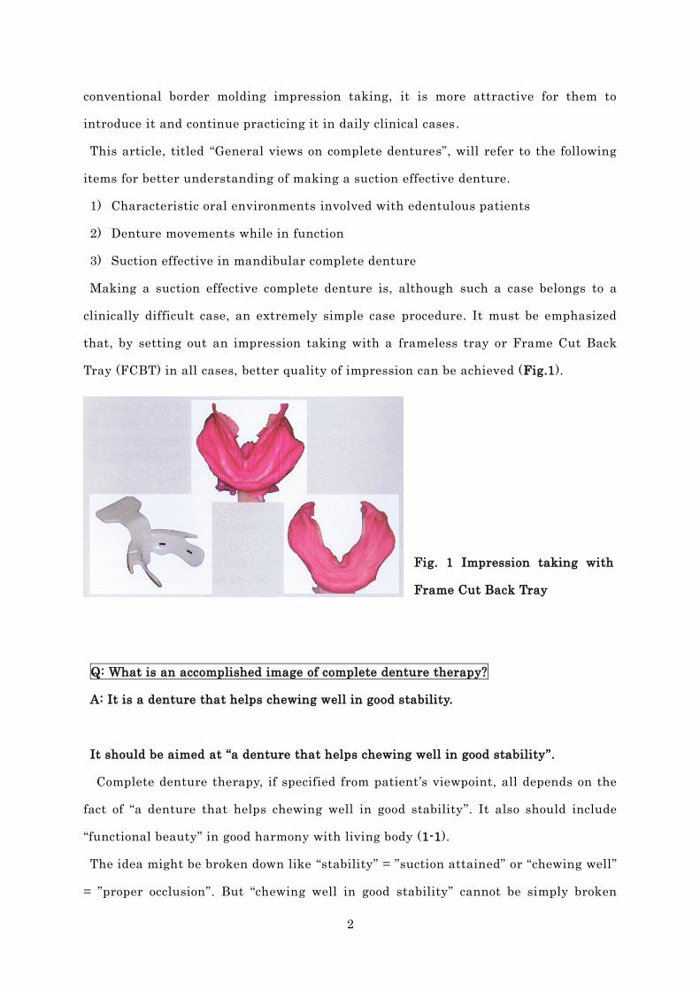
Making a suction effective complete denture is, although such a case belongs to a
clinically difficult case, an extremely simple case procedure. It must be emphasized
that, by setting out an impression taking with a frameless tray or Frame Cut Back
Tray (FCBT) in all cases, better quality of impression can be achieved (Fig.1).
Fig. 1 Impression taking with
Frame Cut Back Tray
Q: What is an accomplished image of complete denture therapy?
A: It is a denture that helps chewing well in good stability.
It should be aimed at “a denture that helps chewing well in good stability”.
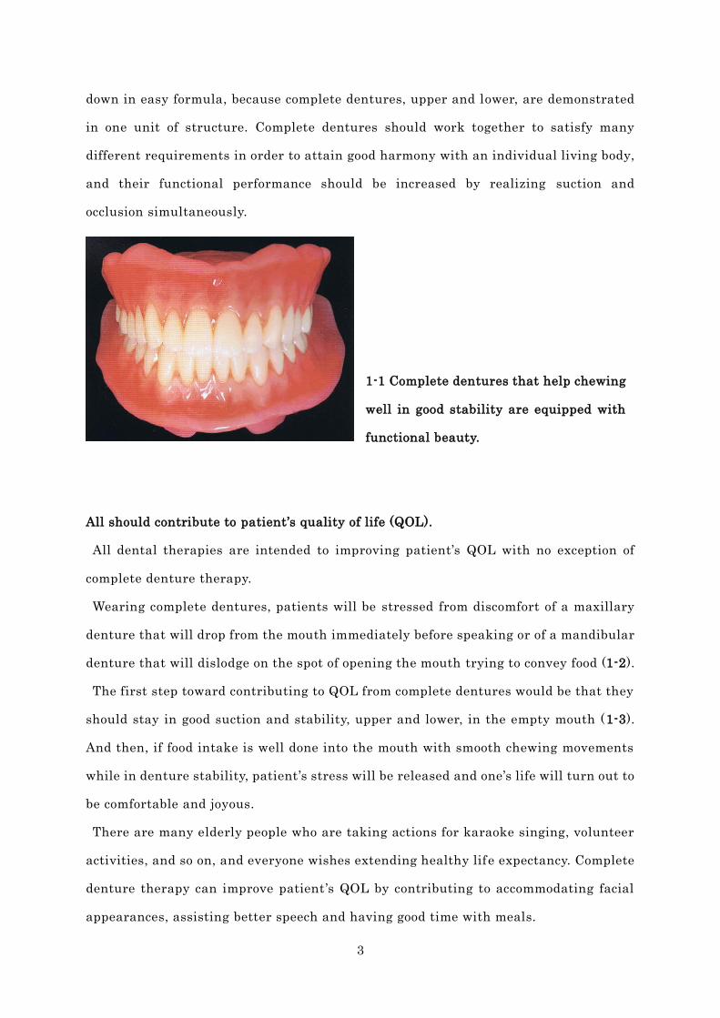
Complete denture therapy, if specified from patient’s viewpoint, all depends on the
fact of “a denture that helps chewing well in good stability”. It also should include
“functional beauty” in good harmony with living body (1-1).
The idea might be broken down like “stability” = ”suction attained” or “chewing well”
= ”proper occlusion”. But “chewing well in good stability” cannot be simply broken
3
down in easy formula, because complete dentures, upper and lower, are demonstrated
in one unit of structure. Complete dentures should work together to satisfy many
different requirements in order to attain good harmony with an individual living body,
and their functional performance should be increased by realizing suction and
occlusion simultaneously.
1-1 Complete dentures that help chewing
well in good stability are equipped with
functional beauty.
All should contribute to patient’s quality of life (QOL).
All dental therapies are intended to improving patient’s QOL with no exception of
complete denture therapy.
Wearing complete dentures, patients will be stressed from discomfort of a maxillary
denture that will drop from the mouth immediately before speaking or of a mandibular
denture that will dislodge on the spot of opening the mouth trying to convey food (1-2).
The first step toward contributing to QOL from complete dentures would be that they
should stay in good suction and stability, upper and lower, in the empty mouth (1-3).
And then, if food intake is well done into the mouth with smooth chewing movements
while in denture stability, patient’s stress will be released and one’s life will turn out to
be comfortable and joyous.
There are many elderly people who are taking actions for karaoke singing, volunteer
activities, and so on, and everyone wishes extending healthy life expectancy. Complete
denture therapy can improve patient ’s QOL by contributing to accommodating facial
appearances, assisting better speech and having good time with meals.
4
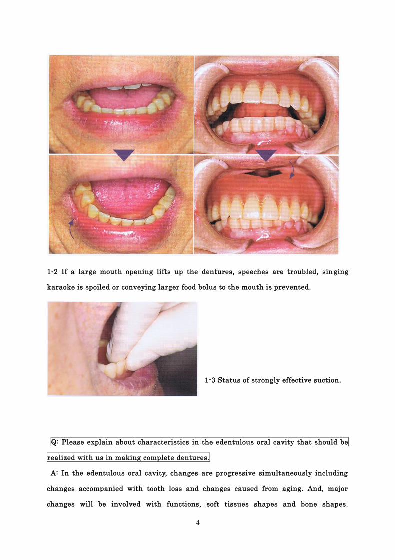
1-2 If a large mouth opening lifts up the dentures, speeches are troubled, singing
karaoke is spoiled or conveying larger food bolus to the mouth is prevented.
1-3 Status of strongly effective suction.
Q: Please explain about characteristics in the edentulous oral cavity that should be
realized with us in making complete dentures.
A: In the edentulous oral cavity, changes are progressive simultaneously including
changes accompanied with tooth loss and changes caused from aging. And, major
changes will be involved with functions, soft tissues shapes and bone shapes.
5
Consequently, in complete denture making, the oral environments must be observed
closely in different individual patients. And degree of difficulty on each oral part must
be assessed in advance.
Changes of functions
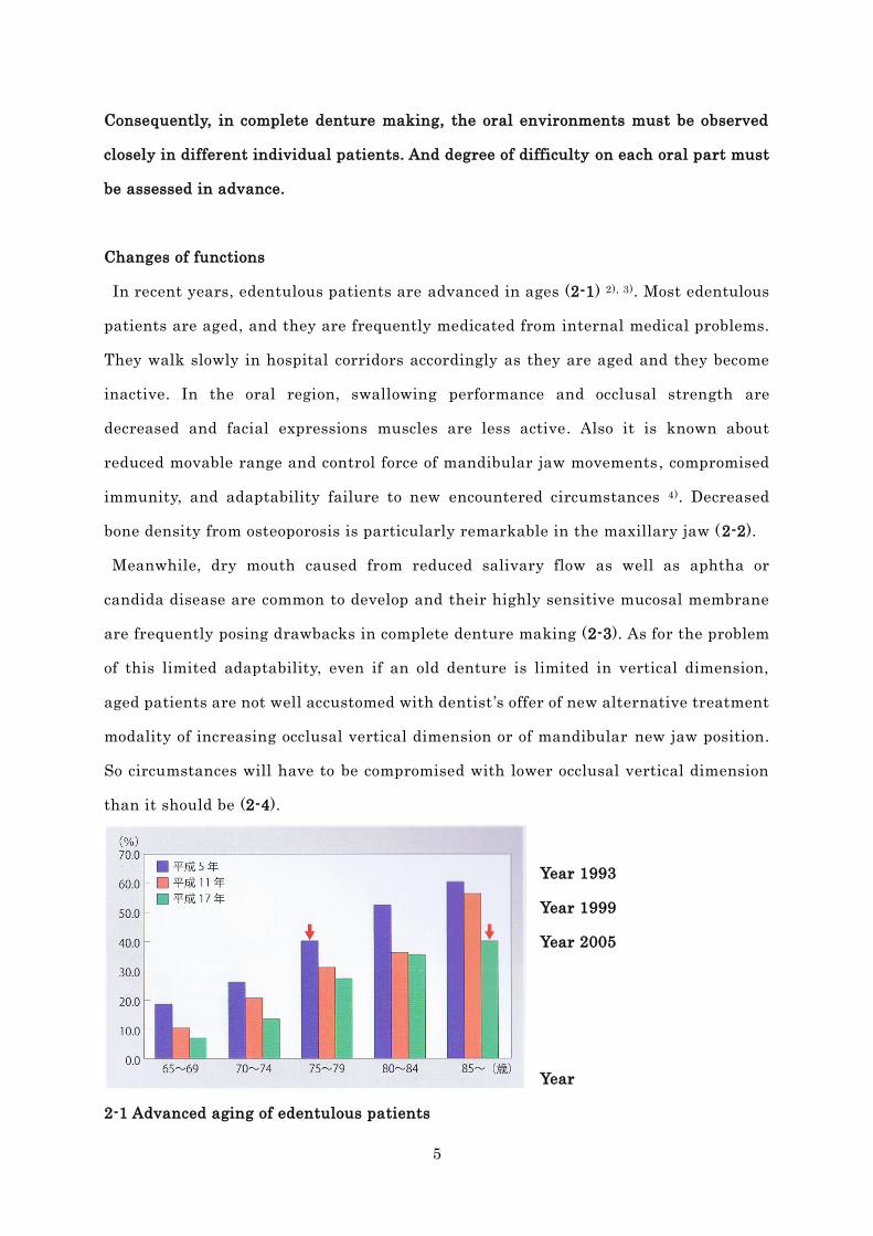
In recent years, edentulous patients are advanced in ages (2-1) 2), 3). Most edentulous
patients are aged, and they are frequently medicated from internal medical problems.
They walk slowly in hospital corridors accordingly as they are aged and they become
inactive. In the oral region, swallowing performance and occlusal strength are
decreased and facial expressions muscles are less active. Also it is known about
reduced movable range and control force of mandibular jaw movements , compromised
immunity, and adaptability failure to new encountered circumstances 4). Decreased
bone density from osteoporosis is particularly remarkable in the maxillary jaw (2-2).
Meanwhile, dry mouth caused from reduced salivary flow as well as aphtha or
candida disease are common to develop and their highly sensitive mucosal membrane
are frequently posing drawbacks in complete denture making (2-3). As for the problem
of this limited adaptability, even if an old denture is limited in vertical dimension,
aged patients are not well accustomed with dentist ’s offer of new alternative treatment
modality of increasing occlusal vertical dimension or of mandibular new jaw position.
So circumstances will have to be compromised with lower occlusal vertical dimension
than it should be (2-4).
Year 1993
Year 1999
Year 2005
Year
2-1 Advanced aging of edentulous patients
6
Percentage rate of edentulism statistics are almost same at age 75 in 1993 and those
over age 85 in 2005. Although yet unknown whether or not total number of edentulism
has decreased on the whole, the graphic chart does show shifting the average value to
the right hand, indicating advanced aging of edentulous patients. (Kanehira 2009 2),
Statistical analysis committee on the Survey of Dental Diseases 2007 3) )
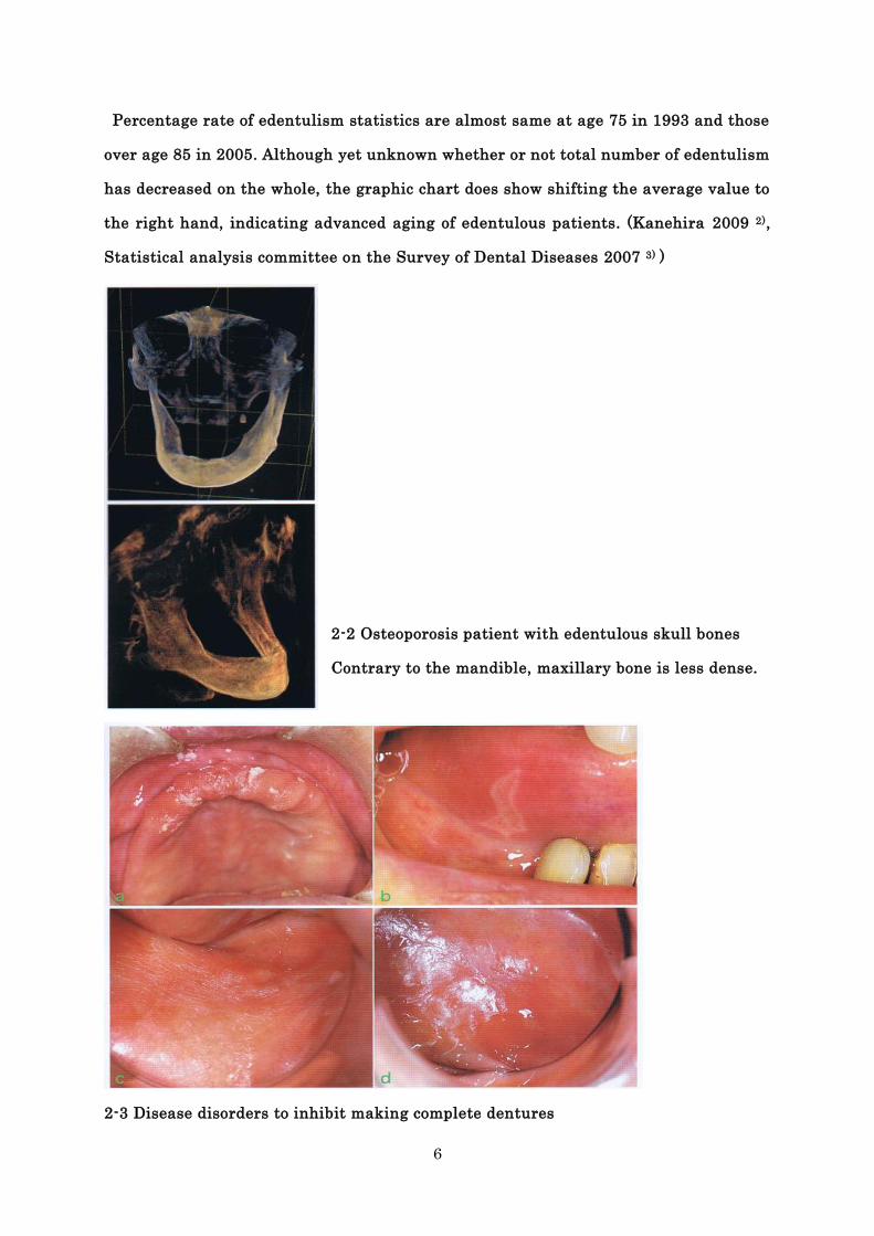
2-2 Osteoporosis patient with edentulous skull bones
Contrary to the mandible, maxillary bone is less dense.
2-3 Disease disorders to inhibit making complete dentures
7
a: Candida disease in the maxillary anterior region , b: Recurrent aphta (anterior side
of tongue, multiple missing teeth case), c: xerostomia, d: buccal mucosa erosion (pain
sensitivity)
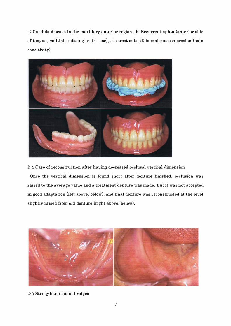
2-4 Case of reconstruction after having decreased occlusal vertical dimension
Once the vertical dimension is found short after denture finished, occlusion was
raised to the average value and a treatment denture was made. But it was not accepted
in good adaptation (left above, below), and final denture was reconstructed at the level
slightly raised from old denture (right above, below).
2-5 String-like residual ridges
8
Changes of soft tissues in the mandible
1) Decreased immobile soft tissues and Double tongue
As the residual ridge is reduced progressively, immobile mucosal membrane cannot be
seen on the buccolingual sides of the alveolar ridge crest in some case (2-5). As long as
sufficient volume of immobile mucosa is present, denture peripheral outlining can be
more easily established for denture support area. But in case like so called “String-like
residual ridge” as shown in 2-5, the ridge crest appears to be already reduced lower in
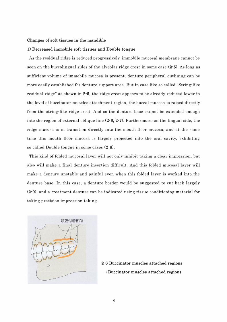
the level of buccinator muscles attachment region, the buccal mucosa is raised directly
from the string-like ridge crest. And so the denture base cannot be extended enough
into the region of external oblique line (2-6, 2-7). Furthermore, on the lingual side, the
ridge mucosa is in transition directly into the mouth floor mucosa, and at the same
time this mouth floor mucosa is largely projected into the oral cavity, exhibiting
so-called Double tongue in some cases (2-8).
This kind of folded mucosal layer will not only inhibit taking a clear impression, but
also will make a final denture insertion difficult. And this folded mucosal layer will
make a denture unstable and painful even when this folded layer is worked into the
denture base. In this case, a denture border would be suggested to cut back largely
(2-9), and a treatment denture can be indicated using tissue conditioning material for
taking precision impression taking.
2-6 Buccinator muscles attached regions
→Buccinator muscles attached regions
9
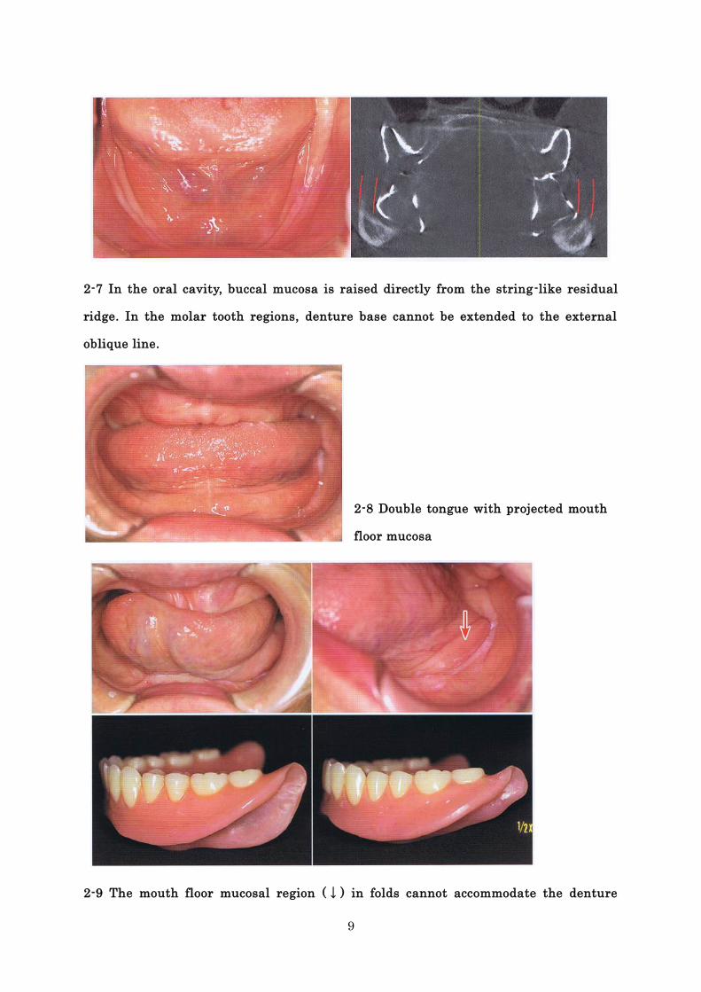
2-7 In the oral cavity, buccal mucosa is raised directly from the string-like residual
ridge. In the molar tooth regions, denture base cannot be extended to the external
oblique line.
2-8 Double tongue with projected mouth
floor mucosa
2-9 The mouth floor mucosal region (↓) in folds cannot accommodate the denture
10
peripheral border, and so in this case the denture base is cut off to the mandibular
mylohyoid line region.
2) Atrophic sublingual folds
While Double tongue is present among aged people, ridge resorption and decreased
salivary flow will be accompanied with getting the sublingual folds unclear. Sublingual
folds are clearly defined among a child and dentulous adult, but aged people are not
clear in many cases. In this case, the mouth floor shows in tension like a tent and it
makes difficult to establish denture base border extension and shape, which will also
make difficult to attain border sealing for effective suction (2-10).
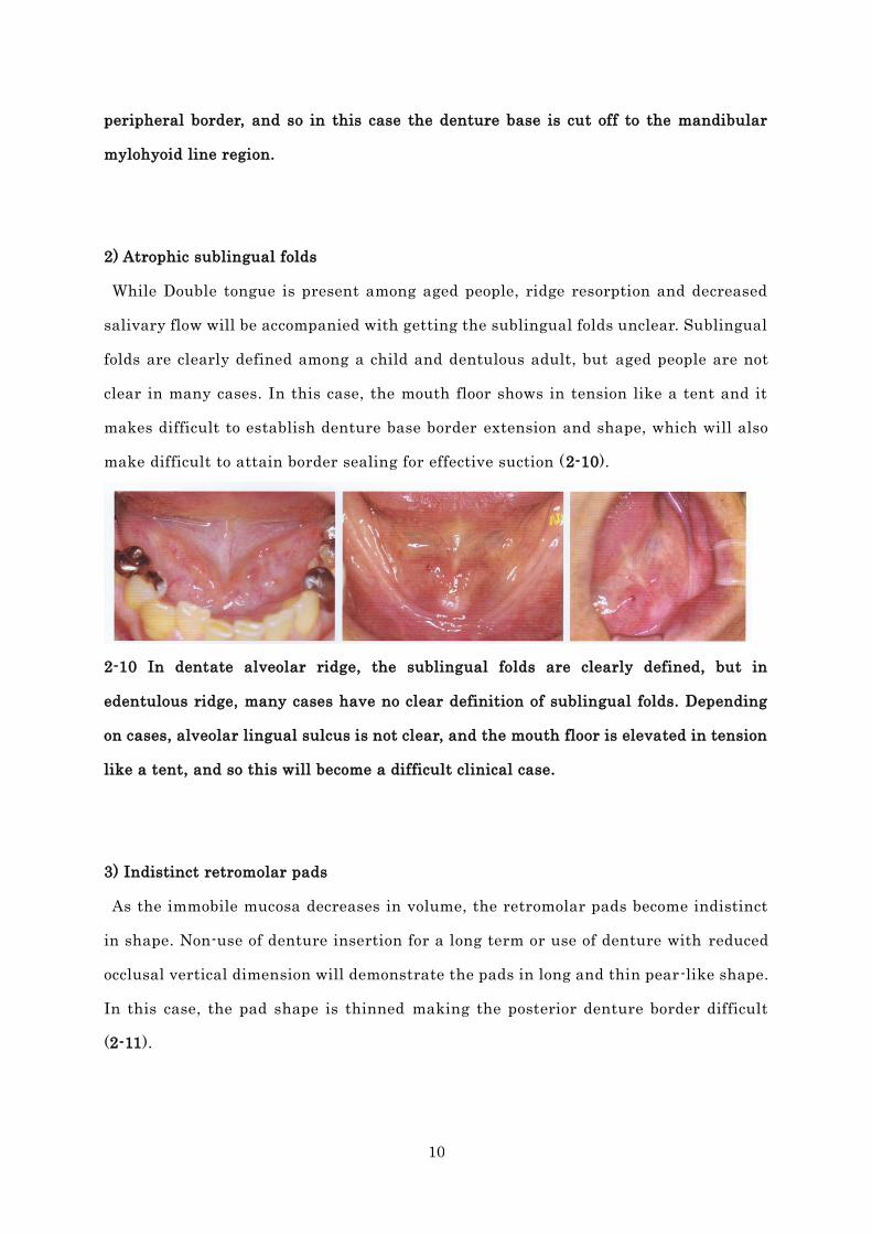
2-10 In dentate alveolar ridge, the sublingual folds are clearly defined, but in
edentulous ridge, many cases have no clear definition of sublingual folds. Depending
on cases, alveolar lingual sulcus is not clear, and the mouth floor is elevated in tension
like a tent, and so this will become a difficult clinical case.
3) Indistinct retromolar pads
As the immobile mucosa decreases in volume, the retromolar pads become indistinct
in shape. Non-use of denture insertion for a long term or use of denture with reduced
occlusal vertical dimension will demonstrate the pads in long and thin pear-like shape.
In this case, the pad shape is thinned making the posterior denture border difficult
(2-11).
11
2-11 Retromolar pads are clearly defined (a), and less clearly defined (b).
4) Retracted tongue root
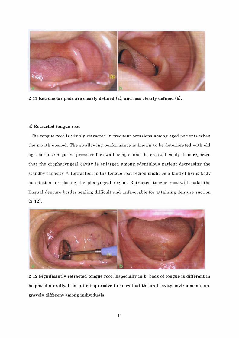
The tongue root is visibly retracted in frequent occasions among aged patients when
the mouth opened. The swallowing performance is known to be deteriorated with old
age, because negative pressure for swallowing cannot be created easily. It is reported
that the oropharyngeal cavity is enlarged among edentulous patient decreasing the
standby capacity 5). Retraction in the tongue root region might be a kind of living body
adaptation for closing the pharyngeal region. Retracted tongue root will make the
lingual denture border sealing difficult and unfavorable for attaining denture suction
(2-12).
2-12 Significantly retracted tongue root. Especially in b, back of tongue is different in
height bilaterally. It is quite impressive to know that the oral cavity environments are
gravely different among individuals.
12
Changes of mandibular bone shapes
1) Where is the ridge crest in the posterior region?
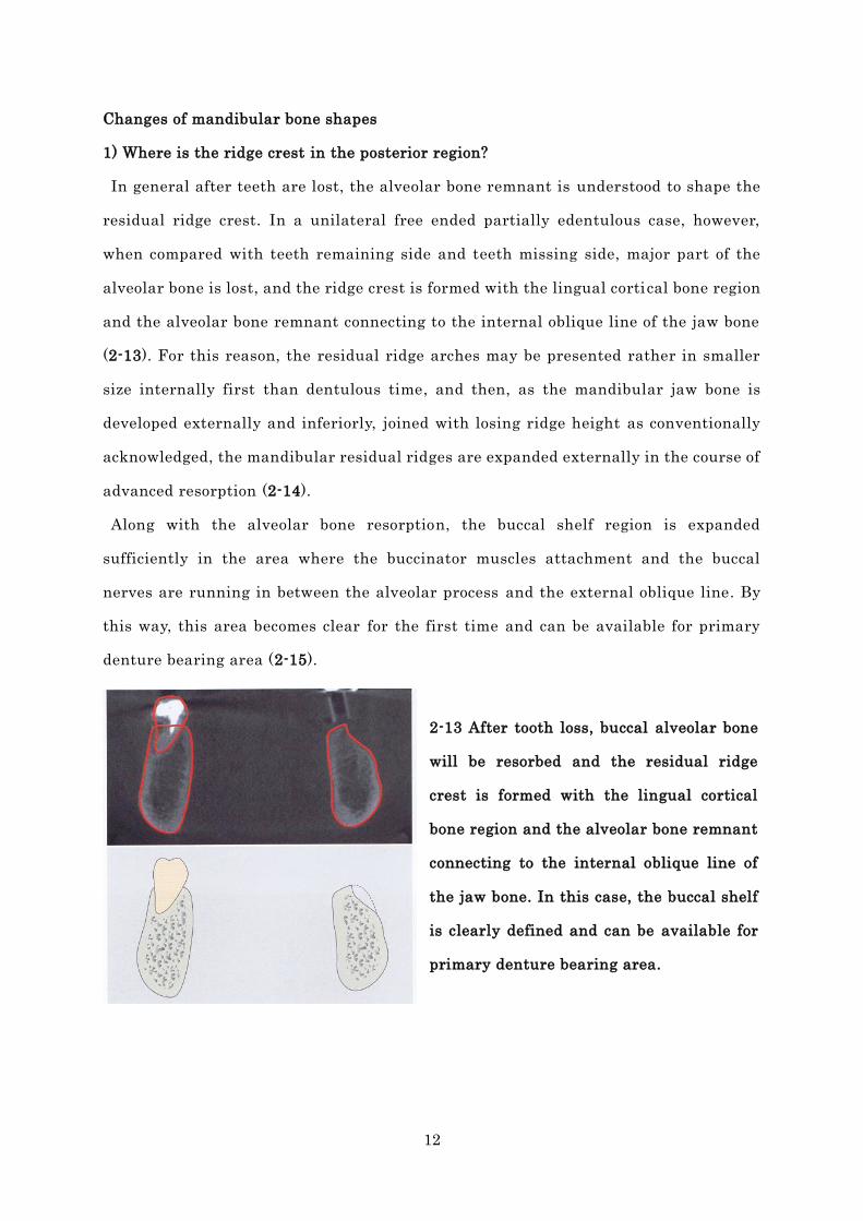
In general after teeth are lost, the alveolar bone remnant is understood to shape the
residual ridge crest. In a unilateral free ended partially edentulous case, however,
when compared with teeth remaining side and teeth missing side, major part of the
alveolar bone is lost, and the ridge crest is formed with the lingual cortical bone region
and the alveolar bone remnant connecting to the internal oblique line of the jaw bone
(2-13). For this reason, the residual ridge arches may be presented rather in smaller
size internally first than dentulous time, and then, as the mandibular jaw bone is
developed externally and inferiorly, joined with losing ridge height as conventionally
acknowledged, the mandibular residual ridges are expanded externally in the course of
advanced resorption (2-14).
Along with the alveolar bone resorption, the buccal shelf region is expanded
sufficiently in the area where the buccinator muscles attachment and the buccal
nerves are running in between the alveolar process and the external oblique line. By
this way, this area becomes clear for the first time and can be available for primary
denture bearing area (2-15).
2-13 After tooth loss, buccal alveolar bone
will be resorbed and the residual ridge
crest is formed with the lingual cortical
bone region and the alveolar bone remnant
connecting to the internal oblique line of
the jaw bone. In this case, the buccal shelf
is clearly defined and can be available for
primary denture bearing area.
13
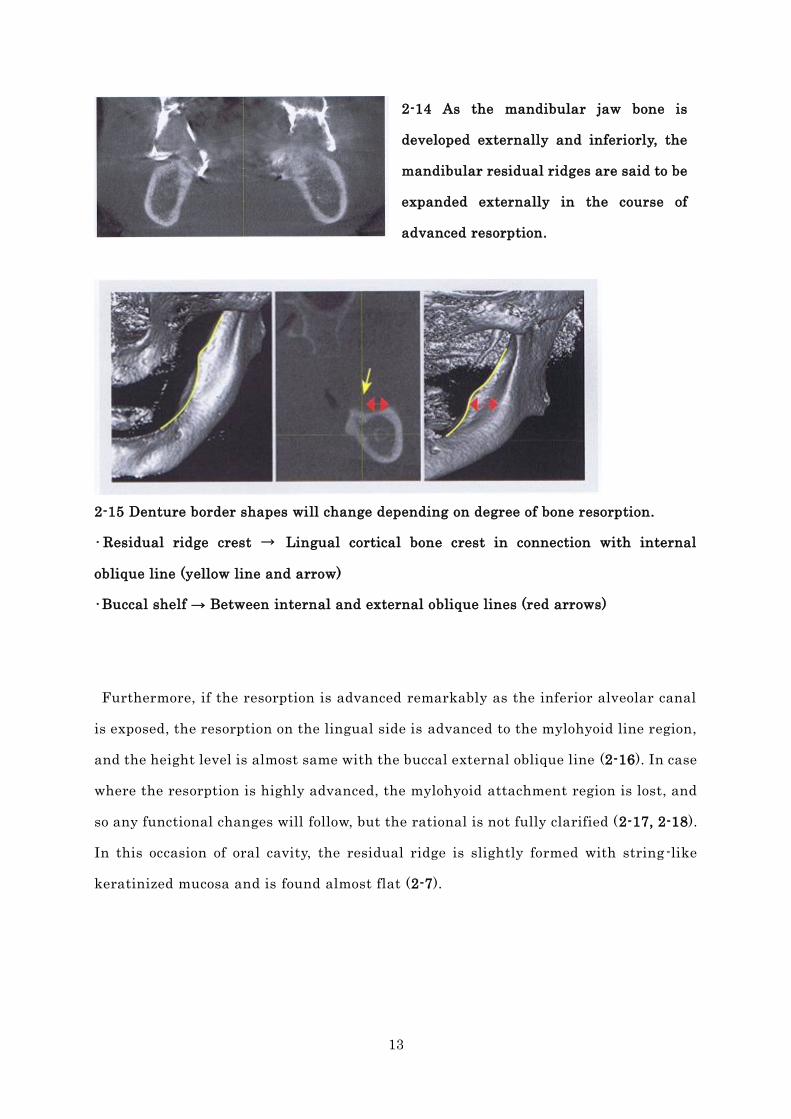
2-14 As the mandibular jaw bone is
developed externally and inferiorly, the
mandibular residual ridges are said to be
expanded externally in the course of
advanced resorption.
2-15 Denture border shapes will change depending on degree of bone resorption.
•Residual ridge crest → Lingual cortical bone crest in connection with internal
oblique line (yellow line and arrow)
•Buccal shelf → Between internal and external oblique lines (red arrows)
Furthermore, if the resorption is advanced remarkably as the inferior alveolar canal
is exposed, the resorption on the lingual side is advanced to the mylohyoid line region,
and the height level is almost same with the buccal external oblique line (2-16). In case
where the resorption is highly advanced, the mylohyoid attachment region is lost, and
so any functional changes will follow, but the rational is not fully clarified (2-17, 2-18).
In this occasion of oral cavity, the residual ridge is slightly formed with string-like
keratinized mucosa and is found almost flat (2-7).
14
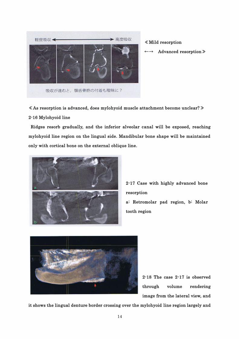
≪Mild resorption
←→ Advanced resorption≫
≪As resorption is advanced, does mylohyoid muscle attachment become unclear?≫
2-16 Mylohyoid line
Ridges resorb gradually, and the inferior alveolar canal will be exposed, reaching
mylohyoid line region on the lingual side. Mandibular bone shape will be maintained
only with cortical bone on the external oblique line.
2-17 Case with highly advanced bone
resorption
a: Retromolar pad region, b: Molar
tooth region
2-18 The case 2-17 is observed
through volume rendering
image from the lateral view, and
it shows the lingual denture border crossing over the mylohyoid line region largely and
15
being extended far below than the mandibular inferior border (↑). In this case, how is
going with the function of mylohyoid?
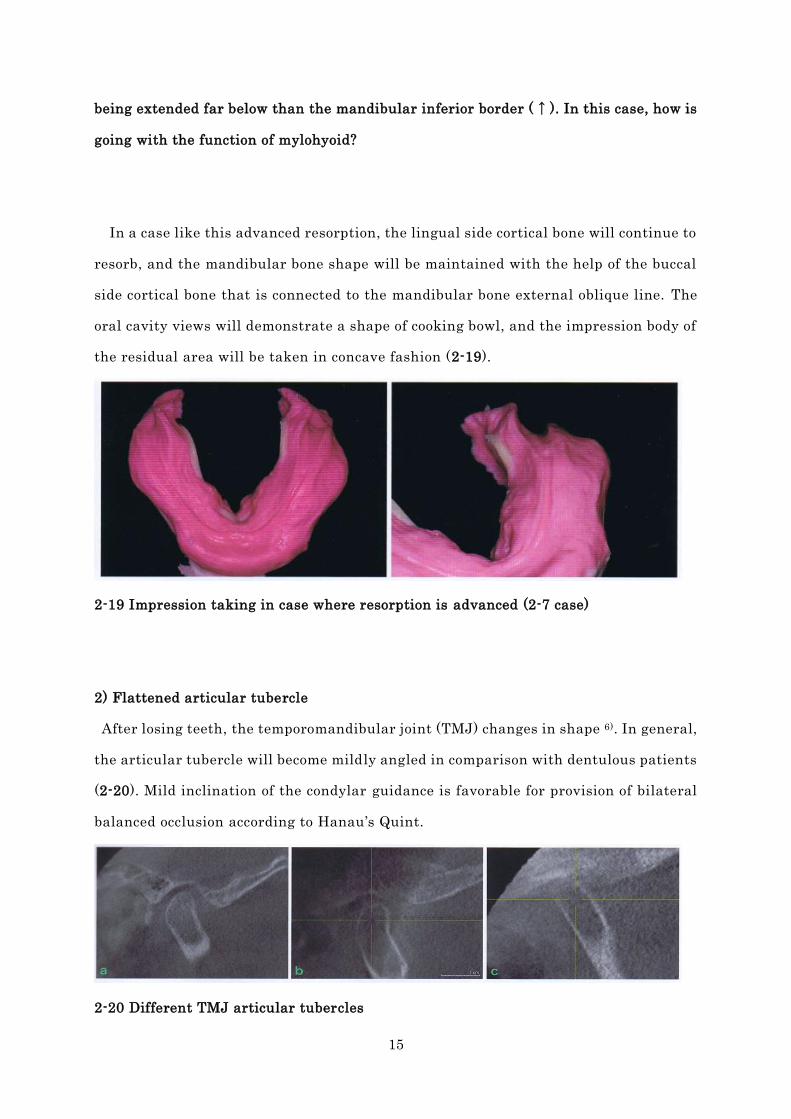
In a case like this advanced resorption, the lingual side cortical bone will continue to
resorb, and the mandibular bone shape will be maintained with the help of the buccal
side cortical bone that is connected to the mandibular bone external oblique line. The
oral cavity views will demonstrate a shape of cooking bowl, and the impression body of
the residual area will be taken in concave fashion (2-19).
2-19 Impression taking in case where resorption is advanced (2-7 case)
2) Flattened articular tubercle
After losing teeth, the temporomandibular joint (TMJ) changes in shape 6). In general,
the articular tubercle will become mildly angled in comparison with dentulous patients
(2-20). Mild inclination of the condylar guidance is favorable for provision of bilateral
balanced occlusion according to Hanau’s Quint.
2-20 Different TMJ articular tubercles
16
a: Dentate jaw, b: Edentulous jaw; the articular tubercle angles become mild.
3) Types of residual ridges
Even in dental implant therapy cases, there can be instances when the bone width is
sufficiently indicated or limitedly indicated from CT scanned radiography. Especially
in case of lady patients, thinned jaw bones are more frequently seen in many cases ,
and there is a difficult case of inserting implants of even 4mm diameter by normal
procedures.
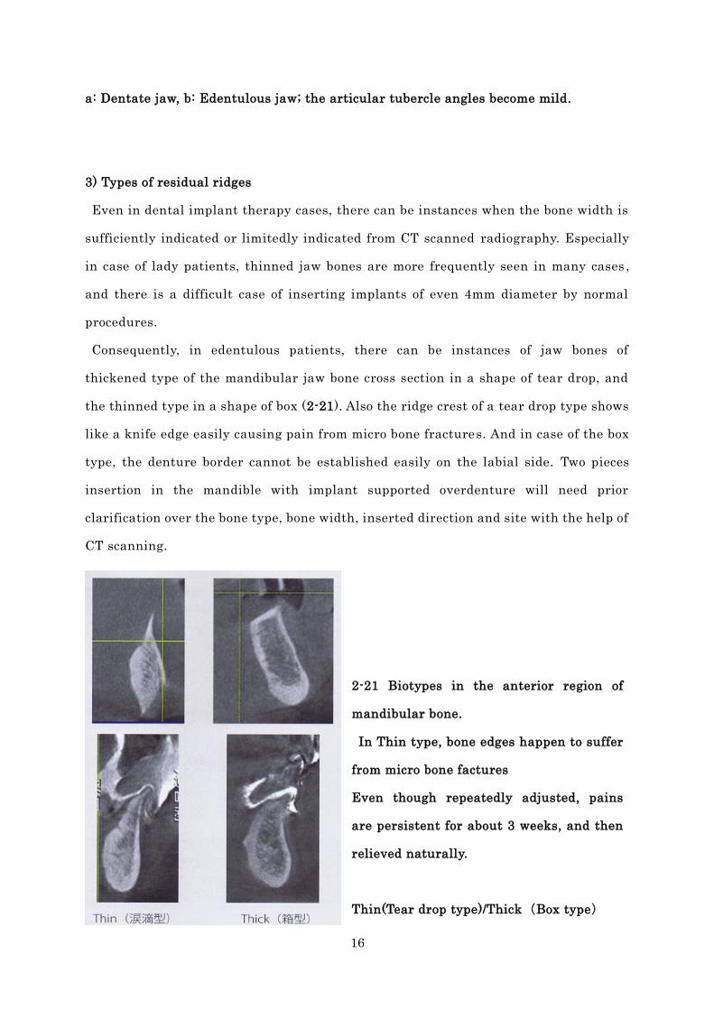
Consequently, in edentulous patients, there can be instances of jaw bones of
thickened type of the mandibular jaw bone cross section in a shape of tear drop, and
the thinned type in a shape of box (2-21). Also the ridge crest of a tear drop type shows
like a knife edge easily causing pain from micro bone fractures. And in case of the box
type, the denture border cannot be established easily on the labial side. Two pieces
insertion in the mandible with implant supported overdenture will need prior
clarification over the bone type, bone width, inserted direction and site with the help of
CT scanning.
2-21 Biotypes in the anterior region of
mandibular bone.
In Thin type, bone edges happen to suffer
from micro bone factures
Even though repeatedly adjusted, pains
are persistent for about 3 weeks, and then
relieved naturally.
Thin(Tear drop type)/Thick(Box type)
17
Q: What makes a denture unstable?
A: The oral cavity continues to move while in function. Since the lips, cheeks and
tongue continue to move when singing a song, talking, taking food and chewing , they
will make a denture unstable, being applied with functional pressures from all
directions. And a denture is made unstable too from direct pressure of food bolus in
chewing.
Changes in oral shapes in function will make a denture unstable.
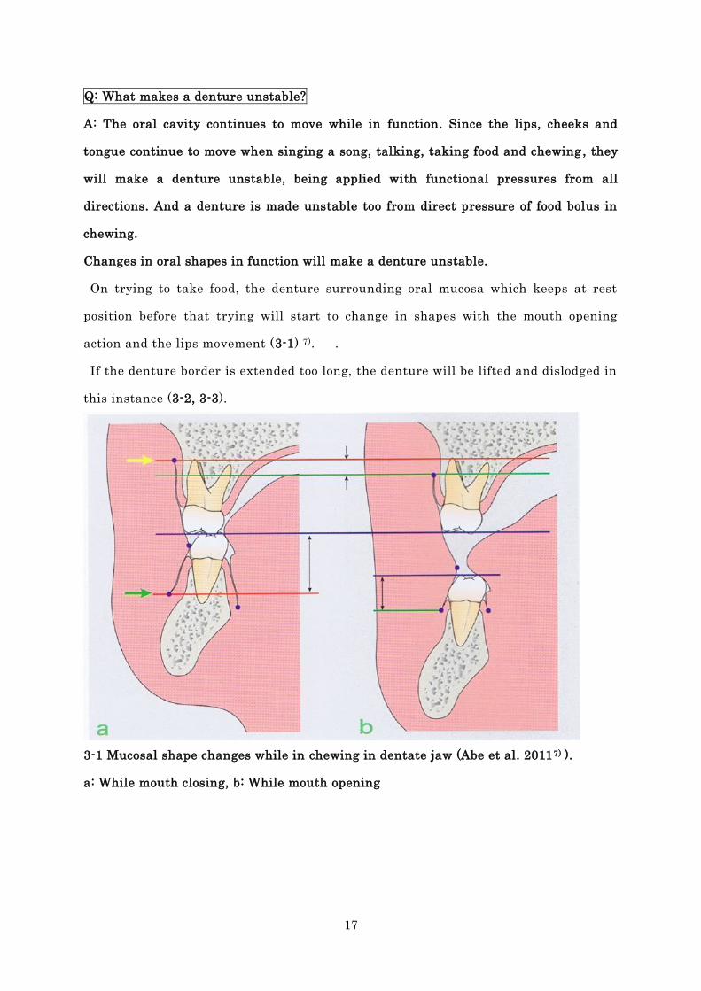
On trying to take food, the denture surrounding oral mucosa which keeps at rest
position before that trying will start to change in shapes with the mouth opening
action and the lips movement (3-1) 7). .
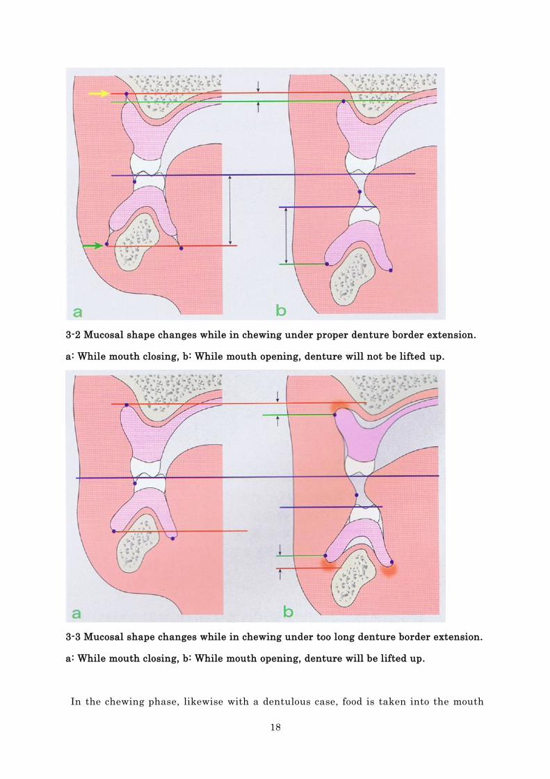
If the denture border is extended too long, the denture will be lifted and dislodged in
this instance (3-2, 3-3).
3-1 Mucosal shape changes while in chewing in dentate jaw (Abe et al. 20117) ).
a: While mouth closing, b: While mouth opening
18
3-2 Mucosal shape changes while in chewing under proper denture border extension.
a: While mouth closing, b: While mouth opening, denture will not be lifted up.
3-3 Mucosal shape changes while in chewing under too long denture border extension.
a: While mouth closing, b: While mouth opening, denture will be lifted up.
In the chewing phase, likewise with a dentulous case, food is taken into the mouth
19
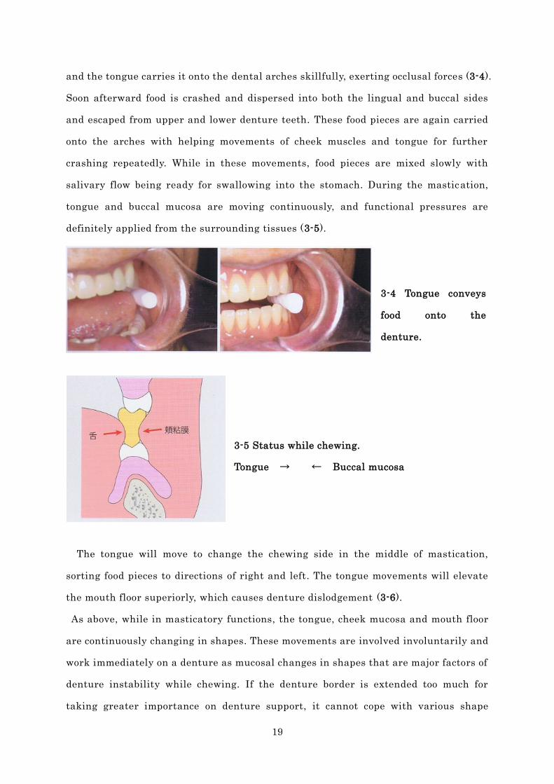
and the tongue carries it onto the dental arches skillfully, exerting occlusal forces (3-4).
Soon afterward food is crashed and dispersed into both the lingual and buccal sides
and escaped from upper and lower denture teeth. These food pieces are again carried
onto the arches with helping movements of cheek muscles and tongue for further
crashing repeatedly. While in these movements, food pieces are mixed slowly with
salivary flow being ready for swallowing into the stomach. During the mastication,
tongue and buccal mucosa are moving continuously, and functional pressures are
definitely applied from the surrounding tissues (3-5).
3-4 Tongue conveys
food onto the
denture.
3-5 Status while chewing.
Tongue → ← Buccal mucosa
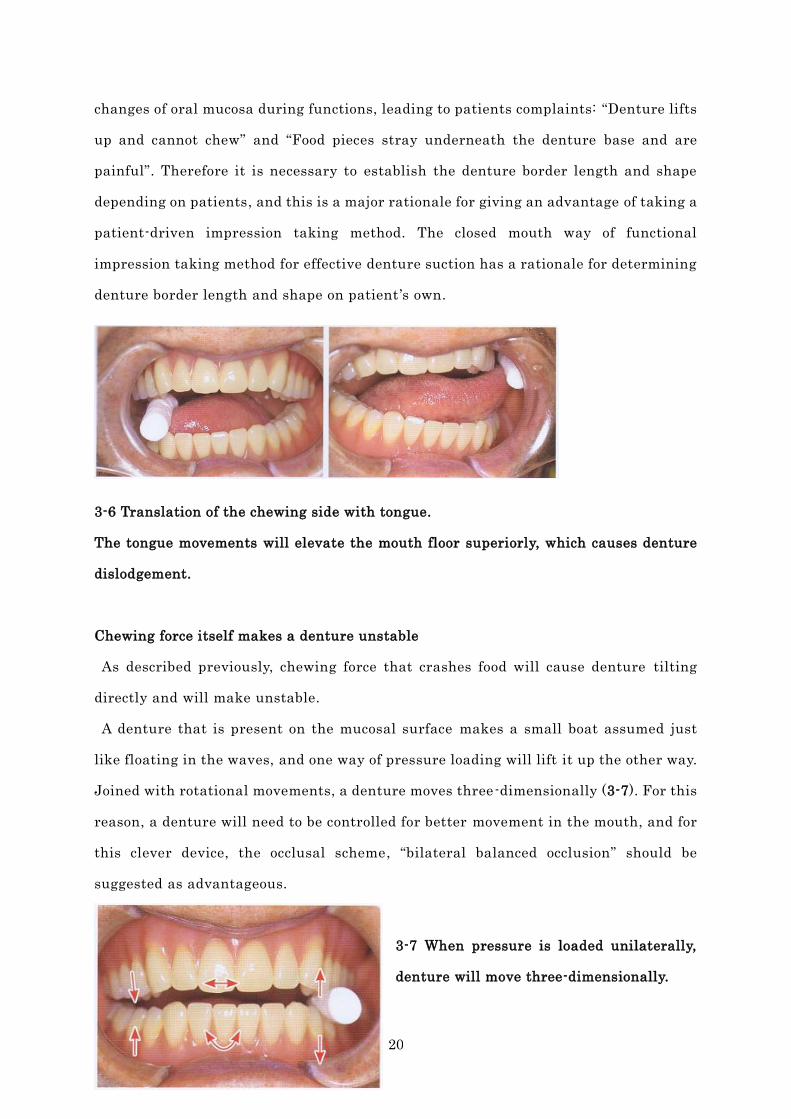
The tongue will move to change the chewing side in the middle of mastication,
sorting food pieces to directions of right and left. The tongue movements will elevate
the mouth floor superiorly, which causes denture dislodgement (3-6).
As above, while in masticatory functions, the tongue, cheek mucosa and mouth floor
are continuously changing in shapes. These movements are involved involuntarily and
work immediately on a denture as mucosal changes in shapes that are major factors of
denture instability while chewing. If the denture border is extended too much for
taking greater importance on denture support, it cannot cope with various shape
20
changes of oral mucosa during functions, leading to patients complaints: “Denture lifts
up and cannot chew” and “Food pieces stray underneath the denture base and are
painful”. Therefore it is necessary to establish the denture border length and shape
depending on patients, and this is a major rationale for giving an advantage of taking a
patient-driven impression taking method. The closed mouth way of functional
impression taking method for effective denture suction has a rationale for determining
denture border length and shape on patient ’s own.
3-6 Translation of the chewing side with tongue.
The tongue movements will elevate the mouth floor superiorly, which causes denture
dislodgement.
Chewing force itself makes a denture unstable
As described previously, chewing force that crashes food will cause denture tilting
directly and will make unstable.
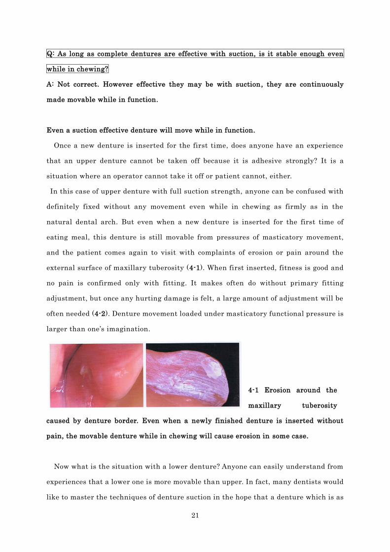
A denture that is present on the mucosal surface makes a small boat assumed just
like floating in the waves, and one way of pressure loading will lift it up the other way.
Joined with rotational movements, a denture moves three-dimensionally (3-7). For this
reason, a denture will need to be controlled for better movement in the mouth, and for
this clever device, the occlusal scheme, “bilateral balanced occlusion” should be
suggested as advantageous.
3-7 When pressure is loaded unilaterally,
denture will move three-dimensionally.
21
Q: As long as complete dentures are effective with suction, is it stable enough even
while in chewing?
A: Not correct. However effective they may be with suction, they are continuously
made movable while in function.
Even a suction effective denture will move while in function.
Once a new denture is inserted for the first time, does anyone have an experience
that an upper denture cannot be taken off because it is adhesive strongly? It is a
situation where an operator cannot take it off or patient cannot, either.
In this case of upper denture with full suction strength, anyone can be confused with
definitely fixed without any movement even while in chewing as firmly as in the
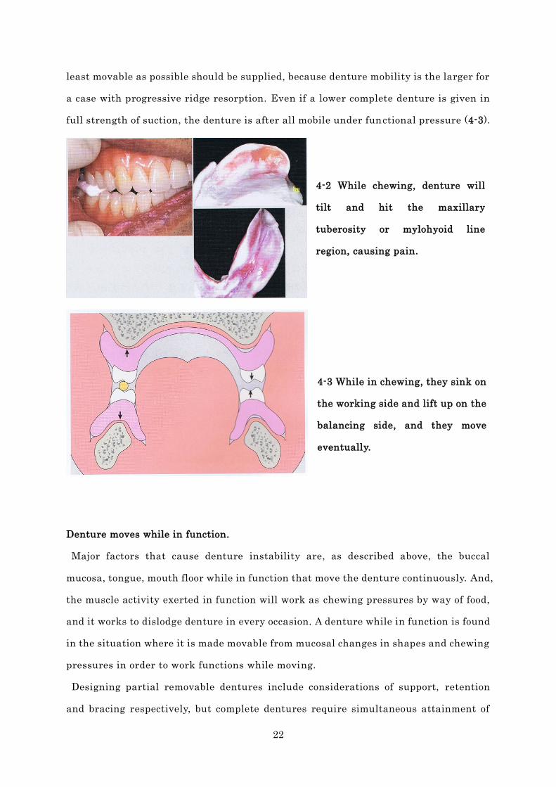
natural dental arch. But even when a new denture is inserted for the first time of
eating meal, this denture is still movable from pressures of masticatory movement,
and the patient comes again to visit with complaints of erosion or pain around the
external surface of maxillary tuberosity (4-1). When first inserted, fitness is good and
no pain is confirmed only with fitting. It makes often do without primary fitting
adjustment, but once any hurting damage is felt, a large amount of adjustment will be
often needed (4-2). Denture movement loaded under masticatory functional pressure is
larger than one’s imagination.
4-1 Erosion around the
maxillary tuberosity
caused by denture border. Even when a newly finished denture is inserted without
pain, the movable denture while in chewing will cause erosion in some case.
Now what is the situation with a lower denture? Anyone can easily understand from
experiences that a lower one is more movable than upper. In fact, many dentists would
like to master the techniques of denture suction in the hope that a denture which is as
22
least movable as possible should be supplied, because denture mobility is the larger for
a case with progressive ridge resorption. Even if a lower complete denture is given in
full strength of suction, the denture is after all mobile under functional pressure (4-3).
4-2 While chewing, denture will
tilt and hit the maxillary
tuberosity or mylohyoid line
region, causing pain.
4-3 While in chewing, they sink on
the working side and lift up on the
balancing side, and they move
eventually.
Denture moves while in function.
Major factors that cause denture instability are, as described above, the buccal
mucosa, tongue, mouth floor while in function that move the denture continuously. And,
the muscle activity exerted in function will work as chewing pressures by way of food,
and it works to dislodge denture in every occasion. A denture while in function is found
in the situation where it is made movable from mucosal changes in shapes and chewing
pressures in order to work functions while moving.
Designing partial removable dentures include considerations of support, retention
and bracing respectively, but complete dentures require simultaneous attainment of
23
support, retention and bracing with one unit of denture body.
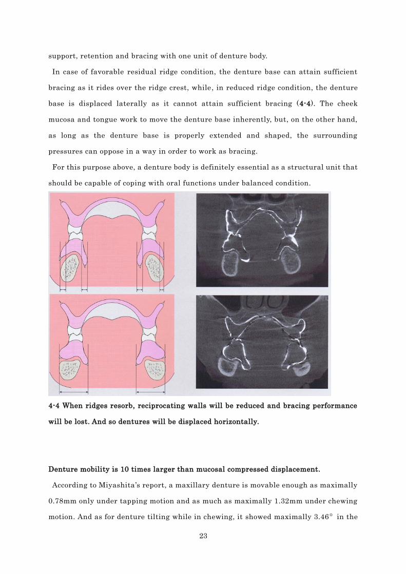
In case of favorable residual ridge condition, the denture base can attain sufficient
bracing as it rides over the ridge crest, while, in reduced ridge condition, the denture
base is displaced laterally as it cannot attain sufficient bracing (4-4). The cheek
mucosa and tongue work to move the denture base inherently, but, on the other hand,
as long as the denture base is properly extended and shaped, the surrounding
pressures can oppose in a way in order to work as bracing.
For this purpose above, a denture body is definitely essential as a structural unit that
should be capable of coping with oral functions under balanced condition.
4-4 When ridges resorb, reciprocating walls will be reduced and bracing performance
will be lost. And so dentures will be displaced horizontally.
Denture mobility is 10 times larger than mucosal compressed displacement.
According to Miyashita’s report, a maxillary denture is movable enough as maximally
0.78mm only under tapping motion and as much as maximally 1.32mm under chewing
motion. And as for denture tilting while in chewing, it showed maximally 3.46º in the
24
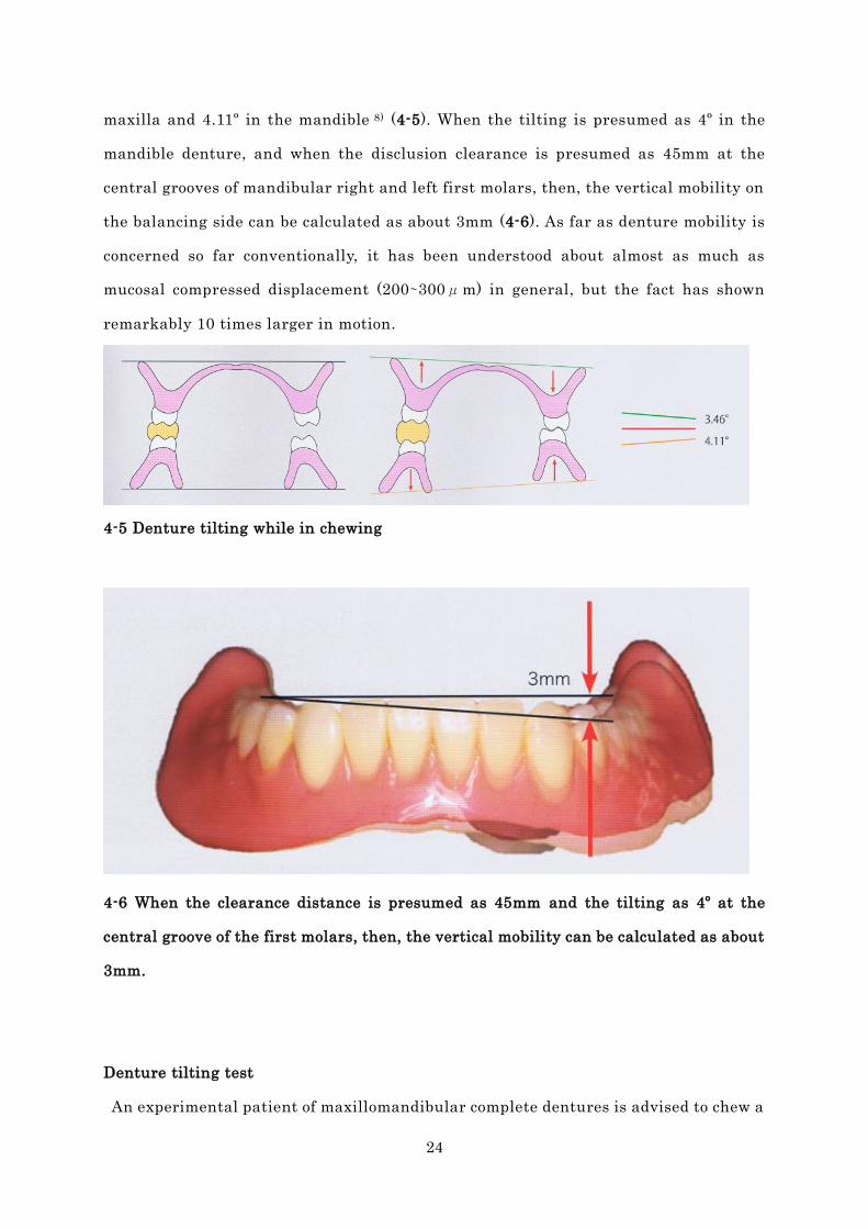
maxilla and 4.11º in the mandible 8) (4-5). When the tilting is presumed as 4º in the
mandible denture, and when the disclusion clearance is presumed as 45mm at the
central grooves of mandibular right and left first molars, then, the vertical mobility on
the balancing side can be calculated as about 3mm (4-6). As far as denture mobility is
concerned so far conventionally, it has been understood about almost as much as
mucosal compressed displacement (200∼300μm) in general, but the fact has shown
remarkably 10 times larger in motion.
4-5 Denture tilting while in chewing
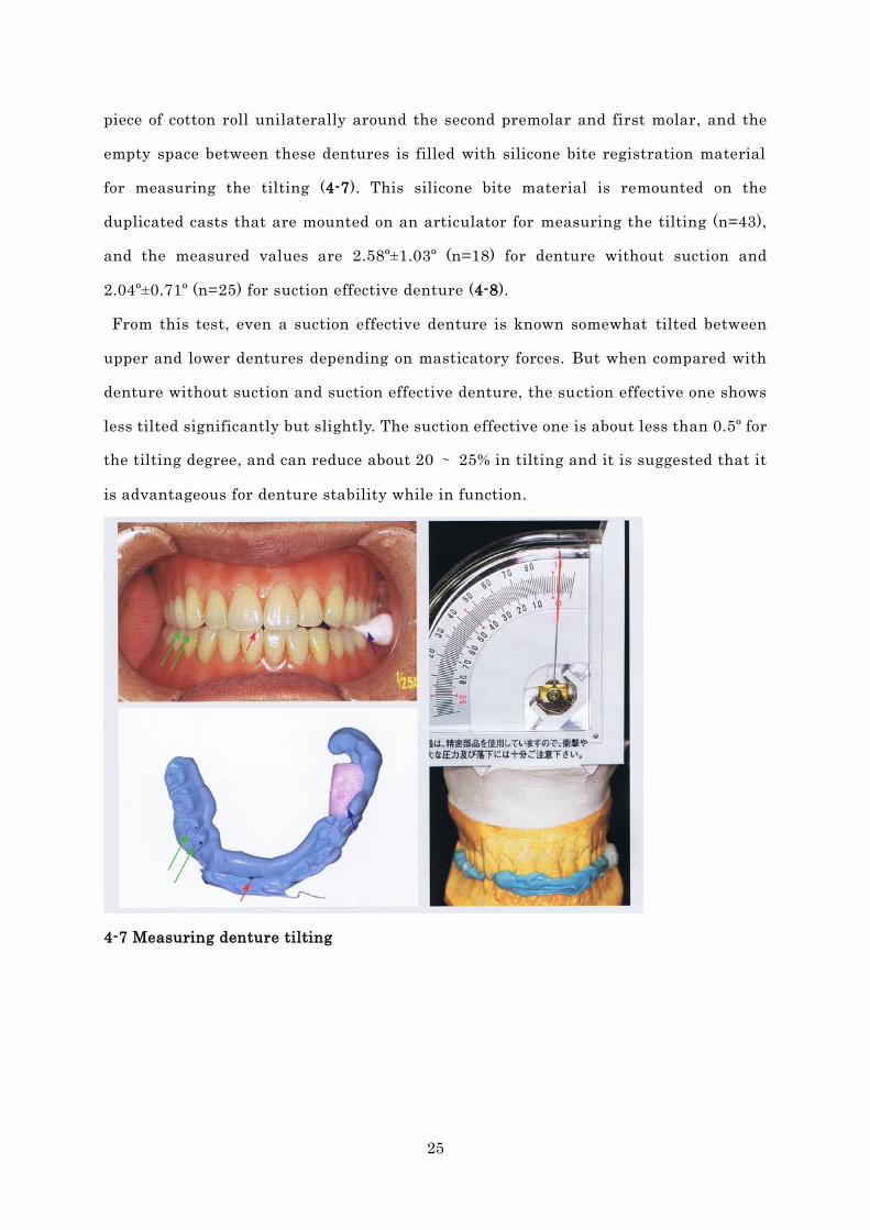
4-6 When the clearance distance is presumed as 45mm and the tilting as 4º at the
central groove of the first molars, then, the vertical mobility can be calculated as about
3mm.
Denture tilting test
An experimental patient of maxillomandibular complete dentures is advised to chew a
25
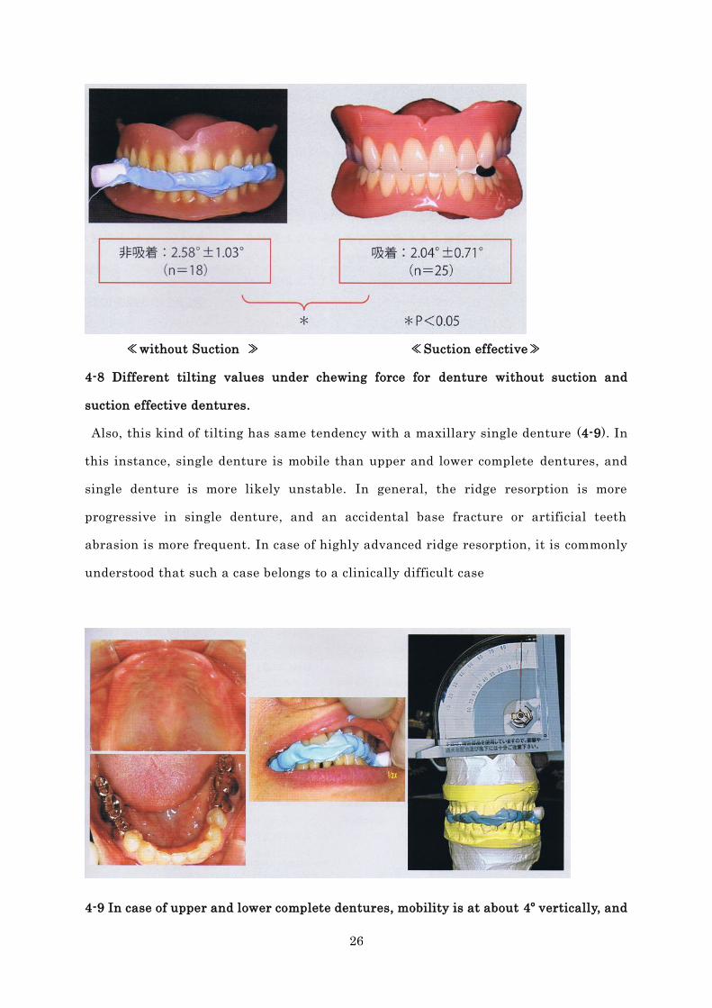
piece of cotton roll unilaterally around the second premolar and first molar, and the
empty space between these dentures is filled with silicone bite registration material
for measuring the tilting (4-7). This silicone bite material is remounted on the
duplicated casts that are mounted on an articulator for measuring the tilting (n=43),
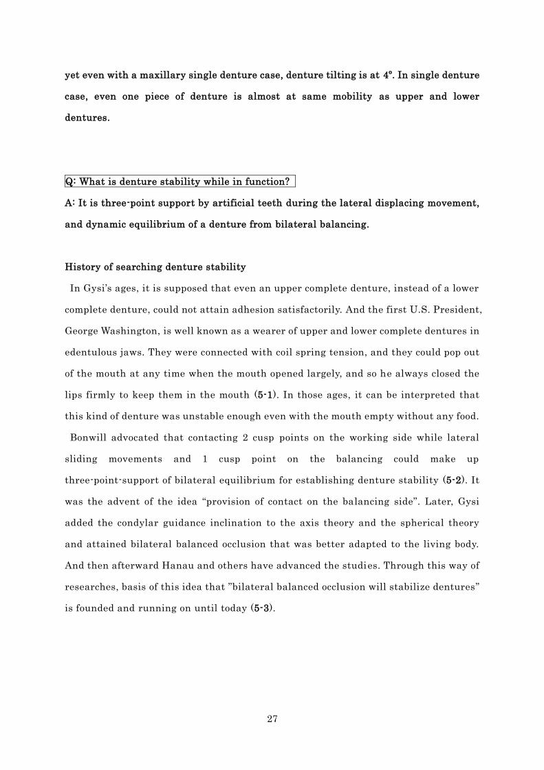
and the measured values are 2.58º±1.03º (n=18) for denture without suction and
2.04º±0.71º (n=25) for suction effective denture (4-8).
From this test, even a suction effective denture is known somewhat tilted between
upper and lower dentures depending on masticatory forces. But when compared with
denture without suction and suction effective denture, the suction effective one shows
less tilted significantly but slightly. The suction effective one is about less than 0.5º for
the tilting degree, and can reduce about 20 ∼ 25% in tilting and it is suggested that it
is advantageous for denture stability while in function.
4-7 Measuring denture tilting
26
≪without Suction ≫ ≪Suction effective≫
4-8 Different tilting values under chewing force for denture without suction and
suction effective dentures.
Also, this kind of tilting has same tendency with a maxillary single denture (4-9). In
this instance, single denture is mobile than upper and lower complete dentures, and
single denture is more likely unstable. In general, the ridge resorption is more
progressive in single denture, and an accidental base fracture or artificial teeth
abrasion is more frequent. In case of highly advanced ridge resorption, it is commonly
understood that such a case belongs to a clinically difficult case
4-9 In case of upper and lower complete dentures, mobility is at about 4º vertically, and
27
yet even with a maxillary single denture case, denture tilting is at 4º. In single denture
case, even one piece of denture is almost at same mobility as upper and lower
dentures.
Q: What is denture stability while in function?
A: It is three-point support by artificial teeth during the lateral displacing movement,
and dynamic equilibrium of a denture from bilateral balancing.
History of searching denture stability
In Gysi’s ages, it is supposed that even an upper complete denture, instead of a lower
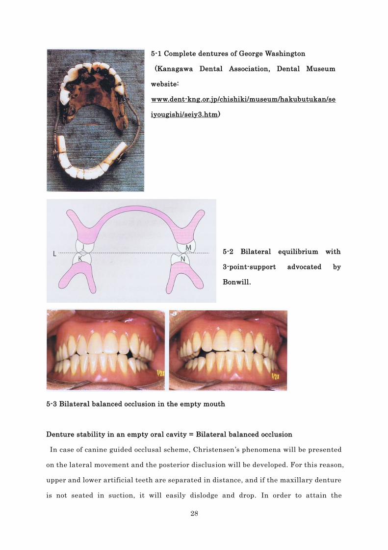
complete denture, could not attain adhesion satisfactorily. And the first U.S. President,
George Washington, is well known as a wearer of upper and lower complete dentures in
edentulous jaws. They were connected with coil spring tension, and they could pop out
of the mouth at any time when the mouth opened largely, and so he always closed the
lips firmly to keep them in the mouth (5-1). In those ages, it can be interpreted that
this kind of denture was unstable enough even with the mouth empty without any food.
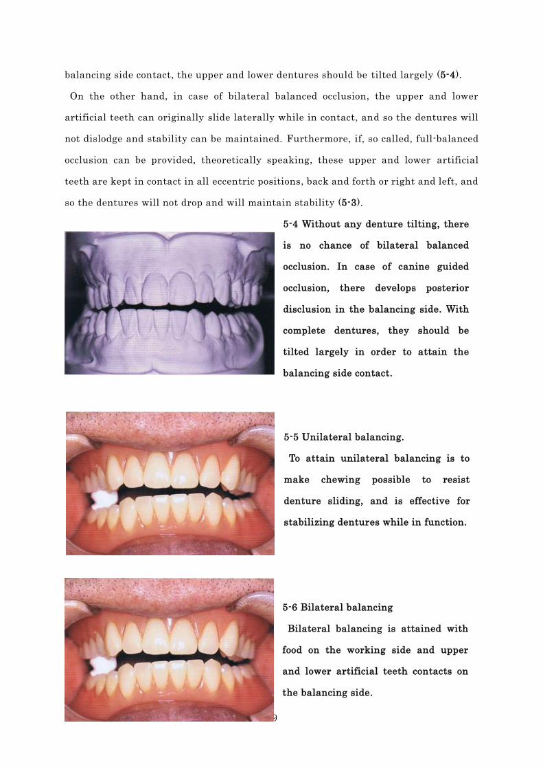
Bonwill advocated that contacting 2 cusp points on the working side while lateral
sliding movements and 1 cusp point on the balancing could make up
three-point-support of bilateral equilibrium for establishing denture stability (5-2). It
was the advent of the idea “provision of contact on the balancing side”. Later, Gysi
added the condylar guidance inclination to the axis theory and the spherical theory
and attained bilateral balanced occlusion that was better adapted to the living body.
And then afterward Hanau and others have advanced the studies. Through this way of
researches, basis of this idea that ”bilateral balanced occlusion will stabilize dentures”
is founded and running on until today (5-3).
28
5-1 Complete dentures of George Washington
(Kanagawa Dental Association, Dental Museum
website:
www.dent-kng.or.jp/chishiki/museum/hakubutukan/se
iyougishi/seiy3.htm)
5-2 Bilateral equilibrium with
3-point-support advocated by
Bonwill.
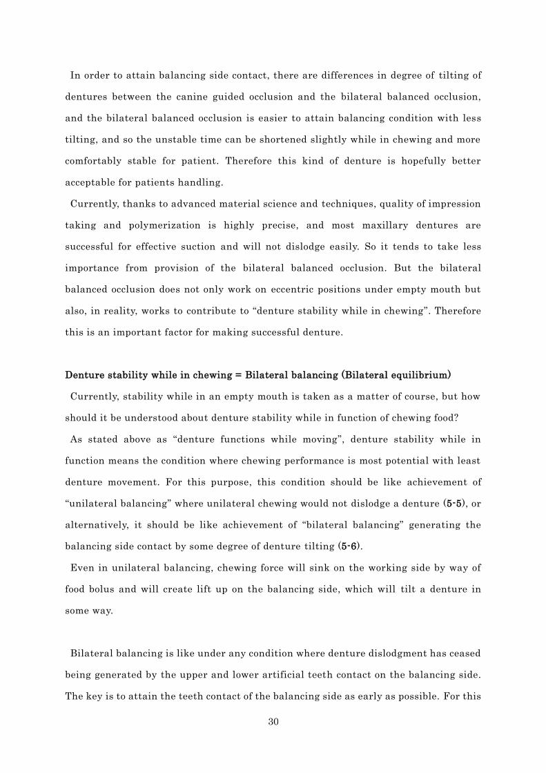
5-3 Bilateral balanced occlusion in the empty mouth
Denture stability in an empty oral cavity = Bilateral balanced occlusion
In case of canine guided occlusal scheme, Christensen ’s phenomena will be presented
on the lateral movement and the posterior disclusion will be developed. For this reason,
upper and lower artificial teeth are separated in distance, and if the maxillary denture
is not seated in suction, it will easily dislodge and drop. In order to attain the
29
balancing side contact, the upper and lower dentures should be tilted largely (5-4).
On the other hand, in case of bilateral balanced occlusion, the upper and lower
artificial teeth can originally slide laterally while in contact, and so the dentures will
not dislodge and stability can be maintained. Furthermore, if, so called, full-balanced
occlusion can be provided, theoretically speaking, these upper and lower artificial
teeth are kept in contact in all eccentric positions, back and forth or right and left, and
so the dentures will not drop and will maintain stability (5-3).
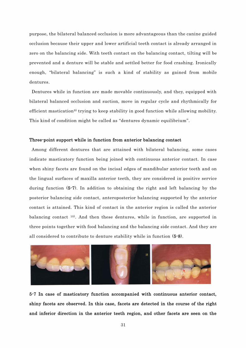
5-4 Without any denture tilting, there
is no chance of bilateral balanced
occlusion. In case of canine guided
occlusion, there develops posterior
disclusion in the balancing side. With
complete dentures, they should be
tilted largely in order to attain the
balancing side contact.
5-5 Unilateral balancing.
To attain unilateral balancing is to
make chewing possible to resist
denture sliding, and is effective for
stabilizing dentures while in function.
5-6 Bilateral balancing
Bilateral balancing is attained with
food on the working side and upper
and lower artificial teeth contacts on
the balancing side.
30
In order to attain balancing side contact, there are differences in degree of tilting of
dentures between the canine guided occlusion and the bilateral balanced occlusion,
and the bilateral balanced occlusion is easier to attain balancing condition with less
tilting, and so the unstable time can be shortened slightly while in chewing and more
comfortably stable for patient. Therefore this kind of denture is hopefully better
acceptable for patients handling.
Currently, thanks to advanced material science and techniques, quality of impression
taking and polymerization is highly precise, and most maxillary dentures are
successful for effective suction and will not dislodge easily. So it tends to take less
importance from provision of the bilateral balanced occlusion. But the bilateral
balanced occlusion does not only work on eccentric positions under empty mouth but
also, in reality, works to contribute to “denture stability while in chewing”. Therefore
this is an important factor for making successful denture.
Denture stability while in chewing = Bilateral balancing (Bilateral equilibrium)
Currently, stability while in an empty mouth is taken as a matter of course, but how
should it be understood about denture stability while in function of chewing food?
As stated above as “denture functions while moving”, denture stability while in
function means the condition where chewing performance is most potential with least
denture movement. For this purpose, this condition should be like achievement of
“unilateral balancing” where unilateral chewing would not dislodge a denture (5-5), or
alternatively, it should be like achievement of “bilateral balancing” generating the
balancing side contact by some degree of denture tilting (5-6).
Even in unilateral balancing, chewing force will sink on the working side by way of
food bolus and will create lift up on the balancing side, which will tilt a denture in
some way.
Bilateral balancing is like under any condition where denture dislodgment has ceased
being generated by the upper and lower artificial teeth contact on the balancing side.
The key is to attain the teeth contact of the balancing side as early as possible. For this
31
purpose, the bilateral balanced occlusion is more advantageous than the canine guided
occlusion because their upper and lower artificial teeth contact is already arranged in
zero on the balancing side. With teeth contact on the balancing contact, tilting will be
prevented and a denture will be stable and settled better for food crashing. Ironically
enough, “bilateral balancing” is such a kind of stability as gained from mobile
dentures.
Dentures while in function are made movable continuously, and they, equipped with
bilateral balanced occlusion and suction, move in regular cycle and rhythmically for
efficient mastication9) trying to keep stability in good function while allowing mobility.
This kind of condition might be called as “dentures dynamic equilibrium”.
Three-point support while in function from anterior balancing contact
Among different dentures that are attained with bilateral balancing, some cases
indicate masticatory function being joined with continuous anterior contact. In case
when shiny facets are found on the incisal edges of mandibular anterior teeth and on
the lingual surfaces of maxilla anterior teeth, they are considered in positive service
during function (5-7). In addition to obtaining the right and left balancing by the
posterior balancing side contact, anteroposterior balancing supported by the anterior
contact is attained. This kind of contact in the anterior region is called the anterior
balancing contact 10). And then these dentures, while in function, are supported in
three points together with food balancing and the balancing side contact. And they are
all considered to contribute to denture stability while in function (5-8).
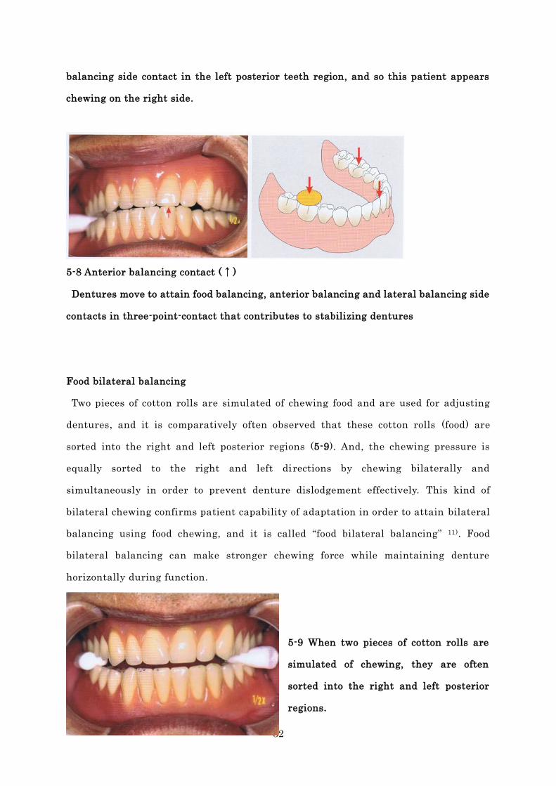
5-7 In case of masticatory function accompanied with continuous anterior contact,
shiny facets are observed. In this case, facets are detected in the course of the right
and inferior direction in the anterior teeth region, and other facets are seen on the
32
balancing side contact in the left posterior teeth region, and so this patient appears
chewing on the right side.
5-8 Anterior balancing contact (↑)
Dentures move to attain food balancing, anterior balancing and lateral balancing side
contacts in three-point-contact that contributes to stabilizing dentures
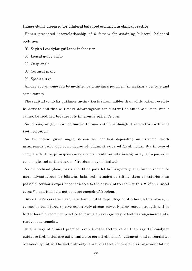
Food bilateral balancing
Two pieces of cotton rolls are simulated of chewing food and are used for adjusting
dentures, and it is comparatively often observed that these cotton rolls (food) are
sorted into the right and left posterior regions (5-9). And, the chewing pressure is
equally sorted to the right and left directions by chewing bilaterally and
simultaneously in order to prevent denture dislodgement effectively. This kind of
bilateral chewing confirms patient capability of adaptation in order to attain bilateral
balancing using food chewing, and it is called “food bilateral balancing” 11). Food
bilateral balancing can make stronger chewing force while maintaining denture
horizontally during function.
5-9 When two pieces of cotton rolls are
simulated of chewing, they are often
sorted into the right and left posterior
regions.
33
Hanau Quint prepared for bilateral balanced occlusion in clinical practice
Hanau presented interrelationship of 5 factors for attaining bilateral balanced
occlusion.
① Sagittal condylar guidance inclination
② Incisal guide angle
③ Cusp angle
④ Occlusal plane
⑤ Spee’s curve
Among above, some can be modified by clinician ’s judgment in making a denture and
some cannot.
The sagittal condylar guidance inclination is shown milder than while patient used to
be dentate and this will make advantageous for bilateral balanced occlusion, but it
cannot be modified because it is inherently patient’s own.
As for cusp angle, it can be limited to some extent, although it varies from artificial
teeth selection.
As for incisal guide angle, it can be modified depending on artificial teeth
arrangement, allowing some degree of judgment reserved for clinician. But in case of
complete denture, principles are non-contact anterior relationship or equal to posterior
cusp angle and so the degree of freedom may be limited.
As for occlusal plane, basis should be parallel to Camper ’s plane, but it should be
more advantageous for bilateral balanced occlusion by tilting them as anteriorly as
possible. Author ’s experience indicates to the degree of freedom within 2∼3º in clinical
cases 11), and it should not be large enough of freedom.
Since Spee’s curve is to some extent limited depending on 4 other factors above, it
cannot be considered to give excessively strong curve. Rather, curve strength will be
better based on common practice following an average way of teeth arrangement and a
ready made template.
In this way of clinical practice, even 4 other factors other than sagittal condylar
guidance inclination are quite limited to permit clinician ’s judgment, and so requisites
of Hanau Quint will be met duly only if artificial teeth choice and arrangement follow
34
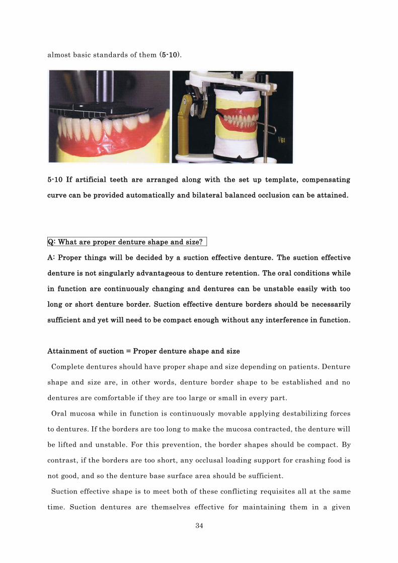
almost basic standards of them (5-10).
5-10 If artificial teeth are arranged along with the set up template, compensating
curve can be provided automatically and bilateral balanced occlusion can be attained.
Q: What are proper denture shape and size?
A: Proper things will be decided by a suction effective denture. The suction effective
denture is not singularly advantageous to denture retention. The oral conditions while
in function are continuously changing and dentures can be unstable easily with too
long or short denture border. Suction effective denture borders should be necessarily
sufficient and yet will need to be compact enough without any interference in function.
Attainment of suction = Proper denture shape and size
Complete dentures should have proper shape and size depending on patients. Denture
shape and size are, in other words, denture border shape to be established and no
dentures are comfortable if they are too large or small in every part.
Oral mucosa while in function is continuously movable applying destabilizing forces
to dentures. If the borders are too long to make the mucosa contracted, the denture will
be lifted and unstable. For this prevention, the border shapes should be compact. By
contrast, if the borders are too short, any occlusal loading support for crashing food is
not good, and so the denture base surface area should be sufficient.
Suction effective shape is to meet both of these conflicting requisites all at the same
time. Suction dentures are themselves effective for maintaining them in a given
35
position, and moreover they are compact enough to prevent from dislodging against
moving mucosa, and necessarily sufficient surface area is equipped.
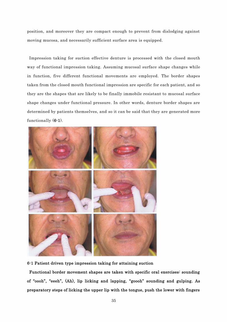
Impression taking for suction effective denture is processed with the closed mouth
way of functional impression taking. Assuming mucosal surface shape changes while
in function, five different functional movements are employed. The border shapes
taken from the closed mouth functional impression are specific for each patient, and so
they are the shapes that are likely to be finally immobile resistant to mucosal surface
shape changes under functional pressure. In other words, denture border shapes are
determined by patients themselves, and so it can be said that they are generated more
functionally (6-1).
6-1 Patient driven type impression taking for attaining suction
Functional border movement shapes are taken with specific oral exercises: sounding
of “oooh”, “eeeh”, (Ah), lip licking and lapping, “goooh” sounding and gulping. As
preparatory steps of licking the upper lip with the tongue, push the lower with fingers
36
in order not to lift it up at the mouth opening.
In order to take an operator driven impression taking like conventional method of
border molding method, impression should be taken necessarily assuming shape
changes of these mucosal surface shape changes. Unless an operator is equipped with
experience and skills, comfortable border shapes cannot be taken properly. In case of
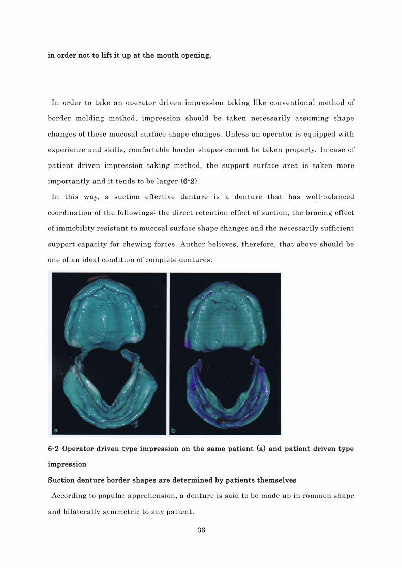
patient driven impression taking method, the support surface area is taken more
importantly and it tends to be larger (6-2).
In this way, a suction effective denture is a denture that has well-balanced
coordination of the followings: the direct retention effect of suction, the bracing effect
of immobility resistant to mucosal surface shape changes and the necessarily sufficient
support capacity for chewing forces. Author believes, therefore, that above should be
one of an ideal condition of complete dentures.
6-2 Operator driven type impression on the same patient (a) and patient driven type
impression
Suction denture border shapes are determined by patients themselves
According to popular apprehension, a denture is said to be made up in common shape
and bilaterally symmetric to any patient.
37
Conversely when details are studied carefully, residual ridge appearances and
mucosal surface shapes while in function are different from individual patients, and so,
as a matter of fact, there is absolutely nothing same in denture shapes, and even
within the same patient there is nothing symmetrical.
In order to make up the shapes of suction denture border, the peripheral border seal
must be made to apply proper pressures to the mucosa throughout the denture margin
entirely, and so it is necessary to judge the location and shape of where to
accommodate the denture border correctly. This kind of difficult subject should not be
mastered by limited resources of clinician ’s experience and skills, but this difficulty
can be solved automatically by taking a snap impression of the closed mouth method
using the Frame Cut Back Tray initially, and by being followed with the patient driven
type impression taking method. Thinking of asking patient so that the denture border
can be determined by patient ’s own in this way, any clinician can attain a suction
effective denture in almost all cases.
Q: What is the reason for making the bite taking of complete denture patient
difficult?
A: Bite taking of complete denture patient is determined by patient’s own occlusal
plane, occlusal vertical dimension, and horizontal mandibular jaw position. Reasons
for what makes difficult are because it needs to take this method by putting together
both factors of patient’s own shapes and of functions. So this method cannot be taken
from every patient in full confidence yet in good advance, but it is developed through
gradual steps from obscurity. Upper and lower residual ridges cannot determine upper
and lower dentures easily in three dimensional positions, even though they are
connected with TMJ and other oral tissues. In order to take bite taking successfully,
one should try carefully to set up basic standard, to evaluate functions, and to
accommodate within the area of patient’s own ‘strike zone’.
38
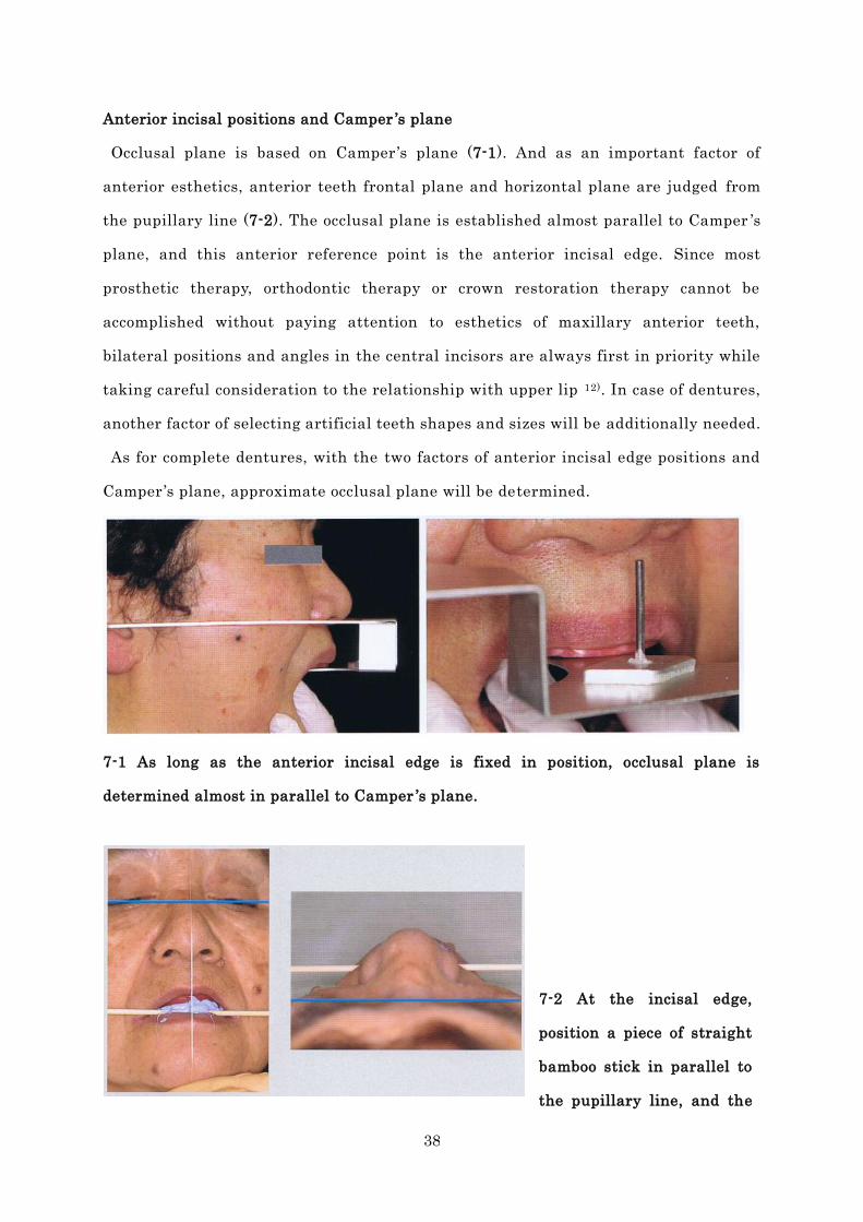
Anterior incisal positions and Camper ’s plane
Occlusal plane is based on Camper ’s plane (7-1). And as an important factor of
anterior esthetics, anterior teeth frontal plane and horizontal plane are judged from
the pupillary line (7-2). The occlusal plane is established almost parallel to Camper ’s
plane, and this anterior reference point is the anterior incisal edge. Since most
prosthetic therapy, orthodontic therapy or crown restoration therapy cannot be
accomplished without paying attention to esthetics of maxillary anterior teeth,
bilateral positions and angles in the central incisors are always first in priority while
taking careful consideration to the relationship with upper lip 12). In case of dentures,
another factor of selecting artificial teeth shapes and sizes will be additionally needed.
As for complete dentures, with the two factors of anterior incisal edge positions and
Camper ’s plane, approximate occlusal plane will be determined.
7-1 As long as the anterior incisal edge is fixed in position, occlusal plane is
determined almost in parallel to Camper ’s plane.
7-2 At the incisal edge,
position a piece of straight
bamboo stick in parallel to
the pupillary line, and the
39
frontal plane can be recorded as in the right photo. Maxillary anterior teeth can be
arranged by coordinating the labial surfaces to patient ’s facial front.
Reasons for why bite taking is not done well in an edentulous jaw
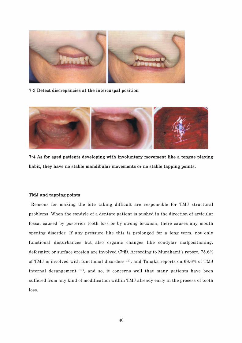
If there is any discrepancy between bite taking records, touch them with finger
surface while having patient close upper and lower artificial teeth. With this, you can
feel the discrepancy as displacement of dentures from the touching senses (7-3). Like
in this case, are the errors of bite taking records that prevent smooth intercuspation
responsible for clinician ’s reading mistakes at the stage of trial insertion or finishing?
Errors of bite taking records are caused by method failure of constructing a biteplate,
its poor handling, or incorrect situation of reproducibility of occlusion to a given
position. These causes are categorized on problems of clinicians or patients.
As for errors involved with clinicians, causes behind cannot be detected incidentally.
They are causes of ill fitting of biteplates being difficult to keep a given position and of
interfering of upper and lower wax rims or biteplates somewhere in the way of guiding
mandibular positions.
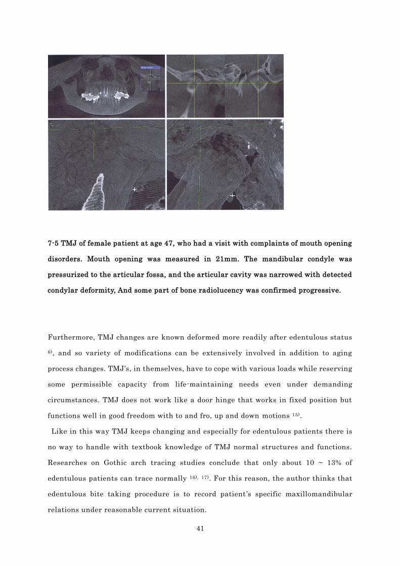
As for errors of patients side, when the patient driven type is taken on the upper and
lower wax rims asking “close them, please” or “bite them, please”, then, inability of
controlling mandibular jaw positions from central nerve system problems may cause
accidental discrepancy in the anterior or lateral directions. Especially in case of any
involuntary motion like tongue habit, there may be a cause of unstable mandibular jaw
position (7-4). Also, in case when a bite is taken on the operator driven type of
impression taking, issues will include how comfortably patient is relaxed in advance or
how much clinician is equipped with guiding skills. The biggest problem is, however,
concerned with disorders with TMJ as in the followings.
40
7-3 Detect discrepancies at the intercuspal position
7-4 As for aged patients developing with involuntary movement like a tongue playing
habit, they have no stable mandibular movements or no stable tapping points.
TMJ and tapping points
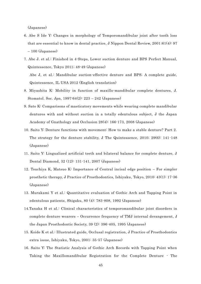
Reasons for making the bite taking difficult are responsible for TMJ structural
problems. When the condyle of a dentate patient is pushed in the direction of articular
fossa, caused by posterior tooth loss or by strong bruxism, there causes any mouth
opening disorder. If any pressure like this is prolonged for a long term, not only
functional disturbances but also organic changes like condylar malpositioning,
deformity, or surface erosion are involved (7-5). According to Murakami ’s report, 75.6%
of TMJ is involved with functional disorders 13), and Tanaka reports on 68.6% of TMJ
internal derangement 14), and so, it concerns well that many patients have been
suffered from any kind of modification within TMJ already early in the process of tooth
loss.
41
7-5 TMJ of female patient at age 47, who had a visit with complaints of mouth opening
disorders. Mouth opening was measured in 21mm. The mandibular condyle was
pressurized to the articular fossa, and the articular cavity was narrowed with detected
condylar deformity, And some part of bone radiolucency was confirmed progressive.
Furthermore, TMJ changes are known deformed more readily after edentulous status
6), and so variety of modifications can be extensively involved in addition to aging
process changes. TMJ’s, in themselves, have to cope with various loads while reserving
some permissible capacity from life-maintaining needs even under demanding
circumstances. TMJ does not work like a door hinge that works in fixed position but
functions well in good freedom with to and fro, up and down motions 15).
Like in this way TMJ keeps changing and especially for edentulous patients there is
no way to handle with textbook knowledge of TMJ normal structures and functions.
Researches on Gothic arch tracing studies conclude that only about 10 ~ 13% of
edentulous patients can trace normally 16), 17). For this reason, the author thinks that
edentulous bite taking procedure is to record patient ’s specific maxillomandibular
relations under reasonable current situation.

42
Bite taking in clinical practice is in reality to judge the reproducible tapping points
based on the functional examination of Gothic arch tracings after removing engram,
and to determine the mandibular jaw position as best compromised points 16) (7-6).
7-6 With the Gothic arch tracings, tapping points are verified for proper judgment.
Tapping points taken with reproducibility and stability should be employed.
3 Essences and 4 Steps in denture construction
Complete denture construction can be consolidated into 3 essences : denture base
shape, bite taking and finalization of denture with artificial teeth arrangement.
“Denture base shape” means “impression taking” in other words, and “bite taking” is
“vertical and horizontal mandibular jaw position”. And, “denture finalization with
artificial teeth arrangement” means equally to “a laboratory bench work to integrate
those two above to realize esthetics and functionality and to finish them through
polymerization”. Complete dentures are perfected by consolidating these 3 essences
into one unit of structures (7-7).
By contrast, at dentists chair side, complete denture construction includes major 4

43
steps: impression taking, bite taking, insertion trial and finishing. Even if a treatment
denture is employed, time will be taken similarly for taking impressions and bites, and
so 3 essences above are eventually proceeding with these 4 steps.
Good quality denture for individual patient is the one that dentist judges about 3
essences with discretion based on basic and standard, and that dental technician
proceeds with error-free laboratory work while adding best values to finishing
dentures (7-7).
7-7 Impressions are taken by dentist, stone casts are made to develop denture border
shapes, and bite taking determines maxillomandibular relation record for mounting
them on an articulator. Dental technician arranges artificial teeth in order to have
both ways of function and esthetics. Through polymerization, precise dentures are
finished. (Dental laboratory work: Mr.Tetsuya Sudo, Defy Lab)
At the conclusion
Do you think that denture suction is not only added value for patients but also
success criterion for dentists now? In other words, it is one of indications to evaluate

44
the degree of success of complete dentures, and as described so far, as long as suction is
effective, the entire peripheral seal is confirmed by variously different oral conditions
depending on individual patient, and they are evidences that retention, bracing and
support are all well harmonized. Even if suction is not effective, as long as this denture
is finished through a series of established procedures for attaining suction, such a
denture should be accepted for a patient with favorable results. If one of functions,
that is, “denture suction” is enhanced, denture performances in total will be improved
eventually.
How many dentists and dental technicians in the world are supplying their own made
dentures with confidence? We, as goodwill suppliers of dentures in daily practice, are
continuously addressing questions, “what denture is an ideal complete denture?” and
we should never cease asking it.
Reference
1. Abe J: Lower complete denture suction that anyone can get, Tokyo: Hyoron Publ.,
2004 (Japanese)
2. Kanehira H: Partial dentures now and the future, J Osaka Dent Univ 2009:180:1-5
(Japanese)
3. Edited by Statistical analysis committee on the survey of dental disease:
Comprehensive guide to the survey of dental disease (2005) , Oral Health Association
of Japan, Tokyo, 2007 (Japanese)
4. Minagi S et al.: A New Design Prosthesis for the Occlusal and Masticatory
Functional Rehabilitation for Edentulous Patient : A Case of Dysphagia Patient
Treated by a New Concept of Occluso-Swallow Prosthesis and Masticato-Swallow
Prosthesis, Japanese journal of gerodontology, 2009; 24(3): 293-299 (Japanese)
5. Tamada Y, Furuya J: Effect of wearing complete dentures on hyoid bone position and
pharyngeal diameter, Dental Journal of Iwate Medical University, 2012;36:141-152
45
(Japanese)
6. Abe S Ide Y: Changes in morphology of Temporomandibular joint after tooth loss
that are essential to know in dental practice, J Nippon Dental Review, 2001;61(4): 97
– 100 (Japanese)
7. Abe J. et al.: Finished in 4-Steps, Lower suction denture and BPS Perfect Manual,
Quintessence, Tokyo 2011; 48-49 (Japanese)
Abe J, et al.: Mandibular suction-effective denture and BPS; A complete guide,
Quintessence, IL USA 2012 (English translation)
8. Miyashita K: Mobility in function of maxillo-mandibular complete dentures, J.
Stomatol. Soc. Jpn, 1997:64(2): 223 – 242 (Japanese)
9. Sato K: Comparisons of masticatory movements while wearing complete mandibular
dentures with and without suction in a totally edentulous subject, J the Japan
Academy of Gnathology and Occlusion 28(4): 166-173, 2008 (Japanese)
10. Saito Y: Denture functions with movement: How to make a stable denture? Part 2.
The strategy for the denture stability, J The Quintessence, 2010; 29(6): 141 -148
(Japanese)
11. Saito Y: Lingualized artificial teeth and bilateral balance for complete denture, J
Dental Diamond, 32 (12): 131-141, 2007 (Japanese)
12. Tsuchiya K, Matsuo K: Importance of Central incisal edge position – For simpler
prosthetic therapy, J Practice of Prosthodontics, Ishiyaku, Tokyo, 2010: 43(1): 17-36
(Japanese)
13. Murakami Y et al.: Quantitative evaluation of Gothic Arch and Tapping Point in
edentulous patients, Shigaku, 80 (4): 783-808, 1992 (Japanese)
14.Tanaka H et al.: Clinical characteristics of temporomandibular joint disorders in
complete denture wearers – Occurrence frequency of TMJ internal derangement, J
the Japan Prosthodontic Society, 39 (2): 396-405, 1995 (Japanese)
15. Koide K et al.: Illustrated guide, Occlusal registration, J Practice of Prosthodontics
extra issue, Ishiyaku, Tokyo, 2001: 35-57 (Japanese)
16. Saito Y: The Statistic Analysis of Gothic Arch Records with Tapping Point when
Taking the Maxillomandibular Registration for the Complete Denture - The
46
Relation of the Tracing between the Quantitative Evaluation and the Morphological
Evaluation by the Gothic Arch Score, J. Acad. Gnathol. Occlusion, 2009: 29 (4): 252
- 265 (Japanese)
17. Mizokami T, Omatsu M: Gothic Arch tracing method combined with tapping point
recording in clinical cases of edentulous jaws and its advantage, Dental Diamond,
10: 246-251, 1985 (Japanese)
18. Saito Y: Diagnostic significance of Gothic Arch tracing, J Practice in Prosthodontics
2010: 43(1): 39 – 53 (Japanese)
【End】