Embed Size (px)
Citation preview
ARTICLE IN PRESS
acta histochemica 112 (2010) 215–221
Contents lists available at ScienceDirect
acta histochemica
0065-12
doi:10.1
n Corr
E-m1 Th
journal homepage: www.elsevier.de/acthis
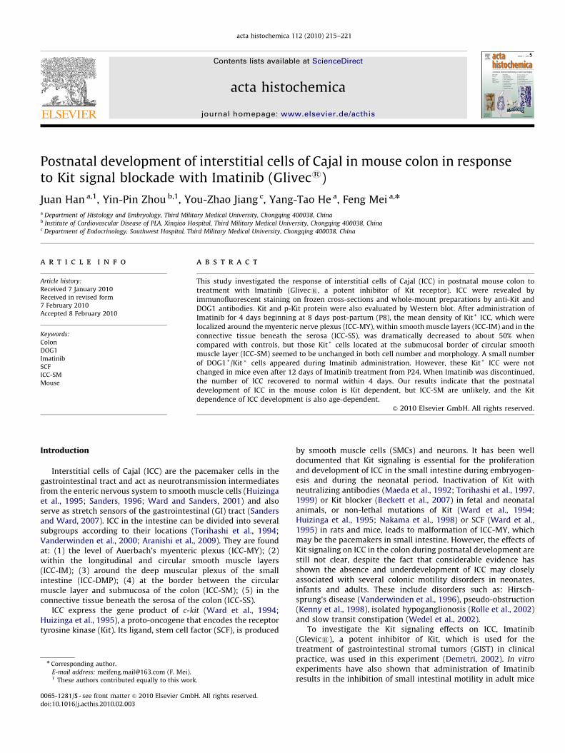
Postnatal development of interstitial cells of Cajal in mouse colon in responseto Kit signal blockade with Imatinib (Glivecs)
Juan Han a,1, Yin-Pin Zhou b,1, You-Zhao Jiang c, Yang-Tao He a, Feng Mei a,n
a Department of Histology and Embryology, Third Military Medical University, Chongqing 400038, Chinab Institute of Cardiovascular Disease of PLA, Xinqiao Hospital, Third Military Medical University, Chongqing 400038, Chinac Department of Endocrinology, Southwest Hospital, Third Military Medical University, Chongqing 400038, China
a r t i c l e i n f o
Article history:
Received 7 January 2010
Received in revised form
7 February 2010
Accepted 8 February 2010
Keywords:
Colon
DOG1
Imatinib
SCF
ICC-SM
Mouse
81/$ - see front matter & 2010 Elsevier Gmb
016/j.acthis.2010.02.003
esponding author.
ail address: [email protected] (F. Mei).
ese authors contributed equally to this work
a b s t r a c t
This study investigated the response of interstitial cells of Cajal (ICC) in postnatal mouse colon to
treatment with Imatinib (Glivecs, a potent inhibitor of Kit receptor). ICC were revealed by
immunofluorescent staining on frozen cross-sections and whole-mount preparations by anti-Kit and
DOG1 antibodies. Kit and p-Kit protein were also evaluated by Western blot. After administration of
Imatinib for 4 days beginning at 8 days post-partum (P8), the mean density of Kit+ ICC, which were
localized around the myenteric nerve plexus (ICC-MY), within smooth muscle layers (ICC-IM) and in the
connective tissue beneath the serosa (ICC-SS), was dramatically decreased to about 50% when
compared with controls, but those Kit+ cells located at the submucosal border of circular smooth
muscle layer (ICC-SM) seemed to be unchanged in both cell number and morphology. A small number
of DOG1+/Kit� cells appeared during Imatinib administration. However, these Kit+ ICC were not
changed in mice even after 12 days of Imatinib treatment from P24. When Imatinib was discontinued,
the number of ICC recovered to normal within 4 days. Our results indicate that the postnatal
development of ICC in the mouse colon is Kit dependent, but ICC-SM are unlikely, and the Kit
dependence of ICC development is also age-dependent.
& 2010 Elsevier GmbH. All rights reserved.
Introduction
Interstitial cells of Cajal (ICC) are the pacemaker cells in thegastrointestinal tract and act as neurotransmission intermediatesfrom the enteric nervous system to smooth muscle cells (Huizingaet al., 1995; Sanders, 1996; Ward and Sanders, 2001) and alsoserve as stretch sensors of the gastrointestinal (GI) tract (Sandersand Ward, 2007). ICC in the intestine can be divided into severalsubgroups according to their locations (Torihashi et al., 1994;Vanderwinden et al., 2000; Aranishi et al., 2009). They are foundat: (1) the level of Auerbach’s myenteric plexus (ICC-MY); (2)within the longitudinal and circular smooth muscle layers(ICC-IM); (3) around the deep muscular plexus of the smallintestine (ICC-DMP); (4) at the border between the circularmuscle layer and submucosa of the colon (ICC-SM); (5) in theconnective tissue beneath the serosa of the colon (ICC-SS).
ICC express the gene product of c-kit (Ward et al., 1994;Huizinga et al., 1995), a proto-oncogene that encodes the receptortyrosine kinase (Kit). Its ligand, stem cell factor (SCF), is produced
H. All rights reserved.
.
by smooth muscle cells (SMCs) and neurons. It has been welldocumented that Kit signaling is essential for the proliferationand development of ICC in the small intestine during embryogen-esis and during the neonatal period. Inactivation of Kit withneutralizing antibodies (Maeda et al., 1992; Torihashi et al., 1997,1999) or Kit blocker (Beckett et al., 2007) in fetal and neonatalanimals, or non-lethal mutations of Kit (Ward et al., 1994;Huizinga et al., 1995; Nakama et al., 1998) or SCF (Ward et al.,1995) in rats and mice, leads to malformation of ICC-MY, whichmay be the pacemakers in small intestine. However, the effects ofKit signaling on ICC in the colon during postnatal development arestill not clear, despite the fact that considerable evidence hasshown the absence and underdevelopment of ICC may closelyassociated with several colonic motility disorders in neonates,infants and adults. These include disorders such as: Hirsch-sprung’s disease (Vanderwinden et al., 1996), pseudo-obstruction(Kenny et al., 1998), isolated hypoganglionosis (Rolle et al., 2002)and slow transit constipation (Wedel et al., 2002).
To investigate the Kit signaling effects on ICC, Imatinib(Glevics), a potent inhibitor of Kit, which is used for thetreatment of gastrointestinal stromal tumors (GIST) in clinicalpractice, was used in this experiment (Demetri, 2002). In vitro
experiments have also shown that administration of Imatinibresults in the inhibition of small intestinal motility in adult mice

ARTICLE IN PRESS
J. Han et al. / acta histochemica 112 (2010) 215–221216
(Shimojima et al., 2005) and humans (Popescu et al., 2006), andthe disappearance of ICC in organ cultures of small intestinaltissue from the fetal and neonatal mice.
Therefore, we have used immunofluorescent staining andWestern blot methods to investigate the alterations of ICC inthe mouse colon at P8 (neonatal) and P24 (young) after Imatinibtreatment.
Materials and methods
Animals
BALB/C mice were purchased from the Animal Center of theThird Military Medical University (Chongqing, China) and pairedto produce offspring. Imatinib mesylate (Glivecs) was purchasedfrom Novartis Pharma AG (Basel, Switzerland). Mice wereintragastrically administered Imatinib at a dosage of 0.5 mg g�1
per day and animals were divided into 6 groups: (1) mice at P8were killed 6 h after one dose of drug (n=3); (2) mice at P8 weretreated with the drug for 1 day (n=5); (3) mice at P8 were treatedwith the drug for 4 days (n=8); (4) mice were administrated thedrug for 4 days from P8 then it was withdrawn for 4 days (n=5);(5) mice at P24 were treated with the drug for 4 days (n=5); (6)mice at P24 were treated with the drug for 12 days (n=5). Glucosein water was given to 21 mice as controls. All experiments wereperformed in accordance with our University Health Guide for theCare and Use of Laboratory Animals.
Immunofluorescence
The entire colon from the ileo-cecal junction to the pelvic brimwas carefully removed. For whole-mount preparations, the colonfrom groups 2–6 (n=25) and controls (n=15) was inflated withacetone for 30 min, then the mucosa was removed by sharpdissection using a dissection microscope. For frozen sections, thecolon from group 3 (n=3) and control (n=3) was divided intothree segments of identical length from oral to anal end:proximal, middle and distal colon, then each part was placedinto optimal cutting temperature compound (OCT), and quicklyfrozen with liquid nitrogen. Longitudinal sections (6–8 mm thick)were cut with a cryostat (Leica CM 1850, Leica Microsystems,Wetzlar, Germany) and fixed with 100% acetone for 15 min (4 1C).The immunostaining procedures have been described previously.In short, whole mount preparations or frozen sections wereincubated with primary antibody overnight at 4 1C and thensecondary antibodies for 1 h at room temperature (Table 1).Sections were counterstained with DAPI. The stained results weredetected by BX51 fluorescence microscope (Olympus, Tokyo,Japan) or TCS SP5 confocal laser scanning microscope (Leica
Table 1The antibodies used in the study.
Antigen Clone Supplier Dilution Isotype Conjugated
Kit ACk2 eBioscience 1:100 IF Rat IgG Purified
Kit Polyclone Santa Cruz 1:1000 WB Goat IgG Purified
DOG1 Polyclone Abcam 1:100 IF Rabbit lgG Purified
a-SMA 1A4 Boster 1:100 IF Mouse lgG Purified
p-Kit Polyclone Cell Signal 1:1000 WB Rabbit lgG Purified
b-actin Polyclone Santa Cruz 1:1000 WB Mouse IgG Purified
Anti-rat Polyclone Zymed 1:100 IF Goat IgG Cy3
Anti-rabbit Polyclone Zymed 1:100 IF Goat IgG Cy5
Anti-mouse Polyclone Dako 1:100 IF Goat IgG FITC
Anti-goat Polyclone Dako 1:2000 WB Donkey IgG HRP
Anti-rabbit Polyclone Dako 1:3000 WB Goat IgG HRP
Anti-mouse Polyclone Dako 1:1000 WB Goat IgG HRP
Microsystems, Wetzlar, Germany) with an excitation wavelengthappropriate for FITC(488 nm), Cy3(552 nm), Cy5 (625 nm) andDAPI(380 nm).
Western blotting
Total protein was extracted from smooth muscle layers of thecolon from group 1 (n=3) and control (n=3) using RIPA lysisbuffer (50 mM tris pH 7.4, 150 mM NaCl, 1% triton X-100, 1%sodium deoxycholate, 0.1% SDS, 1 mM EDTA, 1 mM sodiumorthovanadate, 10 mM NaF), containing 10 mg/ml aprotinin,10 mg/ml leupeptin and 1 mM phenylmethylsulphonyl fluoride.The protein concentration was measured using a BCA ProteinAssay Kit (Beyotime Biotechnology, Jiangsu, China). Total protein(100 mg) was separated on 7.5% SDS-PAGE and transferred to aPVDF membrane at 350 mA for 1.5 h by using semi-dry transfercell (Bio-Rad, Hercules, CA, USA). Then the membrane wasincubated in blocking buffer consisting of 5% non-fat dry milkfor 2 h at room temperature, and then with primary antibodiesovernight at 4 1C and secondary antibodies subsequently (Table 1).Protein–antibody complexes were detected with an ECL Westernblotting detection and analysis system (GE Healthcare LifeSciences, Amersham Biosciences, Bucks., UK).
Measurements and statistical analysis
Six intestinal segments were sampled in a random mannerfrom experimental animals for whole mount preparations. Afterimmunofluorescent staining assessment, photomicrographs ofboth types of Kit positive cells were taken in 10 random fields(�200 magnification, 0.2607 mm2) per whole-mount preparationwith a digital camera (SPOT, Diagnostic Instruments, SterlingHeights, MI, USA) mounted on a BX51 fluorescence microscope(Olympus, Tokyo, Japan). The numbers of Kit+ cells were countedwith Image-Pro Plus 5.0 (Media Cybernetics, Silver Spring, MD,USA). Data were expressed as means7S.E.M. The n value reportedin the text refers to the number of animal used. Differences in thedata were evaluated by student t test, and Po0.05 was taken as astatistically significant difference.
Results
Kit signaling blockade by Imatinib administration
To evaluate the Kit blocking effects of Imatinib, Western blotwas applied to assay the alterations of phosphorylated Kit protein(p-Kit) and Kit. The results showed that the amount of p-Kit wasobviously decreased and Kit expression was persistent afterImatinib treatment for 6 h (Fig. 1), that means Imatinib caninhibit Kit signaling by inhibition of Kit phosphorylation and thisdrug can be used in vivo to investigate the effects of Kit blockadeon ICC.
Fig. 1. Western blotting showed that Kit and p-Kit can be detected in control. Six
hours after one dose of Imatinib, p-Kit was nearly not detected as compared with
Kit expression.

ARTICLE IN PRESS
J. Han et al. / acta histochemica 112 (2010) 215–221 217
Distribution alterations of ICC in neonatal mouse colon
To show clearly the alterations of ICC distribution after Kitblockade, Kit/a-smooth muscle actin (a-SMA) double labelingwas carried out on sections of proximal colon. The results showedKit+ ICC were seen apparently between the submucosa andcircular muscle layer (ICC-SM), at the level of Auerbach’s plexus(ICC-MY), within the smooth muscle layers (ICC-IM) and in theconnective tissue beneath the serosa (ICC-SS) in the controls(Figs. 2A–C). However, after administration of the drug for 4 daysbeginning at P8, the Kit+ ICC-MY, ICC-IM and ICC-SS weredramatically decreased, apart from Kit+ ICC-SM that were stillpresent (Fig. 2D–F).
Morphological changes of ICC in neonatal mouse colon
To clarify further the alterations in morphology and cellnumber, Kit immunofluorescent staining on whole mountpreparations of proximal colon was performed. Numerous Kit+
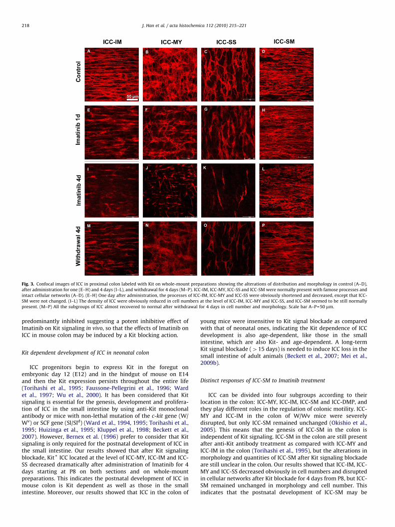
ICC-MY, ICC-SS and ICC-SM with round cell bodies and longprocesses were seen, and ICC-IM in parallel with smooth musclecells (Figs. 3A–D). After administration for 1 day, Kit+ ICC-IM, ICC-MY and ICC-SS were characterized by shortened processes anddisrupted cellular network (Fig. 3E–G). After treatment for 4 dayswith Imatinib, the cellular network of ICC-MY was more obviouslydisrupted and ICC-IM and ICC-SS were greatly decreased in cellnumber (Fig. 3I–K). After withdrawal of the drug, ICC-MY, ICC-IMand ICC-SS mainly recovered to normal within 4 days (Figs. 3M–O).However, it is noteworthy that Kit+ ICC-SM seemed neitherto change in morphology nor in cell number during drugadministration and withdrawal (Fig. 3D, H, L and P).
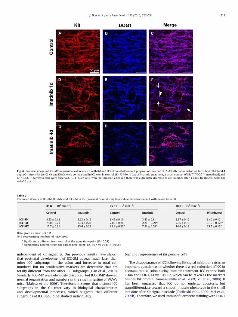
Kit expression may be down-regulated in the condition of Kitsignaling blockade, and DOG1, a Ca(2+)-activated Cl(�) channel,which is also named Ano1 or TMEM16A, was also the marker ofICC. So we investigated the expression of DOG1 as cellularmarkers of ICC. Immunofluorescent staining showed that theDOG1+ were also Kit+ in controls (Figs. 4A–C), and a smallnumber of Kit�/DOG1+ and Kitweak/DOG1+ cells were observedafter 1 day administration (Fig. 4D–E). After administration for 4days, a dramatic reduction of Kit+ ICC were observed, which werealso DOG1 positive (Figs. 4G–I). One explanation of this resultmight be that down-regulation of Kit expression is involved in theprocess of ICC lost after Kit blockade.
Fig. 2. Confocal images of ICC in the proximal colon labeled with Kit after Imatinib tre
located between (ICC-MY) and within the longitudinal and circular smooth muscle lay
(ICC-SM), and in the connective tissue beneath serosa of colon (ICC-SS) in control. (D–
while ICC-SM were normal present. Scale bar A–F=40 mm.
Density alteration of ICC in neonatal mouse colon
To understand better the degree of Kit+ ICC reduction, wecounted the cell numbers of ICC subgroups individually in theproximal colon on whole-mount preparations. The resultsindicated that Kit+ ICC-IM in the proximal colon decreased from7.9070.21�102 to 2.2170.09�102 mm�2 (Table 2) at 4 daysafter drug administration from P8, and similarly the mean densityof Kit+ ICC-MY decreased from 17.770.21�102 mm�2 to a valueless than a half (7.5170.09�102 mm�2) (Table 2). The cellnumbers of both Kit+ ICC-IM and ICC-MY nearly recovered tonormal within 4 days. However, those Kit+ ICC-SM were notchanged in density during drug administration and withdrawal(Table 2).
Effects of Kit blockade on ICC in young mouse colon
Kit immunofluorescent staining showed that the Kit+ ICC-MY,ICC-IM, ICC-SM and ICC-SS showed no change in either cellnumbers or morphology after either treatment for 4 days(Figs. 5A–D) or 12 days with Imatinib as compared withcontrols (Fig. 5E–H) from P24. It can be interpreted that ICC inyoung mice may be much less sensitive to Kit blockade thanneonatal ones.
Discussion
The present study demonstrates that: (1) postnatal develop-ment of ICC in the mouse colon is dependent on Kit signaling, butthis may not be the case with ICC-SM; (2) ICC become insensitiveto Kit blockade in older mice; (3) Imatinib can be used as a potentKit signal blocker in vivo.
Kit signal blockade effects by Imatinib
Imatinib was developed as a potent inhibitor of Bcr–Abltyrosine kinase in chronic myeloid leukemia (CML), platelet-derived growth factor receptor kinase (PDGFR) and also Kittyrosine kinase (Manley et al., 2002). ICC express neither Bcr–Abltyrosine kinase, nor PDGFR (Iino et al., 2009). To further confirmthe Kit blocking effect by intragastric administration of Imatinib,Western blotting was carried out to assay the level of Kit proteinphosphorylation (p-Kit). The results showed that p-Kit was
atment for 4 days from P8 on longitudinal sections. (A–C) Kit positive cells were
ers (ICC-IM), at the border between circular muscle layer and submucosa of colon
F) After treatment for 4 days, ICC-MY, ICC-IM and ICC-SS were greatly decreased,
ARTICLE IN PRESS
Fig. 3. Confocal images of ICC in proximal colon labeled with Kit on whole-mount preparations showing the alterations of distribution and morphology in control (A–D),
after administration for one (E–H) and 4 days (I–L), and withdrawal for 4 days (M–P). ICC-IM, ICC-MY, ICC-SS and ICC-SM were normally present with famose processes and
intact cellular networks (A–D). (E–H) One day after administration, the processes of ICC-IM, ICC-MY and ICC-SS were obviously shortened and decreased, except that ICC-
SM were not changed. (I–L) The density of ICC were obviously reduced in cell numbers at the level of ICC-IM, ICC-MY and ICC-SS, and ICC-SM seemed to be still normally
present. (M–P) All the subgroups of ICC almost recovered to normal after withdrawal for 4 days in cell number and morphology. Scale bar A–P=50 mm.
J. Han et al. / acta histochemica 112 (2010) 215–221218
predominantly inhibited suggesting a potent inhibitive effect ofImatinib on Kit signaling in vivo, so that the effects of Imatinib onICC in mouse colon may be induced by a Kit blocking action.
Kit dependent development of ICC in neonatal colon
ICC progenitors begin to express Kit in the foregut onembryonic day 12 (E12) and in the hindgut of mouse on E14and then the Kit expression persists throughout the entire life(Torihashi et al., 1995; Faussone-Pellegrini et al., 1996; Wardet al., 1997; Wu et al., 2000). It has been considered that Kitsignaling is essential for the genesis, development and prolifera-tion of ICC in the small intestine by using anti-Kit monoclonalantibody or mice with non-lethal mutation of the c-kit gene (W/Wv) or SCF gene (Sl/Sld) (Ward et al., 1994, 1995; Torihashi et al.,1995; Huizinga et al., 1995; Kluppel et al., 1998; Beckett et al.,2007). However, Bernex et al. (1996) prefer to consider that Kitsignaling is only required for the postnatal development of ICC inthe small intestine. Our results showed that after Kit signalingblockade, Kit+ ICC located at the level of ICC-MY, ICC-IM and ICC-SS decreased dramatically after administration of Imatinib for 4days starting at P8 on both sections and on whole-mountpreparations. This indicates the postnatal development of ICC inmouse colon is Kit dependent as well as those in the smallintestine. Moreover, our results showed that ICC in the colon of
young mice were insensitive to Kit signal blockade as comparedwith that of neonatal ones, indicating the Kit dependence of ICCdevelopment is also age-dependent, like those in the smallintestine, which are also Kit- and age-dependent. A long-termKit signal blockade (415 days) is needed to induce ICC loss in thesmall intestine of adult animals (Beckett et al., 2007; Mei et al.,2009b).
Distinct responses of ICC-SM to Imatinib treatment
ICC can be divided into four subgroups according to theirlocation in the colon: ICC-MY, ICC-IM, ICC-SM and ICC-DMP, andthey play different roles in the regulation of colonic motility. ICC-MY and ICC-IM in the colon of W/Wv mice were severelydisrupted, but only ICC-SM remained unchanged (Okishio et al.,2005). This means that the genesis of ICC-SM in the colon isindependent of Kit signaling. ICC-SM in the colon are still presentafter anti-Kit antibody treatment as compared with ICC-MY andICC-IM in the colon (Torihashi et al., 1995), but the alterations inmorphology and quantities of ICC-SM after Kit signaling blockadeare still unclear in the colon. Our results showed that ICC-IM, ICC-MY and ICC-SS decreased obviously in cell numbers and disruptedin cellular networks after Kit blockade for 4 days from P8, but ICC-SM remained unchanged in morphology and cell number. Thisindicates that the postnatal development of ICC-SM may be
ARTICLE IN PRESS
Fig. 4. Confocal images of ICC-MY in proximal colon labeled with Kit and DOG1 on whole-mount preparations in control (A–C), after administration for 1 days (D–F) and 4
days (G–I) from P8. (A–C) Kit and DOG1 were co-localized in ICC well in control. (D–F) After 1 day of Imatinib treatment, a small number of Kitweak/DOG+ (arrowhead) and
Kit�/DOG1+ (arrows) cells were observed. (G–F) Such cells were not present, although there was a dramatic decrease of cell number after 4 days’ treatment. Scale bar
A–I=50 mm.
Table 2The mean density of ICC-IM, ICC-MY and ICC-SM in the proximal colon during Imatinib administration and withdrawal from P8.
24 h (�102 mm�2) 96 h (�102 mm�2) 48 h (�102 mm�2)
Control Imatinib Control Imatinib Control Withdrawal
ICC-SM 3.5370.12 3.6270.12 3.4570.10 3.4270.11 3.3770.12 3.4070.12
ICC-IM 7.9070.21 7.5470.22 7.8670.20 2.2170.09ab 7.9670.18 5.1670.12ab
ICC-MY 17.770.21 15.670.22a 15.470.20b 7.5170.09ab 14.670.18 13.570.12b
Data given as mean7S.E.M.
N=3 representing numbers of mice used.
a Significantly different from control at the same time-point (Po0.05).b Significantly different from the earlier time-point (i.e., 96 h vs 24 h) (Po0.05).
J. Han et al. / acta histochemica 112 (2010) 215–221 219
independent of Kit signaling. Our previous results have shownthat postnatal development of ICC-SM appear much later thanother ICC subgroups in the colon and increase in total cellnumbers, but no proliferative markers are detectable that aretotally different from the other ICC subgroups (Han et al., 2010).Similarly, ICC-MY were obviously disrupted, but ICC-DMP showednormal organization and numbers in the small intestine of W/Wvmice (Malysz et al., 1996). Therefore, it seems that distinct ICCsubgroups in the GI tract vary in biological characteristicsand developmental processes, which suggests that differentsubgroups of ICC should be studied individually.
Loss and reappearance of Kit positive cells
The disappearance of ICC following Kit signal inhibition raises animportant question as to whether there is a real reduction of ICC inneonatal mouse colon during Imatinib treatment. ICC express bothCD44 and DOG1, as well as Kit, which can be taken as the markersbesides Kit protein (Gomez-Pinilla et al., 2009; Yu et al., 2009). Ithas been suggested that ICC do not undergo apoptosis, buttransdifferentiate toward a smooth muscle phenotype in the smallintestine after Kit signal blockade (Torihashi et al., 1999; Mei et al.,2009b). Therefore, we used immunofluorescent staining with DOG1

ARTICLE IN PRESS
Fig. 5. Confocal images of ICC in the proximal colon of mice in control (A–D) and Imatinib treatment for 4 days (E–H) from P24 on whole mount preparations, and neither
ICC subgroup changed after administration of Imatinib for 4 days. Scale bar, A–H=50 mm.
J. Han et al. / acta histochemica 112 (2010) 215–221220
antibody, and the results showed a small number of Kit�/DOG1+
and Kitweak/DOG1+ appeared after day 1 of Imatinib administrationand then these cells could not been observed after treatment for 4days. Moreover, immunofluorescent staining with anti-CD44 anti-body also showed a similar alteration (data not shown). Theseresults may suggest that the reduced Kit+ ICC were not a realreduction of ICC, but they may cease expressing DOG1 and CD44 aswell as Kit and transdifferentiate to a kind of intermediate cells. Sonew cellular markers, which ICC persistently express during Kitsignal blockade, and electronic microscope, gold standard for theidentification of ICC, are necessary to investigate the fate of lost ICC.
Our previous results have shown that proliferation is involvedin the recovery of ICC after loss in adult animals (Mei et al., 2006,2009a, 2009b) and ICC in mouse colon and small intestineproliferate during postnatal development (Mei et al., 2009c; Hanet al., 2010). The present study showed that ICC recovered tonormal after withdrawal of Imatinib and whether proliferation isinvolved in this recovery process requires further investigation.
Our study provides evidence that the postnatal development ofICC in the colon is Kit dependent, suggesting that insufficiency ofKit signaling would lead to the underdevelopment of ICC, whichmight be one of the mechanisms underlying the motilitydisorders of neonates and infants.
Acknowledgements
We thank Wei Sun and Li-ting Wang (Central laboratory, ThirdMilitary Medical University) for their help with the confocal laserscanning microscopy. This study was supported in part by theNatural Science Foundation Project of CQ CSTC 2009BB5320.
References
Aranishi H, Kunisawa Y, Komuro T. Characterization of interstitial cells of Cajal inthe subserosal layer of the guinea-pig colon. Cell Tissue Res 2009;335:323–329.
Beckett EA, Ro S, Bayguinov Y, Sanders KM, Ward SM. Kit signaling is essential fordevelopment and maintenance of interstitial cells of Cajal and electricalrhythmicity in the embryonic gastrointestinal tract. Dev Dyn 2007;236:60–72.
Bernex F, De Sepulveda P, Kress C, Elbaz C, Delouis C, Panthier JJ. Spatial andtemporal patterns of c-Kit-expressing cells in WlacZ/+ and WlacZ/WlacZmouse embryos. Development 1996;122:3023–3033.
Demetri GD. Identification and treatment of chemoresistant inoperable ormetastatic GIST: experience with the selective tyrosine kinase inhibitorImatinib mesylate (STI571). Eur J Cancer 2002;38(Suppl 5):S52–S59.
Faussone-Pellegrini MS, Matini P, Stach W. Differentiation of enteric plexuses andinterstitial cells of Cajal in the rat gut during pre- and postnatal life. Acta Anat(Basel) 1996;155:113–125.
Gomez-Pinilla PJ, Gibbons SJ, Bardsley MR, Lorincz A, Pozo MJ, Pasricha PJ, et al.Ano1 is a selective marker of interstitial cells of Cajal in the human and mousegastrointestinal tract. Am J Physiol Gastrointest Liver Physiol 2009;296:G1370–G1381.
Han J, Shen WH, Jiang YZ, Yu B, He YT, Li N, et al. Distribution, development andproliferation of interstitial cells of Cajal in murine colon: an immunohisto-chemical study from neonatal to adult life. Histochem Cell Biol 2010;133:163–175.
Huizinga JD, Thuneberg L, Kluppel M, Malysz J, Mikkelsen HB, Bernstein A. W/Kitgene required for interstitial cells of Cajal and for intestinal pacemakeractivity. Nature 1995;373:347–349.
Iino S, Horiguchi K, Horiguchi S, Nojyo Y. c-Kit-negative fibroblast-like cellsexpress platelet-derived growth factor receptor alpha in the murine gastro-intestinal musculature. Histochem Cell Biol 2009;131:691–702.
Kenny SE, Vanderwinden JM, Rintala RJ, Connell MG, Lloyd DA, Vanderhaegen JJ,et al. Delayed maturation of the interstitial cells of Cajal: a new diagnosis fortransient neonatal pseudoobstruction. Report of two cases. J Pediatr Surg1998;33:94–98.
Kluppel M, Huizinga JD, Malysz J, Bernstein A. Developmental origin and Kit-dependent development of the interstitial cells of cajal in the mammaliansmall intestine. Dev Dyn 1998;211:60–71.
Maeda H, Yamagata A, Nishikawa S, Yoshinaga K, Kobayashi S, Nishi K, et al.Requirement of c-Kit for development of intestinal pacemaker system.Development 1992;116:369–375.
Malysz J, Thuneberg L, Mikkelsen HB, Huizinga JD. Action potential generation inthe small intestine of W mutant mice that lack interstitial cells of Cajal. Am JPhysiol 1996;271:G387–G399.
Manley PW, Cowan-Jacob SW, Buchdunger E, Fabbro D, Fendrich G, Furet P, et al.Imatinib: a selective tyrosine kinase inhibitor. Eur J Cancer 2002;38(Suppl5):S19–S27.
Mei F, Guo S, He YT, Zhu J, Zhou DS, Niu JQ, et al. Apoptosis of interstitial cells ofCajal, smooth muscle cells, and enteric neurons induced by intestinal ischemiaand reperfusion injury in adult guinea pigs. Virchows Arch 2009a;454:401–409.
Mei F, Han J, Huang Y, Jiang ZY, Xiong CJ, Zhou DS. Plasticity of interstitial cells ofcajal: a study in the small intestine of adult guinea pigs. Anat Rec (Hoboken)2009b;292:985–993.
Mei F, Zhu J, Guo S, Zhou DS, Han J, Yu B, et al. An age-dependent proliferation isinvolved in the postnatal development of interstitial cells of Cajal in the smallintestine of mice. Histochem Cell Biol 2009c;131:43–53.
Mei F, Yu B, Ma H, Zhang HJ, Zhou DS. Interstitial cells of Cajal could regenerateand restore their normal distribution after disrupted by intestinal transectionand anastomosis in the adult guinea pigs. Virchows Arch 2006;449:348–357.
Nakama A, Hirota S, Okazaki T, Nagano K, Kawano S, Hori M, et al. Disturbedpyloric motility in Ws/Ws mutant rats due to deficiency of c-Kit-expressinginterstitial cells of Cajal. Pathol Int 1998;48:843–849.
Okishio Y, Takeuchi T, Fujita A, Suenaga K, Fujinami K, Munakata S, et al.Ascending contraction and descending relaxation in the distal colon of micelacking interstitial cells of Cajal. J Smooth Muscle Res 2005;41:163–174.
Popescu LM, Vidulescu C, Curici A, Caravia L, Simionescu AA, Ciontea SM, et al.Imatinib inhibits spontaneous rhythmic contractions of human uterus andintestine. Eur J Pharmacol 2006;546:177–181.
Rolle U, Yoneda A, Solari V, Nemeth L, Puri P. Abnormalities of C-Kit-positivecellular network in isolated hypoganglionosis. J Pediatr Surg 2002;37:709–714.

ARTICLE IN PRESS
J. Han et al. / acta histochemica 112 (2010) 215–221 221
Sanders KM. A case for interstitial cells of Cajal as pacemakers and mediators ofneurotransmission in the gastrointestinal tract. Gastroenterology 1996;111:492–515.
Sanders KM, Ward SM. Kit mutants and gastrointestinal physiology. J Physiol2007;578:33–42.
Shimojima N, Nakaki T, Morikawa Y, Hoshino K, Kitajima M. Imatinib blocksspontaneous mechanical activities in the adult mouse small intestine: possibleinhibition of c-Kit signaling. Pharmacology 2005;74:95–99.
Torihashi S, Gerthoffer WT, Kobayashi S, Sanders KM. Identification andclassification of interstitial cells in the canine proximal colon by ultrastructureand immunocytochemistry. Histochemistry 1994;101:169–183.
Torihashi S, Nishi K, Tokutomi Y, Nishi T, Ward S, Sanders KM. Blockade of Kitsignaling induces transdifferentiation of interstitial cells of cajal to a smoothmuscle phenotype. Gastroenterology 1999;117:140–148.
Torihashi S, Ward SM, Nishikawa S, Nishi K, Kobayashi S, Sanders KM.c-Kit-dependent development of interstitial cells and electricalactivity in the murine gastrointestinal tract. Cell Tissue Res 1995;280:97–111.
Torihashi S, Ward SM, Sanders KM. Development of c-Kit-positive cells and theonset of electrical rhythmicity in murine small intestine. Gastroenterology1997;112:144–155.
Vanderwinden JM, Rumessen JJ, Bernex F, Schiffmann SN, Panthier JJ. Distributionand ultrastructure of interstitial cells of Cajal in the mouse colon,using antibodies to Kit and Kit(W-lacZ) mice. Cell Tissue Res 2000;302:155–170.
Vanderwinden JM, Rumessen JJ, Liu H, Descamps D, De Laet MH, Vanderhaeghen JJ.Interstitial cells of Cajal in human colon and in Hirschsprung’s disease.Gastroenterology 1996;111:901–1110.
Ward SM, Burns AJ, Torihashi S, Harney SC, Sanders KM. Impaired development ofinterstitial cells and intestinal electrical rhythmicity in steel mutants. Am JPhysiol 1995;269:C1577–C1585.
Ward SM, Burns AJ, Torihashi S, Sanders KM. Mutation of the proto-oncogene c-Kitblocks development of interstitial cells and electrical rhythmicity in murineintestine. J Physiol 1994;480:91–97.
Ward SM, Harney SC, Bayguinov JR, McLaren GJ, Sanders KM. Development ofelectrical rhythmicity in the murine gastrointestinal tract is specificallyencoded in the tunica muscularis. J Physiol 1997;505:241–258.
Ward SM, Sanders KM. Physiology and pathophysiology of the interstitial cell ofCajal: from bench to bedside. I. Functional development and plasticity ofinterstitial cells of Cajal networks. Am J Physiol Gastrointest Liver Physiol2001;281:G602–G611.
Wedel T, Spiegler J, Soellner S, Roblick UJ, Schiedeck TH, Bruch HP, et al. Entericnerves and interstitial cells of Cajal are altered in patients with slow-transitconstipation and megacolon. Gastroenterology 2002;123:1459–1467.
Wu JJ, Rothman TP, Gershon MD. Development of the interstitial cell of Cajal:origin, Kit dependence and neuronal and nonneuronal sources of Kit ligand. JNeurosci Res 2000;59:384–401.
Yu B, Han J, He YT, Guo S, Li SF, Mei F. Immunohistochemical study of CD44immunopositive cells in the muscular layers of the gastrointestinal tract inadult guinea pigs and mice. Acta Histochem 2009;111:382–390.