Embed Size (px)
Citation preview
Posterior patterning by the Caenorhabditis elegans even-skipped homolog vab-7 Julie Ahringer 1
Medical Research Council Laboratory of Molecular Biology, Cambridge CB2 2QH, UK
Patterning of the posterior end in animals is not well understood. Homologs of Drosophi la even-sk ipped (eve) have a similar posterior expression pattern in many animals, and in vertebrates they are linked physically to the "posterior" ends of homeotic clusters (HOM-C), suggesting a conserved role in posterior development. However, the function of this posterior expression is not known. Here I show that the Caenorhabdi t is elegans gene vab-7 encodes an eve homolog that is required for posterior development and expressed in a pattern strikingly similar to that of vertebrate eve genes. Using a four-dimensional recording system, I found that posterior body muscles and the posterior epidermis are patterned abnormally in vab-7 mutants, but commitment to muscle and epidermal fates is normal. Furthermore, vab-7 activity is required for the complete expression of the most posterior HOM-C gene egl-5 in muscle cells, supporting the idea that eve homologs may act with the HOM-C to determine posterior cell fates. The conservation of sequence and expression pattern between vab-7 and eve homologs in other animals argues that most eve genes have posterior mesodermal and ectodermal patterning functions.
[Key Words." C. elegans; pattern formation; lineage analysis; HOX genes]
Received January 31, 1996; revised version accepted March 21, 1996.
During development, maternal (and paternal) products are important for setting up early asymmetries in the embryo (Slack 1991). Later, in most if not all animals, an evolutionarily conserved cluster of homeotic genes (the HOM-C) helps pattern the central body along the ante- rior-posterior {a/p) axis (for review, see McGinnis and Krumlauf 1992; Kenyon 1994). How the extremities are patterned is still largely mysterious.
One conserved gene suggested to have a role in poste- rior patterning is even-skipped (eve). This is based on its posterior expression in many organisms and its physical linkage to the "posterior" ends of vertebrate HOX clusters (Ruiz i Altaba and Melton 1989a,b; Bastian and Gruss 1990; D'Esposito et al. 1991; Faiella et al. 1991; Bastian et al. 1992; Dush and Martin 1992; Patel et al. 1992; Joly et al. 1993; Dolld et al. 1994). In most animals, eve genes have three major phases of expression: (1) in the developing posterior mesoderm and ectoderm, (2) in developing posterior structures such as the tail bud in mouse, and (3) in the developing nervous system lthe segmental expression of Drosophila eve (Macdonald et al. 1986; Frasch et al. 1987) is unusual]. The shared pos- terior expression pattern suggests that these genes have a conserved function in posterior development, but nei- ther the function of this expression nor any functional
1Present address: University of Cambridge, Department of Genetics, Downing Street, Cambridge CB2 3EH, UK.
interaction with the posterior part of the HOX cluster has yet been reported.
Caenorhabditis e]egans also has a homeotic cluster that has been shown to be important for establishing cell fates along the a/p axis (for review, see Salser and Kenyon 19941. The functions of these genes have been studied mainly during postembryonic development. Zy- gotic patterning in the embryo has been largely ignored because of difficulties in analysis. In particular, it was not possible to routinely follow cell lineages in mutant embryos because of the number of simultaneous cell di- visions to be watched. This problem has been overcome by the development of a "four-dimensional" (4D) video recording s~ stem that can be used to look for alterations in the divisions, migrations, and differentiation of al- most any cell in the embryo (Hird and White 1993). As the embryonic cell lineage is essentially invariant in C. elegans (Sulston et al. 1983), this type of data is a good measure of pattern alteration, and is especially useful where defects may not be obvious or interpretable from looking at a differentiated embryo.
Here, I show that the C. elegans gene vab-7 encodes an eve homolog. Using the 4D recording system, I found that vab-7 functions in posterior muscle and epidermal patterning in the embryo. In addition, posterior muscle cells require vab-7 activity for complete expression of the most posterior HOM-C gene egl-5. Because the pos- terior expression of vab-7 is similar to that of eve in other organisms, it is likely that eve genes have posterior patterning functions in most animals.
1120 GENES & DEVELOPMENT 10:1120-1130 ~ 1996 by Cold Spring Harbor Laboratory Press ISSN 0890-9369/96 $5.00
Cold Spring Harbor Laboratory Press on February 7, 2022 - Published by genesdev.cshlp.orgDownloaded from
Posterior patterning in C. elegans
R e s u l t s
vab-7 encodes an eve homolog
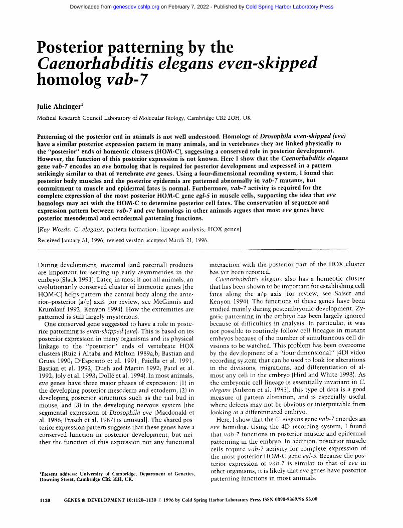
A sequence highly similar to the eve homeo box of Drosophila was identified by the C. elegans sequencing consort ium during the partial sequencing of yeast artifi- cial chromosome (YAC) Y52D3. Figure 1A shows an al ignment of this sequence with eve homologs from other organisms. The C. elegans sequence has >~80% amino acid ident i ty wi th any other eve homeo domain; the closest non-eve homeo domain is from Drosophila rough (Tomlinson et al. 1988), with 59% identity.
I searched the genetic map in the region of Y52D3 for a previously identified gene that could correspond to this sequence. Mutat ions in the gene vab-7 (for variable ab- normal) result in posterior defects (J. Culotti, pers. comm.), which made it a good candidate. Two experi- ments showed that vab-7 does encode the eve homolog. First, DNAs containing the eve gene rescue the vab-7 mutan t phenotype (Fig. 1B). Second, two alleles of vab-7
(e1562 and ed7) each carry a muta t ion in the homeo do- main (arrows in Fig. 1A, circled in Fig. 1C). The e1562 mutat ion introduces a stop codon in the fifth amino acid of the homeo domain. This t runcat ion should remove about two-thirds of the protein, including most of the homeo domain, and is predicted to cause a strong reduc- tion of vab-7 function, consistent with the results of genetic experiments (see Materials and methods). The ed7 mutat ion changes an arginine to a cysteine near the end of the recognition helix, in a residue that makes a phosphate contact in the Antp (C39S) homeo domain - DNA complex (Billeter et al. 1993; see Gehring et al. 1994 for a review of homeo domain structure). This same arginine residue is changed to a hist idine by the temper- ature-sensitive ID 19 muta t ion in Drosophila eve (Frasch et al. 19881. The vab-7(ed7) muta t ion is not temperature sensitive and probably retains partial activity (see be- lowl.
To identify more of the vab-7 coding region, I isolated and sequenced a cDNA clone IFig. 1C). This cDNA is
Figure 1. vab-7 sequence and genomic structure. (A1 Alignment of the vab-7 ho- meo domain with those from other eve ho- mologs. Residues identical with the vab-7 sequence are shaded. (eve_dml Drosophila eve (Macdonald et al. 1986; Frasch et al. 19871; (eve_sa} Schistocerca eve {Patel et al. 1992}; (evxl_mm! Mus musculus evxl {Bas- t(an and Gruss 1990; Dush and Martin 1992); (xhox3_xll Xenopus laevis Xhox3 IRuiz i Altaba and Melton 1989a}; (eveC_af) Acropora formosa la corall eveC (Miles and Miller 1992}. (B} vab-7 genomic region and rescuing DNAs. M142 is a cosmid; pJA11, plA12, and pAJ17 are genomic clones de- rived from M142. pJA15 is a vab-7::lacZ translational fusion. DNAs are represented by open rectangles indicating approximate region of overlap, l+) The DNA has vab-7 rescuing activity; t - 1 The DNA has no res- cuing activity. Shaded boxes above the open boxes indicate the regions of adjacent genes predicted by the GENEFINDER program (P. Green, pers. comm.1. Boxes connected by lines represent exons of the vab-7 cDNA (sequence shown in C}; the homeo domain is indicated in black. (C1 Sequence of a par- tial vab-7 cDNA. The open reading frame is shown in one-letter code above the nucle- or(de sequence; a possible start methionine is boxed at amino acid 17. A polyalanine stretch is underlined, the homeo domain is boxed, and solid triangles mark the loca- tions of introns. Amino acids changed by vab-7 mutations are circled: vab-7(e1562) is a C---* T change at nucleotide 264 [codon change CGA---* TGA, R (amino acid 88) to Stop]. vab-7(ed7) is a C --+ T change at nu- cleotide 405 [codon change CGT---, GTG, R (amino acid 1341 to CI.
GENES & DEVELOPMENT 1121
Cold Spring Harbor Laboratory Press on February 7, 2022 - Published by genesdev.cshlp.orgDownloaded from
Ahringer
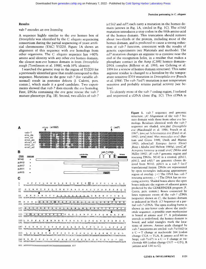
Figure 2. vab-7 phenotype. (A) Wild-type first-stage (L1) larvae. (B) vab-7(eI562) mutant L1 larvae. The vab-7 mutant larva has a severely disorganized posterior end and uncoordinated move- ment. Anterior, left.
880 bp long and has one long open reading frame, begin- ning at the 5' end of the clone and ending with a stop codon at position 700. Therefore, it appears to encode the carboxy-terminal end of the protein. Using the GENE- FINDER program (P. Green, pets. comm.), no additional 5' exons could be predicted in the genomic sequence; however, an in-frame meth ionine that could be the start of the protein is found at position 17 (boxed in Fig. 1C). The eDNA ends wi th a run of A residues, most of which are found in the genomic sequence, suggesting that this is not the true 3' end of the transcript. There is no obvi- ous polyadenylation signal in the clone, but one that might be used is located 400 bp further downstream in the genomic sequence. From Northern blotting, vab-7 RNA is est imated to be 1.4 kb (Fig. 3A, below), therefore this cDNA clone is miss ing - 5 0 0 nucleotides.
Besides the homeo box, the amino acid sequence shows two additional features in common with other eve homologs: a polyalanine stretch (underlined in Fig. 1C) and a region rich in serine and proline residues (at the carboxyl terminus of VAB-7; Fig. 1C).
vab-7 mutants have posterior defects
Figure 2 shows the phenotype caused by the strong vab-7 allele e1562. Mutants have a severely disorganized pos- terior region and uncoordinated movement . Despite these defects, most (85%) vab-7(e1562) animals are via- ble. The lethali ty may result from the inabil i ty to defe- cate, as the intest ines of dead larvae are extremely bloated. Two other alleles of vab-7 have weaker effects. vab-7(ed7) mutants have a similar movement defect, but a slightly less disorganized posterior region than vab- 7(e1562); vab-7(ed6) mutants have nearly wild-type movement , but have a bobbed tail.
The vab-7 movement defect shows an interesting fea- ture. When wild-type animals are touched on the head (or tail), they move backward (or forward), propelling themselves by the alternate contraction and relaxation of dorsal and ventral body wall muscles. When vab- 7(e1562) mutants back up in response to head touch, they flex both dorsally and ventrally, although more slowly and less smoothly than wild type. However, when vab-7(el 562) mutants move forward in response to tail touch, they curl ventrally and become locked in this
position for several seconds. This difference in behavior when moving forward versus backing suggests that at least part of the vab-7 movement defect has a neuronal origin, as the same muscles are used for movement in both directions; as yet, it is not known which neurons might be affected.
Dorsal posterior muscle and epidermal precursors express vab-7 RNA
vab-7 RNA is expressed primari ly in embryos, L1 and L2 larvae (Fig. 3A). To see where in the embryo vab-7 is expressed, RNA in situ hybridization was done. vab-7 RNA is first detected at the 100-cell stage, in four cells at the posterior end (Cxxp cells; Fig. 3B-D). These four cells are descendants of the C blastomere (one of six so-called founder cells), and give rise to primari ly dorsal posterior body wall muscles (hereafter "muscle" will be used to mean body wall muscle) and epidermis (also called the hypodermis in C. elegans), as well as two neurons (Fig. 3Dt. vab-7 RNA is also detected in daughters of these cells; however, in situ hybridization staining of older embryos was variable (not shown), possibly because the level of expression is reduced.
To look more easily at vab-7 expression, I made a translational fusion with lacZ (see Fig. 1B, pJA15). Early
Figure 3. vab-7 expression. (A} Developmental Northern blot with RNA from E (embryosl, L1 to L4 larvae, and A (adults). ITopl vab-7 probe; Ibottom} act-I probe as a loading control. (B.CI Earliest vab-7 RNA detected by in situ hybridization. Shown are two focal planes of a 100-cell stage embryo with four positive cells, two in B and two in C. [D} Partial lineage of the C blastomere. Cells are named by adding "a" to an anterior daughter and "p" to a posterior daughter; horizontal lines show cell divisions, vertical lines indicate time. (Q) Cells positive in B and C; below are their names and the cell types they produce.
1122 GENES & DEVELOPMENT
Cold Spring Harbor Laboratory Press on February 7, 2022 - Published by genesdev.cshlp.orgDownloaded from
Posterior patterning in C. elegans
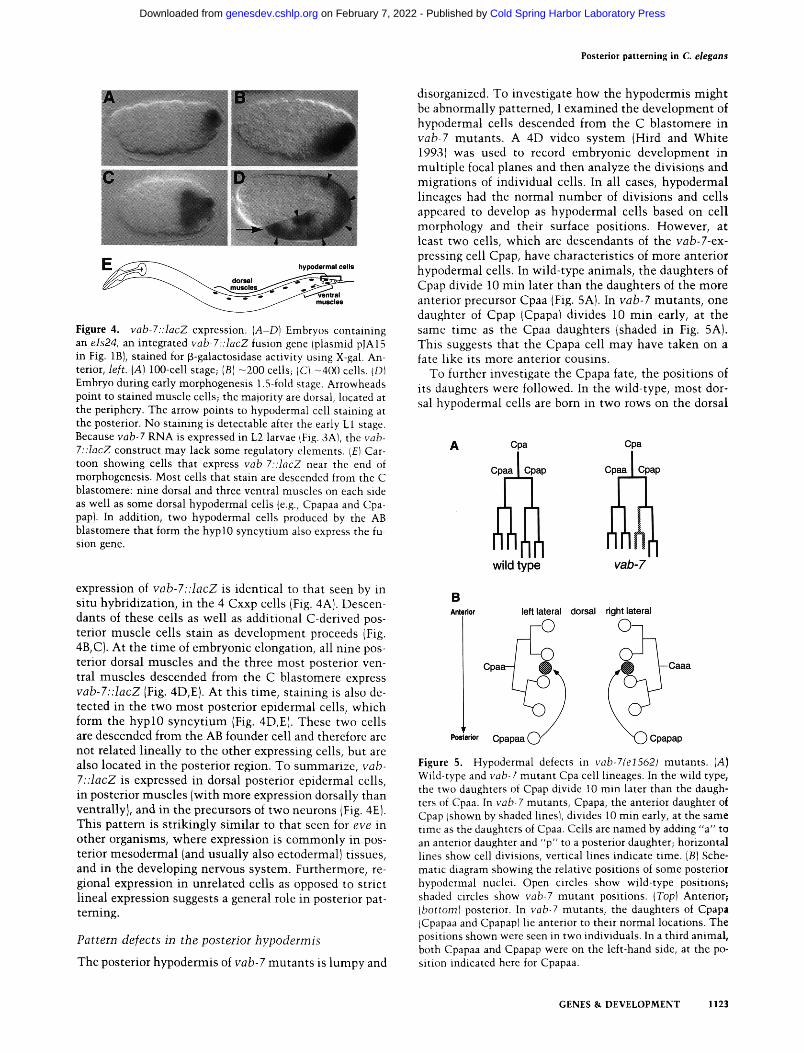
Figure 4. vab-7::lacZ expression. (A-D) Embryos containing an els24, an integrated vab-7::lacZ fusion gene (plasmid pJA15 in Fig. 1B), stained for p-galactosidase activity using X-gal. An- terior, left. (A) 100-cell stage; (B) -200 cells; IC) -400 cells. (DI Embryo during early morphogenesis 1.5-fold stage. Arrowheads point to stained muscle cells; the majority are dorsal, located at the periphery. The arrow points to hypodermal cell staining at the posterior. No staining is detectable after the early L1 stage. Because vab-7 RNA is expressed in L2 larvae ffig. 3A), the vab- 7::lacZ construct may lack some regulatory elements. IE) Car- toon showing cells that express vab-7::lacZ near the end of morphogenesis. Most cells that stain are descended from the C blastomere: nine dorsal and three ventral muscles on each side as well as some dorsal hypodermal cells (e.g., Cpapaa and Cpa- pap}. In addition, two hypodermal cells produced by the AB blastomere that form the hypl0 syncytium also express the fu- sion gene.
disorganized. To investigate how the hypodermis might be abnormally patterned, I examined the development of hypodermal cells descended from the C blastomere in vab-7 mutants . A 4D video system (Hird and White 1993} was used to record embryonic development in multiple focal planes and then analyze the divisions and migrations of individual cells. In all cases, hypodermal lineages had the normal number of divisions and cells appeared to develop as hypodermal cells based on cell morphology and their surface positions. However, at least two cells, which are descendants of the vab-7-ex- pressing cell Cpap, have characteristics of more anterior hypodermal cells. In wild-type animals, the daughters of Cpap divide 10 min later than the daughters of the more anterior precursor Cpaa (Fig. 5A). In vab-7 mutants , one daughter of Cpap (Cpapa} divides 10 min early, at the same time as the Cpaa daughters (shaded in Fig. 5A). This suggests that the Cpapa cell may have taken on a fate like its more anterior cousins.
To further investigate the Cpapa fate, the positions of its daughters were followed. In the wild-type, most dor- sal hypodermal cells are born in two rows on the dorsal
expression of vab-7::lacZ is identical to that seen by in situ hybridization, in the 4 Cxxp cells (Fig. 4A). Descen- dants of these cells as well as additional C-derived pos- terior muscle cells stain as development proceeds (Fig. 4B, C). At the t ime of embryonic elongation, all nine pos- terior dorsal muscles and the three most posterior ven- tral muscles descended from the C blastomere express vab-7::lacZ (Fig. 4D,E). At this time, staining is also de- tected in the two most posterior epidermal cells, which form the hyp l0 syncyt ium (Fig. 4D,E). These two cells are descended from the AB founder cell and therefore are not related lineally to the other expressing cells, but are also located in the posterior region. To summarize, vab- 7::lacZ is expressed in dorsal posterior epidermal cells, in posterior muscles (with more expression dorsally than ventrallyl, and in the precursors of two neurons IFig. 4E}. This pattern is strikingly similar to that seen for eve in other organisms, where expression is commonly in pos- terior mesodermal (and usually also ectodermal} tissues, and in the developing nervous system. Furthermore, re- gional expression in unrelated cells as opposed to strict lineal expression suggests a general role in posterior pat- terning.
Pattern defects in the posterior hypodermis
The posterior hypodermis of vab-7 mutants is lumpy and
Figure 5. Hypodermal defects in vab-7~e1562) mutants. (A} Wild-type and vab-7 mutant Cpa cell lineages. In the wild type, the two daughters of Cpap divide 10 min later than the daugh- ters of Cpaa. In vab-7 mutants, Cpapa, the anterior daughter of Cpap lshown by shaded lines), divides 10 min early, at the same time as the daughters of Cpaa. Cells are named by adding "a" to an anterior daughter and "p" to a posterior daughter; horizontal lines show cell divisions, vertical lines indicate time. (B) Sche- matic diagram showing the relative positions of some posterior hypodermal nuclei. Open circles show wild-type positions; shaded circles show vab-7 mutant positions. (Top) Anterior; ibottoml posterior. In vab-7 mutants, the daughters of Cpapa ICpapaa and Cpapap) lie anterior to their normal locations. The positions shown were seen in two individuals. In a third animal, both Cpapaa and Cpapap were on the left-hand side, at the po- sition indicated here for Cpapaa.
GENES & DEVELOPMENT 1123
Cold Spring Harbor Laboratory Press on February 7, 2022 - Published by genesdev.cshlp.orgDownloaded from
Ahringer
surface (Sulston et al. 1983). These nuclei then migrate in between each other across the dorsal midl ine to the opposite side, and eventual ly stop at left and right lateral positions. In these rows, the Cpapa daughters lie just posterior to the granddaughters of Cpaa and Caaa (final wild-type positions are shown by open circles in Fig. 5B}. In vab-7 mutants , the Cpapa daughters (Cpapaa and Cpa- pap) are located anterior to their normal positions (filled circles in Fig. 5B). Most often, they first migrate anteri- orly to the Caaa and Cpaa daughters, and then interdig- itate along wi th them. Therefore, by two criteria, the early cell division t ime and the anterior location of its daughters, the posterior Cpapa hypodermal cell appears to be transformed into a more anterior hypodermal cell. Other posterior hypodermal cells are also sometimes ab- normal ly positioned (not shown). The malformed poste- rior region of vab-7 mutants is l ikely to result at least partially from incorrect hypodermal cell locations, but affected cells may also produce the wrong type of hypo- dermal tissue.
Posterior muscles in vab-7 mutan t s are disorganized
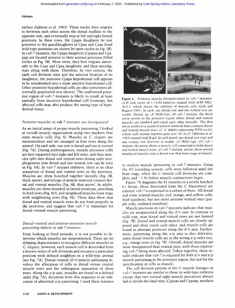
As an init ial assay of proper muscle patterning, I looked at overall muscle organization using two markers that stain muscle cells (Fig. 6). Muscles in C. elegans are mononucleate and are arranged in four rows along the animal. On each side, one row is dorsal and one is ventral (Fig. 7A). During embryogenesis, muscle precursor cells are first separated into right and left sides, and then mus- cles split into dorsal and ventral rows during early mor- phogenesis (one dorsal and one ventral row can be seen in Fig. 6A). In vab-7 mutan t embryos, there is no clear separation of dorsal and ventral rows in the posterior. Muscles are often bunched together laterally (Fig. 6B, thick arrow), and strings of muscle material connect dor- sal and ventral muscles (Fig. 6B, thin arrow). In adults, muscles are often stranded in lateral positions, attaching to both rows (Fig. 6D), and misplaced muscles form rings with neighboring rows (Fig. 6E). These data show that dorsal and ventral muscle rows do not form properly in the posterior, and suggest that vab-7 is important for dorsal-ventral muscle patterning.
Dors al-ven tral an d an terior-pos terior m u scle patterning defects in vab-7 mutan t s
From looking at fixed animals, it is not possible to de- termine which muscles are mispositioned. There are no defining characteristics to recognize different muscles in C. elegans; however, each muscle cell is descended from a known series of cell divisions and occupies a particular position wi th defined neighbors in a wild-type animal (see Fig. 7A). Dorsal-ventral (d/v) muscle patterning in- volves the allocation of cells to dorsal versus ventral muscle rows and the subsequent separation of these rows. Along the a/p axis, muscles are found in a defined order (Fig. 7A); deviation from this sequence is an indi- cation of abnormal a/p patterning. I used these features
Figure 6. Posterior muscle disorganization in vab-7 mutants. (A.B1 Left views of 1.5-fold embryos stained with mAb NE2/ 4C6.3, which shows the outlines of muscle cells (Goh and Bogaert 19911. In each, one dorsal row and one ventral row are visible. Dorsal, up. IA1 Wild type. IBI vab-7 mutant; the thick arrow points to the posterior region where dorsal and ventral muscles are jumbled and touch each other laterally. The thin arrow points to a strand of muscle material that connects dorsal and ventral muscle rows. (C-E} Adults expressing PD56 (cc561, a body wall myosin reporter gene lur2c-54::]acZ; Okkema et al. 1993) stained with X-gal. In each panel, one dorsal row (top) and one ventral row (bottoml is visible. ICI Wild type. (D} vab-7 mutant; the arrow shows a muscle cell connected to both dorsal and ventral muscle rows. IEI yah-7 mutant; arrows show several misplaced muscles near a dorsal row that form rings of muscle.
to analyze muscle patterning in vab-7 mutants . Using the 4D recording system, cells were followed until the bean stage, when the C muscle cell divisions are com- plete and - 1 hr before muscle contractions begin.
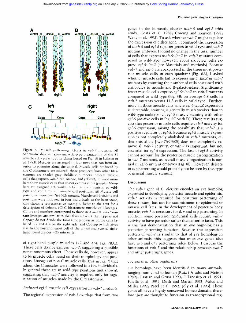
Figure 7A diagrams the 81 muscles in a newly hatched L1 larvae; those descended from the C blastomere are colored, vab-7 is expressed in a subset of these: All dorsal and some ventral muscles (red, orange, and yellow cells, bold numbers), but not more anterior ventral ones (pur- ple cells, outlined numbers}.
Muscle positions in vab-7 mutants indicate that mus- cles are mispatterned along the d/v axis. In contrast to wild type, neat dorsal and ventral rows are not formed IFig. 7B}. Dorsal and ventral muscle cells are closely op- posed and often touch each other, and muscle cells are found in aberrant positions along the d/v axis. Further- more, patterning along the a/p axis is also defective; some dorsal muscle cells are in the wrong a/p order (see, e.g., orange rows in Fig. 7B}. Overall, dorsal muscles are more disorganized than ventral ones, with those express- ing vab-7 being most affected. Taken together, these re- sults indicate that vab-7 is required for both d/v and a/p muscle patterning in the posterior region, but not for the specification of cells as muscle.
The cell division pattern of the C muscle lineages in vab-7 mutants are similar to those in wild-type embryos except that two ventral right muscle precursors always fail to divide the final t ime (Cppaaa and Cppaap, mothers
1124 GENES & DEVELOPMENT
Cold Spring Harbor Laboratory Press on February 7, 2022 - Published by genesdev.cshlp.orgDownloaded from
Posterior patterning in C. elegans
Figure 7. Muscle patterning defects in vob-7 mutants. {A) Schematic diagram showing wild-type organization of the 81 muscle cells present at hatching (based on Fig. 15 in Sulston et al. 198,31. Muscles are arranged in four rows that run from an- terior to posterior along the animal. Muscle cells produced by the C blastomere are colored; those produced from other blas- tomeres are shaded gray. Boldface numbers indicate muscle cells that express vab-7 (red, orange, and yellowl; outlined num- bers show muscle cells that do not express vab-7 Ipurplet. Num- bers are assigned arbitrarily to facilitate comparison of wild- type and vab-7 mutant muscle cell positions. LBI Muscle cell positions in one vab-7(e1562) mutant. Muscle cell divisions and positions were followed in four individuals to the bean stage; this shows a representative example. Refer to the text for a description of defects. (C) C blastomere muscle cell lineages. Colors and numbers correspond to those in A and B. vab-7 mu- tant lineages are similar to that shown except that Cppaaa and Cppaap do not divide the final time {shown as purple cells la- beled 1/2 and 3/4 on the right sidel and Cppppp Iwhich gives rise to the posterior-most cell of the dorsal and ventral right- hand rowsl divides -25 min early.
of right-hand purple muscles 1/2 and 3/4; Fig. 7B,C). These cells do not express vab-7, suggesting a possible nonautonomous effect. These cells do, however, appear to be muscle cells based on their morphology and posi- tions. Lineages of non-C muscle cells (gray in Fig. 7) that adjoin the C muscles were followed in a few individuals. In general these are in wild-type positions {not shown), suggesting that vab-7 activity is required only for orga- nization of muscles made by the C blastomere.
Reduced egl-5 muscle cell expression in vab-7 mutants
The regional expression of vab-7 overlaps that from two
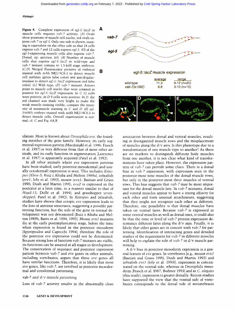
genes in the homeotic cluster mab-5 and egl-5 (this study; Costa et al. 1988; Cowing and Kenyon 1992; Wang et al. 19931. To ask whether vab-7 might regulate the expression of either gene, I compared the expression of mab-5 and egI-5 reporter genes in wild-type and vab-7 mutant embryos. ! found no change in the total number of cells that express mab-5::lacZ in vab-7 mutan ts com- pared to wild-type; however, about six fewer cells ex- press egl-5::lacZ (see Materials and methods). Because rob-7 and egl-5 are coexpressed in the three most poste- rior muscle cells in each quadrant (Fig. 8A), I asked whether muscle cells fail to express egl-5::lacZ in vab-7 mutants by counting the number of cells costained with antibodies to muscle and 13-galactosidase. Significantly fewer muscle cells express egl-5::lacZ in vab-7 mutants compared to wild type (Fig. 8B; on average 6.8 cells in rob-7 mutants versus 11.3 cells in wild type). Further- more, in those muscle cells where egl-5::lacZ expression is detectable, staining is generally much weaker than in wild-type embryos {cf. egl-5 muscle staining with other egl-5 positive cells in Fig. 8C with D). These results sug- gest that posterior muscle cells require vab-7 activity for eg]-5 expression, raising the possibility that vab-7 is a positive regulator of egl-5. Because egl-5 muscle expres- sion is not completely abolished in vab-7 mutants , ei- ther this allele [vab-Tle1562)] does not completely re- move all vab-7 activity, or vab-7 is important, but not essential for egl-5 expression. The loss of egl-5 activity cannot account for the posterior muscle disorganization in yah-7 mutants, as overall muscle organization is nor- mal in egl-5 mutan t embryos IFig. 8E). However, defects in a/p patterning would probably not be seen by this type of general muscle staining.
D i s c u s s i o n
The vab-7 gene of C. elegans encodes an eve homolog expressed in developing posterior muscle and epidermis. vab-7 activity is required for posterior patterning of these tissues, but not for commi tmen t to epidermal or muscle cell fates. In the development of posterior body muscle, vab-7 is necessary for d/v and a/p patterning. In addition, some posterior epidermal cells require vab-7 activity to have posterior rather than anterior fates. This is the first demonstrat ion that an eve homolog has a posterior patterning function. Because the expression pattern of vab-7 is similar to that of eve homologs in other animals, this suggests that most eve genes also have a/p and d /v patterning roles. Below, I discuss the functions of vab-7 and the relationship between vab-7 and other patterning genes.
eve genes in other organisms
eve homologs have been identified in many animals, ranging from coral to human {Ruiz i Altaba and Melton 1989a; Bastian and Gruss 1990; D'Esposito et al. 1991; Faiella et al. 1991; Dush and Martin 1992; Miles and Miller 1992; Patel et al. 1992; Joly et al. 1993). These genes all have a highly conserved homeo domain, there- fore they are thought to function as transcriptional reg-
GENES & DEVELOPMENT 1125
Cold Spring Harbor Laboratory Press on February 7, 2022 - Published by genesdev.cshlp.orgDownloaded from
Ahringer
Figure 8. Complete expression of egl-5::lacZ in muscle cells requires vab-7 activity. (A) Ovals show positions of muscle cell nuclei; red ovals ex- press vab-7 or egl-5. Only one side is shown; stain- ing is equivalent on the other side so that 24 cells express vab-7 and 12 cells express egI-5. All of the egl-5-expressing muscle cells also express vab-7. Dorsal, up; anterior, left. (B) Number of muscle cells that express egl-5::tacZ in wild-type and vab-7 mutant comma to 1.5-fold stage embryos. {C,D) Merged fluorescence pictures of embryos stained with mAb NE2/4C6.3 to detect muscle cell outlines (green false color} and anti-f~-galac- tosidase to detect egl-5::lacZ expression (red false color). (C} Wild type; {D) vab-7 mutant. Arrows point to muscle cell nuclei that were counted as positive for egJ-5::lacZ expression. In C 12 cells were positive; in D 9 cells were positive. In D. the red channel was made very bright to make the weak muscle staining visible; compare the inten- sity of nonmuscle staining in C and D. (El egl- 5(n945) embryo stained with mAb NE2/4C6.3 to detect muscle cells. Overall organization is nor- mal; cf. C and Fig. 6A, B.
ulators. Most is known about Drosophila eve, the found- ing member of the gene family. However, its early seg- mental expression pattern {Macdonald et al. 1986; Frasch et al. 1987) is very different from that of most other an- imals, and its early function in segmentat ion (Lawrence et al. 1987) is apparently acquired (Patel et al. 19921.
In all other animals where eve expression patterns have been studied, early posterior mesodermal (and usu- ally ectodermal) expression is seen. This includes Xeno- pus (Xhox-3; Ruiz i Altaba and Melton 1989a), zebrafish (evel; Joly et al. 1993), mouse (evxl; Bastian and Gruss 1990; Dush and Mart in 1992; evx2 is expressed in the posterior at a later time, in a manner similar to that of Hoxd-13; Doll4 et al. 1994), and grasshopper (even- skipped; Patel et al. 1992). In Xenopus and zebrafish, studies have shown that ectopic eve expression leads to the loss of anterior structures, suggesting a possible pat- terning function, but the role of the gene in normal de- velopment was not determined (Ruiz i Altaba and Mel- ton 1989b; Barro et al. 1994, 1995). Mouse evxI mutants die at the early post implantat ion stage, before the t ime when expression is found in the posterior mesoderm (Spyropoulos and Capecchi 1994), therefore the role of the posterior eve expression could not be determined. Because strong loss of function vab-7 mutants are viable, its functions can be assayed at all stages in development. The conservation of sequence and posterior expression pattern between vab-7 and eve genes in other animals, including vertebrates, argues that these eve genes all have similar functions. Therefore, it is likely that most eve genes, like vab-7, are involved in posterior mesoder- real and ectodermal patterning.
vab-7 and d / v muscle patterning
Loss of vab-7 activity results in the abnormally close
association between dorsal and ventral muscles, result- ing in disorganized muscle rows and the misplacement of muscles along the d/v axis. Is this phenotype due to a transformation of one muscle type to another? As there are no markers to distinguish different body muscles from one another, it is not clear what kind of transfor- mations have taken place. However, the expression pat- tern of vab-7 can provide some clues. There is a dorsal bias in yah-7 expression, with expression seen in the posterior-most nine muscles of the dorsal muscle rows, but only in the posterior-most three muscles of ventral rows. This bias suggests that vab-7 may be most impor- tant for the dorsal muscle fate. In vab-7 mutants , dorsal and ventral muscles appear to have a strong affinity for each other and form unusual a t tachments , suggesting that they might not recognize each other as different. Therefore, one possibility is that dorsal muscles have taken on ventral fates. Because vab-7 is expressed in some ventral muscles as well as dorsal ones, it could also be that the time or level of vab-7 protein expression de- termines different fates along the d /v axis. Finally, it is likely that other genes act in concert with vab-7 for pat- terning. Identification of interacting genes and detailed studies of the requirement for vab-7 in different muscles will help to explain the role of vab-7 in d /v muscle pat- terning.
A d/v bias in posterior mesoderm expression is a gen- eral feature of eve genes. In vertebrates [e.g., mouse evxl (Bastian and Gruss 1990; Dush and Mart in 1992) and zebrafish evel {Joly et al. 1993)], expression is concen- trated on the ventral side, whereas in Drosophila meso- derm (Frasch et al. 1987; Bodmer 1993) and in C. elegans (this study), expression is greater dorsally. Recent studies have supported the view that the ventral side of verte- brates corresponds to the dorsal side of invertebrates
1126 GENES & DEVELOPMENT
Cold Spring Harbor Laboratory Press on February 7, 2022 - Published by genesdev.cshlp.orgDownloaded from
Posterior patterning in C. elegans
{Holley et al. 1995); the different d/v biases in eve ex- pression are consistent with this idea.
a/p axial patterning by vab-7 and interaction with the HOM-C
The most distinctive defects in vab-7 mutants are in d/v muscle patterning. However, two phenotypes indicate that vab-7 is also important for a/p patterning. First, in vab-7 mutants two dorsal posterior hypodermal cells have fates similar to those of more anterior hypodermal cells. Second, some dorsal posterior muscles are disor- dered along the a/p axis. In both of these cases, cells undergo abnormal migrations, with the hypodermal cells moving a significant distance anteriorly. The loss of vab-7 activity could result in the transformation to an- terior cell fates or may cause cells to have confused in- structions that result in aberrant anterior positions.
Although little is known in C. elegans about external positional cues that might guide cells along the a/p axis, it is clear that genes in the HOM-C help give cells ap- propriate fates (for review, see Salser and Kenyon 1994). These homeot ic genes are homologous to related genes in probably all animals, and are most often arranged on the chromosome in the order in which they act along the a/p axis (for review, see McGinnis and Kmmlauf 1992; Kenyon 1994). In mice and humans, eve genes are phys- ically l inked to the "posterior" ends of two homeotic clusters (Faiella et al. 1991; D'Esposito et al. 1991; Dush and Martin 1992; Bastian et al. 19921, and it has been argued that this is the ancestral arrangement (Doll4 et al. 1994). This finding, coupled with their expression over- lapping and posterior to that of homeotic genes suggests that eve genes might be involved in a/p patterning, pos- sibly acting in concert with the HOM-C. vab-7 is not tightly l inked to the C. elegans HOM-C [although it is on the same chromosome {III)], but like vertebrate eve genes, its expression is posterior to and overlaps with that of the HOM-C genes. Here, I showed that the most posterior C. elegans HOM-C gene egl-5 requires vab-7 activity for complete expression in muscle cells. This interaction wi th the HOM-C lends support to the idea that eve genes in general may function as a posterior part of this global a/p patterning system. Loss of egl-5 activ- ity cannot account for the d/v muscle patterning defects in vab-7 mutants, as muscle rows in egl-5 mutants are well separated. However, it is possible that the a/p mus- cle patterning defects in vab-7 mutants are attributable to loss of egl-5 expression; experiments are underway to test this idea.
Zygotic embryonic patterning in C. elegans
The identif ication and study of numerous maternal gene products in C. elegans has led to an increasingly detailed view of early patterning events (for review, see Wood and Edgar 1994; Bowerman 1995). How zygotic patterning is init iated in the embryo is largely unknown. It is assumed that the HOM-C genes will be important for embryonic a/p patterning, as they begin to be expressed in mid-
embryogenesis (Cowing and Kenyon 1992; Clark et al. 1993; Wang et al. 1993), but their functions have so far only been investigated postembryonically. With the in- troduction of the 4D video microscope, full advantage can be taken of the invariant lineage as a powerful tool for the study of embryonic defects. One challenge will be to understand how maternal products ini t iate zygotic patterning in C. elegans, and the vab-7 gene will provide a new entry point for such studies. Given the simplici ty of C. elegans, further work on vab-7 is l ikely to lead to the identification of additional genes important for ani- mal patterning.
Mater ia l s and m e t h o d s
Genetic and phenotypic analyses
Worm culture and genetic analyses were as described (Bren- ner 1974; Wood 1988). The following mutations were used: wild type IBristoll N2; LGII: /em-l(hcl 7ts); LGIII: vab-7(I562}, vab-7(ed6L vab-Tted7L tD/2. unc-32{e189), dpy-18(e499); LGV: mulsl3 [egl-5..lacZ + rol-6(d)], cc56 {unc-54::lacZ + sup- 7} muls3 (mab-5::lacZ + rol-6(d)l. Extrachromosomal array: m uEx 19 [egl-5::lacZ + rol-6fd)l.
vab-7(e 1562) was isolated after ethylmethanesulfonate (em$) mutagenesis by J. Culotti lpers, comm.), vab-7(ed6) and vab- 7(ed7; are also ems-induced mutations and were isolated in a noncomplementation screen with deficiency tDf2 (which un- covers vab-7} by M. Maduro and D. Pilgrim (pets. comm.).
Percent lethality of vab-7(eI562) was determined for three broods at 25~ of 279 total progeny, 38 died t14%; 31 died as larvae and 7 as embryos}. The phenotype of vab-7(e1562) over a deficiency was determined as follows. Females of genotype fera- l (hcl 7ts); vab-7~e1562) dpy-I8(e499) raised at 25~ were mated to single males obtained from crossing tDf2/unc-32(e189) dpy- 18~e499! hermaphrodites with wild-type males. Progeny from crosses yielding no Dpy animals (tDf2 does not remove dpy-18) were scored as Vab or wild type. Of 531 progeny, 293 were scored as wild-type 185 were Vab and reached adulthood, and 53 died (3 as embryos and 50 as larvae). Assuming the Vab and dead animals were of genotype fem-l(hcl 7ts)/ +; vab-7(e1562) dpy- 18(e499J/tDf2, 22% 153/238} of vab-7(eI562)/tDf2 animals died Icompared with 14% of vab-7(e1562) homozygotes above). Scor- ing was conservative and probably included some Vabs in the wild-type class, which will slightly overestimate the lethality of vab-7(e1562)/tDf2. Overall, both the percent lethality and the phenotypes of vab-7(e 1562) homozygotes are similar to those of vab-7te1562)/tDf2, indicating that vab-7(e1562) is a strong loss of function allele. The phenotype of vab-7(ed6) over either vab- 7fe1562) or the deficiency tDf2 is stronger than that of vab- 7fed6) homozygotes. The phenotype of vab-7(ed7) in trans to vab-7(eI562) or tDf2 is only slightly stronger than that of vab- 7(ed7) homozygotes.
vab-7 cloning and sequencing
Subclones of cosmid M142 were made as follows, pJA12 is the 20-kb PstI fragment from M142 cloned into the PstI site of pBSKS- {Stratagene). pJA17 is a 14.1-kb SmaI-PstI fragment from M142 made by digesting pJA12 with Sinai (which cuts once in the polylinker 5' to vab-7 and once in the M142 se- quencel and religating it. pJA11 is the 8.3-kb SacI fragment from M142 cloned into the 5acI site of pBSKS .
For testing DNAs for rescuing activity, a cosmid or plasmid at 10 ~.g/ml was injected together with 100 p.g/ml of marker plas-
GENES & DEVELOPMENT 1127
Cold Spring Harbor Laboratory Press on February 7, 2022 - Published by genesdev.cshlp.orgDownloaded from
Ahringer
mid pRF4 {containing a rol-6(d) gene that causes a dominant rolling phenotype; Mello et al. 1991) using described procedures (Fire 1986; Mello et al. 1991). For M142, pJA12, and pJAll , rescue was tested in heritable lines that were generated. The 2/2 lines containing M142 each rescued the posterior defects and uncoordinated phenotype of vab-7(eI562). 5/5 lines con- taining pJA12 rescued, and 0/3 lines containing pJA11 rescued. Plasmid pJA17 was tested only for rescue in the F~ progeny of injected hermaphrodites. Of 12 F~ rolling hermaphrodites, 7 were rescued.
One cDNA was identified after screening 1 mill ion phage from an embryonic cDNA library in lambda gtl 1 {made by P. Okkema). This phage grew very poorly, therefore the insert was amplified by PCR and cloned into pBSKS- two independent times. The sequence was determined on both strands from one clone, and on one strand from another clone using Sequenase (U.S. Biochemical). Software from the Staden package (Staden 1994) was used for sequence assembly. To identify the positions of introns, the sequence of most of the genomic region corre- sponding to the cDNA was assembled by alternately sequencing regions of pJAll using vab-7-specific primers and Sequenase (U.S. Biochemical) and then obtaining overlapping sequence reads from YAC Y52D3 from the sequencing consort ium {not shown). Recently, unfinished cosmid M142 sequence has be- come available, and the diagram in Figure 1B showing the M 142 genomic organization is based on this sequence. The GENE- FINDER program (P. Green, pets. comm.) was used to predict genes within M142; gray boxes in Figure 1A indicate regions of predicted adjacent genes. GENEFINDER predicted the exons found in the vab-7 cDNA described above, but did not predict any additional exons.
To look for mutat ions associated with vab-7 alleles, the ho- meo domain was sequenced from each. vab-7-specific primers were used to PCR amplify each of the two exons containing homeo domain sequence along with some intron sequence from homozygous mutan t animals. For each mutant , 10 worms were picked into 50 ~1 of 1 x Promega Taq buffer containing 100 ~g/ml of proteinase K {Sigma), incubated for 1 hr at 65~ then heated to 95~ for 15 rain to inactivate the proteinase; 5-10 ~1 of this DNA was used for each PCR reaction. The amplified products were purified using Wizard PCR kit tPromega) and sequenced directly with Sequenase and either a ~2P-labeled primer or with an unlabeled primer and [3~S]dATP. Any changes found were confirmed by sequencing an independent PCR prod- uct. One change was found in vab-7(e1562) and one in vab- 7(edT) (shown in Fig. 1), but no changes were found in the ho- meo domain of vab-7(ed6).
vab-7 appears to be a single copy gene based on Southern blotting; only vab-7 DNA was detected using the vab-7 homeo box as a probe at high stringency {67~ 5 x SSC). At low strin- gency (52~ 5x SSC), many additional bands were visible at differing intensities, probably corresponding to the numerous other homeo domain-containing genes {not shownl.
vab-7 expression
For the Northern blot, - 1 ~g of poly(A)+ RNA was used per lane (less for the embryonic and adult lanes). Preparation of poly(A} + RNA, formaldehyde/agarose electrophoresis, and blot- ting were carried out as described {Rosenquist and Kimble 1988) except that Hybond membrane (Amersham) was used. The vab-7 homeo domain sequence was used as a probe, and the blot was reprobed with act-l-specific DNA {Files et al. 1983; insert from plasmid pT3/T7-18-103, from M. Krause, National Insti- tutes of Health, Bethesda, MD). In situ hybridization was as in Seydoux and Fire (1994) also with a vab-7 homeo domain probe.
The vab-7::lacZ reporter gene contains DNA from the up- stream PstI site in p|A12 to a XhoI site in the second exon of the cDNA (a total of 13.8 kb}, fused in-frame to the lacZ-coding sequence. This was made as follows: pJA12 was digested with XhoI, filled in with Klenow, and digested with PstI. The insert was ligated to pPD16.43 (Fire et al. 1990) that had been digested with XbaI, filled in using Klenow, and then digested with PstI. This plasmid, designated pIA17, was injected into wild-type hermaphrodites at 50 ~g/ml together with pRF4 at 100 ~g/ml las in Fire 1986; Mello et al. 19911 and three heritable lines containing extrachromosomal arrays were obtained. These were assayed for p-galactosidase activity by staining with X-gal (as in Fire et al. 1990; Wang et al. 1993), and all three showed the same pattern of expression. An integrated version, els24 (on chromo- some II!, was isolated after t reatment of one of these arrays with X-rays 13500 fads). The pattern of expression of els24 is the same as that of the extrachromosomal arrays, and it was used for characterization of vab-7 reporter gene expression pattern in Figure 3. els24 does not rescue the vab-7(1562) mutan t pheno- type.
The cells that express vab-7 RNA by in situ hybridization were identified based on their positions in the embryo. As the early expression from els24 is indistinguishable from the early in situ pattern, cell identification was confirmed in els24 em- bryos as follows, els24 embryos stuck to polylysine-coated cov- erslips were mounted in a mixture of fixative and staining so- lution 150 mM NaPi (pH 7.5}, 2 mM MgCI~, 5 mM KFe/5 mM KFo, 1 ~g/ml of DAPI, 0.004% SDS, 5% sucrose, 0.125% X-gal, 0.7% glutyraldehyde; G. Seydoux, pets. comm.1 on 3% agar pads also containing this solution. Development was recorded up to the 150-cell stage using the four-dimensional microscope. The egg- shell was then permeabilized using a laser to s imultaneously fix the embryo and allow detection of p-galactosidase activity. Af- ter incubation overnight at room temperature, the positions of positive cells were recorded, and these were identified unam- biguously based on their lineage, which was determined from the earlier recording. In t 50-cell stage embryos, both daughters of four cells la total of 8) stained; Caap, Capp, Cpap, Cppp. Later staining cells in els24 were identified by double staining with markers. For muscle, els24 was double stained with mAb NE8/ 4C6.2 IGoh and Bogaert 1991} and anti-p-galactosidase (Cappel); for hypodermal cells, double staining was with anti-LIN-26 polyclonal rabbit serum (which stains all epidermal cells; M. Labouesse, pets. comm.i and anti-p-galactosidase (Promega). The cells positive with two antibodies were identified in comma to twofold embryos based on their positions Ibased on Sulston et al. 1983).
Lineage analyses
Embryonic lineages were followed using a four-dimensional video recording system IHird and White 1993). Recordings usu- ally began at the 4- to 12-cell stage and ended as the embryo began to move lafter most cell divisions are complete); 20-25 focal planes were recorded every 30 sec. For analysis, an em- bryo-shaped grid printed on a transparency was made contain- ing 10 rows and 10 columns, and this was pasted to the screen while recordings were played. The position on the grid, the t ime in the recording, and the focal plane were noted for all cell divisions within the C lineage plus most other muscle lineages and many other posterior cells in four vab-7(e1562) embryos. For Figure 7B, muscle cell positions shown were determined at the bean stage. Muscle cell positions in the other three embryos were similarly defective, although positions varied from animal to animal; data for these are available upon request.
1128 GENES & DEVELOPMENT
Cold Spring Harbor Laboratory Press on February 7, 2022 - Published by genesdev.cshlp.orgDownloaded from
Posterior patterning in C. elegar~s
Reporter gene and muscle marker assays
Antibody staining of embryos was as in Albertson (1984}. [3-Ga- lactosidase activity from reporter genes was detected either us- ing X-gal (as in Fire et al. 1990 for adults or Wang et al. 1993 for embryos) or using an anti-~-galactosidase antibody (Cappel or Promega). Overall muscle organization was assayed in adults with a body wall myosin reporter gene called PD56(cc56) (Okkema et al. 1993; an unc-54::lacZ fusion gene} and in em- bryos by staining with mAb NE8/4C6.3, which stains the out- lines of body wall muscle cells (Goh and Bogaert 1991).
mab-5 expression was assayed using muls3, a mab-5::lacZ fusion gene (Cowing and Kenyon 1992). Embryos were stained with X-gal and total stained cells counted. An average of 13 cells stained in both wild-type (n= 18) and vab-7(e1562) mutant (n= 15) comma stage embryos. The total number of cells ex- pressing egl-5::lacZ was counted using muExI9 (Wang et al. 1993L an extrachromosomal array that is lost mitotically, re- sulting in variable staining. At the comma stage, an average of 19 cells (range 9-31, n =28)expressed egl-5::lacZ in wild type versus 13 cells (range 3-21, n =26)in vab-7(e1562) mutants. At the threefold stage, an average of 26 cells (range 15-32, n =33} expressed egl-5::lacZ in wild type versus 18 cells (range 13-27, n=39) in vab-7(e1562) mutants, muls l3 (an integrated version of muExl9; S. Salser and C. Kenyon, pers. comm.) was used to count the number of muscle cells that express egl-5::]acZ. Em- bryos were costained with mAb NE8/4C6.3 and rabbit anti-[3- galactosidase polyclonal serum (Cappel}, and the number of cells staining with both were counted in wild type and vab- 7(e1562) mutant comma to 1.5-fold stage embryos. Nonmuscle staining of egl-5::lacZ was of similar intensity in wild-type and vab-7 mutant embryos, but muscle cell staining was often very weak in vab-7 mutants. To aid in identifying these weakly stained muscle cells, a confocal microscope was used; this was unnecessary for wild-type embryos, egl-5::lacZ-expressing cells are missing from all four muscle rows (not shown}.
A c k n o w l e d g m e n t s
I thank the following people for sending me reagents, strains, and protocols: A. Coulson for cosmids, A. Fire for PD56, M. Krause for an act-1 plasmid, M. Labouesse for anti-LIN-26 an- tibody, M. Maduro and D. Pilgrim for vab-7 alleles, P. Okkema for cDNA libraries, S. Salser and C. Kenyon for HOM-C::lacZ strains, and G. Seydoux for in situ and lacZ staining protocols. I am also grateful to M. Berks and the rest of the C. elegans sequencing consortium for Y52D3 and M142 sequence, and to R. Durbin for help with genefinding. R. Durbin, M. Freeman, J. Hodgkin, P. Lawrence, and J-P. Vincent gave me helpful com- ments on the manuscript. Finally, I thank the Cell Biology di- vision for laboratory space and colleagues for discussions.
N o t e added in proof
The EMBL accession number for the vab-7 cDNA sequence is X96636.
References
Albertson, D.G. 1984. Formation of the first cleavage spindle in nematode embryos. Dev. Biol. 101" 61-72.
Barro, O., C. Joly, H. Condamine, and H. Boulekbache. 1994. Widespread expression of the Xenopus homeobox gene Xhox3 in zebrafish eggs causes a disruption of the anterior- posterior axis. Int. J. Dev. Biol. 38: 613-622.
Barro, O., S. Vriz, J.S. Joly, C. Joly, H. Condamine, and H.
Boulkebache. 1995. Widespread expression of the eveI gene in zebrafish embryos affects the anterior-posterior axis pat- tern. Dev. Genet. 17: 117-128.
Bastian, H. and P. Gruss. 1990. A murine even-skipped homo- logue, Evx I, is expressed during early embryogenesis and neurogenesis in a biphasic manner. EMBO J. 9: 1839-1852.
Bastian, H., P. Gruss, D. Duboule, and J.C. Izpisuabelmonte. 1992. The murine even-skipped-like gene evx-2 is closely linked to the Hox-4 complex, but is transcribed in the oppo- site direction. Mamm. Genome 3: 241-243.
Billeter, M., Y.Q. Qian, G. Otting, M. Muller, W. Gehring, and K. Wuthrich. 1993. Determination of the NMR solution structure of an Antennapedia homeodomain-DNA complex. I. Mol. Biol. 234: 1084-1094.
Bodmer, R. 1993. The gene tinman is required for specification of the heart and visceral muscles in Drosophila. Develop- ment 118: 719-729.
Bowerman, B. 1995. Determinants of blastomere identity in the early C. elegans embryo. BioEssays 17: 405-414.
Brenner, S. 1974. The genetics of Caenorhabditis elegans. Ge- netics 77: 71-94.
Clark, S.G., A.D. Chisholm, and H.R. Horvitz. 1993. Control of cell fates in the central body region of C. elegans by the homeobox gene 1m-39. Cell 74: 43-55.
Costa, M., M. Weir, A. Coulson, J. Sulston, and C. Kenyon. 1988. Posterior pattern formation in C. elegans involves po- sition-specific expression of a gene containing a homeobox. Cell 55: 747-756.
Cowing, D.W. and C. Kenyon. 1992. Expression of the homeotic gene mab-5 during Caenorhabc!itis elegans embryogenesis. Development 116: 481-490.
D'Esposito, M., F. Morelli, D. Acampora, E. Migliaccio, A. Sim- eone, and E. Boncinelli. 1991. EVX2, a human homeobox gene homologous to the even-skipped segmentation gene, is localized at the 5' end of HOX4 locus on chromosome 2. Genomics 10: 43-50.
Dolle, P., V. Fraulob, and D. Duboule. 1994. Developmental expression of the mouse Evx-2 gene: Relationship with the evolution of the HOM/Hox complex. (Suppl.} Development: 143-153.
Dush, M.K. and G.R. Martin. 1992. Analysis of mouse Evx genes: Evx-I displays graded expression in the primitive streak. Dev. Biol. 151: 273-287.
Faiella, A., M. D'Esposito, M. Rambaldi, D. Acampora, S. Bal- sofiore, A. Stornaiuolo, A. Mallamaci, E. Migliaccio, M. Gulisano, A. Simeone, et al. 1991. Isolation and mapping of EVX1, a human homeobox gene homologous to even- skipped, localized at the 5' end of HOX1 locus on chromo- some 7. Nucleic Acids Res. 19: 6541-6545.
Files, J.G., S. Cart, and D. Hirsh. 1983. Actin gene family of Caenorhabditis elegans. I. Mol. Biol. 164: 355-375.
Fire, A. 1986. Integrative transformation of C. elegans. EMBO ]. 5: 2673-2680.
Fire, A., H.S. White, and D. Dixon. 1990. A modular set of lacZ fusion vectors for studying gene expression in Caenorhab- ditis elegans. Gene 93: 189-198.
Frasch, M., T. Hoey, R. Christine, H. Doyle, and M. Levine. 1987. Characterization and localization of the even-skipped protein of Drosophila. EMBO ]. 6: 749-759.
Frasch, M., R. Warrior, I. Tugwood, and M. Levine. 1988. Mo- lecular analysis of even-skipped mutants in Drosophila de- velopment. Genes & Dev. 2: 824-1838.
Gehring, W.J., Y.Q. Qian, M. Billeter, K. Furukubotokunaga, A.F. Schier, D. Resendezperez, M. Affolter, G. Otting, and K. Wuthrich. 1994. Homeodomain-DNA recognition. Cell 78:211-223.
GENES & DEVELOPMENT 1129
Cold Spring Harbor Laboratory Press on February 7, 2022 - Published by genesdev.cshlp.orgDownloaded from
Ahringer
Goh, P.-Y. and T. Bogaert. 1991. Positioning and maintenance of embryonic body wall muscle attachments in C. elegans re- quires the mup-I gene. Development 111: 667-681.
Hird, S.N. and J.G. White. 1993. Cortical and cytoplasmic flow polarity in early embryonic cells of Caenorhabditis elegans. J. Celt Biol. 121: 1343-1355.
Holley, S.A., P.D. Jackson, Y. Sasai, B. Lu, E.M. Derobertis, F.M. Hoffmann, and E.L. Ferguson. 1995. A conserved system for dorsal-ventral patterning in insects and vertebrates involv- ing sog and chordin. Nature 376: 249-253.
Joly, J.-S., C. Joly, S. Schulte-Merker, H. Boulekbache, and H. Condamine. 1993. The ventral and posterior expression of the zebrafish homeobox gene evel is perturbed in dorsalized and mutant embryos. Development 119: 1261-1275.
Kenyon, C. 1994. If birds can fly, why can't we? Homeotic genes and evolution. Cell 78: 175-180.
Lawrence, P.A., P. Johnston, P. Macdonald, and G. Struhl. 1987. Borders of parasegments in Drosophila embryos are delim- ited by the fushi tarazu and even-skipped genes. Nature 328: 440--442.
Macdonald, P., P.W. Ingham, and G. Struhl. 1986. Isolation, structure, and expression of even-skipped: A second pair- rule gene of Drosophila containing a homoebox. Ceil 47: 721-734.
McGinnis, W. and R. Krumlauf. 1992. Homeobox genes and axial patterning. Cell 68: 283-302.
Mello, C.C., J.M. Kramer, D. Stinchcomb, and V. Ambros. 1991. Efficient gene transfer in C. elegans: Extrachromosomal maintenance and integration of transforming sequences. EMBO ]. 10: 3959-3970.
Miles, A. and D.J. Miller. 1992. Genomes of diploblastic organ- isms contain homeoboxes: Sequence of eveC, an even- skipped homologue from the cnidarian Acropora formosa. Proc. R. Soc. Lond. Biol. 248: 159-161.
Okkema, P.G., S.W. Harrison, V. Plunger, A. Aryans, and A. Fire. 1993. Sequence requirements for myosin gene expres- sion and regulation in Caenorhabditis elegans. Genetics 135: 385-404.
Patel, N.H., E.E. Ball, and C.S. Goodman. 1992. Changing role of even-skipped during the evolution of insect pattern forma- tion. Nature 357: 339-342.
Rosenquist, T.A. and J. Kimble. 1988. Molecular cloning and transcript analysis of fern-3, a sex-determination gene in C. elegans. Genes & Dev. 2: 606-616.
Ruiz i Altaba, A. and D.A. Melton. 1989a. Bimodal and graded expression of the Xenopus homeobox gene Xhox3 during embryonic development. Development 106:173-183.
1989b. Involvement of the Xenopus homeobox gene Xhox3 in pattern formation along the anterior-posterior axis. Cell 57:317-326.
Salser, S.J. and C. Kenyon. 1994. Patterning C. elegans: Ho- meotic cluster genes, cell fates and cell migrations. Trends Genet. 10: 159-164.
Seydoux, G. and A. Fire. 1994. Soma-germline asymmetry in the distributions of embryonic RNAs in Caenorhabditis ele- gans. Development 120" 2823-2834.
Slack, J.M.W. 1991. From egg to embryo. Cambridge University Press, Cambridge, UK.
Spyropoulos, D.D. and M.R. Capecchi. 1994. Targeted disrup- tion of the even-skipped gene, evxl, causes early postim- plantation lethality of the mouse conceptus. Genes & Dev. 8" 1949-1961.
Staden, R. 1994. Staden. Methods Mol. Biol. 25: 9-170. Sulston, J.E., E. Schierenberg, J.G. White, and J.N. Thomson.
1983. The embryonic cell lineage of the nematode Cae- norhabditis elegans. Dev. Biol. 100:64-119.
Tomlinson, A., B.E. Kimmel, and G.M. Rugin. 1988. rough, a Drosophila homeobox gene required in photoreceptors R2 and R5 for inductive interactions in the developing eye. Cell 55: 771-784.
Wang, B.B., I.M. Muller, J. Austin, N.T. Robinson, A. Chisholm, and C. Kenyon. 1993. A homeotic gene cluster patterns the anteroposterior body axis of C. elegans. Cell 74.29-42.
Wood, W.B. 1988. The Nematode Caenorhabditis elegans. Cold Spring Harbor Laboratory, Cold Spring Harbor, NY.
Wood, W.B. and L.G. Edgar. 1994. Patterning in the C. elegans embryo. Trends Genet. 10: 49-54.
1130 GENES & DEVELOPMENT
Cold Spring Harbor Laboratory Press on February 7, 2022 - Published by genesdev.cshlp.orgDownloaded from
10.1101/gad.10.9.1120Access the most recent version at doi: 10:1996, Genes Dev.
J Ahringer homolog vab-7.Posterior patterning by the Caenorhabditis elegans even-skipped
References
http://genesdev.cshlp.org/content/10/9/1120.full.html#ref-list-1
This article cites 44 articles, 11 of which can be accessed free at:
License
ServiceEmail Alerting
click here.right corner of the article or
Receive free email alerts when new articles cite this article - sign up in the box at the top
Copyright © Cold Spring Harbor Laboratory Press
Cold Spring Harbor Laboratory Press on February 7, 2022 - Published by genesdev.cshlp.orgDownloaded from