Embed Size (px)
Citation preview

Polymeric synthetic nanoparticles for the induction ofantigen-specific immunological toleranceRoberto A. Maldonadoa, Robert A. LaMothea, Joseph D. Ferraria, Ai-Hong Zhangb, Robert J. Rossib, Pallavi N. Koltea,Aaron P. Griseta, Conlin O’Neila, David H. Altreutera, Erica Browninga, Lloyd Johnstona, Omid C. Farokhzadc,d,Robert Langere,f, David W. Scottb, Ulrich H. von Andriang,h, and Takashi Kei Kishimotoa,1
aSelecta Biosciences, Inc., Watertown, MA 02472; bDepartment of Medicine, Uniformed Services University of the Health Sciences, Bethesda, MD20814; cLaboratory of Nanomedicine and Biomaterials, Department of Anesthesiology, Brigham and Women’s Hospital, Harvard Medical School,Boston, MA 02115; dKing Abdulaziz University, Jeddah 22254, Saudi Arabia; eDepartment of Chemical Engineering, Massachusetts Institute ofTechnology, Cambridge, MA 02139; fThe David H. Koch Institute for Integrative Cancer Research, Cambridge, MA 02139; gDivision of Immunology,Department of Microbiology and Immunobiology, Harvard Medical School, Boston, MA 02115-6017; and hThe Ragon Institute of MassachusettsGeneral Hospital, Massachusetts Institute of Technology and Harvard, Cambridge, MA 02139
Edited by Robert L. Coffman, Dynavax Technologies, Berkeley, CA, and approved November 6, 2014 (received for review May 10, 2014)
Current treatments to control pathological or unwanted immuneresponses often use broadly immunosuppressive drugs. Newapproaches to induce antigen-specific immunological tolerancethat control both cellular and humoral immune responses aredesirable. Here we describe the use of synthetic, biodegradablenanoparticles carrying either protein or peptide antigens anda tolerogenic immunomodulator, rapamycin, to induce durableand antigen-specific immune tolerance, even in the presence ofpotent Toll-like receptor agonists. Treatment with tolerogenicnanoparticles results in the inhibition of CD4+ and CD8+ T-cellactivation, an increase in regulatory cells, durable B-cell toleranceresistant to multiple immunogenic challenges, and the inhibitionof antigen-specific hypersensitivity reactions, relapsing experi-mental autoimmune encephalomyelitis, and antibody responsesagainst coagulation factor VIII in hemophilia A mice, even inanimals previously sensitized to antigen. Only encapsulated rapa-mycin, not the free form, could induce immunological tolerance.Tolerogenic nanoparticle therapy represents a potential novel ap-proach for the treatment of allergies, autoimmune diseases, and pre-vention of antidrug antibodies against biologic therapies.
nanoparticles | immune tolerance | rapamycin | immunotherapy |anti-drug antibodies
Undesired immunogenicity can have a profound impact onhuman health. Allergies, including allergic asthma and se-
vere food allergies, affect ∼20% of the population, and theprevalence has been steadily increasing over the past severaldecades (1). The prevalence of autoimmune diseases, includingmultiple sclerosis and type 1 diabetes, is ∼4.5% (2). Unwantedimmunogenicity can also affect both efficacy and safety of bio-logic drugs (3), particularly in the case of protein replacementtherapies for the treatment of genetic deficiencies, such as he-mophilia A (4) and Pompe Disease (5). Immunomodulatoryagents commonly used to control immunogenicity are oftenbroadly immunosuppressive and typically require chronic ad-ministration that can lead to reactivation of latent pathogens, de-velopment of tumors, and opportunistic infections (6, 7). Therefore,antigen-specific, durable tolerogenic therapy would be highly de-sirable from an efficacy and safety perspective.Multiple techniques for antigen-specific immunotherapy have
been described, although only allergen immunotherapy, whereinlow doses of antigen are delivered in the absence of immuno-modulating agents, is currently used in the clinic (1). Experi-mental approaches have included oral administration of antigen,high dose tolerance, and the use of altered peptide ligands (8).Although these methods have been successful in preclinicalmodels, translation to human clinical trials has been largelydisappointing (8). Alternative strategies to leverage tolerogenicprogramming associated with apoptotic cells include conjugatingantigen to splenocytes (9–12) or synthetic microparticles (13, 14)
or targeting antigen to the surface of red blood cells (15). Otherapproaches include loading particles with MHC complexes thatpresent relevant peptides in the absence of costimulation (16,17), liposomal copresentation of antigen with a ligand specificfor the negative signaling receptor CD22 on B cells (18), code-livery of peptide antigen with an aryl hydrocarbon receptor ag-onist (19), and cotreatment with pharmacological agents, such asmethotrexate (20). A major concern for antigen immunotherapyis the ability to induce and maintain tolerance in the presence ofproinflammatory stimuli caused by tissue stress, injury, or con-current infections. We sought to develop an antigen-specifictolerogenic technology that could control both T-cell– andB-cell–mediated immunity and that was durable over time and tomultiple challenges with the antigen, even in the presence ofstrong innate immune stimulants.Dendritic cells (DCs) are an attractive target for immuno-
therapies due to their central role in antigen presentation to Tcells and their ability to induce and control regulatory responses
Significance
Synthetic nanoparticles containing either protein or peptideantigen and the immunosuppressant rapamycin are capable ofinducing durable and specific resistance to mounting immuneresponses toward the antigen. This immunological toleranceoperates on lymphocytes even after multiple immunogenicchallenges with the antigen and adding enhancers of immuneresponses (adjuvants). As a result, the animals treated withthese tolerogenic nanoparticles (tNPs) show reduced allergichypersensitivity disorders, protection from disease relapse ina model of multiple sclerosis, and prevention of inhibitoryantidrug antibody responses in an animal model of hemophiliaA. These results show the potential for nanocarriers to modifythe immunoreactivity of a given molecule by providing tol-erogenic instructions to the immune system, thereby pre-venting or reversing pathological and neutralizing immuneresponses.
Author contributions: R.A.M., R.A.L., J.D.F., A.-H.Z., D.W.S., U.H.v.A., and T.K.K. designedresearch; R.A.M., R.A.L., J.D.F., A.-H.Z., R.J.R., P.N.K., A.P.G., C.O., D.H.A., and E.B. per-formed research; A.P.G., C.O., D.H.A., L.J., O.C.F., and R.L. contributed new reagents/analytic tools; R.A.M., R.A.L., J.D.F., A.-H.Z., R.J.R., P.N.K., A.P.G., C.O., D.H.A., E.B., L.J.,O.C.F., D.W.S., U.H.v.A., and T.K.K. analyzed data; and R.A.M. and T.K.K. wrote the paper.
Conflict of interest statement: U.H.v.A., O.C.F., and R.L. are co-founders and shareholdersof Selecta Biosciences. The other authors—except for D.W.S., A.-H.Z., and R.J.R.—areemployees and shareholders of Selecta Biosciences. D.W.S. received financial supportfrom Selecta Biosciences.
This article is a PNAS Direct Submission.
Freely available online through the PNAS open access option.1To whom correspondence should be addressed. Email: [email protected].
This article contains supporting information online at www.pnas.org/lookup/suppl/doi:10.1073/pnas.1408686111/-/DCSupplemental.
E156–E165 | PNAS | Published online December 29, 2014 www.pnas.org/cgi/doi/10.1073/pnas.1408686111

to secure self-tolerance (21–25). Thomson and colleagues (26,27) demonstrated that treating DCs with rapamycin, an inhibitorof the mTOR pathway, induces a tolerogenic DC phenotypecapable of inducing Treg differentiation and antigen-specificimmune tolerance that is resistant to the proinflammatory cas-cade triggered by TLR signaling. However, conventional therapywith free rapamycin requires chronic systemic administration,resulting in broad immunosuppression due to its direct effect onlymphocytes (28), whereas low doses of rapamycin may para-doxically augment effector T-cell memory (29). Thus, it would bedesirable to transiently target rapamycin’s effects to DCs andother antigen-presenting cells (APCs) at the time of antigen en-counter. Nanoparticles (NPs) are an ideal mechanism to deliverantigen (16, 30, 31) and drugs (32) to APCs, as these cells arekeyed to capture and internalize nanoparticulates such as viruses.Here we describe the development of tolerogenic NPs (tNPs)
using materials and compounds that have been well validated inthe clinic. These self-assembling, biodegradable poly(lactide-coglycolide) (PLGA) tNPs containing either protein or peptideantigens and rapamycin are capable of inducing durable antigen-specific tolerance that control adaptive immune responses andwithstand multiple immunogenic challenges with antigen. Wedemonstrate that either s.c. or i.v. administration of tNPs inhibitsthe activation of antigen-specific CD4+ and CD8+ T cells and Bcells while inducing antigen-specific Tregs and Bregs. Swiss JackLambert (SJL) mice immunized with the myelin proteolipidprotein 139–151 peptide in complete Freud’s adjuvant (PLP139–
151/CFA) and treated therapeutically with a single dose of tNPsat the peak of disease are completely protected from developingrelapsing paralysis. In hemophilia A animals, administration oftNP before or after the establishment of an anti-factor VIII(FVIII) antibody response led to a significant reduction of theneutralizing antibody response against FVIII. Treatment of micewith tNP prevents both cellular and humoral immunity even inthe presence of potent TLR agonists. These effects are de-pendent on the presence of the encapsulated rapamycin (not freein solution).
ResultsInduction of CD4+ T-Cell Tolerance Using tNPs. PLGA tNPs con-taining rapamycin and chicken ovalbumin (OVA) or rapa-mycin and the MHC class II-restricted OVA peptide 323–339(OVA323–339) were prepared using a water-in-oil-in-water doubleemulsion process (Fig. 1A). To investigate whether tNPs couldinhibit CD4+ T-cell activation, T cells specific for the OVA323–339peptide (OTII T cells) were adoptively transferred into naiveanimals. Mice were then immunized s.c. with OVA323–339 admixedwith a TLR7/8 agonist, R848, and concomitantly treated i.v. withtNP or control NPs. Four days postimmunization, the number ofOTII T cells in the draining popliteal lymph node (popLN) ofanimals that were immunized but not treated was ∼6.5-foldgreater than that of their naïve, unimmunized counterpart (Fig.1B). The i.v. administration of tNPs completely abrogated theproliferation of OTII T cells, whereas empty NPs admixed withOVA323–339 peptide had little effect. Of note, in these experi-ments, the tNP treatment was administered i.v., whereas theimmunization was provided locally by s.c. administration. To testthe direct, local effect of tNP treatment, subsequent experimentsevaluated s.c. administration of tNPs (Fig. 1C). Control NPscontaining OVA323–339 only (NP[OVA323–339]) induced the ac-tivation and proliferation of OTII T cells in the draining popLN,whereas no expansion was observed after treatment with tNPcontaining rapamycin and OVA323–339. However, an increase inthe percentage of Foxp3+CD25+ OTII T cells was evident aftertNP treatment, indicating induction of T regulatory cells. Un-expectedly, the opposite trends were observed when an equiva-lent amount of free rapamycin (10 μg) was admixed with NP[OVA323–339] (Fig. 1C). These results suggest that administration
of tNP led to the formation of MHC class II-restricted regulatoryT cells that inhibited the activation of CD4 T-cell immunity andthat codelivery of antigen with encapsulated rapamycin wascritical. To avoid the possible direct suppressive activity ofresidual encapsulated rapamycin on CD4 T-cell activation, micewere treated i.v. with tNP 6 d before the OTII T-cell transfer toallow time for the inhibitory effect of rapamycin to dissipate(Fig. 1D). Immunized animals without previous treatmentshowed robust accumulation of the OTII T cells following im-munization with free OVA323–339+R848 s.c. (Fig. 1D). Treat-ment of mice with tNP before OTII T-cell transfer and im-munization inhibited the subsequent expansion of OTII T cells,whereas injections of control NPs containing rapamycin only(NP[Rapa]) had little to no effect. These results indicate thatadministration of tNP can suppress CD4+OTII T-cell responsesthrough the induction of endogenous MHC class II-restrictedsuppressive cells.
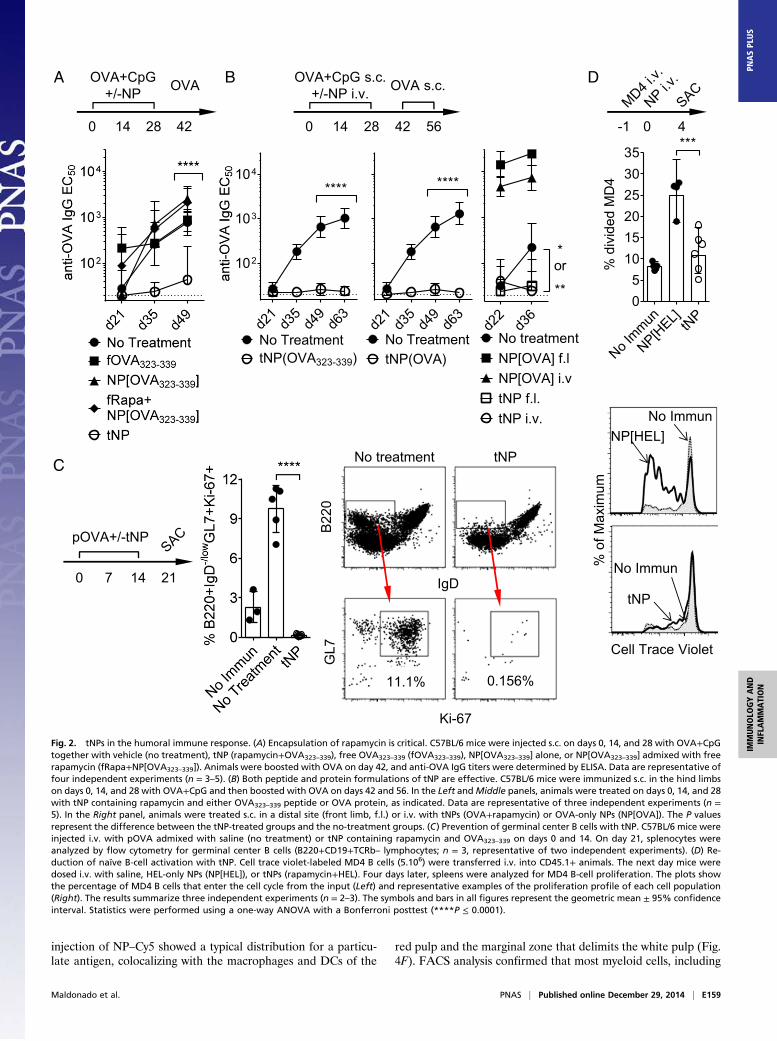
Inhibition of Humoral Immunity by tNP. We next investigated theeffect of tNP treatment during the development of a humoralresponse. Mice immunized s.c. with an admix of OVA anda TLR9 agonist, CpG oligonucleotides (OVA+CpG), showeda mounting antibody response with increasing anti-OVA titerswith each successive boost (Fig. 2 A and B). Concomitanttreatment with tNP administered either s.c (Fig. 2A) or i.v. (Fig.2B) inhibited the anti-OVA antibody response, even when thetNPs were admixed with CpG (Fig. 2A). The inhibition of thehumoral response was dependent on the encapsulation of rap-amycin, as providing the OVA323–339 peptide alone in free orencapsulated form (NP[OVA323–339]) or just free rapamycin (alladmixed to OVA+CpG) did not affect the anti-OVA response(Fig. 2A). Importantly, both peptide- and protein-containingtNPs were similarly efficacious (Fig. 2B, Left and Middle panels,respectively). In contrast, control NP[OVA] administered s.c.or i.v. enhanced the humoral response (Fig. 2B, Right).We next evaluated splenic B-cell activation and function in
mice immunized i.v. with an immunogenic particulate form ofOVA (pOVA). Approximately 10–12% of B cells (B220+) foundin the local draining LN acquired a typical germinal centerphenotype, expressing low levels of IgD, high levels of GL7, andthe proliferation marker Ki67 (Fig. 2C). In contrast, when ani-mals were concomitantly administered tNP (containing rapa-mycin+OVA323–339) s.c., the proportion of activated germinalcenter B cells was similar to or below the levels found in naïveanimals. To determine the impact of tNP treatment directly onB cells, B cells from MD4 B-cell receptor (BCR) transgenicanimals were adoptively transferred into CD45.1+ animals. Thetransgenic BCR is specific for hen egg lysozyme (HEL), and thepresence of this protein results in cell activation of MD4 B cellsin vivo and in vitro (33). HEL-only particles injected i.v. inducedthe activation of MD4 B cells in the spleen 4 d later, howeverproviding tNP containing similar amounts of HEL (together withrapamycin) did not (Fig. 2D). These results show that treatmentwith tNP inhibited B-cell activation and differentiation intoantibody-producing cells.
Durability of the tNP Treatment Effect. Mice were injected s.c. withtNP or saline admixed with OVA+CpG (days 0, 14, and 28) andOVA (days 42, 56, 70, 84, and 98; Fig. 3A). Animals treatedconcurrently with tNPs showed a substantial delay in the de-velopment of anti-OVA antibodies, and the titers remained∼37-fold lower than those observed in the immunized controls. Ofthe 53 animals that were treated with tNP (nine independentexperiments), 15 showed no detectable titers and 38 showed lowtiters at days 91–119 compared with animals in the untreatedgroup (Inset table in Fig. 3A). Only three animals (5%) of tNP-treated animals showed titers greater than 2,000, compared with59 animals (92%) in the control group. To test the durability of
Maldonado et al. PNAS | Published online December 29, 2014 | E157
IMMUNOLO
GYAND
INFLAMMATION
PNASPL
US

tolerance induced by tNP treatment, animals were given threebiweekly doses of tNPs concurrently with OVA+CpG immuniza-tions and then boosted 14 times with OVA in the absence offurther tNP treatment (Fig. 3B). Anti-OVA titers in the tNP-treated animals did not become detectable until day 58 andremained ∼30–35-fold lower than that of untreated animals evenin the absence of tNP treatment between days 42–234. Anti-OVAantibody titers remained well controlled in the tNP-treated ani-mals even following challenge with OVA+CpG on days 239 and253. Importantly, therapeutic administration of tNPs in mice withpreexisting anti-OVA antibody titers attenuated further boostingwith antigen even when the tNPs were coadministered with CpG(Fig. 3C).
Antigen-Specific Tolerance Induction by tNP Administration.Animalsfrom Fig. 3B that had shown a long-lasting inhibition of anti-OVA responses were immunized and boosted with keyholelimpet hemocyanin (KLH). The tNP-treated mice showed a ro-bust anti-KLH response (Fig. 4A) similar to that of the controlanimals, indicating they were not chronically immunosuppressed.To further test the specificity of the effect and show that theimmunosuppressive effects of rapamycin are not broad or sys-temic, animals were injected s.c. with OVA+CpG ± tNP in theright limbs and KLH+CpG in the left limbs (Fig. 4B). MixingtNP (containing OVA323–339 and rapamycin) with OVA+CpGcompletely blocked the development of anti-OVA responses buthad no effect on the anti-KLH response initiated in the con-tralateral limb (Fig. 4B). Moreover, a single injection of tNPcontaining OVA and rapamycin administered 14 d before re-peated immunization with pOVA and KLH selectively inhibitedthe antibody response to OVA, but not KLH (Fig. 4C). Incontrast, pretreatment at day –14 with control NP[Rapa] hadno significant effect on the response to either OVA or KLH. Inaddition, animals that were treated three times every 2 wk withtNP+pOVA showed no detectable antibody response even at day111 after five additional injections of pOVA in the absence ofadditional tNP treatment (Fig. 4D). Naïve animals in whichpOVA immunizations were delayed until day 42 developeda robust anti-OVA response by day 84 after three injections ofpOVA. These results further support the notion that tNPtreatment did not lead to systemic immunosuppression and in-duced durable tolerance rather than merely delaying the immuneresponse.
tNP Trafficking. To determine the fate of tNP after injection invivo, cyanine 7 (Cy7) and Cy5 fluorescently labeled NPs weredeveloped. Whole-body imaging by 3D fluorescence-based to-mography (34) showed that fluorescent NPs rapidly and selec-tively accumulated in the liver and the spleen following i.v.administration (Fig. 4E, Left and Fig. S1 A and B). In contrast,fluorescent NPs administered s.c. in the hind limb showed rapidand selective accumulation in the draining LNs (Fig. 4E, Right).Immunohistochemical analysis of the spleen 24 h after i.v.
-1 0 7
-7 -1 0 4
D
323-3
39
323-3
39
C
B
-2 0 4
Rapamycin
Antigen
A
No Immun
izatio
n
No Trea
tmen
t
NP[Rap
a] tNP
Fig. 1. tNPs in the CD4+ T-cell response. (A) tNP platform. Modular, syn-thetic, self-assembling tolerogenic PLGA NPs contain antigen and rapamycin.(B) NPs containing antigen and rapamycin (but not antigen alone) are tol-erogenic. OTII T cells were transferred into CD45.1 mice on day –2. At day 0,animals received s.c. immunizations with vehicle (no immunization) orOVA323–339 admixed with R848. At the same time, animals were injected i.v.with saline (no treatment), empty NPs admixed with OVA323–339 (NP[Empty]+OVA323–339), or tNPs (rapamycin+OVA323–339). Four days later, popLNs wereharvested and the total number of OTII T cells were quantified by flowcytometry (n = 4 per group). Data are representative of two independentexperiments. (C) Encapsulation of rapamycin is critical for the control of CD4+T-cell responses by tNP. OTII T cells were transferred into CD45.1+ mice1 d before s.c. injection of tNPs (containing rapamycin and OVA323–339) or
NP[OVA323–339] alone or NP[OVA323–339] admixed with free rapamycin(fRapa+NP[OVA323–339]). Seven days later, popLNs were harvested, andtotal number of OTII cells was enumerated (Left) and the percentage ofFoxp3+CD25+ OTII T cells was determined (Right). Data are cumulative oftwo independent experiments (n = 5). (D) Induction of antigen-specificMHC class II-restricted CD4 T-cell tolerance using tNP. C57BL/6 mice weretreated i.v. on day –7 with NP[Rapa] or tNPs (rapamycin+OVA323–339). Onday –1, purified naïve CFSE-labeled CD4+OTII T cells were administeredi.v., and then mice were immunized with OVA323–339+R848 s.c. in the hindlimbs on day 0. On day 4, popLNs were harvested and analyzed asin B. Data are representative of two independent experiments (n = 3 or4 per group). All statistical analyses were performed using a one-wayANOVA with a Bonferroni posttest (***P ≤ 0.001).
E158 | www.pnas.org/cgi/doi/10.1073/pnas.1408686111 Maldonado et al.
injection of NP–Cy5 showed a typical distribution for a particu-late antigen, colocalizing with the macrophages and DCs of the
red pulp and the marginal zone that delimits the white pulp (Fig.4F). FACS analysis confirmed that most myeloid cells, including
d21
d35
d49
d63
****
tNP(OVA)No Treatment
d21
d35
d49
d63
102
103
104
****
tNP(OVA323-339)No Treatment
B
0 14 28 42 56
OVA+CpG s.c.+/-NP i.v. OVA s.c.A
0 14 28 42
OVA
C
0 7 14 21
pOVA+/-tNP
OVA+CpG+/-NP
d22
d36
No treatmentNP[OVA] f.l
tNP f.l.NP[OVA] i.v
tNP i.v.
*or
**
% d
ivid
ed M
D4
No Immun
NP[HEL] tN
P05
101520253035
***
D
No treatment tNP
Ki-67
GL7
IgD
B22
0
0.156%11.1%
Cell Trace Violet
No ImmunNP[HEL]
No Immun
tNP%
of M
axim
um
-1 0 4
Fig. 2. tNPs in the humoral immune response. (A) Encapsulation of rapamycin is critical. C57BL/6 mice were injected s.c. on days 0, 14, and 28 with OVA+CpGtogether with vehicle (no treatment), tNP (rapamycin+OVA323–339), free OVA323–339 (fOVA323–339), NP[OVA323–339] alone, or NP[OVA323–339] admixed with freerapamycin (fRapa+NP[OVA323–339]). Animals were boosted with OVA on day 42, and anti-OVA IgG titers were determined by ELISA. Data are representative offour independent experiments (n = 3–5). (B) Both peptide and protein formulations of tNP are effective. C57BL/6 mice were immunized s.c. in the hind limbson days 0, 14, and 28 with OVA+CpG and then boosted with OVA on days 42 and 56. In the Left andMiddle panels, animals were treated on days 0, 14, and 28with tNP containing rapamycin and either OVA323–339 peptide or OVA protein, as indicated. Data are representative of three independent experiments (n =5). In the Right panel, animals were treated s.c. in a distal site (front limb, f.l.) or i.v. with tNPs (OVA+rapamycin) or OVA-only NPs (NP[OVA]). The P valuesrepresent the difference between the tNP-treated groups and the no-treatment groups. (C) Prevention of germinal center B cells with tNP. C57BL/6 mice wereinjected i.v. with pOVA admixed with saline (no treatment) or tNP containing rapamycin and OVA323–339 on days 0 and 14. On day 21, splenocytes wereanalyzed by flow cytometry for germinal center B cells (B220+CD19+TCRb– lymphocytes; n = 3, representative of two independent experiments). (D) Re-duction of naïve B-cell activation with tNP. Cell trace violet-labeled MD4 B cells (5.106) were transferred i.v. into CD45.1+ animals. The next day mice weredosed i.v. with saline, HEL-only NPs (NP[HEL]), or tNPs (rapamycin+HEL). Four days later, spleens were analyzed for MD4 B-cell proliferation. The plots showthe percentage of MD4 B cells that enter the cell cycle from the input (Left) and representative examples of the proliferation profile of each cell population(Right). The results summarize three independent experiments (n = 2–3). The symbols and bars in all figures represent the geometric mean ± 95% confidenceinterval. Statistics were performed using a one-way ANOVA with a Bonferroni posttest (****P ≤ 0.0001).
Maldonado et al. PNAS | Published online December 29, 2014 | E159
IMMUNOLO
GYAND
INFLAMMATION
PNASPL
US

macrophages, conventional DC (cDC), and plasmacytoid DC(pDC), contained particles (Fig. S1C) (16, 30, 31). These resultsshow that our NPs efficiently deliver their payload to APCs inthe lymphoid organs.
Effects of tNP in Animal Models of Hypersensitivity. Animals re-peatedly immunized i.v. with pOVA developed anaphylactoidresponses after three weekly i.v. infusions of pOVA, reminiscentof the antibody-mediated hypersensitivity reactions observed insome patients who develop antidrug antibodies (35–37). Un-treated animals with high titers against OVA from Fig. 4Cexhibited strong hypersensitivity to pOVA administered i.v.,whereas animals that received a single prophylactic treatment oftNP showed no anti-OVA titers and were largely protected fromanaphylaxis (Fig. 5A). Similarly, animals immunized with OVA+CpG and challenged with OVA s.c. experienced a significantdelayed-type hypersensitivity (DTH) response on the day afterchallenge, whereas animals that received concomitanttNP treatment during immunization showed no inflammation(Fig. 5B). In addition, prophylactic treatment with tNP (con-taining OVA+rapamycin) administered 4 d before immunizationto induce an anti-OVA CD8 T-cell response inhibited the sub-sequent in vivo killing of labeled target cells loaded withOVA257–264 (SIINFEKL), an MHC class I-restricted peptide ofOVA (Fig. 5C). Next, the efficacy of tNP was tested in animalswith allergic disorders. Animals treated with tNP during oral(Fig. 5D) or i.p. sensitization (Fig. 5H) showed complete in-hibition of both anti-OVA IgG and IgE antibody responses.
Immunization with OVA and aluminum hydroxide gel (Alum) i.p. followed by intragastric (i.g.) or intranasal (i.n.) challenge withOVA induced the development of oral allergy or allergic airwayinflammation, respectively. Sensitized animals treated thera-peutically with tNP i.v. at the time of oral challenge showeda reduction in anaphylaxis (sickness score, diarrhea, and tem-perature drop; Fig. 5E). Serum levels of mouse mast cell pro-tease 1 (mMCP1) in recently challenged animals were alsoreduced by prior treatment with tNP (Fig. 5F). No effect wasobserved on the levels of serum anti-OVA IgE concentrationusing this therapeutic protocol. In animals with allergic airwayinflammation induced by challenge with OVA i.n. (Fig. 5G),therapeutic tNP treatment after establishment of the allergicreaction reduced the number of lymphocytes (CD8+ and CD4+T cells and B cells) and eosinophils in the bronchoalveolar la-vage (BAL) fluid. Treatment with tNPs also resulted in de-creased numbers of splenic T cells with an activated phenotype(CXCR5+TCRβ+CD19–CD45R–) and an increased proportionof cells with a B regulatory phenotype (CD19+CD45R+IL-10+)compared with saline-treated animals. Similar to animals withoral allergies, no significant effect was observed on the IgE levelsusing this therapeutic protocol (Fig. 5G). However, concurrenttreatment with tNP during immunization (days, 0, 7, and 14)suppressed the IgE response and completely blocked the re-cruitment of lymphocytes to the airways in the presence of OVA(Fig. 5H). Importantly, no adverse events were observed even whentNPs were administered i.v. to animals with high serum levels ofantigen-specific IgG and/or IgE. Together these results show that
Days
anti-
OV
A Ig
G E
C50
d21 d33-35 d47-55 d56-63 d70-77 d91-119
102
103
104
****
***
*
No TreatmenttNP ****
0 14 28 42 - 98
OVA+CpG+/-NP s.c.
NP+/-OVA s.c.Biweekly
d91-119 No treat tNPND 0 15EC50<1,000 5 38EC50>2,000 36 3EC50>5,000 23 0
A
B
0 14 28 42
NP i.v. +/-OVA+CpG s.c.
224 234 239 253 267
OVA s.c.Biweekly
Days0 50 100 150 200 250
102
103
104
No treatment tNP
tNP
** or ****
C
0 14 28 42
OVA+CpG s.c.
192 213
NP+/-OVA+CpG s.c.Biweekly
Fig. 3. Sustained and robust control of antibodyresponses with tNP. (A) Long-term effect of tNP.C57BL/6 animals were immunized s.c. with OVA+CpG on days 0, 14, and 28 and boosted with OVAevery 14 days until at least day 77 with or withouttNP (containing rapamycin+OVA323–339). The figuredepicts the anti-OVA IgG titers for the indicatedrange of days. Each dot represents the average titerfor each group taken from nine independentexperiments (n = 51 and 53 animals per group). Thetable shows the number of animals in each groupwith the indicated level of anti-OVA titers at the lastbleed day (between day 91 and day 119). (B) Dura-bility of tNP therapy. Mice were immunized s.c. withOVA+CpG and treated i.v. with vehicle (no treat-ment) or tNP (rapamycin+OVA323–339) on days 0, 14,and 28. Animals were boosted with OVA every 14d from day 42 to day 224 without any further tNPtreatment. The treated group received another in-jection of tNP on day 234. The animals were sub-sequently challenged with OVA+CpG s.c. on days239 and 253, followed by a boost of OVA s.c. on day267 (n = 5). (C) Therapeutic tNP treatment inhibitsfurther boosting of antigen titers. Animals wereimmunized s.c. with OVA+CpG on days 0 and 14without any treatment. At day 21, anti-OVA titerswere measurable in all animals. The untreatedcontrol group continued to receive injections ofOVA+CpG every 2 wk from days 28–213, whereas thetreated group received biweekly OVA+CpG supple-mented with tNP (rapamycin+OVA323–339) starting atday 21. EC50 was determined by ELISA (n = 5). Thesymbols represent the geometric mean ± 95% con-fidence interval. Statistics were performed usinga two-way ANOVA with a Bonferroni posttest(****P ≤ 0.0001).
E160 | www.pnas.org/cgi/doi/10.1073/pnas.1408686111 Maldonado et al.

treatments with tNP during or after sensitization are safe andcan significantly reduce inflammatory processes associated withhypersensitivity reactions.
Prophylactic and Therapeutic Efficacy of tNP in a Relapsing–RemittingModel of Experimental Autoimmune Encephalomyelitis. Empty NPor tNP containing rapamycin and PLP139–151 were administered
-14 0 14 28
pOVA i.v.+ KLH s.c.
C
D
A
0 14 28 42 … 98
pOVA i.v.+NP i.v.
pOVA i.v.Biweekly
B
0 14 28 42 56
OVA+CpG right limbs +/- tNP right limbs
KLH+CpG left limbs
No Trea
tmen
ttN
Pan
ti-O
VA Ig
G E
C50
anti-KLH IgG
EC50
Liver
Spleen
E
R IP
Injection sites.c.i.v.
25
F250
RP
WP
WPWP
RP RP
B cells
MP
NP-Cy5
µm
µm
Fig. 4. Antigen specificity of tolerance induction by tNP. (A) Antigen specificity of tNP therapy. Animals from Fig. 3B were immunized s.c. with 50 μg of KLH onboth lateral flanks at the base of the tail (b.t.) on days 239, 253, and 267. Anti-KLH IgG and anti-OVA IgG titers from each animal on day 280 are shown. (B)Treatment with tNP does not result in broad immunosuppression. C57BL/6 mice were immunized with OVA+CpG in the right (front and hind) limbs and with KLH+CpG in the left limbs, whereas treated animals received OVA+CpG+tNP injections in the right limbs and KLH+CpG in the left limbs every 2 wk for 8 wk. Anti-OVAand anti-KLH titers were determined by ELISA (n = 5). (C) Prophylactic induction of antigen-specific tolerance. C57BL/6 micewere treated with a single injection i.v.of tNPs (rapamycin+OVA), NP[Rapa], or vehicle (no treatment) on day –14. Mice were immunized with pOVA i.v. and KLH s.c. on days 0, 14, and 28. Anti-OVA andanti-KLH titers were assessed on day 41. The results summarize three independent experiments (n = 5). (D) Durability of prophylactic induction of antigen-specifictolerance. Two groups of C57BL/6 animals (no treatment and tNP-treated) were immunized biweekly with eight injections with pOVA i.v. from day 0 to day 98.The treated group received tNP (rapamycin+OVA) for the first three immunizations only (days 0, 14, and 28). The start of immunization for a third group ofanimals was delayed until day 42 (delayed immunization control). In all figures and panels, the bars and the symbols represent the geometric mean ± 95%confidence interval (n = 5). All P values were calculated using a Bonferroni posttest of a regular one-way or two-way ANOVA test (***P ≤ 0.001). (E) In vivotrafficking of tNP. (Left) (i.v. injection) Balb/C animals were immunized with pOVA on days –27 and –13. On day 0, fluorescently tagged Cy7–tNP containing OVAand rapamycin were injected i.v., and the animals were imaged by 3D FMT at 6 h after injection. (Right) (s.c. injection) Naïve Balb/C mice were injected withfluorescently tagged Cy7–tNP s.c. and then imaged 1 h after injection (n = 3). I, iliac; P = popLNs; R, renal. (F) Cellular localization of tNP. Fluorescently tagged NP–Cy5 (red) NPs were injected i.v., and spleens were harvested 24 h later for cryosectioning and immunohistochemical analysis. Shown are representative low andhigh (Lower Right panel) magnification (10× and 100×, respectively) photomicrographs of the distribution of the NPs among the red pulp (RP), the white pulp(WP), and the marginal zone (MZ) delineated by the localization of macrophages (MPs, F4/80, green), DCs (CD11c, dark blue), and B cells (B220, cyan).
Maldonado et al. PNAS | Published online December 29, 2014 | E161
IMMUNOLO
GYAND
INFLAMMATION
PNASPL
US

G
0 5 12 13 14 15 18 25 32 46 47 48
OVA+Alum OVA i.n. SACsentinels
Treatment OVA i.n.
% B
reg
0
10
20
30
40
SpleenIL-10+ Bregs
SpleenCXCR5+ T cells
BAL CD8+
BAL Eos
BAL B
BAL CD4+
H
mMCP1F0 14 28 30 32 34 36 38
A
Ana
phyl
axis
Sco
re
No Immun
No Treat
tNP
i.v. Anaphylaxis
Hin
d Li
mb
Sw
ellin
g (m
m)
No Immun
No Trea
ttN
P
E
DTH
Mock I
mmun
No Treat
tNP
0
1
2
3
4**
nsOral Anaphylaxis Diarrhea Body Temperature
OVA+Alum OVA i.g. +/- NP i.v.
B
IgE
BAL Total
IgE
Oral sensitizationD
anti-
OV
A Ig
E (m
g/m
l)
No Trea
ttN
P
0 7 14 21-24 25
OVA+Alum+/- NP i.v. OVA i.n.
In vivo CTL C
Fig. 5. Prevention and amelioration of hypersensitivity disorders by tNP. (A) Inhibition of anaphylactic response to i.v. antigen. Anaphylactic reactions inresponse to the third i.v. injection of pOVA in animals described in Fig. 4C were assessed by three blinded scorers (0, no symptom; 1, lethargy; 2, lethargy andinability to right; 3, moribund). The results summarize three independent experiments (n = 5). (B) Inhibition of anti-OVA DTH response. C57BL/6 mice wereimmunized s.c. with OVA+CpG in the front limbs and treated i.v. with tNP or saline on days 0, 14, and 28. Animals were challenged s.c. on day 34 with 25 μg ofOVA in one hind paw and saline in the other. The next day the difference in thickness between the two limbs was determined using a caliper. Data arerepresentative of two independent experiments (n = 5). (C) In vivo cytotoxic T-lymphocyte (CTL) assay. Animals were treated with tNP or PBS on days 0 and 5and then immunized with NP-encapsulated OVA and CpG on day 9. On day 14, animals received a mixture of CFSE-labeled target cells loaded with the MHCclass I-restricted OVA peptide OVA257-264 SIINFEKL and labeled control cells. The next day, spleens were harvested and specific lysis of target cells was de-termined by flow cytometry (n = 3). (D) Inhibition of orally induced IgG and IgE responses using tNP. BALB/c mice were sensitized i.g. with OVA (5 mg) andcholera toxin (CTx, 10 μg) weekly for 10 wk. Mice were treated i.v. with saline (no treatment) or tNP (rapamycin+OVA). Anti-OVA titers were assessed on day83 (n = 5). (E) Inhibition of anaphylactic response to an oral allergen. Balb/C mice were immunized with OVA+Alum on days 0 and 14 and challenged six timeswith OVA by i.g. gavage every 2 d from day 28 until day 38. Animals were treated i.v. with saline (no treatment) or tNP (rapamycin+OVA) during each oralchallenge. Mock immunized animals received Alum. Animals were assessed by three blinded scorers for anaphylaxis, and the percent of animals with diarrhea.The changes in body temperature were also recorded. Anti-OVA IgE was assessed on day 38. Data are representative of two independent experiments (n = 5).(F) Inhibition of mMCP1 release. Balb/C animals were immunized with OVA+Alum and then challenged orally with OVA while receiving treatment with tNPs.Serum mMCP1 levels were assessed 1 h after the last (sixth) challenge by ELISA (n = 5). (G) Effect of therapeutic treatment with tNP on allergic airway in-flammation. Balb/C animals were injected with OVA+Alum on days 0 and 5 and then challenged with OVA i.n. on days 12, 13, and 14. A sentinel group ofanimals killed on day 15 were confirmed to have allergic airways. At days 18, 25, and 32, animals received i.v. treatments of saline, NP[OVA323–339], or tNPs(rapamycin+OVA323–339). BAL fluid was collected after challenge and assessed for T cells, B cells, and eosinophils. Splenocytes were restimulated overnight withOVA and assessed for TCRβ+CXCR5+ T cells and CD19+B220+IL-10+ cells (Bregs). (H) Inhibition of IgE responses and airway reactivity using tNP. BALB/c animalswere injected with OVA+Alum on days 0, 7, and 14 and then challenged daily with OVA i.n. from day 21–24. Animals were treated i.v. with saline (notreatment), NP[OVA323–339], or tNP (rapamycin+OVA323–339) on days 0, 7, and 14. The results are representative of two independent experiments(n = 5). The bars in all figures and panels represent the geometric mean (±95% confidence interval). All P values were calculated using a Bonferroniposttest of a regular one-way or two-way ANOVA test (***P ≤ 0.001).
E162 | www.pnas.org/cgi/doi/10.1073/pnas.1408686111 Maldonado et al.
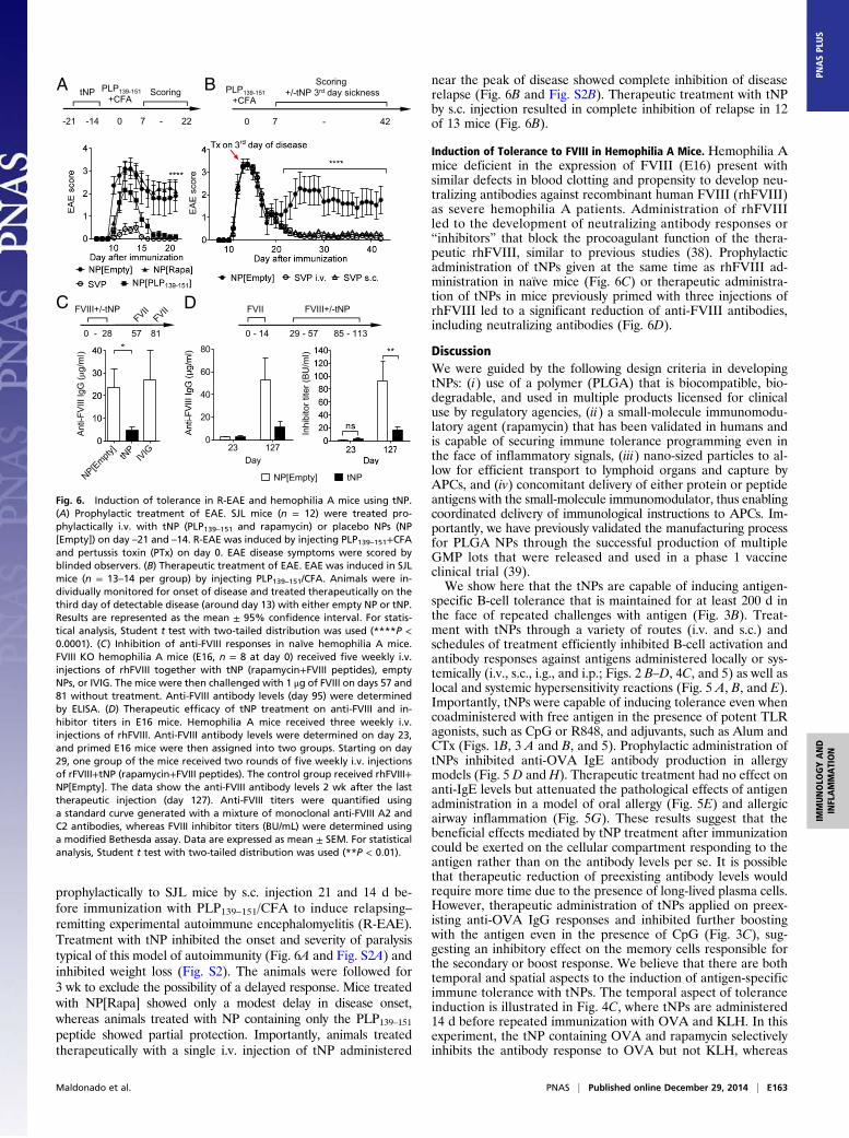
prophylactically to SJL mice by s.c. injection 21 and 14 d be-fore immunization with PLP139–151/CFA to induce relapsing–remitting experimental autoimmune encephalomyelitis (R-EAE).Treatment with tNP inhibited the onset and severity of paralysistypical of this model of autoimmunity (Fig. 6A and Fig. S2A) andinhibited weight loss (Fig. S2). The animals were followed for3 wk to exclude the possibility of a delayed response. Mice treatedwith NP[Rapa] showed only a modest delay in disease onset,whereas animals treated with NP containing only the PLP139–151peptide showed partial protection. Importantly, animals treatedtherapeutically with a single i.v. injection of tNP administered
near the peak of disease showed complete inhibition of diseaserelapse (Fig. 6B and Fig. S2B). Therapeutic treatment with tNPby s.c. injection resulted in complete inhibition of relapse in 12of 13 mice (Fig. 6B).
Induction of Tolerance to FVIII in Hemophilia A Mice. Hemophilia Amice deficient in the expression of FVIII (E16) present withsimilar defects in blood clotting and propensity to develop neu-tralizing antibodies against recombinant human FVIII (rhFVIII)as severe hemophilia A patients. Administration of rhFVIIIled to the development of neutralizing antibody responses or“inhibitors” that block the procoagulant function of the thera-peutic rhFVIII, similar to previous studies (38). Prophylacticadministration of tNPs given at the same time as rhFVIII ad-ministration in naïve mice (Fig. 6C) or therapeutic administra-tion of tNPs in mice previously primed with three injections ofrhFVIII led to a significant reduction of anti-FVIII antibodies,including neutralizing antibodies (Fig. 6D).
DiscussionWe were guided by the following design criteria in developingtNPs: (i) use of a polymer (PLGA) that is biocompatible, bio-degradable, and used in multiple products licensed for clinicaluse by regulatory agencies, (ii) a small-molecule immunomodu-latory agent (rapamycin) that has been validated in humans andis capable of securing immune tolerance programming even inthe face of inflammatory signals, (iii) nano-sized particles to al-low for efficient transport to lymphoid organs and capture byAPCs, and (iv) concomitant delivery of either protein or peptideantigens with the small-molecule immunomodulator, thus enablingcoordinated delivery of immunological instructions to APCs. Im-portantly, we have previously validated the manufacturing processfor PLGA NPs through the successful production of multipleGMP lots that were released and used in a phase 1 vaccineclinical trial (39).We show here that the tNPs are capable of inducing antigen-
specific B-cell tolerance that is maintained for at least 200 d inthe face of repeated challenges with antigen (Fig. 3B). Treat-ment with tNPs through a variety of routes (i.v. and s.c.) andschedules of treatment efficiently inhibited B-cell activation andantibody responses against antigens administered locally or sys-temically (i.v., s.c., i.g., and i.p.; Figs. 2 B–D, 4C, and 5) as well aslocal and systemic hypersensitivity reactions (Fig. 5 A, B, and E).Importantly, tNPs were capable of inducing tolerance even whencoadministered with free antigen in the presence of potent TLRagonists, such as CpG or R848, and adjuvants, such as Alum andCTx (Figs. 1B, 3 A and B, and 5). Prophylactic administration oftNPs inhibited anti-OVA IgE antibody production in allergymodels (Fig. 5 D and H). Therapeutic treatment had no effect onanti-IgE levels but attenuated the pathological effects of antigenadministration in a model of oral allergy (Fig. 5E) and allergicairway inflammation (Fig. 5G). These results suggest that thebeneficial effects mediated by tNP treatment after immunizationcould be exerted on the cellular compartment responding to theantigen rather than on the antibody levels per se. It is possiblethat therapeutic reduction of preexisting antibody levels wouldrequire more time due to the presence of long-lived plasma cells.However, therapeutic administration of tNPs applied on preex-isting anti-OVA IgG responses and inhibited further boostingwith the antigen even in the presence of CpG (Fig. 3C), sug-gesting an inhibitory effect on the memory cells responsible forthe secondary or boost response. We believe that there are bothtemporal and spatial aspects to the induction of antigen-specificimmune tolerance with tNPs. The temporal aspect of toleranceinduction is illustrated in Fig. 4C, where tNPs are administered14 d before repeated immunization with OVA and KLH. In thisexperiment, the tNP containing OVA and rapamycin selectivelyinhibits the antibody response to OVA but not KLH, whereas
0 - 28 57 81
FVIII+/-tNPC
0 - 14 29 - 57 85 - 113
FVIII+/-tNPFVIID
Anti-
FVIII
IgG
(g/
ml)
NP[Empty]
tNP
IVIG
Day23 127
0
20
40
60
80
NP[Empty] tNP
Inhi
bito
r tite
r (BU
/ml)
EA
E s
core
-21 -14 0 7 - 22
tNP ScoringPLP139-151+CFA
0 7 - 42
Scoring+/-tNP 3rd day sicknessPLP139-151
+CFAA B
Fig. 6. Induction of tolerance in R-EAE and hemophilia A mice using tNP.(A) Prophylactic treatment of EAE. SJL mice (n = 12) were treated pro-phylactically i.v. with tNP (PLP139–151 and rapamycin) or placebo NPs (NP[Empty]) on day –21 and –14. R-EAE was induced by injecting PLP139–151+CFAand pertussis toxin (PTx) on day 0. EAE disease symptoms were scored byblinded observers. (B) Therapeutic treatment of EAE. EAE was induced in SJLmice (n = 13–14 per group) by injecting PLP139–151/CFA. Animals were in-dividually monitored for onset of disease and treated therapeutically on thethird day of detectable disease (around day 13) with either empty NP or tNP.Results are represented as the mean ± 95% confidence interval. For statis-tical analysis, Student t test with two-tailed distribution was used (****P <0.0001). (C) Inhibition of anti-FVIII responses in naïve hemophilia A mice.FVIII KO hemophilia A mice (E16, n = 8 at day 0) received five weekly i.v.injections of rhFVIII together with tNP (rapamycin+FVIII peptides), emptyNPs, or IVIG. The mice were then challenged with 1 μg of FVIII on days 57 and81 without treatment. Anti-FVIII antibody levels (day 95) were determinedby ELISA. (D) Therapeutic efficacy of tNP treatment on anti-FVIII and in-hibitor titers in E16 mice. Hemophilia A mice received three weekly i.v.injections of rhFVIII. Anti-FVIII antibody levels were determined on day 23,and primed E16 mice were then assigned into two groups. Starting on day29, one group of the mice received two rounds of five weekly i.v. injectionsof rFVIII+tNP (rapamycin+FVIII peptides). The control group received rhFVIII+NP[Empty]. The data show the anti-FVIII antibody levels 2 wk after the lasttherapeutic injection (day 127). Anti-FVIII titers were quantified usinga standard curve generated with a mixture of monoclonal anti-FVIII A2 andC2 antibodies, whereas FVIII inhibitor titers (BU/mL) were determined usinga modified Bethesda assay. Data are expressed as mean ± SEM. For statisticalanalysis, Student t test with two-tailed distribution was used (**P < 0.01).
Maldonado et al. PNAS | Published online December 29, 2014 | E163
IMMUNOLO
GYAND
INFLAMMATION
PNASPL
US

NPs containing only rapamycin have no effect on either re-sponse. In contrast, Fig. 4B illustrates the spatial aspect of an-tigen-specific tolerance induction, where tNP and OVA+CpGare administered in the left flank and KLH+CpG is administeredat the same time in the right flank. Because the tNP bio-distribution following s.c. administration is localized to thedraining LNs (Fig. 4E, Right), the tNP selectively inhibits theimmune reaction against OVA in the LNs, draining the left flankwithout affecting the immune response to KLH in the right flank.The precise mechanism of action of our tNPs remains to be
determined. Nanoparticulates have been shown to be selectivelyendocytosed by APCs, such as macrophages and DCs (16, 30,31). The codelivery of antigen and rapamycin provide an in-struction set that could allow for the conditioning of tolerogenicDCs capable of inducing CD4+ Treg (22, 23). In support of thisnotion, prophylactic treatment with OVA323–339–loaded tNPs 7 dbefore transfer of OTII T cells resulted in complete inhibitionof OTII T-cell expansion in response to immunization withOVA323–339+R848, whereas treatment with control NPs con-taining only rapamycin had no effect (Fig. 1D). These experi-ments show that the inhibitory effect induced by the tNPs is moredurable than that expected from transient immunosuppressionby rapamycin and is dependent on the presence of antigen, in-dicating the formation of OVA323–339–specific regulatory cellsthat can control the activation of the subsequently transferredOTII T cells. Indeed, tNP treatment induced an increase in thepercentage of FoxP3+ antigen-specific T cells (Fig. 1B). Im-portantly, we found that admixing free rapamycin with NP-encapsulated OVA (or OVA323–339) did not result in immunetolerance. In fact, free rapamycin, administered at an equivalentdose as that used in tNP treatment, showed the opposite trendwith respect to inhibition of OVA-specific T-cell expansion andinduction of antigen-specific Tregs, perhaps due to the directeffect of free rapamycin on enhancing effector memory T-cellformation (40). Moreover, free rapamycin showed a consistenttrend toward enhancing anti-OVA–specific antibody responses.Similarly, NPs loaded with protein antigen in the absence ofrapamycin were very immunogenic. These results indicate thatthe biology of both the rapamycin and the antigen can be pro-foundly influenced by the context in which they are delivered.Tolerance induction using NPs was also demonstrated by Yeste
et al. (19), using gold NPs loaded with 2-(1′H-indole-3′-carbonyl)-thiazole-4-carboxylic acid methyl ester (or ITE, a ligand for the arylhydrocarbon receptor) and MOG35–55 (MHC class II-restrictedpeptide from myelin oligodendrocyte glycoprotein) in a model ofEAE. In contrast, Getts et al. (13) demonstrated tolerogenic activityof PLP139–151 conjugated to polystyrene or PLGA microparticles inthe absence of any immunomodulator. Induction of tolerance withNPs containing only antigen was not observed by us (Fig. 2B) or byYeste et al. (19). However, the larger, highly negatively chargedparticles used by Getts et al. (13) may induce tolerance through analternative mechanism via MARCO+ macrophages.One of the potential risks of antigen-based immunotherapy is
that administration during an ongoing inflammatory event couldpotentially override tolerogenic programming, resulting in dis-ease exacerbation. This is a phenomenon well known in allergyimmunotherapy in which administration of the antigen can resultin adverse events, including anaphylaxis (41). We have attemptedto mitigate this risk by using an immunomodulatory agent,rapamycin, which can induce and maintain the tolerogenicprogramming, even in the presence of potent TLR agonists.Another potential advantage of our tNPs is the ability toincorporate whole protein antigen, which is an important con-sideration for clinical application in a heterogeneous humanpopulation, particularly for large complex proteins, like FVIII,that contain many MHC class II epitopes. NPs containing pro-tein antigen without rapamycin are immunogenic (Fig. 2 B andD), whereas the encapsulation of rapamycin in the NPs, but not
admixed free rapamycin, induces tolerance. To our knowledge,the only other NPs capable of presenting and inducing toleranceto whole protein antigens are the liposomes bearing the CD22ligand described by Macauley et al. (18), which induce B-cell, butnot T-cell, tolerance. Finally, it is notable that our tNPs are ca-pable of inhibiting immune responses when administered eitheri.v. or s.c., whereas other NP approaches appear to be limited tosystemic administration (13, 19, 32). More experimentation isnecessary to compare these technologies and determine whetherthey can be used in similar applications.The emergence of NP-based therapies for the induction of
antigen-specific immune tolerance holds considerable promisefor the future of immunotherapy. Tolerogenic therapies could bebeneficial for the treatment of allergic asthma, life-threateningfood allergies, and autoimmune disease (8). A single therapeuticdose of tNP administered at the peak of disease completelyinhibited disease relapse in EAE, a model of human multiplesclerosis (Fig. 6B). Antigen-specific tolerance could be useful inpreventing antidrug antibodies, which can compromise efficacyand safety of biologics. A prominent example is the neutralizingantibody responses to FVIII, which results in major complica-tions for up to 30% of hemophilia A patients (42). Currentlypatients that develop inhibitors are treated with large and fre-quent doses of FVIII, which is extremely costly. Here we showthat adding tNP to the therapeutic regimen of FVIII for a limitednumber of injections allows for the establishment of tolerance(Fig. 6 C and D). Protein therapies are the fastest growingsegment of new drug approvals, and a proliferation of novelengineered proteins, such as multivalent antibodies and anti-body-drug conjugates, may be at increased risk for immunoge-nicity (43). tNPs could potentially also be applied to preventimmunogenicity associated with vectors used for gene therapy.The ability to translate antigen-specific therapy to human clinicalpractice would represent a breakthrough that could potentiallybenefit patients across a wide spectrum of clinical indications.
Materials and MethodsComplete details of materials are provided in SI Materials and Methods.
tNP and NP Manufacturing. tNPs containing both antigen and rapamycin wereprepared using a water-in-oil-in-water double emulsion solvent evaporationmethod (44, 45). Briefly, PLGA, pegylated polylactic acid (PLA-PEG), andrapamycin were dissolved in dichloromethane to form the oil phase. Anaqueous solution of antigen (OVA protein, OVA323–339 peptide, FVIII75–89peptide, FVIII1723–1737 peptide, FVIII2191–2210 peptide, or PLP139–151 peptide)was then added to the oil phase and emulsified by sonication (BransonDigital Sonifier 250A). Following emulsification of the antigen solution intothe oil phase, a double emulsion was created by adding an aqueous solutionof polyvinylalcohol and sonicating a second time. The double emulsion wasadded to a beaker containing phosphate buffer solution and stirred at roomtemperature for 2 h to allow the dichloromethane to evaporate. Whencreating NPs containing rapamycin but no antigen, or NPs without any encap-sulated agents, a similar oil-in-water single emulsion process was used. Theresulting NPs were washed twice by centrifuging at 75,600 × g and 4 °C followedby resuspension of the pellet in PBS. Fluorescent Cy5- and Cy7-containing NPswere manufactured as described above using PLGA–Cy5 or PLGA–Cy7 conjugate,respectively. PLGA with a butyl amine end group was prepared from PLGA–acid,which was then treated with Cy7–acid or Cy5–acid in the presence of a couplingagent (O-(Benzotriazol-1-yl)-N,N,N′,N′-tetramethyluronium tetrafluoroborate) toafford the conjugates.
Animal Models. Animal procedures involving hemophilia A mice, EAE, and invivo imaging were approved by the Institutional Animal Care and UseCommittee of the Uniformed Services University of the Health Sciences,Hooke Laboratories, and Molecular Research Imaging, respectively. All othermouse experiments were approved by the Institutional Animal Care and UseCommittee of Avastus Preclinical Services, following local and nationalguidelines and regulations.Relapsing EAE model. EAE was induced by injection of SJL mice s.c. at four sitesin the back with PLP139–151 emulsified in CFA (Hooke Laboratories) followed2 h later by i.p. injection of 154 ng of pertussis toxin. Blinded EAE scores
E164 | www.pnas.org/cgi/doi/10.1073/pnas.1408686111 Maldonado et al.

were assessed daily starting from day 7, and body weight was measuredthree times per week. EAE was scored on a 0–5 scale as follows: 0, no obviouschanges in motor functions of the mouse in comparison with nonimmunizedmice; 1, limp tail; 2, limp tail and weakness of hind legs; 3, limp tail andcomplete paralysis of hind legs (most common) or limp tail with paralysisof one front and one hind leg; 4, complete hind leg and partial front legparalysis; 5, death or euthanized because of severe paralysis.Allergic airway model. BALB/c female mice aged 8–10 wk were sensitized by i.p.injections of 10 μg Ova adsorbed to 4 mg of Alum on days 0 and 5. On days 7,8, and 9, mice were administered 25 μg Ova i.n. Mice were treated with tNPor PBS on days 25, 32, and 39. Finally, mice were administered PBS for thecontrol group or 25 μg OVA i.n. for 3 consecutive days from days 46–48 andwere killed 1 h after the last challenge by a lethal dose of isofluorane. Lungswere lavaged three times with 1 mL of ice-cold PBS containing 3 mM EDTA.Oral allergy model. BALB/c female mice were sensitized by i.p. injectionsof 500 μg OVA adsorbed to 4 mg of Alum on day 0 and then 50 μg OVAadsorbed to 4 mg of Alum on day 14. On days 27, 29, 31, 34, and 36, micewere treated i.v. with tNP or PBS and challenged with 50 mg of OVA by i.g.gavage. Mice were evaluated for sickness by three blinded scorers as pre-viously described (46, 47). Rectal temperatures and liquid stool scoreswere measured (0, dry; 1, wet) 1 h after the final challenge.In vivo CTL assay. Test animals were immunized with NPs containing OVAprotein and CpG oligodeoxynucleotides (ODN). Five days later, spleen cellsharvested from naïve animals were loaded with low levels of carboxy-fluorescein diacetate succinimidyl diester (CFSE) and the OVA257–264 MHCclass I-restricted peptide of OVA (CFSElow, SIINFEKL+ Targets), whereas
another portion of cells were loaded with high levels of CFSE alone (CFSEhigh,SIINFEKL− Control). A 1:1 mixture of these labeled target cells was injectedinto the recipient naïve and test animals to monitor specific killing of SIIN-FEKL peptide-presenting cells (CFSElow) relative to their control (CFSEhigh).
In Vivo FMT Imaging. In vivo 3D fluorescence tomography (FMT) imaging wasperformed by Molecular Imaging on the Perkin-Elmer FMT 2500 LX QuantitativeTomography Imaging System. Mice were injected with Cy7-labeled NPs andanesthesitized with 2% (vol/vol) isoflurane gas just before imaging. A scanningregion was manually positioned over the body from the head to chest for scan1 and from chest to hind limbs for scan 2. Each animal was scanned in thatspecific region of interest using a medium (3 mm) source density. Images werereconstructed and analyzed using Perkin-Elmer True Quant software.
Fluorescence Imaging. Frozen sections were fixed with cold acetone andstained for the indicated markers and visualized at room temperature usingan upright epifluorescencemicroscope Leica DMI6000B system equippedwitha Leica DFC340FX. Images were processed using ImageJ software.
ACKNOWLEDGMENTS. The authors gratefully acknowledge the excellentsupport of Aditi Chalishazar, Dr. Lynelle Pittet, and the ELISA team for ELISAanalyses and Dr. Fen-ni Fu, Dr. William Kuhlman, Dr. Victor Chan, RayDamien, and Dr. Nelly Viseux for analytical characterization of nanoparticles.We thank Dr. Suzana Marusic for support with the EAE studies. This workwas partially funded by a grant from the Juvenile Diabetes ResearchFoundation.
1. Burks AW, et al. (2013) Update on allergy immunotherapy: American Academy ofAllergy, Asthma & Immunology/European Academy of Allergy and Clinical Immu-nology/PRACTALL consensus report. J Allergy Clin Immunol 131(5):1288–1296, e1283.
2. Hayter SM, Cook MC (2012) Updated assessment of the prevalence, spectrum and casedefinition of autoimmune disease. Autoimmun Rev 11(10):754–765.
3. Nechansky A, Kircheis R (2010) Immunogenicity of therapeutics: A matter of efficacyand safety. Expert opin drug discov 5(11):1067–1079.
4. Zhang AH, Skupsky J, Scott DW (2009) Factor VIII inhibitors: Risk factors and methodsfor prevention and immune modulation. Clin Rev Allergy Immunol 37(2):114–124.
5. Banugaria SG, Patel TT, Kishnani PS (2012) Immune modulation in Pompe diseasetreated with enzyme replacement therapy. Expert Rev Clin Immunol 8(6):497–499.
6. Carbone J, del Pozo N, Gallego A, Sarmiento E (2011) Immunological risk factors forinfection after immunosuppressive and biologic therapies. Expert Rev Anti Infect Ther9(4):405–413.
7. Riminton DS, Hartung HP, Reddel SW (2011) Managing the risks of immunosuppres-sion. Curr Opin Neurol 24(3):217–223.
8. Bluestone JA, Bour-Jordan H (2012) Current and future immunomodulation strategies torestore tolerance in autoimmune diseases. Cold Spring Harb Perspect Biol 4(11):pii: a007542.
9. Smarr CB, Hsu CL, Byrne AJ, Miller SD, Bryce PJ (2011) Antigen-fixed leukocytes tol-erize Th2 responses in mouse models of allergy. J Immunol 187(10):5090–5098.
10. Miller SD, Wetzig RP, Claman HN (1979) The induction of cell-mediated immunity andtolerance with protein antigens coupled to syngeneic lymphoid cells. J Exp Med149(3):758–773.
11. Scott DW, Long CA (1976) Role of self-carriers in the immune response and tolerance.I. B-cell unresponsiveness and cytotoxic T-cell immunity induced by haptenated syn-geneic lymphoid cells. J Exp Med 144(5):1369–1374.
12. Battisto JR, Bloom BR (1966) Dual immunological unresponsiveness induced by cellmembrane coupled hapten or antigen. Nature 212(5058):156–157.
13. Getts DR, et al. (2012) Microparticles bearing encephalitogenic peptides induce T-celltolerance and ameliorate experimental autoimmune encephalomyelitis. Nat Bio-technol 30(12):1217–1224.
14. Getts DR, et al. (2014) Therapeutic inflammatory monocyte modulation usingimmune-modifying microparticles. Sci Transl Med 6(219):219ra217.
15. Kontos S, Kourtis IC, Dane KY, Hubbell JA (2013) Engineering antigens for in situerythrocyte binding induces T-cell deletion. Proc Natl Acad Sci USA 110(1):E60–E68.
16. Irvine DJ, Swartz MA, Szeto GL (2013) Engineering synthetic vaccines using cues fromnatural immunity. Nat Mater 12(11):978–990.
17. Tsai S, et al. (2010) Reversal of autoimmunity by boosting memory-like auto-regulatory T cells. Immunity 32(4):568–580.
18. Macauley MS, et al. (2013) Antigenic liposomes displaying CD22 ligands induceantigen-specific B cell apoptosis. J Clin Invest 123(7):3074–3083.
19. Yeste A, Nadeau M, Burns EJ, Weiner HL, Quintana FJ (2012) Nanoparticle-mediatedcodelivery of myelin antigen and a tolerogenic small molecule suppresses experi-mental autoimmune encephalomyelitis. Proc Natl Acad Sci USA 109(28):11270–11275.
20. Joseph A, Munroe K, Housman M, Garman R, Richards S (2008) Immune toleranceinduction to enzyme-replacement therapy by co-administration of short-term, low-dosemethotrexate in a murine Pompe disease model. Clin Exp Immunol 152(1):138–146.
21. Steinman RM, Hawiger D, Nussenzweig MC (2003) Tolerogenic dendritic cells. AnnuRev Immunol 21:685–711.
22. Maldonado RA, von Andrian UH (2010) How tolerogenic dendritic cells induce reg-ulatory T cells. Adv Immunol 108:111–165.
23. Thomson AW (2010) Tolerogenic dendritic cells: All present and correct? Am JTransplant 10(2):214–219.
24. Manicassamy S, Pulendran B (2011) Dendritic cell control of tolerogenic responses.
Immunol Rev 241(1):206–227.25. Cobbold SP, Adams E, Nolan KF, Regateiro FS, Waldmann H (2010) Connecting the mech-
anisms of T-cell regulation: Dendritic cells as the missing link. Immunol Rev 236:203–218.26. Taner T, Hackstein H, Wang Z, Morelli AE, Thomson AW (2005) Rapamycin-treated,
alloantigen-pulsed host dendritic cells induce ag-specific T cell regulation and prolong
graft survival. Am J Transplant 5(2):228–236.27. Fischer R, Turnquist HR, Taner T, Thomson AW (2009) Use of rapamycin in the in-
duction of tolerogenic dendritic cells. Handb Exp Pharmacol (188):215–232.28. Powell JD, Pollizzi KN, Heikamp EB, Horton MR (2012) Regulation of immune re-
sponses by mTOR. Annu Rev Immunol 30:39–68.29. Araki K, Ellebedy AH, Ahmed R (2011) TOR in the immune system. Curr Opin Cell Biol
23(6):707–715.30. Bachmann MF, Jennings GT (2010) Vaccine delivery: A matter of size, geometry, ki-
netics and molecular patterns. Nat Rev Immunol 10(11):787–796.31. Metcalfe SM, Fahmy TM (2012) Targeted nanotherapy for induction of therapeutic
immune responses. Trends Mol Med 18(2):72–80.32. Look M, et al. (2013) Nanogel-based delivery of mycophenolic acid ameliorates sys-
temic lupus erythematosus in mice. J Clin Invest 123(4):1741–1749.33. Goodnow CC, et al. (1988) Altered immunoglobulin expression and functional si-
lencing of self-reactive B lymphocytes in transgenic mice. Nature 334(6184):676–682.34. Vasquez KO, Casavant C, Peterson JD (2011) Quantitative whole body biodistribution of
fluorescent-labeled agents by non-invasive tomographic imaging. PLoS ONE 6(6):e20594.35. Liu E, et al. (2002) Anti-peptide autoantibodies and fatal anaphylaxis in NOD mice in
response to insulin self-peptides B:9-23 and B:13-23. J Clin Invest 110(7):1021–1027.36. Krieckaert C, Rispens T, Wolbink G (2012) Immunogenicity of biological therapeutics:
From assay to patient. Curr Opin Rheumatol 24(3):306–311.37. Vultaggio A, Maggi E, Matucci A (2011) Immediate adverse reactions to biologicals:
From pathogenic mechanisms to prophylactic management. Curr Opin Allergy Clin
Immunol 11(3):262–268.38. Qian J, Borovok M, Bi L, Kazazian HH, Jr, Hoyer LW (1999) Inhibitor antibody de-
velopment and T cell response to human factor VIII in murine hemophilia A. Thromb
Haemost 81(2):240–244.39. Selecta Biosciences (2011) Safety and Pharmacodynamics of SEL-068 Vaccine in
Smokers and Non-Smokers. Available at www.ClinicalTrials.gov.40. Araki K, et al. (2009) mTOR regulates memory CD8 T-cell differentiation. Nature
460(7251):108–112.41. Smarr CB, Bryce PJ, Miller SD (2013) Antigen-specific tolerance in immunotherapy of
Th2-associated allergic diseases. Crit Rev Immunol 33(5):389–414.42. Scott DW, Pratt KP, Miao CH (2013) Progress toward inducing immunologic tolerance
to factor VIII. Blood 121(22):4449–4456.43. Enever C, Batuwangala T, Plummer C, Sepp A (2009) Next generation immunother-
apeutics—Honing the magic bullet. Curr Opin Biotechnol 20(4):405–411.44. Astete CE, Sabliov CM (2006) Synthesis and characterization of PLGA nanoparticles.
J Biomater Sci Polym Ed 17(3):247–289.45. Bi L, et al. (1995) Targeted disruption of the mouse factor VIII gene produces a model
of haemophilia A. Nat Genet 10(1):119–121.46. Bailón E, et al. (2012) A shorter and more specific oral sensitization-based experi-
mental model of food allergy in mice. J Immunol Methods 381(1-2):41–49.47. Kasper CK, et al. (1975) Proceedings: A more uniform measurement of factor VIII
inhibitors. Thromb Diath Haemorrh 34(2):612.
Maldonado et al. PNAS | Published online December 29, 2014 | E165
IMMUNOLO
GYAND
INFLAMMATION
PNASPL
US