Embed Size (px)
Citation preview

Proc. Nati. Acad. Sci. USAVol. 84, pp. 8414-8418, December 1987Biophysics
Picosecond kinetics of fluorescence and absorbance changes inphotosystem II particles excited at low photon density
(excitation trapping/charge separation/electron transfer/pheophytin a/cyanobacterium)
GUNTHER H. SCHATZ, HELMUT BROCK, AND ALFRED R. HOLZWARTHMax-Planck-Institut fur Strahlenchemie, Stiftstrasse 34-36, D-4330 Muilheim a.d. Ruhr, Federal Republic of Germany
Communicated by Pierre Joliot, June 11, 1987
ABSTRACT Oxygen-evolving photosystem II particles(from Synechococcus) with about 80 chlorophyll molecules perprimary electron donor (P6m) were used for a correlated studyof picosecond kinetics of fluorescence and absorbance changes,detected by the single-photon-timing technique and by apump-probe apparatus, respectively. Chlorophyll fluorescencedecays were biexponential with lifetimes i = 80 ± 20 ps andT2 = 520 + 120 ps in open reaction centers and Tj = 220 + 30ps and x2 = 1.3 + 0.15 ns in closed reaction centers. Thecorresponding fluorescence yield ratio F.IU,/FO was 3-4. Ab-sorbance changes were monitored in the spectral range of620-700 nm after excitation at 675 nm with 10-ps pulsessufficiently weak (<7 x 10¹² photons/cm2 per pulse) to avoidsinglet-singlet annihilation. With open reaction centers, theabsorbance changes could be fit to the sum of three exponen-tials. The associated absorbance difference spectra were at-tributed to (t) exciton trapping and charge separation (x = 100± 20 ps), (it) the electron-transfer step P& I- QA -_ PZ;0 I QA(where I is the primary electron acceptor and QA is the firstquinone acceptor) (x = 510 ± 50 ps), and (iii) the reduction ofP40 by the intact donor side (x > 10 ns). With closed reactioncenters, the absorbance changes were biexponential withlifetimes il = 170-260 ps and x2 = 1.6-1.75 ns, The results areexplained in terms of a kinetic model that assumes P60 toconstitute a shallow trap. The results show that QA reductionin these photosystem II particles decreases both the apparentrate and the yield of the primary charge separation by a factorof2-3 and increases the mean lifetime of excitons in the antennaby a factor of 34. Thus, we conclude that the long-lived,nanosecond chlorophyll fluorescence is not charge-recombina-tion luminescence but rather emission from equilibrated excit-ed states of antenna chlorophylls.
The processes of excitation trapping, charge separation, andprimary charge stabilization have been resolved by applica-tion of picosecond spectroscopy to reaction centers (RCs)isolated from purple bacteria. For the RCs of photosystem II(PSII), the situation is much less clear because availablepreparations enriched in PSII and still active in chargestabilization (I- QA -- I Q-, where I is the primary electronacceptor and QA is the first quinone acceptor) contain arelatively large amount ofantenna pigments bound to the RC.This imposes several experimental difficulties on measure-ments of absorbance changes with picosecond time resolu-tion: (i) the relative signal amplitude is small, due to strongbackground absorption, and (ii) the threshold for singlet-singlet annihilation within the antenna is shifted to muchlower values of photon fluence per pulse as compared toisolated RCs. The latter problem was evident in a recentstudy (1), which revealed the transient reduction of pheo-phytin a as intermediate electron acceptor I in PSII, as
proposed by Klimov et al. (2), but not the kinetics ofexcitation trapping and PgjPl- formation (where P680 is theprimary electron donor). It was proposed (2, 3) that in theRCs of PSII, the reduction of QA should induce a delayedfluorescence after charge recombination of P68o0-, to whichthe long-lived (2-4 ns) fluorescent component observed withclosed PSII was attributed (4, 5). A direct experimental testof this hypothesis by absorbance spectroscopy has to differ-entiate between the contributions from the radical pair(P680I-) and the excited states of the primary donor (P&0)and/or antenna chlorophyll (Chl*). However, all these con-tributions show much closer spectral overlap in PSII (1) ascompared to the RC of purple bacteria.
Therefore, we have chosen the following approach in orderto gain more insight into the primary processes in PSII. (i) Wecorrelated measurements of picosecond kinetics of absor-bance changes with those of fluorescence decays obtainedunder almost identical conditions, using intact, oxygen-evolving PSII particles with about 80 Chl a molecules perPS1. (ii) To avoid artificial contributions by singlet-singletannihilation, we applied a type of picosecond spectrometerthat allows us to measure picosecond absorbance transientsat extremely low excitation fluence (<2 pJ/cm2 per pulse).Our data from both absorbance and fluorescence with openand closed RCs of PSII can be explained in terms of a kineticmodel that accounts for all primary processes. Thus, thekinetics of trapping, charge separation, and charge stabiliza-tion could be characterized.
MATERIALS AND METHODS
Fluorescence decay kinetics were measured at 685 nm witha single-photon-timing system as described (6) upon excita-tion with laser pulses [675 nm, 10-ps full width at halfmaximum (FWHM)] of 400-kHz repetition rate and a fluenceE < 0.1 IJ/cm2 per pulse. Absorbance transients weredetermined by the pump-probe technique. The pump pulse(675 nm, 10-ps FWHM, 2-pJ/cm2 pulse, if not stated other-wise) and the probe pulse (620-700 nm, <15-ps FWHM,0.02-p.J/cm2 pulse) were provided by two synchronizedcavity-dumped (400 kHz) dye lasers (Spectra Physics, SantaClara, CA), which were both pumped by a mode-locked CWneodymium/yttrium/aluminum garnet laser (Spectra Phys-ics). Both dye-laser beams were focused (focal length, 80mm) onto the cuvette (optical pathlength, 1 mm). The timedelay between the pulses was variable from -300 ps to 1500ps by means of an optical delay line. Noise reduction wasaccomplished by modulation of both the pump and the probepulse train by mechanical choppers and 2-fold lock-in am-plification of the probe signal. Together with a restriction ofthe solid angle ofthe probe beam to -5 x 10⁻⁴, this techniquehelped to suppress fluorescence contributions below the
Abbreviations: Chl, chlorophyll; I, primary electron acceptor; P680,primary electron donor; QA, first quinone acceptor; PSII, photosys-tem II; RC, reaction center.
8414
The publication costs of this article were defrayed in part by page chargepayment. This article must therefore be hereby marked "advertisement"in accordance with 18 U.S.C. §1734 solely to indicate this fact.
Dow
nloa
ded
by g
uest
on
Apr
il 14
, 202
0

Proc. Natl. Acad. Sci. USA 84 (1987) 8415
noise level. The planes of polarization of the pump and probebeam were adjusted to the "magic angle" (54.7°) to avoid anyinterference from depolarization processes. The sample wasrapidly (400-500 ml/min) cycled through a flow cuvette (1mm x 1 mm) to prevent the accumulation of RCs closed bythe laser pulses. A full description of the apparatus will bepublished elsewhere.The PSII particles were prepared from a thermophilic
cyanobacterium (Synechococcus sp.) as described in refs. 7and 8, and were characterized by about 80 Chl molecules perPSII RC (8, 9) and full oxygen-evolving capacity (7). Theywere suspended in 50 ml of buffer [containing 500 mMsucrose, 10 mM MgCl2, 2 mM K2HPO4, and 20 mM 4-mor-pholineethanesulfonic acid/NaOH (pH 6.0)] at a Chi con-centration of 20-30 AM for absorbance or 10 AM forfluorescence measurements. In all measurements FO condi-tions (open PSII centers) were established by addition of 2mM K3Fe(CN)6 in darkness, and Fma, conditions (closedPSII centers), by addition of 2 mM sodium dithionite undernitrogen in ambient light. The temperature was stabilized at150C.Data analysis of the absorbance changes was performed by
a deconvolution procedure based on a global fitting algorithm(10, 11) and using the cross-correlation ofthe two laser pulsesas system response. By the global procedure the signalsmeasured at different wavelengths (Xk) are simultaneouslyfitted to a sum of exponential functions plus a constant,
n
A(Xk, t) = Ao(Xk) + > A(Xk)Jexp(-t/i), [1]
by free-running optimizations for all amplitudes and life-times. However, a dependence of the lifetimes ri on thewavelength is excluded. Compared to a conventional analy-sis, this procedure has a much stronger potential for multi-component resolution, because of the reduced number offreeparameters (11). Plots of the resulting amplitudes A(XAk)versus the wavelength yield the decay-associated absorbancedifference spectra (see Figs. 4 and 6). The values of AOrepresent an offset of the baselines (generally <<10-4) in theindividual experiments.
RESULTS
Fluorescence Decay Kinetics. Typical fluorescence decaykinetics of the PSII particles with open and closed RCs areshown in Fig. 1 A and B. Comparison of the weightedresiduals of errors shows that, in both cases, two exponentialdecay components are not sufficient to fit the experimentaldata. Characteristic results from three-exponential fits ofindependent measurements on six sample preparations (Ta-ble 1) show that the main contribution to the F. kineticsderives from a rapidly decaying (60-100 ps) component. The1.8-ns component of minor amplitude (<10%) is attributed tosample impurities-possibly allophycocyanin, which wasalways present to a small extent in our PSII preparations (7).We noted a 3-fold increase of the relative amplitude of the1.8-ns component when the excitation wavelength was shift-ed to 650 nm, preferentially exciting allophycocyanin. There-fore, we attribute this kinetic component to pigments thatwere impaired in the excitation transfer from allophycocy-anin to the RC of PSIL. There may be several such pigments,as indicated by an alternative fit with four exponentialcomponents. This had mainly the effect of replacing the1.8-ns component by a 1.1-ns and a 2.4-ns component.Ignoring such long-lived component(s) with generally lessthan 10o of the total amplitude, we see that the fluorescencedecay kinetics ofthe Chl molecules coupled to the RC appearto be biexponential. Closing the RC of the PSII particles byreduction of QA with dithionite [or with 20 AM 3-(3,4-
78 I -.0E
-1 .2x ei.loAn~~~~~ 2exponentials Chi2: 3.845- 74.
0E
-/.
4
v)
c
(-30C.)so
1 2 3 4 5 6 7 8 1 2 3 4 5 6 7 8Time, ns Time, ns
FIG. 1. Fluorescence decay at 685 nm in PS11 particles with 2 mMK3Fe(CN)6 (A, open reaction centers) or 2 mM sodium dithionite (B,closed reaction centers); excitation wavelength was 675 nm. The brokenline shows the system response. Weighted residuals of errors (sigma)between experimental curves and fits with two or three components,respectively, are shown above the fluorescence decay curves.
dichlorophenyl)methylurea plus light; data not shown] re-sults in an increase of the lifetimes by a factor of 2-3 and ina decrease ofthe amplitude ratio a1/a2 by a factor of 3-4 (seeTable 1). Concomitantly, the total Chl fluorescence yieldincreases by a factor of 3-4.
In the case ofPSII the absorbance changes associated withthe primary processes have strong spectral overlap withthose caused by Chl*-Chl* annihilation. Therefore, our firstaim was to determine the threshold of excitation density atwhich such artifacts become negligible. We studied thedependence of the absorbance changes at 685 nm on thefluence of the exciting pulse from 20 to 2 PJ/cm2 per pulse(different from the values given in ref. 12, where the beamdiameter was underestimated). At high excitation densitiesthe observed kinetics (data not shown) were dominated by a
component of lifetime 20-40 ps, which was not observed atlow excitation density or by fluorescence (Table 1).We analyzed all absorbance changes by the global
analysist in terms of four exponentials with two of them keptfixed to the Chl* fluorescence lifetimes (see Table 1). Theresulting amplitudes of the four components are plottedversus the excitation fluence (E) on a logarithmic scale in Fig.2 and show saturation curves of two different types: (i) thosewith almost constant slopes within our range ofE and (ii) onewith a clearly distinct, nonlinear dependence. The latter isassociated with a lifetime of 25 ps and attributed to Chl*-Chl*annihilation. It was dominant atE - 10 pJ/cm2 per pulse andmarkedly decreased at values of E - 3 pJ/cm2 per pulse.Therefore, we chose an excitation fluence E - 2 PJ/cm2 perpulse for the subsequent experiments.Absorbance Changes in Open PSI1 Centers. These were
probed in the wavelength range from 620 to 700 nm. Typicalkinetic traces are shown in Fig. 3. All data were submitted tothe global analysis and simultaneously fitted to a sum ofthreeexponentials. The resulting lifetimes (Table 2) show goodaccordance of r1 and r2 with the corresponding fluorescencelifetimes (see Table 1). The resulting amplitudes (see Eq. 1)are plotted as the absorbance difference spectra for each
tDue to the global fitting procedure, the time constant for annihila-tion was not allowed to increase with decreasing flash intensities,and the resulting lifetimes were a priori dominated by the experi-ments at high intensities. Nevertheless, it follows from Fig. 2 thatthe contribution by the annihilation process is negligible at fluenceE s 2 AJ/cm2 per pulse.
q~~,,yr~~~~d ,-'E.,
2exponentials Chi2: 2.223
sw 1 -!"'"I ' W1 71I'- "Ierrs I` 3 exponentials Chi2: 1.183
.8 'I - -...
.0i AJ& A J, JLIJJ. iji
IMF In.
Biophysics: Schatz et al.
rR wr
n
11 Tyr [PIP .11.1 .. T' 1111T ITT "Irr 3 e Chi 1.186
Dow
nloa
ded
by g
uest
on
Apr
il 14
, 202
0

Proc. Natl. Acad. Sci. USA 84 (1987)
Table 1. Lifetimes (Tr) and relative amplitudes (a,) from athree-exponential analysis of the fluorescence-decaycomponents in PS11 particles at F. and Fmax
F. Fmax
i x, ps a, % x, pS a, %
1 80± 20 72 ± 5 220 ± 30 44 ± 42 520 ± 120 20 ± 3 1300 ± 150 47 ± 43 1800 ± 100 8 ± 2 4000 ± 500 9 ± 2
Excitation wavelength was 675 nm; emission wavelength was 685nm. Values are means ± SEM from 6 samples.
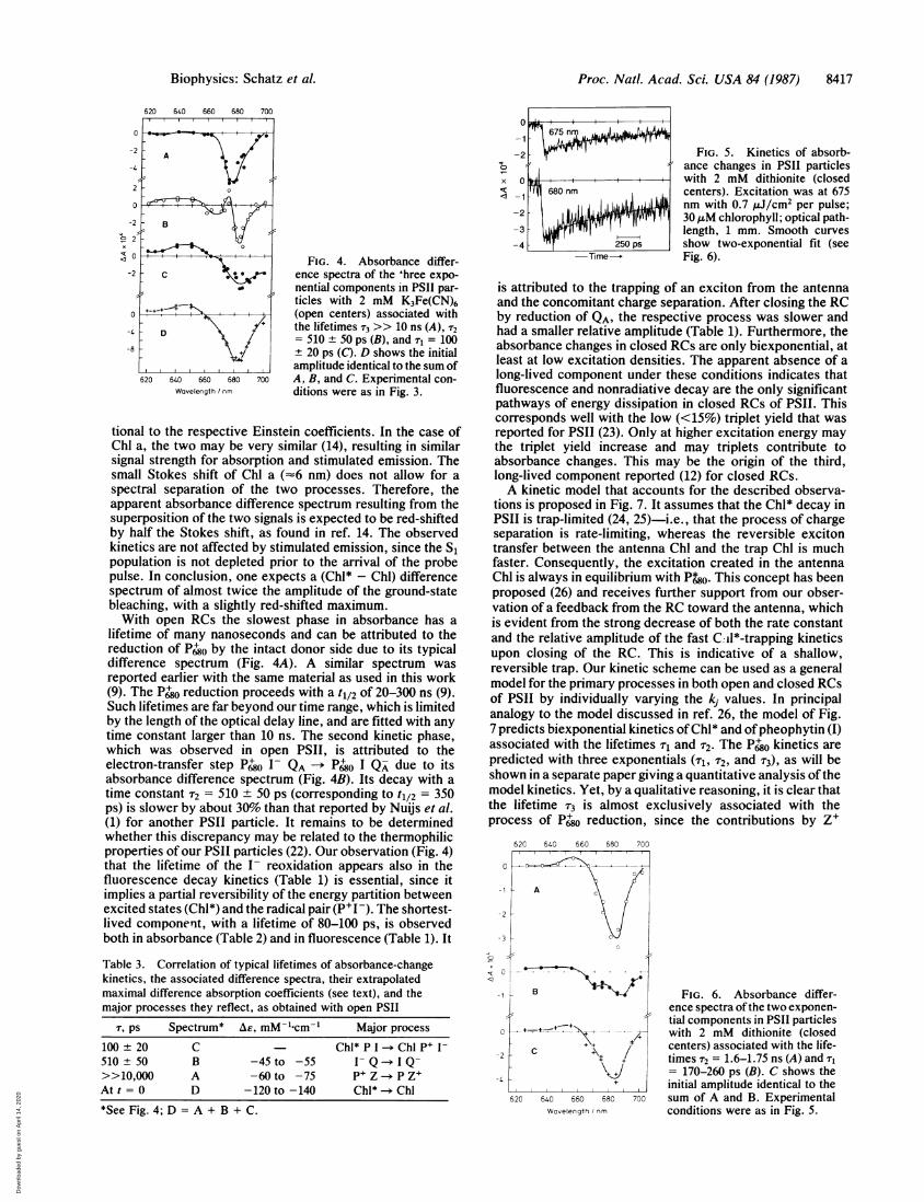
kinetic component in Fig. 4. At first glance, the differencespectra of the long- and intermediate-lifetime components(Fig. 4 A and B) show the typical features of the (P'0 - P680)and (pheophytin- - pheophytin) difference spectra, respec-tively, which are well known (refs. 9 and 13 and referencestherein). The sum of all component spectra is shown in Fig.4D. It is defined (see Eq. 1) as the absorbance differencespectrum at time zero and is therefore attributed to the (Sl -SO) difference spectrum of Chl a, the initially excited pigment(where S, is the first excited singlet state and So is the groundsinglet state). Similar spectra were reported in refs. 1 and 14.Thus, in a first approach, one can assign the differentcomponents to the processes listed in Table 3.The difference absorption coefficients (Ar) for the compo-
nent spectra in Figs. 4 A, B, and D that would be obtainedafter saturating excitation can be roughly estimated from aplot of 1/AA vs. 1E (see ref. 12) and are given in Table 3 forthe wavelengths of maximal bleaching. The value obtainedfor AE(P+ - P) agrees well with that reported in ref. 9 (-67mM-'-cm-' at 680 nm). However, the extrapolated value ofAE(Chl* - Chl) appears to be larger than the Chl ground-stateabsorption coefficient, e(Chl), by a factor of -2. Thisapparent contradiction is most easily explained by an addi-tional contribution, by stimulated emission from Chl*, to the(Chl* - Chl) bleaching signals (see Discussion).Absorbance Changes in Closed PSIH Centers. These were
first measured at the same excitation energy as for opencenters (E = 2 ,uJ/cm2 per pulse). The resulting kinetics(reported in ref. 12 and not shown here) were analyzed interms of two or three exponentials. In both cases the lifetimeof the shortest-lived component (110 or 120 ps, respectively)was much shorter than the shortest fluorescence lifetime(Table 1) under Fmax conditions. This indicated that, in closedRCs, the contribution by rapid Chl* annihilation processes isno longer negligible at the fluence given above. Due to theincrease of the mean exciton lifetime in Fmax by a factor of3-4 with respect to F, the probability of singlet-singletannihilation is also expected to increase by a similar factor.Reduction of the excitation fluence by a factor of 3, to -0.7YJ/cm2 per pulse, appeared to be necessary. The signals (Fig.
I /1013 photons cm-2 pulse-0.3 1 3 10
1 2 5 10 20
El:i] cm-2 pulse-'
FIG. 2. Amplitudes of four ex-ponential components fitted to ab-sorbance changes observed inP1SII particles with open centers [2mM K3Fe(CN)6; Chl concentra-tion, 30 AM] vs. the fluence (E) ofthe pump pulse plotted on a loga-rithmic scale. Excitation was at675 nm, and detection at 685 nm;optical pathlength was 1 mm. Thekinetic analysis was performed bythe global analysis (see text) withtwo free-running lifetimes [Tj = 25ps (A, solid curve) and T4 >> 10 ns(+, dashed curve)] and two fixedlifetimes [T2 = 90 ps (O) and T3 =500 ps (e), dotted curve].
0
x";c
FIG. 3. Kinetics of absorb-ance changes in PSII particleswith 2 mM K3Fe(CN)6 (opencenters). Excitation was at 675nm with 2 pj/cm2 per pulse; 30,4M chlorophyll; optical path-length, 1 mm. Smooth curvesshow three-exponential fit (seeFig. 4).
5) decreased by about the same factor, of course. They werewell fitted (global analysis) by two exponentials with thelifetimes T1 = 170-260 ps and r2 = 1.6-1.75 ns. These are inaccord with the fluorescence lifetimes (Table 1). The asso-ciated absorbance difference spectra are shown in Fig. 6 Aand B. The fast (170- to 260-ps) phase is associated with adifference spectrum (Fig. 6B) similar to that of the 80- to120-ps phase in open centers (Fig. 4C). This may indicate theprocess of trapping and charge separation in closed PSII. Thespectra representing the initial absorbance difference due to(Chl* - Chl) are almost identical for open (Fig. 4D) andclosed (Fig. 6C) PSII. Only upon excitation with higherintensities were more than two exponentials required todescribe the kinetics (see ref. 12 and Discussion).
DISCUSSIONThis study shows that picosecond absorbance changes inPSII particles can reflect the primary processes in the RC(exciton trapping, charge separation, and charge stabiliZa-tion), as well as the processes within the antenna (singlet-singlet annihilation and stimulated emission). Singlet-singletannihilation in photosynthetic-pigment domains is known(see, for example, ref. 15) to shorten the lifetimes of excitedstates. In our sample, this was observed upon excitation with675-nm pulses of more than 3 x 10¹³ photons/cm2, corre-sponding to one excitation per 150-200 Chl molecules or toan absorbed photon/RC ratio 0.2. Only at lower excitationdensity does the contribution by singlet-singlet annihilationbecome negligible. This is a major difference with respect tohigh-intensity experiments (as in ref. 1) where, under stan-dard conditions, the absorbed photon/RC ratio was 5 and,thus, Chl*-Chl* annihilation was the dominant processmonitored. Stimulated emission from the first excited singletstate (Sl) is expected to add to the detected intensity of thetransmitted probe beam. The spectrum of stimulated emis-sion should be similar to that of spontaneous fluorescence.This was observed in the picosecond absorbance changes ofbacterial RCs containing bacteriochlorophyll a (16-20) orbacteriochlorophyll b (21). The amplitude contributions byground-state absorption and stimulated emission are propor-
Table 2. Lifetimes (T,) associated with the absorbance changes inPSII particles with open (FO) and closed (Fmax) RCs
T, PS
i F. Fmax1 80, 120 170, 2602 460, 560 1600, 17503 >>1,000'*
Excitation wavelength was 675 nm. Values are shown for twoexperiments.*Beyond our accessible time range.
-20
0Cx
-15
-10
A
+ +
1 --5
0
8416 Biophysics: Schatz et al.
Dow
nloa
ded
by g
uest
on
Apr
il 14
, 202
0
Proc. Natl. Acad. Sci. USA 84 (1987) 8417
FIG. 4. Absorbance differ-ence spectra of the 'hree expo-nential components in PS11 par-ticles with 2 mM K3Fe(CN)6(open centers) associated withthe lifetimes T3 >> 10 ns (A), r2
= 510 + 50 ps (B), and Tg = 10020 ps (C). D shows the initial
amplitude identical to the sum ofA, B, and C. Experimental con-ditions were as in Fig. 3.
tional to the respective Einstein coefficients. In the case ofChi a, the two may be very similar (14), resulting in similarsignal strength for absorption and stimulated emission. Thesmall Stokes shift of Chi a (-6 nm) does not allow for aspectral separation of the two processes. Therefore, theapparent absorbance difference spectrum resulting from thesuperposition of the two signals is expected to be red-shiftedby half the Stokes shift, as found in ref. 14. The observedkinetics are not affected by stimulated emission, since the Sipopulation is not depleted prior to the arrival of the probepulse. In conclusion, one expects a (Chl* - Chl) differencespectrum of almost twice the amplitude of the ground-statebleaching, with a slightly red-shifted maximum.With open RCs the slowest phase in absorbance has a
lifetime of many nanoseconds and can be attributed to thereduction of P680 by the intact donor side due to its typicaldifference spectrum (Fig. 4A). A similar spectrum wasreported earlier with the same material as used in this work(9). The P'0 reduction proceeds with a tl/2 of 20-300 ns (9).Such lifetimes are far beyond our time range, which is limitedby the length of the optical delay line, and are fitted with anytime constant larger than 10 ns. The second kinetic phase,which was observed in open PSII, is attributed to theelectron-transfer step P0 I- QA -* P680 I Q- due to its
absorbance difference spectrum (Fig. 4B). Its decay with atime constant T2 = 510 + 50 ps (corresponding to t1/2 = 350ps) is slower by about 30% than that reported by Nuijs et al.(1) for another PSII particle. It remains to be determinedwhether this discrepancy may be related to the thermophilicproperties of our PSII particles (22). Our observation (Fig. 4)that the lifetime of the I- reoxidation appears also in thefluorescence decay kinetics (Table 1) is essential, since itimplies a partial reversibility of the energy partition betweenexcited states (Chl*) and the radical pair (P'I-). The shortest-lived component, with a lifetime of 80-100 ps, is observedboth in absorbance (Table 2) and in fluorescence (Table 1). It
Table 3. Correlation of typical lifetimes of absorbance-changekinetics, the associated difference spectra, their extrapolatedmaximal difference absorption coefficients (see text), and themajor processes they reflect, as obtained with open PS11
r, ps Spectrum* AE, mM-,-cm-, Major process
100 ± 20 C Chl* P I-Chl P+ I-
510 ± 50 B -45 to -55 I- Q > I Q->>10,000 A -60 to -75 P+ Z *P ZAt t = 0 D -120 to -140 Chl* Chl
*See Fig. 4; D = A + B + C.
FIG. 5. Kinetics of absorb-ance changes in PSII particleswith 2 mM dithionite (closedcenters). Excitation was at 675nm with 0.7 pJ/cm2 per pulse;30 ,gM chlorophyll; optical path-length, 1 mm. Smooth curvesshow two-exponential fit (seeFig. 6).
is attributed to the trapping of an exciton from the antennaand the concomitant charge separation. After closing the RCby reduction of QA, the respective process was slower andhad a smaller relative amplitude (Table 1). Furthermore, theabsorbance changes in closed RCs are only biexponential, atleast at low excitation densities. The apparent absence of along-lived component under these conditions indicates thatfluorescence and nonradiative decay are the only significantpathways of energy dissipation in closed RCs of PSII. Thiscorresponds well with the low (<15%) triplet yield that wasreported for PSII (23). Only at higher excitation energy maythe triplet yield increase and may triplets contribute toabsorbance changes. This may be the origin of the third,long-lived component reported (12) for closed RCs.A kinetic model that accounts for the described observa-
tions is proposed in Fig. 7. It assumes that the Chl* decay inPSII is trap-limited (24, 25)-i.e., that the process of chargeseparation is rate-limiting, whereas the reversible excitontransfer between the antenna Chl and the trap Chl is muchfaster. Consequently, the excitation created in the antennaChl is always in equilibrium with P&0. This concept has beenproposed (26) and receives further support from our obser-vation of a feedback from the RC toward the antenna, whichis evident from the strong decrease of both the rate constantand the relative amplitude of the fast Ci18-trapping kineticsupon closing of the RC. This is indicative of a shallow,reversible trap. Our kinetic scheme can be used as a generalmodel for the primary processes in both open and closed RCsof PSII by individually varying the kj values. In principalanalogy to the model discussed in ref. 26, the model of Fig.7 predicts biexponential kinetics of Chl* and ofpheophytin (I)associated with the lifetimes T1 and T2. The P'0 kinetics arepredicted with three exponentials (T1, T2, and T3), as will beshown in a separate paper giving a quantitative analysis ofthemodel kinetics. Yet, by a qualitative reasoning, it is clear thatthe lifetime T3 is almost exclusively associated with theprocess of P' reduction, since the contributions by Z'
620 640 660 680 700
-1 A
-2-
-3
q *--
-1 S2KFIG.6. Absorbance differ-
ence spectra ofthe two exponen-tial components in PSII particles
0 A+ with 2 mM dithionite (closed+ +
centers) associated with the life-- + times T2 = 1.6-1.75 ns (A) and Tr
\+J = 170-260 ps (B). C shows the-4 + initial amplitude identical to the
620 640 660 680 700 sum of A and B. ExperimentalWavelength conditions were as in Fig. 5.
0
x
620 640 660 680 700
0L
AA
2 B 0
-82
620 640 660 680 700Wavelength /nm
Biophysics: Schatz et al.
Dow
nloa
ded
by g
uest
on
Apr
il 14
, 202
0

Proc. Natl. Acad. Sci. USA 84 (1987)
B (Ch*)
h-V ~,% KA
A Chi)
A ID)
K1
V-K-1
C (Chl) K2e. D (Chi)
K3
E IDh
FIG. 7. Kinetic model of the primary reactions in open RCs ofPSII. In this scheme the rate constants k4 describe kA = kd + k,,d, theradiationless plus the radiative decay of Chl*; k1, the apparentprimary charge separation; k-1, charge recombination to the excitedstate; k2 and k3, the processes ofcharge stabilization. For closed PSIIRCs, the rate constant k2 is replaced by k2 leading to (as yet)unspecified product(s) instead of D.
(where Z is the secondary electron donor of PSII) formationto the absorbance changes in the observed spectral region arenegligible (8). Correspondingly, the absorbance differencespectrum shown in Fig. 4A is a pure (P6g0 - P680) spectrumin good accordance with a previously published spectrum (9).The lifetimes T1 and T2 are associated with observablechanges in the state of all three constituents (Chl, P, and I).Hence, the time-resolved absorbance difference spectra fromFigs. 4 and 6 are expected to be composite spectra withcontributions that depend on the relative values of the rateconstants kj. However, such additional contributions areunlikely to explain the obvious difference between thespectrum in Fig. 4B and the (I- - I) spectra reported fromsteady-state measurements (2, 27), particularly the zerotransition near 670 nm. Such differences could be attributedto electrochromic shifts induced by the presence of P680and/or the concomitant change in the electric charge on QA,which are inherent to the transient (I- - I) spectrum of Fig.4B.The spectrum shown in Fig. 4C (associated with r3) is
apparently the most complex one. It contains contributionsby the decay of Chl* and by the rise of both P'0 and I-. At-697 nm, where the latter two contributions vanish (see Fig.4 C and B), the amplitude is a measure of Chl* that becomestrapped. The energy-normalized amplitudes at this wave-length show (Fig. 6B) that 50-70% fewer excitons are trappedin closed PSII centers as compared to open centers (Fig. 4C).This means that the yield of P6PI- formation is reduced byabout the same factor as a consequence ofQA being reduced.This result is ofmajor importance for the understanding ofthemechanisms of primary processes in PSII. It indicates thatthe yield of the radical pair P+ OI- is controlled by the redoxstate of QA. A similar conclusion was already derived froma model analysis of Chl-fluorescence kinetics of unfraction-ated thylakoids (26). The control of P+ I- formation by theredox state ofQA may be a consequence of the electrical fieldcreated by the negative charge on QA and the smaller distancefrom QA to I than from QA to P680.
CONCLUSIONSThe kinetic analysis presented here indicates that excited-state kinetics in open and in closed RCs of small PSIIparticles are biexponential. In both cases, the rapid phasereflects the equilibration of energy between the antenna andthe radical pair, whereas the slow phase is associated with theoverall decay of the resulting equilibrium by deactivation ofthe antenna and the forward reaction of the radical pair. Thecharacteristic changes upon reduction ofQA are the increased
lifetime of Chl* and the decreased yield of P6jol-. Conse-quently, one can conclude that the emission from PSII withclosed RCs is primarily "prompt fluorescence" of antennaChl*. This result is in conflict with the hypothesis (2, 3) thata delayed luminescence after charge recombination fromP6PI- should account for the long-lived fluorescence-decaycomponent in closed PSII. This hypothesis has already beenquestioned solely on the basis of the fluorescence-decayproperties in green algae (26, 28).We thank Ms. B. Kalka and Mr. U. Pieper for preparing the PSII
particles, Mr. P. Klein-Bolting and Mr. W. Schuster for data-analysisprograms, and Mr. V. Seeling for help with construction of the lasersystem. We are grateful to Prof. K. Schaffner for his stimulatinginterest and to the Deutsche Forschungsgemeinschaft for partialfinancial support.
1. Nuijs, A. M., van Gorkom, H. J., Plijter, J. J. & Duysens,L. N. M. (1986) Biochim. Biophys. Acta 848, 167-175.
2. Klimov, V. V., Klevanik, A. V., Shuvalov, V. A. & Krasnov-sky, A. A. (1977) FEBS Lett. 82, 183-186.
3. Klimov, V. V. & Krasnovskii, A. A. (1982) Biophysics 27,186-198.
4. Karukstis, K. K. & Sauer, K. (1983) J. Cell. Biochem. 23,131-158.
5. Mauzerall, D. C. (1985) Biochim. Biophys. Acta 809, 11-16.6. Holzwarth, A. R., Wendler, J. & Haehnel, W. (1985) Biochim.
Biophys. Acta 807, 155-167.7. Schatz, G. H. & Witt, H. T. (1984) Photobiochem. Photobio-
phys. 7, 1-14.8. Schatz, G. H. and van Gorkom, H. J. (1985) Biochim. Bio-
phys. Acta 810, 283-294.9. Schlodder, E., Brettel, K., Schatz, G. H. & Witt, H. T. (1984)
Biochim. Biophys. Acta 765, 178-185.10. Knorr, F. J. & Harris, J. M. (1981) Anal. Chem. 53, 272-276.11. Holzwarth, A. R., Wendler, J. & Suter, G. W. (1987) Biophys.
J. 51, 1-12.12. Holzwarth, A. R., Brock, H. & Schatz, G. H. (1987) in Prog-
ress in Photosynthesis Research, ed. Biggins, J. (Nijhoff,Dordrecht, The Netherlands), Vol. 1, pp. 61-65.
13. Parson, W. W. & Ke, B. (1982) in Photosynthesis: EnergyConversion by Plants and Bacteria, ed. Govindjee (Academic,New York), Vol. 1, pp. 331-385.
14. Klug, D. R., Gore, B. L., Giorgi, L. B. & Porter, G. (1987) inProgress in Photosynthesis Research, ed. Biggins, J. (Nijhoff,Dordrecht, The Netherlands), Vol. 1, pp. 95-98.
15. Breton, J. & Geacintov, N. E. (1980) Biochim. Biophys. Acta594, 1-32.
16. Becker, M., Middendorf, D., Woodbury, N. W., Parson,W. W. & Blankenship, R. E. (1986) in Ultrafast Phenomena,eds. Fleming, G. R. & Siegman, A. E. (Springer, Berlin), Vol.5, pp. 374-378.
17. Woodbury, N. W., Becker, M., Middendorf, D. & Parson,W. W. (1985) Biochemistry 24, 7516-7521.
18. Martin, J. L., Breton, J., Hoff, A. J., Migus, A. & Antonetti,A. (1986) Proc. Natl. Acad. Sci. USA 83, 957-961.
19. Shuvalov, V. A. & Duysens, L. N. M. (1986) Proc. Natl.Acad. Sci. USA 83, 1690-1694.
20. Shuvalov, V. A., Vasmel, H., Amesz, J. & Duysens, L. N. M.(1986) Biochim. Biophys. Acta 851, 361-368.
21. Breton, J., Martin, J. L., Migus, A., Antonetti, A. & Orszag,A. (1986) Proc. Natl. Acad. Sci. USA 83, 5121-5125.
22. Schatz, G. H. & Witt, H. T. (1984) Photobiochem. Photobio-phys. 7, 77-89.
23. Kramer, H. & Mathis, P. (1980) Biochim. Biophys. Acta 593,319-329.
24. Pearlstein, R. M. (1982) in Photosynthesis: Energy Conversionby Plants and Bacteria, ed. Govindjee (Academic, New York),Vol. 1, pp. 293-330.
25. Pearlstein, R. M. (1982) Photochem. Photobiol. 35, 835-844.26. Schatz, G. H. & Holzwarth, A. R. (1986) Photosynth. Res. 10,
309-318.27. Klimov, V. V., Shuvalov, V. A. & Heber, U. (1985) Biochim.
Biophys. Acta 809, 345-350.28. Haehnel, W., Holzwarth, A. R. & Wendler, J. (1983) Photo-
chem. Photobiol. 37, 435-443.
8418 Biophysics: Schatz et al.
Dow
nloa
ded
by g
uest
on
Apr
il 14
, 202
0


![Psii Pengkelasan Dan Justifikasi Penggunaan Media Dalam Pengajaran Dan[1]](https://img.dokumen.tips/doc/110x75/5571fa6b4979599169922dfb/psii-pengkelasan-dan-justifikasi-penggunaan-media-dalam-pengajaran-dan1.jpg)


