Embed Size (px)
Citation preview

223
http://journals.tubitak.gov.tr/biology/
Turkish Journal of Biology Turk J Biol(2015) 39: 223-232© TÜBİTAKdoi:10.3906/biy-1406-10
Phytofabrication of biomolecule-coated metallic silver nanoparticles using leaf extracts of in vitro-raised bamboo species and its anticancer activity against human PC3 cell lines
Kalamegam KALAIARASI1, Govindaraj PRASANNARAJ1,2, Shivendra Vikram SAHI3, Perumal VENKATACHALAM1,2,*1Plant Genetic Engineering and Molecular Biology Lab, Department of Biotechnology, Periyar University,
Periyar Palkalai Nagar, Salem, Tamil Nadu, India2Centre for Nanoscience and Nanotechnology, Periyar University, Salem, Tamil Nadu, India
3Department of Biology, Western Kentucky University, Bowling Green, KY, USA
* Correspondence: [email protected]
1. IntroductionCancer is a malignant, aggressive growth that invades surrounding tissues and metastasizes to other areas of the body. Cancer is literally a runaway cell growth, and the cellular replication machinery is unable to shut down. Prostate cancer is the most common cancer in men after skin cancer and is the second most common cause of cancer death among men (913,000 new cases, 13.8% of the total), after lung cancer. Nearly three-quarters of the registered cases occur in developed countries (658,000 cases) (Ferlay et al., 2010). Normal prostate epithelium contains luminal epithelial cells, basal cells, and a small component of neuroendocrine cells that are scattered throughout the prostate (Huang et al., 2007; Yuan et al., 2007; Sun et al., 2009). The majority of prostate cancers are classified as adenocarcinomas characterized by an absence of basal cells and uncontrolled proliferation of malignant tumor cells, with features of luminal differentiation including glandular formation and the expression of androgen receptors and prostate-specific antigens (Tai et al., 2011). Incidence rates of prostate cancer vary by more
than 25-fold worldwide; the highest rates are in Australia and New Zealand (104.2 per 100,000), and incidence rates are relatively high in certain developing regions such as the Caribbean, South America, and sub-Saharan Africa. The lowest age-standardized incidence rate is estimated to be in South Central Asia (4.1 per 100,000) (Ferlay et al., 2010). New therapies are crucial in the fight against stubborn prostate cancers that spread to all organs of the body and aggressive tumors that stop responding to standard treatments. In addition, information on the biological response of human cells to green synthesized metallic silver nanoparticles (AgNPs) is also very limited.
The exploitation of various plant materials for the biosynthesis of nanoparticles (NPs) is considered a green technology and does not involve any harmful chemicals. AgNPs are important materials that have been studied extensively. Such nanoscale materials possess unique electrical, optical, and biological properties and are thus applied in catalysis, biosensing, imaging, drug delivery, nanodevice fabrication, and medicinal applications (Ahamed et al., 2010, Tolaymat et al., 2010). Due to
Abstract: The present study describes the biosynthesis of metallic silver nanoparticles (AgNPs) from silver ions using in vitro-grown leaf samples of Bambusa arundinacea (Ba) and Bambusa nutans (Bn) and their anticancer activity against human prostatic cancer cell lines (PC3). The metallic AgNPs were synthesized at room temperature by treating the leaf extracts of Bambusa arundinacea and Bambusa nutans with 2 mM silver nitrate using the boiled method. The characterization of synthesized BaAgNPs and BnAgNPs was performed using UV-Vis spectrum (absorption peak at 365 nm), FTIR, TEM (spherical shape), SEM (size of 30–90 nm), and EDX analysis. Cell viability assays were carried out to determine the cytotoxicity effect of AgNPs on the PC3 cancer cell line and the Vero normal cell line. The IC50 values for BaAgNP- and BnAgNP-treated PC3 cancer cells were found to be 73.57 µg/mL and 84.88 µg/mL while the values for treated Vero cells were found to be 93.58 µg/mL and 96.41 µg/mL, respectively. The percentages of the apoptotic bodies by AO/EtBr staining were found to be 76% and 62% for BaAgNPs and BnAgNPs, respectively. The results strongly suggest that synthesized BaAgNPs show potential anticancer activity against human PC3 cell lines compared to BnAgNPs.
Key words: Anticancer activity, biological synthesis, bamboo, silver nanoparticles, electron microscopy, PC3 human prostate cancer cell lines
Received: 04.06.2014 Accepted: 14.10.2014 Published Online: 01.04.2015 Printed: 30.04.2015
Research Article

KALAIARASI et al. / Turk J Biol
224
strong antimicrobial activity, AgNPs are also used in clothing, the food industry, sunscreens, and cosmetics (Vigneshwaran et al., 2007; Kokura et al., 2010; Martinez-Gutierrez et al., 2010). Although different techniques such as UV irradiation, aerosol technologies, lithography, laser ablation, ultrasonic fields, and photochemical reduction have been used successfully to produce metal NPs, they remain expensive and sometimes involve the use of hazardous chemicals (Okitsu et al., 2001; Naik et al., 2002; Narayanan and Sakthivel, 2010). The mechanisms for AgNP-induced toxicity may be related to mitochondrial damage, oxidative stress, DNA damage, and induction of apoptosis (Sukirtha et al., 2012). Consequently, green synthesis of nanoparticles has received increasing attention due to the growing need for environmentally benign technology in NP synthesis.
Bambusa arundinacea (Retz.) Wild, the fastest-growing perennial plant on earth, is native to India and cultivated throughout the tropics, commonly known as thorny bamboo, or “mulmunkil” in Tamil. Bamboo leaves have been used clinically in the treatment of hypertension, arteriosclerosis, cardiovascular disease, and cancer (Zhang et al., 2007). Bambusa nutans Wall. ex Munro is a medium-sized woody bamboo that is naturally distributed throughout various states of India up to an altitude of 1500 m. B. nutans is one of the priority species of bamboos identified by the National Bamboo Mission formulated by the Ministry of Agriculture, Government of India, for commercial cultivation, and it is exploited at the present time for the pulp and paper industry in India. Culms can measure 6–15 m in height and 5–10 cm in diameter and are smooth, straight, and thick-walled. B. nutans is used for various purposes, including timber, house construction, scaffolding, and paper mats. It is reported that B. arundinacea plants possess flavonoids, phenols, steroids, glycosides, alkaloids, and phytosterols, which could be used for various pharmaceutical applications (Nirmala et al., 2011). Bamboo leaf extract has exhibited anticancer (Seki and Maeda, 2010), antiinflammatory, and antiulcer properties (Muniappan and Sundararaj, 2003); has antimicrobial activity (Vijaykumar et al., 2010); and has decreased bone erosion, spleen enlargement, and rheumatoid factor (Rathod et al., 2012). B. arundinacea seeds showed significant antidiabetic activity (Macharla and Goli, 2011). Moulton et al. (2010) showed that green synthesized AgNPs were potentially biocompatible with human keratinocyte cells. The green synthesis of AgNPs includes selection of solvent medium, reducing agent, and nontoxic stabilizing compound/material, also called a capping agent, and prevents aggregation of the NPs. As these NPs result in significantly low toxicity on adoption of this technique, they can be used for encapsulation of drug molecules. In recent reports, both silver and gold NPs have been synthesized using leaf extracts of Azadirachta indica
(Shankar et al., 2004), black tea leaf (Begum et al., 2009), Ocimum sanctum (Philip, 2010), Annona squamosa (Vivek et al., 2012), Rauvolfia tetraphylla (Kalaiarasi et al., 2013), and Caesalpinia coriaria (Jeeva et al., 2014). Although there were many reports on biosynthesis of NPs using various plant extracts, no report has been published on the synthesis of AgNPs in B. arundinacea and B. nutans species until now. The major objective of the present study was to develop a rapid method for biological synthesis of bioactive molecules loaded with metallic AgNPs from in vitro-grown leaf samples of B. arundinacea and B. nutans and to evaluate their anticancer activity against human PC3 cell lines.
2. Materials and methods2.1. Collection and preparation of bamboo leaf extractThe leaf samples for NP synthesis were collected from in vitro-raised Bambusa arundinacea (Ba) and Bambusa nutans (Bn) acclimatized under greenhouse conditions and established in the Biodiversity Garden, Periyar University, Salem. Mature, green, and undamaged leaves were selected and washed thoroughly with tap water and later washed with sterile distilled water in order to remove adhering dust particles. The bamboo leaves were used to prepare the aqueous extract and AgNPs were synthesized by boiled method from B. arundinacea and B. nutans. The sterile leaves from both the species (5 g) were cut into fine pieces and were boiled with 25 mL of sterile distilled water for 15 min at 60 °C. The extract was filtered through filter paper (Whatman No. 1) and the filtrate was used as the reducing agent for silver nitrate (AgNO3).2.2. Biosynthesis of silver NPs According to the methodology followed by Kalaiarasi et al. (2013), an aqueous AgNO3 solution (2 mM) was prepared and used for the synthesis of AgNPs. Boiled leaf extracts of both B. arundinacea and B. nutans (10 mL) were taken separately and added into 90 mL of 2 mM AgNO3 solution for the reduction of Ag+ ions into Ag0 and kept overnight at room temperature in the dark to minimize the photoactivation of silver nitrate. After the incubation period, the sample color changed from light green to brown, indicating the synthesis of AgNPs.2.3. Characterization of AgNPsUV-Vis spectra were recorded as a function of the reaction time on a Systronics 2203 spectrophotometer. The bioreduction of silver ions and formation and stability of AgNPs in an aqueous colloidal solution was monitored periodically with UV-Vis spectroscopy (200–700 nm) at room temperature. After completion of the synthesis of AgNPs, the reaction mixture was centrifuged at 8000 rpm for 10 min, and the NP pellet was dissolved in sterile distilled water and washed thrice by centrifugation to remove impurities. The purified AgNPs were examined for

KALAIARASI et al. / Turk J Biol
225
the presence of functional groups using Fourier transform infrared spectroscopy (FTIR) analysis. The FTIR analysis for AgNPs was carried out using an RXI model FTIR spectrophotometer from PerkinElmer. The dried samples were compressed into a thin KBr disc under a pressure of 7845 kPa for 2 min and all spectra were recorded within the range of 4000 to 400 cm–1 in transmittance mode. X-ray diffraction (XRD) measurement of bioreduced AgNPs was carried out and crystalline metallic silver was confirmed. For this experiment, the plant broth–mediated AgNPs were spread onto a glass substrate and examined on a Shimadzu Model XRD 6000 operated at a voltage of 40 keV and a current of 30 mA with CuKα radiation (λ = 1.54056 Å) in a θ–2θ configuration. The shapes of the freeze-dried AgNPs were analyzed by scanning electron microscopy (SEM; JEOL-JSM 6390). For transmission electron microscopy (TEM) analysis, the aqueous suspension of AgNPs was prepared by placing a drop of the suspension on carbon-coated Cu2 grids. The films on the TEM grids were allowed to stand for 2 min, after which the extra solution was removed using a blotting paper and the grid was allowed to dry prior to measurement. TEM observations were performed on a Philips-TECNAI 10 instrument attached to an Olympus camera operated at an accelerating voltage of 100 kV. The size distribution of the resulting NPs was estimated on the basis of TEM micrographs with the assistance of Sigma Scan Pro software (SPSS Inc., Version 4.01.003). Energy dispersive X-ray (EDX) spectroscopy analysis was performed with an OXFORD INCA penta FETX3 model 7582 EDX spectrometer to confirm the elemental composition of the samples.2.4. Cell culture and maintenanceTo study the cytotoxic effect of the synthesized NPs on cancer cell lines, a human prostate cancer cell line (PC3) and an African monkey kidney control cell line (Vero) were purchased from the National Centre for Cell Sciences, Pune, India. The cells were maintained in Dulbecco’s modified Eagle’s medium supplemented with 2 mM L-glutamine and balanced salt solution and adjusted to contain 1.5 g/L sodium carbonate, 0.1 mM nonessential amino acids, 1 mM sodium pyruvate, 2 mM L-glutamine, 1.5 g/L glucose, 10 mM (4-(2-hydroxyethyl)-1-piperazine ethane sulfonic acid) HEPES, and 10% fetal bovine serum (GIBCO). Penicillin and streptomycin (1 IU/1 µg) were added at 1 mL/L. The cells were maintained in 25-cm2 culture flasks (Corning) at 37 °C with 5% CO2 in a humidified CO2 incubator.2.5. In vitro cytotoxicity and IC50The inhibitory concentration (IC50) value was evaluated using MTT [3-(4,5-dimethylthiazol-2-yl)-2,5-diphenyltetrazolium bromide] assay. The cultured cells (approximately 1 × 10⁴ cells/well) were seeded into 96-well culture plates and incubated for 48 h. PC3 cells were treated
with series of 10–100 µg/mL concentrations of BaAgNPs and BnAgNPs. A stock concentration (5 mg/mL) of MTT (Sigma), a yellow tetrazole, was prepared and the culture medium was removed, and 100 µL of MTT was added in both AgNP-treated and control wells and incubated at 37 °C for 4–6 h. After incubation, purple formazan crystals were observed at the bottom of the wells. These crystals were dissolved with 100 µL of dimethyl sulfoxide and read at 620 nm in a multiwell ELISA plate reader (Thermo Multiskan EX) (Suresh et al., 2013). The optical density (OD) value was used to calculate the percentage of cell viability by using the following formula.
Percentage of cell viability = OD value of experimental AgNPs treated sampleOD value of experimental untreated sample
×100
2.6. Acridine orange and ethidium bromide stainingApproximately 5 µL of dye mixture (100 mg/mL acridine orange (AO) and 100 mg/mL ethidium bromide (EtBr) in phosphate buffered saline (PBS)) was mixed with 9 mL of cell suspension (1 × 10⁵ cells/mL) on clean microscopic cover slips. The cancer cells were collected, washed with PBS (pH 7.2), and stained with 1 mL of AO/EtBr. After incubation for 2–3 min, the cells were visualized under a fluorescence microscope (Nikon Eclipse) at 40× magnification with an excitation filter set at 480 nm (Vivek et al., 2012). The percentage of apoptotic cells was determined by the following formula.
Percentage of apoptotic cells = Total number of apoptotic cells Total number of normal and apoptotic cells
×100
2.7. Statistical analysisAll statistical analyses were carried out using one-way ANOVA with Dunnett’s posttest using GraphPad InStat and Prism version 3.00 for Windows (GraphPad Software).
3. Results and discussion3.1. Biosynthesis and characterization of NPsThe bioreduction of silver ions was visually confirmed by color change from green to brown for AgNPs during the exposure of bamboo leaf extract, while no color change was observed in the control AgNO3 solution without leaf extract. The addition of bamboo leaf extracts to 2 mM aqueous silver nitrate solution changed the color after 4 h of reaction due to the production of AgNPs (Figures 1A–1C). These color changes arose because of the excitation of surface plasmon vibrations with the AgNPs (Shankar et al., 2004).
The most widely used, simple, and sensitive technique for the observation of NP synthesis is UV-Vis spectroscopy. AgNP suspension exhibits an intense golden yellow color due to the surface plasmon resonance (SPR), which results from the collective oscillations of their conduction band

KALAIARASI et al. / Turk J Biol
226
electrons in response to electromagnetic waves. Under the UV region, AgNPs give a characteristic absorbance band due to the excitation mode of their surface plasmons, which is dependent on the size of NPs. These SPR bands undergo redshift or blueshift depending on the quantum size effects (Asharani et al., 2008; Thomas et al., 2008). Consequently, absorbance peaks can be used as tools to predict particle size and stability. Smaller AgNPs will have an absorbance maximum around 400 nm, which increases with size and disappears when particle size falls outside nano dimensions. The production of AgNPs from leaf extracts of B. arundinacea and B. nutans was analyzed in the range of 200 nm to 700 nm. Results revealed a peak at 420 nm in both leaf extracts, indicating silver nanoparticle production. The UV-Vis spectrum results showed that the maximum absorbance of green synthesized AgNPs was observed at 420 nm. A narrow absorption peak at 210 nm showed that the green synthesized AgNPs had a narrow size distribution with smaller-sized particles predominating. A change in color was also associated with well-defined peaks characterized by maxima centered at 280 nm. A variety of biomolecules were postulated to be involved in biological NP synthesis (Pimprikar et al., 2009).
The FTIR measurements were performed to identify the presence of possible biomolecules responsible for bioreduction of silver ions and capping of the bioreduced AgNPs synthesized using bamboo leaf broths. The lyophilized powders of the AgNPs were then subjected to FTIR spectroscopy analysis. The FTIR spectra of in vitro boiled leaf sample BaAgNPs showed intense peaks at 3396 cm–1, representing the NH stretching (aliphatic and aromatic amines), 3211 cm–1 for S–H stretching, 2286 cm–1 for C=C stretching (alkenes), 1618 cm–1 for NH bend, 1402 cm–1 for C=C stretching, 1281 cm–1 for N–O nitro
stretching, 974 cm–1 for C–H (alkene), 913 cm–1 for O–H bend (carboxylic acid), 604 cm–1 for C–H bend (alkynes), and 526 cm–1 for C–Br stretching (alkyl halides). Bamboo species are mainly composed of pectin, cellulose and hemicellulose, flavonoids, lactones, and phenolic acids. The functional groups associated with these polymers, as well as the proteinaceous matter, may be involved in reducing the Ag+ to Ag0. All the biological components were known to interact with metal salts via these functional groups and mediate their reduction to NPs (Bar et al., 2009; Ganesh Babu and Gunasekaran, 2009). The FTIR spectra of in vitro boiled leaf sample BnAgNPs showed intense peaks at 3913 cm–1, 3790 cm–1, 3731 cm–1, 3400 cm–1, 3215 cm–1, 2835 cm–1, 1612 cm–1, 1400 cm–1, 973 cm–1, 913 cm–1, and 590 cm–1. The peak produced at 3790 cm–1 indicates N–H stretching (aliphatic and aromatic amines), 3731 cm–1 N–H stretching, 3400 cm–1 2° amine N–H stretching, 3215 cm–1 O–H stretching (phenol), 2835 cm–1 C=O stretching (aldehyde), 1612 cm–1 C=O stretching (ketone), 1400 cm–1 N=O stretching, 973 cm–1 C–H (alkene), 913 cm–1 O–H bend (carboxylic acid), and 590 cm–1 C–Br stretching (alkyl halides) (Figures 2A and 2B). It is evident that the differences in the absorption bands, the small shift in band position, and the capping series could be different for each synthesized BaAgNP and BnAgNP.
XRD analysis revealed the distinctive facets (210, 122, 231, 142, 220, and 311 planes) of the AgNPs. XRD studies were carried out to confirm the crystalline nature of the silver particles; the XRD patterns are illustrated in Figures 3A and 3B. XRD patterns showed intense peaks in the whole spectrum of 2θ values ranging from 20 to 80. The XRD pattern was observed to be 27.88°, 32.29°, 46.29°, 54.92°, and 77.28° for boiled in vitro leaf extracts of B. arundinacea and 27.74°, 32.14°, 46.17°, 54.77°,
Figure 1. Color changes after the addition of leaf extracts with aqueous silver nitrate solution: (A) control, (B) B. arundinacea, and (C) B. nutans.

KALAIARASI et al. / Turk J Biol
227
56.47°, 64.56°, and 77.35° for B. nutans. A correspondence to Bragg’s reflection sets of lattice planes was observe, according to JCPDS no. 04-0783 for silver, which may be indexed to (210), (122), (231), (142), (311) for B. arundinacea and (210), (122), (231), (142), (241), (220), (311) for B. nutans, and it indicates the face-centered cubic structure of the AgNPs. The present result is in good agreement with earlier reports (Jayaseelan and Rahuman, 2012). It suggests that the prepared AgNPs were biphasic in nature. The slight shift in the peak positions indicated the presence of strain in the crystal structure, which is a characteristic feature of nanocrystallites. Thus, the XRD patterns were strong evidence in favor of the UV-Vis spectra for the presence of silver nanocrystals.
SEM analysis confirmed the synthesis of AgNPs in the reaction mixture. At room temperature, the addition
of 2 mM aqueous silver nitrate solution to the plant extract caused the precipitation of NPs. These precipitates were observed under microscopic studies for the surface morphology of NPs. SEM images of BaAgNPs and BnAgNPs derived from boiled leaf extracts are depicted in Figures 4A and 4B. The size of the synthesized AgNPs obtained ranged between 32 to 46.65 nm and 41.40 to 98.67 nm for BaAgNPs and BnAgNPs, respectively. TEM images for the biosynthesized AgNPs showed that the particles were spherically shaped with an average size ranging from 30 nm to 90 nm for both of the AgNPs (Figures 5A and 5B). The presence of silver atoms in BaAgNPs and BnAgNPs was further confirmed using EDX spectroscopy. The EDX profiles showed a strong silver signal, along with other weak signals of oxygen and chlorine, which may have originated from the bioactive molecules bound
A B
Figure 2. FTIR spectral analysis of: (A) aqueous boiled in vitro leaf extracts of B. arundinacea and synthesized AgNPs, (B) aqueous extracts of B. nutans and synthesized AgNPs.
bA B
Figure 3. XRD pattern of biosynthesized AgNPs using in vitro leaf extracts of (A) B. arundinacea and (B) B. nutans.

KALAIARASI et al. / Turk J Biol
228
to the surface of the NPs. The optical absorption peak was observed at 2.5 and 3.0 keV for B. arundinacea and at 2.6 and 3.0 keV for B. nutans, which is typical for the absorption of silver nanocrystallites due to SPR (Figures 6A and 6B). It has been reported that NPs synthesized using leaf extracts are surrounded by a thin layer of some capping organic materials from the plant leaf broth. The weak signal elements may have originated from the biomolecules bound to the surface of the biosynthesized BaAgNPs and BnAgNPs. The signals were most likely due to the X-ray emission from carbohydrates/proteins/enzymes present in the cell wall of the biomass (Mishra et al., 2010). The percentage of the silver ions present in the synthesized NPs was found to be 90% and 85% for BaAgNPs and BnAgNPs, respectively. The optical absorption peaks observed at 0.2, 3, 3.3, and 3.7 keV are typical for the absorption of the silver nanocrystallites due to SPR. It was reported that individual spherical AgNPs synthesized using alfalfa sprouts showed absorption peaks in the range
of 2.5–4 keV (Gardea-Torresdey et al., 2003). The EDX spectrum shows the characteristics of silver peaks on the surface of the NPs, suggesting successful AgNPs synthesis using plant leaf extracts of B. arundinacea and B. nutans. Most recently, Jeeva et al. (2014) obtained spherical and triangular silver NPs at 2–4 keV using Caesalpinia coriaria leaf extracts. 3.2. Determination of cytotoxicity effect of AgNPsIn vitro cytotoxic concentration was assessed using MTT reduction. After treating PC3 and Vero cells for 48 h with BaAgNPs and BnAgNPs in a concentration ranging from 10 to 100 µg/mL, the inhibitory concentration of 50% (IC50) against the growth of treated cancer and normal cancer cells was calculated. The percent of cell viability obtained with continuous exposure for 48 h is depicted in Figure 7. Cell viability was also found to decrease with increasing concentrations of the biosynthesized BaAgNPs and BnAgNPs. The IC50 values of the PC3 cells and Vero cells were 73.57 and 84.88 µg/mL and 93.58 and 96.41
Figure 4. SEM images of synthesized silver nanoparticles using in vitro leaf extracts of (A) B. arundinacea and (B) B. nutans.
Figure 5. TEM images of synthesized silver nanoparticles using in vitro leaf extracts of (A) B. arundinacea and (B) B. nutans confirming the spherically shaped nanocrystals.

KALAIARASI et al. / Turk J Biol
229
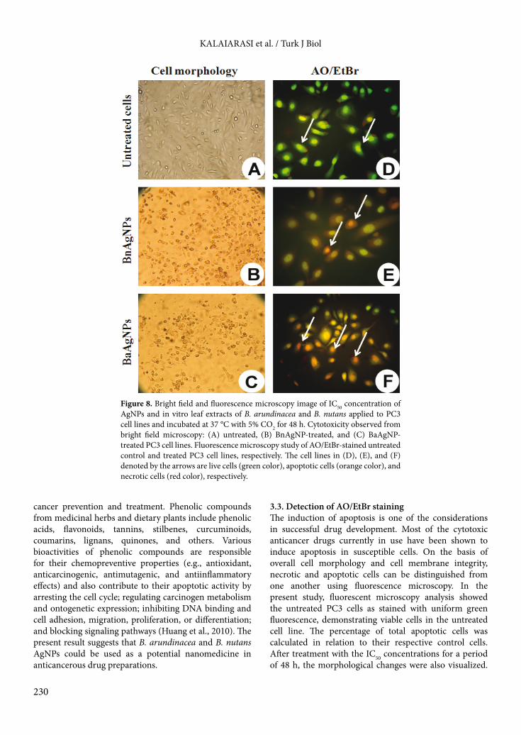
µg/mL for BaAgNPs and BnAgNPs, respectively. The effect of NPs on the growth of normal cell lines (Vero) did not exhibit significant cytotoxicity at their lower concentrations, whereas cytotoxicity was increased with increasing concentrations. Upon comparison of the IC50 values of the 2 AgNPs, B. arundinacea NPs appeared to be the most active and showed a higher inhibition rate with lower concentration than B. nutans against PC3 cell lines, due to the presence of flavonoids and phenols. The effect of leaf aqueous extracts of both B. arundinacea and B. nutans against the growth of PC3 cells did not exhibit significant cytotoxicity at their lower concentrations; however, the cytotoxicity increased with increasing concentration from 100 to 160 µg/mL. The most recognizable morphological changes due to the AgNP treatment observed were cytoplasmic condensation, cell shrinkage, and production of numerous cell surfaces bulging at the plasma membrane against PC3 cells (Figures 8A–8C). The mortality data obtained in this study allowed us to predict their potential as cytotoxic agents. It is reported that flavonoid compounds, especially quercetin and genistein, show antitumor activity; these compounds were found to be toxic to cancer cells but had an insignificant effect on normal cells (Baliga and Katiyar, 2006). It has been reported that the flavonoid apigenin holds great potential as a chemopreventive agent for a variety of cancers and exhibits significant activity against UV-induced DNA damage, thus protecting against skin cancer (Satyavani et al., 2011). The cytotoxic effect was identified due to the formation of reactive oxygen species (ROS). ROS typically include the superoxide radical, hydrogen peroxide, and hydroxyl radical, which cause damage to cellular components such as lipids, DNA, and proteins and eventually lead to death (Xia et al., 2006). Natural phenolic compounds play an important role in
A B
Figure 6. EDX spectroscopy of in vitro leaf extracts of (A) B. arundinacea and (B) B. nutans synthesized AgNPs exhibited signs of silver.
A
B
Figure 7. MTT assay results confirming the in vitro cytotoxicity of BaAgNPs and BnAgNPs against (A) PC3 and (B) normal Vero cell lines (CC: cell control).
KALAIARASI et al. / Turk J Biol
230
cancer prevention and treatment. Phenolic compounds from medicinal herbs and dietary plants include phenolic acids, flavonoids, tannins, stilbenes, curcuminoids, coumarins, lignans, quinones, and others. Various bioactivities of phenolic compounds are responsible for their chemopreventive properties (e.g., antioxidant, anticarcinogenic, antimutagenic, and antiinflammatory effects) and also contribute to their apoptotic activity by arresting the cell cycle; regulating carcinogen metabolism and ontogenetic expression; inhibiting DNA binding and cell adhesion, migration, proliferation, or differentiation; and blocking signaling pathways (Huang et al., 2010). The present result suggests that B. arundinacea and B. nutans AgNPs could be used as a potential nanomedicine in anticancerous drug preparations.
3.3. Detection of AO/EtBr stainingThe induction of apoptosis is one of the considerations in successful drug development. Most of the cytotoxic anticancer drugs currently in use have been shown to induce apoptosis in susceptible cells. On the basis of overall cell morphology and cell membrane integrity, necrotic and apoptotic cells can be distinguished from one another using fluorescence microscopy. In the present study, fluorescent microscopy analysis showed the untreated PC3 cells as stained with uniform green fluorescence, demonstrating viable cells in the untreated cell line. The percentage of total apoptotic cells was calculated in relation to their respective control cells. After treatment with the IC50 concentrations for a period of 48 h, the morphological changes were also visualized.
Figure 8. Bright field and fluorescence microscopy image of IC50 concentration of AgNPs and in vitro leaf extracts of B. arundinacea and B. nutans applied to PC3 cell lines and incubated at 37 °C with 5% CO2 for 48 h. Cytotoxicity observed from bright field microscopy: (A) untreated, (B) BnAgNP-treated, and (C) BaAgNP-treated PC3 cell lines. Fluorescence microscopy study of AO/EtBr-stained untreated control and treated PC3 cell lines, respectively. The cell lines in (D), (E), and (F) denoted by the arrows are live cells (green color), apoptotic cells (orange color), and necrotic cells (red color), respectively.

KALAIARASI et al. / Turk J Biol
231
Orange apoptotic cells containing apoptotic bodies as well as red necrotic cells were also observed (Figures 8D–8F). Microscopic observation showed that the normal viable cells fluoresced green due to the penetration of AO into cell membrane, whereas apoptotic cells were observed as orange-colored bodies because of nuclear shrinkage and blebbing. Necrotic cells fluoresced red due to their loss of membrane integrity via AgNP-induced cytotoxicity. Similar results were also reported earlier by Thangam et al. (2012). The percentage of the apoptotic bodies obtained was 76% and 62% for synthesized BaAgNPs and BnAgNPs, respectively (Figure 9).3.4. ConclusionThe present study documented the first-ever synthesis, characterization, and cytotoxicity of biosynthesized AgNPs from in vitro-propagated leaf extracts of B. arundinacea and B. nutans against PC3 cell lines. FTIR assay confirmed the presence of flavonoids and phenols in the synthesized NPs as well as in the aqueous extracts of both the bamboo species. Structural analysis by XRD, together with the elemental analysis by EDX, strongly suggested the formation of elemental AgNPs. SEM results showed that the shape of the NPs obtained was spherical, ranging from 32 to 99 nm for AgNPs. Collectively, our data suggest that AgNPs possess superior cytotoxic activity compared to the leaf extract. The present study strongly showed that the biosynthesized BaAgNPs exert a potentially cytotoxic effect on human prostate PC3 cancer cells without
affecting the Vero control cells, as opposed to BnAgNPs. However, further research is required to discover the mechanism involved in the cell growth inhibition, thereby permitting the biosynthesized AgNPs to be used as cancer chemopreventive and/or therapeutic agents in the near future.
AcknowledgmentOur sincere thanks to Periyar University for providing a UGC-University Research Fellowship (URF).
AO/EtBr Staining
Untreated BaAgNPs BnAgNPs0
20
40
60
80
100
*
*
PC3 cell lines
Perc
ent o
f apo
ptot
ic c
ells
Figure 9. Histogram representation of the percentage of total observed apoptotic PC3 cancer cells against their IC50 concentration of AgNPs after 48 h of treatment (*P < 0.05).
References
Ahamed M, Al Salhi MS, Siddiqui MKJ (2010). Silver nanoparticle applications and human health. Clin Chim Acta 411: 1841–1848.
Asharani PV, Wu YL, Gong Z, Valiyaveettil S (2008). Toxicity of silver nanoparticles in zebrafish models. Nanotech 19: 255102.
Baliga MS, Katiyar SK (2006). Chemoprevention of photocarcinogenesis by selected dietary botanicals. Photochem Photobiol Sci 5: 243–253.
Bar H, Bhui DK, Sahoo GP, Sarkar P, Pyne S, Misra A (2009). Green synthesis of silver nanoparticles using seed extract of Jatropha curcas. Colloid Surface A 348: 212–216.
Begum NA, Mondal S, Basu S, Laskar RA, Mandal D (2009). Biogenic synthesis of Au and Ag nanoparticles using aqueous solutions of black tea leaf extracts. Colloid Surface B 71: 113–118.
Ferlay J, Shin HR, Bray F, Forman D, Mathers C, Parkin DM (2010). Estimates of worldwide burden of cancer in 2008. Inter J Cancer 127: 2893–2917.
Ganesh Babu MM, Gunasekaran P (2009). Production and structural characterization of crystalline silver nanoparticles from Bacillus cereus isolate. Colloid Surface B 74: 191–195.
Gardea-Torresdey JL, Gomez E, Peralta-Videa JR, Parsons JG, Troiani HE, Jose-Yacaman (2003). Alfalfa sprouts: a natural source for the synthesis of silver nanoparticles. Langmuir 19: 1357–1361.
Huang J, Wu C, Di Sant Agnese PA, Yao JL, Cheng L, Na Y (2007). Function and molecular mechanisms of neuroendocrine cells in prostate cancer. Anal Quant Cytol Histol 29: 128–138.
Huang WY, Cai YZ, Zhang Y (2010). Natural phenolic compounds from medicinal herbs and dietary plants: potential use for cancer prevention. Nutr Cancer 62: 1–20.
Jayaseelan C, Rahuman AA (2012). Acaricidal efficacy of synthesized silver nanoparticles using aqueous leaf extract of Ocimum canum against Hyalomma anatolicum anatolicum and Hyalomma marginatum isaaci (Acari: Ixodidae). Parasitol Res 111: 1369–1378.
Jeeva K, Thiyagarajan M, Elangovan V, Geetha N, Venkatachalam P (2014). Caesalpinia coriaria leaf extracts mediated biosynthesis of metallic silver nanoparticles and their antibacterial activity against clinically isolated pathogens. Ind Crops Prod 52: 714–720.
Kalaiarasi R, Prasannaraj G, Venkatachalam P (2013). A rapid biological synthesis of silver nanoparticles using leaf broth of Rauvolfia tetraphylla and their promising antibacterial activity. Indo Amer J Pharm Res 3: 8052–8062.
Kokura S, Handa O, Takagi T, Ishikawa T, Naito Y, Yoshikawa T (2010). Silver nanoparticles as a safe preservative for use in cosmetics. Nanomed-Nanotechnol 6: 570–574.

KALAIARASI et al. / Turk J Biol
232
Macharla SP, Goli V (2011). Antidiabetic activity of Bambusa arundinaceae seed extract on alloxan induced diabetic rats. Inter J Pharm Res Develop 3: 83–85.
Martinez-Gutierrez F, Olive PL, Banuelos A, Orrantia E, Nino N, Sanchez E (2010). Synthesis, characterization and evaluation of antimicrobial and cytotoxic effect of silver and titanium nanoparticles. Nanomed-Nanotechnol 6: 681–688.
Mishra AN, Bhadauria S, Gaur MS, Pasricha R, Kushwah BS (2010). Synthesis of gold nanoparticles by leaves of zero-calorie sweetener herb (Stevia rebaudiana) and their nanoscopic characterization by spectroscopy and microscopy. Inter J Green Nanotech Phys Chem 1: 118–124.
Moulton MC, Braydich-Stolle LK, Nadagouda MN, Kunzelman S, Hussain SM, Varma RS (2010). Synthesis, characterization and biocompatibility of “green” synthesized silver nanoparticles using tea polyphenols. Nanoscale 2: 763–770.
Muniappan M, Sundararaj T (2003). Anti-inflammatory and antiulcer activities of Bambusa arundinacea. J Ethnopharmacol 88: 161–167.
Naik RR, Stringer SS, Agarwal G, Jones SE, Stone MO (2002). Biomimetic synthesis and patterning of silver nanoparticles. Nat Mater 1: 169–172.
Narayanan KB, Sakthivel N (2010). Biological synthesis of metal nanoparticles by microbes. Adv Colloids Inter Sci 156: 1–13.
Nirmala C, Madho SB, Sheena H (2011). Nutritional properties of bamboo shoots: potentials and prospects for utilization as health food. Compr Rev Food Sci F 10: 153–169.
Okitsu K, Yue A, Tanabe S, Matsumoto H, Yobiko Y (2001). Formation of colloidal gold nanoparticles in an ultrasonic field: control of rate of gold(III) reduction and size of formed gold particles. Langmuir 17: 7717–7720.
Philip D (2010). Green synthesis of silver and gold nanoparticles using Hibiscus rosasinensis. Physica E 42: 1417–1424.
Pimprikar PS, Joshi SS, Kumar AR, Zinjarde SS, Kulkarni SK (2009). Influence of biomass and gold salt concentration on nanoparticle synthesis by the tropical marine yeast Yarrowia lipolytica NCIM 3589. Colloid Surface B 74: 309–316.
Rathod JD, Pathak NL, Patel RG, Jivani NP, Patel LD, Chauhan V (2012). Ameliorative effect of Bambusa arundinacea against adjuvant arthritis–with special reference to bone erosion and tropical splenomegaly. J Drug Deliv Therap 2: 141–145.
Satyavani K, Gurudeeban S, Ramanathan T, Balasubramanian T (2011). Biomedical potential of silver nanoparticles synthesized from calli cells of Citrullus colocynthis (L.) Schrad. J Nanobiotech 9: 2–8.
Seki T, Maeda H (2010). Cancer preventive effect of Kumaizasa bamboo leaf extracts administered prior to carcinogenesis or cancer inoculation. Anticancer Res 30: 111–118.
Shankar SS, Rai A, Ahmad A, Sastry M (2004). Rapid synthesis of Au, Ag and bimetallic Au core–Ag shell nanoparticles using neem (Azadirachta indica) leaf broth. J Colloid Inter Sci 275: 496–502.
Sukirtha R, Priyanka K, Antony JJ, Kamalakkannan S, Thangam R, Gunasekaran P (2012). Cytotoxic effect of green synthesized silver nanoparticles using Melia azedarach against in vitro HeLa cell lines and lymphoma mice model. Process Biochem 47: 273–279.
Sun Y, Niu J, Huang J (2009). Neuroendocrine differentiation in prostate cancer. Am J Transl Res 1: 148–162.
Suresh V, Senthilkumar N, Thangam R, Rajkumar M, Anbazhagan C, Rengasamy R, Gunasekaran P, Kannan S, Palani P (2013). Separation, purification and preliminary characterization of sulfated polysaccharides from Sargassum plagiophyllum and its in vitro anticancer and antioxidant activity. Process Biochem 48: 364–373.
Tai S, Sun Y, Squires JM, Zhang H, Oh WK, Liang CZ, Huang J (2011). PC3 is a cell line characteristic of prostatic small cell carcinoma. Prostate 71: 1668–1679.
Thangam R, Gunasekaran P, Kaveri K, Sridevi G, Sundarraj S, Paulpandi M, Kannan S (2012). A novel disintegrin protein from Naja naja venom induces cytotoxicity and apoptosis in human cancer cell lines in vitro. Process Biochem 47: 1243–1249.
Thomas S, Nair SK, Jamal EMA, Al-Harthi SH, Varma MR, Anantharaman MR (2008). Size-dependent surface plasmon resonance in silver silica nanocomposites. Nanotechnology 19: 075710.
Tolaymat TM, El Badawy AM, Genaidy A, Scheckel KG, Luxton TP, Suidan M (2010). An evidence-based environmental perspective of manufactured silver nanoparticle in syntheses and applications: a systematic review and critical appraisal of peer-reviewed scientific papers. Sci Total Environ 408: 999–1006.
Vigneshwaran N, Kathe AA, Varadarajan PV, Nachane RP, Balasubramanya RJ (2007). Functional finishing of cotton fabrics using silver nanoparticles. J Nanosci Nanotechnol 7: 1893–1897.
Vijaykumar S, Rahul S, Satish V, Shankul K, Sumit G, Ashutosh M (2010). Antibacterial activity of leaves of bamboo. Inter J Pharma Bio Sci 6: 1–5.
Vivek R, Thangam R, Muthuchelian K, Gunasekaran P, Kaveri K, Kannan S (2012). Green biosynthesis of silver nanoparticles from Annona squamosa leaf extract and its in vitro cytotoxic effect on MCF-7 cells. Process Biochem 47: 2405–2410.
Xia T, Kovochich M, Brant J, Hotze M, Sempf J, Oberley T, Sioutas C, Yeh JI, Wiesner MR, Nel AE (2006). Comparison of the abilities of ambient and manufactured nanoparticles to induce cellular toxicity according to an oxidative stress paradigm. Nano Lett 6: 1794–807.
Yuan TC, Veeramani S, Lin MF (2007). Neuroendocrine-like prostate cancer cells: Neuroendocrine transdifferentiation of prostate adenocarcinoma cells. Endocr Relat Cancer 14: 531–547.
Zhang Y, Tie XW, Bao BL, Wu XQ, Zhang Y (2007). Metabolism of flavone C-glucosides and p-coumaric acid from antioxidant of bamboo leaves (AOB) in rats. Br J Nutr 97: 484–494.