Embed Size (px)
Citation preview

Scholars' Mine Scholars' Mine
Masters Theses Student Theses and Dissertations
Summer 2014
Gold-magnetite nanoparticle-biomolecule conjugates: synthesis, Gold-magnetite nanoparticle-biomolecule conjugates: synthesis,
properties and toxicity studies properties and toxicity studies
Akshay Pariti
Follow this and additional works at: https://scholarsmine.mst.edu/masters_theses
Part of the Chemical Engineering Commons, and the Chemistry Commons
Department: Department:
Recommended Citation Recommended Citation Pariti, Akshay, "Gold-magnetite nanoparticle-biomolecule conjugates: synthesis, properties and toxicity studies" (2014). Masters Theses. 7309. https://scholarsmine.mst.edu/masters_theses/7309
This thesis is brought to you by Scholars' Mine, a service of the Missouri S&T Library and Learning Resources. This work is protected by U. S. Copyright Law. Unauthorized use including reproduction for redistribution requires the permission of the copyright holder. For more information, please contact [email protected].


GOLD-MAGNETITE NANOPARTICLE-BIOMOLECULE CONJUGATES:
SYNTHESIS, PROPERTIES AND TOXICITY STUDIES
by
AKSHAY PARITI
A THESIS
Presented to the Faculty of the Graduate School of the
MISSOURI UNIVERSITY OF SCIENCE AND TECHNOLOGY
In Partial Fulfillment of the Requirements for the Degree
MASTER OF SCIENCE IN CHEMICAL ENGINEERING
2014
Approved by
Xinhua Liang, Advisor
Manashi Nath, Co-Advisor
Douglas Ludlow

2014
Akshay Pariti
All Rights Reserved

iii
PUBLICATION THESIS OPTION
This thesis consists of the following article that has been submitted for publication as
follows:
Pages 15-40 have been submitted to MATERIALS RESEARCH EXPRESS.

iv
ABSTRACT
This thesis study focuses on synthesizing and characterizing gold-magnetite
optically active magnetic nanoparticle and its conjugation with biomolecules for
biomedical applications, especially magnetic fluid hyperthermia treatment for cancerous
tissue. Gold nanoparticles have already displayed their potential in the biomedical field.
They exhibit excellent optical properties and possess strong surface chemistry which
renders them suitable for various biomolecule attachments. Studies have showed gold
nanoparticles to be a perfect biocompatible vector. However, clinical trials for gold
mediated drug delivery and treatment studied in rat models identified some problems. Of
these problems, the low retention time in bloodstream and inability to maneuver
externally has been the consequential. To further enhance their potential applications and
overcome the problems faced in using gold nanoparticles alone, many researchers have
synthesized multifunctional magnetic materials with gold at one terminal. Magnetite,
among the investigated magnetic materials is a promising and reliable candidate because
of its high magnetic saturation moment and low toxicity. This thesis showcases a simple
and facile one pot synthesis of gold-magnetite nanoparticles with an average particle size
of 80 nm through hot injection method. The as-synthesized nanoparticles were
characterized by XRD, TEM, Mӧssbauer spectroscopy, SQUID and MTS toxicity
studies. The superparamagnetism of the as-synthesized nanoparticles has an interestingly
high saturation magnetization moment and low toxicity than the literature values reported
earlier. L-cysteine and (-)-EGCG (epigallacatechin-3-gallate) were attached to this
multifunctional nanoparticles through the gold terminal and characterized to show the
particles applicability through Raman, FTIR and UV-Vis spectroscopy.

v
ACKNOWLEDGMENTS
It is a pleasure presenting this thesis which could not have reached fruition with the
support, patience and guidance of the following people. It is to them that I owe my deepest
gratitude. First of all, I would like to thank my advisors Dr. Manashi Nath and Dr. Xinhua
Liang for their insight and guidance throughout the process. Dr. Manashi Nath was very
understanding and patient with me from the beginning. This research work would not have
been complete without her intellectual insight and constructive criticism. I thank Dr. Xinhua
Liang for his constant encouragement and invaluable discussions about my academics and
research. I would also like to thank Dr. Douglas Ludlow for being on my committee along
with my advisors, Dr. Kartik C. Ghosh for SQUID measurements, Dr. Amitava Choudhury
for Mӧssbauer study and Dr. Nuran Ercal for the cell studies.
I am grateful to the entire faculty and staff of the Chemical Engineering and
Chemistry departments of Missouri S&T for helping me grow as a better student over the
past two years. Also, I am indebted to the faculty and staff of the Materials Research Centre
for their instruments access on an as needed basis. I would also like to thank my fellow
graduate students, especially Prachi Desai, Sukhada Mishra and Wipula Liyange for being
there for me through hardships. Without their guidance and training at initial stages, it would
have been very difficult. It is very difficult to mention the names of all the people who were
linked directly or indirectly with this work and I extend my gratitude to all of them.
On a personal note, I would like to dedicate this thesis to my late father, P.V.R.
Moorty and my wonderful mother, Revathi Peri, for her constant support, encouragement and
unconditional love throughout my life for as far as I can remember.

vi
TABLE OF CONTENTS
Page
PUBLICATION THESIS OPTION……………………………………………………..iii
ABSTRACT ....................................................................................................................... iv
ACKNOWLEDGMENTS .................................................................................................. v
LIST OF ILLUSTRATIONS ........................................................................................... viii
LIST OF TABLES .............................................................................................................. x
SECTION
1. INTRODUCTION ...................................................................................................... 1
1.1. GOLD NANOPARTICLES: PROPERTIES AND APPLICATIONS ............... 2
1.1.1. Bio-Sensing .............................................................................................. 5
1.1.2. Hyperthermia ............................................................................................ 6
1.1.3. Contrast Based Imaging ........................................................................... 6
1.1.4. Targeted Drug Delivery ............................................................................ 6
1.2. MAGNETITE NANOPARTICLES: PROPERTIES AND APPLICATIONS... 7
1.2.1. Ferrofluid ................................................................................................ 10
1.2.2. Wastewater Remediation ........................................................................ 11
1.2.3. Biological Applications .......................................................................... 11
1.3. MULTIFUNCTIONAL MAGNETIC MATERIALS ...................................... 12
1.4. RESEARCH PROBLEM .................................................................................. 14
PAPER
I. SUPERPARAMAGNETIC Au-Fe3O4 NANOPARTICLES: ONE-POT
SYNTHESIS, BIOFUNCTIONALIZATION AND TOXICITY EVALUATION ....... 15
1. INTRODUCTION ............................................................................................ 16
2. EXPERIMENTAL SECTION ......................................................................... 18
2.1. Materials .................................................................................................... 18
2.2. Synthesis of bifunctional Au-Fe3O4 nanoparticles .................................... 19
2.3. Purification of crude product ..................................................................... 19
2.4. Characterization of bifunctional Au-Fe3O4 nanoparticles. ........................ 19

vii
2.5. Preparation of L-cysteine modified bifunctional Au-Fe3O4
nanoparticles ............................................................................................. 20
2.6. Characterization of L-cysteine modified bifunctional Au-Fe3O4
nanoparticles ............................................................................................. 21
2.7. Acid ninhydrin assay ................................................................................. 21
2.8. Cell culture ................................................................................................ 21
2.9. Cytotoxicity ............................................................................................... 21
3. RESULTS AND DISCUSSION ...................................................................... 22
4. CONCLUSION ................................................................................................ 34
5. ACKNOWLEDGMENTS ................................................................................ 34
6. REFERENCES ................................................................................................. 34
SECTION
2. CONCLUSION AND FUTURE WORK ................................................................. 41
BIBLIOGRAPHY ............................................................................................................. 43
VITA. ................................................................................................................................ 49

viii
LIST OF ILLUSTRATIONS
Figure Page
1.1. Schematic representation of a localized surface plasmon............................................ 2
1.2. Gold nanoparticles-absorption spectra of various sizes and shapes ............................ 3
1.3. Biomedical applications of gold nanoparticles ............................................................ 5
1.4. Magnetic ordering in (a) paramagnetic, (b) ferromagnetic, (c) anti-ferromagnetic
and (d) ferrimagnetic materials .................................................................................... 8
1.5. Schematic representation of magnetic ordering in bulk ferromagnet material and
single particle domain (in absence and presence of external magnetic field) .............. 8
1.6. Schematic representation of the magnetic energy versus angle between easy axis
of magnetization and applied magnetic field ............................................................. 10
1.7. Schematic representation showing the mechanism of formation of (a) core shell
nanoparticle (polar solvent) and (b) dumbbell shaped nanoparticles (non-polar
solvent) ....................................................................................................................... 13
PAPER
1. PXRD pattern of Au-Fe3O4 bifunctional nanoparticles synthesized at 300°C ............. 22
2. (A) TEM image of the bifunctional nanoparticles showing decoration of Fe3O4
with Au. (B) HRTEM showing the attachment of Au to Fe3O4 and the crystallinity
of the individual regions. Lattice fringes from the Au and Fe3O4 regions
corresponds to <111> and <311> planes, respectively ................................................. 23
3. Absorption spectra of the bifunctional Au-Fe3O4 nanoparticles and Fe3O4
nanoparticles ................................................................................................................. 24
4. (A) ZFC and FC curves of the Au-Fe3O4 nanoparticles under an applied field of
100 Oe. (B) M vs H plots at 5 K, 100 K and 300 K. (C) The Langevin fit at 300 K.
(D) M vs H plots at 5 and 100 K magnified to show the coercive fields ...................... 26
5. Mössbauer spectrum of Au-Fe3O4 nanoparticles collected at room temperature
with zero magnetic field showing the two characteristic sextets corresponding
to Fe3+
and Fe2+
states ................................................................................................... 29
6. Scheme showing the functionalization of Au-Fe3O4 bifunctional nanoparticles by
attachment of L-cysteine to the Au-terminal ................................................................ 30
7. (A) FTIR spectra of (1) pure L-cysteine and (2) L-cysteine modified-bifunctional
Au-Fe3O
4 nanoparticles (B) Raman spectra of (1) bifunctional Au-Fe
3O
4
nanoparticles and (2) L-cysteine modified Au-Fe3O
4 nanoparticles ............................ 31
ix
8. (A) Magnetic separation of the nanoparticles from the L-cysteine solution.
(B) Color of supernatant after adding acid ninhydrin and (C) after heating in
water bath for 15 minutes. (D) Standard curve of absorbance versus L-cysteine
concentration ................................................................................................................. 33
9. Cytotoxicity of Au-Fe3O4 nanoparticles at various concentrations for CHO cells
after 48 h incubation ..................................................................................................... 34
SECTION
2.1. Schematic representation of gold magnetite nanoparticle conjugation with
EGCG ........................................................................................................................ 42

x
LIST OF TABLES
Table Page
1.1. Common synthetic methods and capping agents for gold nanoparticles ..................... 4
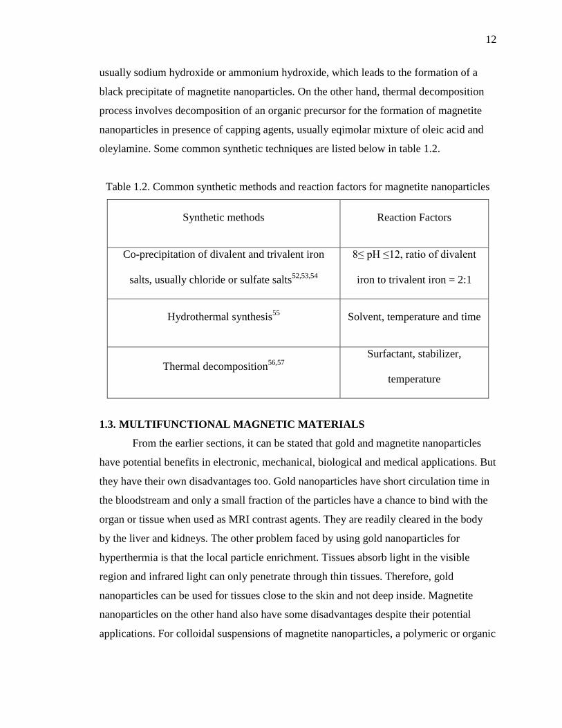
1.2. Common synthetic methods and reaction factors for magnetite nanoparticles ......... 12

1. INTRODUCTION
Nanobiotechnology, the combination of nanotechnology and molecular biology is
a tremendously powerful technology. It holds a huge promise for the design and
development of many types of novel products with potential applications in the fields of
biology and medicine, including early disease detection through advanced noninvasive
medical imaging, treatment through high site-specific drug delivery and protein
purifications. Many metal nanoparticles have been synthesized using various synthetic
methods for this purpose. Among all the candidates investigated, gold nanoparticles have
proven to be extremely useful and reliable candidate. They serve as highly biocompatible
vectors capable of selective and nuclear targeting for site-specific diagnosis and treatment
with extremely low toxicity. Hence, gold nanoparticles were synthesized in different
morphologies including nanoshells, nanorods, tripods, nanocages and nanotubes for
various applications. Despite their numerous advantages, researchers have found some
disadvantages such as the lack of ability to manipulate them externally and low retention
time in the bloodstream.
To overcome these disadvantages and for the potential benefits of multimodal
functionality in biomedical applications, researchers have attached gold with other
magnetic materials. These attached nanomaterials are more precisely described as
multifunctional magnetic nanomaterials, since they exhibit two or more distinctively
different functionalities within a single nanostructure. One of the successful and
promising multifunctional magnetic nanomaterials is the gold-magnetite (Au-Fe3O4)
nanoparticles because magnetite offers high magnetization saturation moment and
susceptibility with low toxic nature. Magnetite nanoparticles by themselves have also
demonstrated their potential for biomedical applications in magnetic resonance imaging
(MRI) and hyperthermia (local heat treatment for destruction of cancer cells). This
research work outlines the synthesis, characterization of gold-magnetite (Au-Fe3O4)
nanoparticles and their conjugation with biomolecules such as L-cysteine and (-)-EGCG
(epigallocatechin-3-gallate).

2
1.1. GOLD NANOPARTICLES: PROPERTIES AND APPLICATIONS
Gold is one of the subjects for most ancient themes of investigation in science.1
Its renaissance now leads to an exponentially increasing number of publications for its
applications in electronic, medical and biological fields, especially in context with the
emerging nanoscience and nanotechnology. Gold nanoparticles are mostly isolated as
stable colloidal solution of clusters of gold atoms in the size range of 1-100 nm. They are
amongst the most stable metal nanoparticles. In this dimensions, gold nanoparticles
exhibit some special optical properties when compared to bulk gold.2,3
For example, the
color changes from yellow to ruby red when bulk gold is converted to gold nanoparticles.
This change in color can be explained using the “surface plasmon resonance” theory
(figure 1.1).
Figure 1.1. Schematic representation of a localized surface plasmon. Reprinted with
permission from reference 4. Copyright 2007 by Annual Reviews.
Surface plasmon resonance5 theory states that when a cluster of gold atoms are hit
by an electromagnetic field, the surface electrons of the gold atoms (occupying the 6s
levels of the gold-atom clusters) present in the conduction band of gold nanoparticles
oscillate back and forth, creating a plasmon band with an absorption peak in the visible
region at 520-550 nm. Gustav Mie proposed an explanation for the phenomenon6 in 1908
which rationalizes the nature of surface plasmon resonance. According to Mie theory, the
total cross section composed of the surface plasmon absorption and scattering is the
summation over all the electric and magnetic oscillations. These surface plasmons were
described quantitatively by solving Maxwell’s equations for spherical particles with
appropriate boundary conditions. Thus, surface plasmon resonance band is absent for

3
gold nanoparticles with size less than 2 nm and greater than 500 nm. The surface plasmon
resonance band maximum and bandwidth are also influenced by the particle shape,
medium dielectric constant and refractive index of the solvent in which gold
nanoparticles are suspended, density of electrons and effective electron mass. For small
spherical particles, the collective oscillation of electrons is called dipole plasmon
resonance of the particle. As the geometry of the nanoparticles changes, these boundary
conditions are no longer valid. Modern day approaches such as discrete dipole
approximation7 can applied to calculate the surface plasmon absorption for arbitrary
geometries. These approaches account for the longitudinal plasmon resonance or
quadrupole plasmon resonance which is in accordance with experimental values. The
increase in intensity and wavelength maximum as the aspect ratio (length divided by
breadth) increases. Figure 1.2 represents the change in absorption spectra as the shape
and size of gold nanoparticles varies.
Figure 1.2. Gold nanoparticles-absorption spectra of various sizes and shapes.
Reproduced from reference 8 with permission of The Royal Society of Chemistry.
Copyright 2005 by Royal Society of Chemistry.

4
Several research groups have fabricated gold nanoparticles with monodispersity
and controlled size by reduction of gold salts in presence of appropriate stabilizers that
prevent particle agglomeration. Electro deposition and physical methods like
sonochemistry, radiolysis have also been employed for their synthesis. Some of the most
common synthetic methods are listed in table 1.1.
Table 1.1. Common synthetic methods and capping agents for gold nanoparticles
Core size
(nm)
Synthetic methods Capping agents
1-2
Reduction of AuCl(PPh3) with diborane or
sodium borohydride.9
Phosphine
(PR3; R= alkyl
groups)
1.5-5
Biphasic reduction of HAuCl4 by sodium
borohydride.10,11
Alkanethiol
10-150 Reduction of HAuCl4 with sodium citrate.12
Citrate
The affinity of gold towards the sulfur head of the thiols and amines make them very
responsive towards biofunctionalization with appropriate biomolecules, leading to
significant medical and biological applications.13,14,15
Figure 1.3 illustrates some of the
biomedical applications of gold nanoparticles and few applications are discussed in detail
later.

5
Figure 1.3. Biomedical applications of gold nanoparticles. Reprinted from reference 14
with permission from Elsevier. Copyright 2008 by Elsevier.
1.1.1. Bio-Sensing. Gold nanoparticles based bio-sensors12
functions by
detecting the change insensitivity of plasmon resonance frequency as it is a very reliable
intrinsic feature. The binding of biomolecules to the surface of gold nanoparticles can
change the plasmon resonance frequency directly. This change in the plasmon resonance
frequency is called plasmon coupling and can be used for colorimetric detection of
analytes. This method was first pioneered by Mirkin et al. who developed an assay for the
detection of DNA.16,17
In this assay, the gold nanoparticles were conjugated with
oligonucleotides containing sticky ends that are complementary to the target sequence
which is to be detected. The initial color of the conjugated gold nanoparticles appeared
red but after hybridization with the target sequence, the colloidal solution appeared
violet-purple. Several other DNA assays have been developed based on this concept for
quantitative analysis and the same concept can be applied for other analytes apart from
DNA. Also, from the change in color one can predict the change in size of the gold
nanoparticles.

6
1.1.2. Hyperthermia. Hyperthermia is a local heat generation technique to kill
cancerous cells by stimulating the nanoparticles with an external stimulus, in this case
light. When gold nanoparticles absorb light, the free electrons on the surface are excited.
This excitation causes collective oscillation of the free electrons. Interaction of these free
electrons with the crystal lattice causes the electrons to relax by transferring the thermal
energy to the lattice. Subsequently, the heat is dissipated to the surrounding
environment.18,19
The ideal average human body temperature is 37°C. Body temperatures
between 37-42°C can lead to fever and above 42°C can be lethal.20
Hyperthermia is based
on this fact and is carried out in two steps. First, gold nanoparticles conjugated with
specific receptors for cancerous cells are enriched in the cancerous tissue (i.e. cell
internalization). Second, the particles are excited by an external stimulus to generate heat
and dissipate to the surroundings for selective killing.
1.1.3. Contrast Based Imaging. Imaging studies are based on the comparisons
of contrast produced by variations in the electron densities from different tissues. Since,
gold nanoparticles have high electron density; they can effectively be used as contrast
enhancing imaging agents. Again, the gold nanoparticles are conjugated with the
appropriate biomolecules to target the specific tissue or organ and administered in to the
body. When the particles are administered and enter the bloodstream, they bind to the
specific organ or tissue through receptor-ligand interactions. The particles bound to the
tissue or organ provides high contrast difference for better imaging. Gold nanoparticles
can be imaged with high signal to noise ratio by X-ray computer tomography with short
exposure time, which helps in reduced damage to the nearby tissues through long
exposed radiation sessions, if needed.21,22
1.1.4. Targeted Drug Delivery. Cells have the ability to ingest nanoparticles
naturally, so nanoparticle incorporation can be specific (via receptor-ligand interaction)
or non-specific.23
After the ingestion, cells store the ingested nanoparticles in
endosomal/lysosomal vesicular structures in them.24
For successful release of the
particles to the cytosol, the nanoparticle surface can be coated with disruptive polymer or
peptide which allows their direct entry in to the cytosol.25,26
Gold nanoparticles can be
modified by conjugation with various drug molecules and coating for release into cytosol
depending on the specific uptake of the targeted cells while retaining their
7
biocompatibility. Generally, they are used in gene therapy27
and anti-cancer drug delivery
to cancerous cells.28
1.2. MAGNETITE NANOPARTICLES: PROPERTIES AND APPLICATIONS
Iron oxide is a generic name given for iron oxides, hydroxides, oxyhyrdoxides
and other related compounds. All together there are 16 different types of compounds
known. Among them, magnetite, Fe3O4 is the most magnetic and naturally abundant
mineral. Loadstone, a naturally magnetized form of magnetite was historically used by
navigators to locate the geographic north. They were also used as pigments in paints and
paleomagnetism (tectonic plate studies). Recently, magnetite nanoparticles have become
important due to their widespread applications such as ferrofluids,29
digital media
recording,30
MRI contrast agents,31
targeted drug delivery,32
magnetic hyperthermia,33
sorbent for wastewater treatment34
etc.
Magnetite has an inverse spinel structure with Fd m space group. The unit cell is
made up of eight formula units. It has oxygen in cubic close packed lattice with Fe3+
occupying the tetrahedral and octahedral sites, where it co-exists with Fe2+
in the
octahedral sites. Thus the formula for magnetite can be precisely written as
tetFe3+
[OctFe3+
Fe2+
]O4. The tetrahedral site is often referred as A site and octahedral site
as B site. There are 16 Fe3+
ions with equal distribution on A and B sites, 8 Fe2+
ions on
the B site and 32 O atoms.
Generally, magnetic materials can be classified as paramagnetic, ferromagnetic,
ferrimagnetic or anitferromagnetic based on the orientation of the magnetic dipoles
within the material in presence of an external magnetic field (figure 1.4). Magnetic
behavior of a material is strongly dependent on size of the material, temperature and the
applied magnetic field. Bulk magnetite is a ferrimagnetic material, however magnetite
nanoparticles exhibit superparamagnetism.35,36
It is the nano-size effect of ferro or
ferrimagnetism. Below a critical size, a ferro or ferrimagnetic material is reduced to a
state which consists of single magnetic domain and with no internal domain boundaries.
It is in a state of unified magnetization within the particle at any applied field. The
direction of collective magnetization of an ensemble can rotate thermally and behave as a
superparamagnet with a very large moment (figure 1.5).

8
Figure 1.4. Magnetic ordering in (a) paramagnetic, (b) ferromagnetic, (c) anti-
ferromagnetic and (d) ferrimagnetic materials.
Figure 1.5. Schematic representation of magnetic ordering in bulk ferromagnet material
and single particle domain (in absence and presence of external magnetic field).

9
Frenkel and Dorfman37
were the first to predict this behavior. Later, Kittel38
and
others estimated the critical dimension for a spherical particle of common ferromagnetic
materials. In 1949, Neel pointed out that if the single domain particles were small
enough, thermal fluctuations could cause its direction of magnetization to undergo a sort
of Brownian motion and also derived an equation for the particles to arrive at thermal
equilibrium in a given time. In 1959, Bean and Livingston39
explained
superparamagnetism using a model system of spherical particles with uniaxial anisotropy
that were first fully magnetized along the easy axis of symmetry. By considering
spherical particles, the magnetic anisotropy can be approximated to be proportional to the
particle volume, V. To approach zero remanence corresponding to thermal equilibrium, a
sufficient number of particles must be reversed by thermal activation. The energy barrier
that separates easy magnetization axes is the magnetic anisotropy energy, KvV, where Kv
is the volume anisotropy constant (figure 1.6). For nanoparticles, the particle size is small
and the magnetic anisotropy energy is comparable with thermal energy (equation 1.1).
Thus, the magnetic moment of the particle may fluctuate behaving as a superparamagnet
but with a total moment exceeding that of the bulk by approximately 1000 times.
…(1.1)
Now applying the Boltzmann distribution for the total magnetization of the
particles ensemble when subjected to external applied field and thermal equilibrium can
be given by Langevin equation (equation 1.2).
[ (
)
(
)] …(1.2)
Where M = magnetization, M0 = saturation magnetization, H = applied magnetic field, µ
= magnetic moment, T = temperature and kB = Boltzmann constant.
Besides, the dependence of magnetization on particle size, shape and composition
it greatly depends on temperature also. The critical temperature above which ferro or
ferrimagnetic particle will behave as superparamagnetic, a state where spontaneous
fluctuations of the particle moment are allowed causing loss of magnetic ordering is
called as blocking temperature (TB)39,40
and can be predicted by equation 1.3 i.e. thermal
energy is equal to the magnetic anisotropy energy.
…(1.3)

10
Figure 1.6. Schematic representation of the magnetic energy versus angle between easy
axis of magnetization and applied magnetic field.
In 1939, Verwey41
observed a phase transition (cubic to triclinic) in magnetite
below 120 K due to first order transition. The resistivity of magnetite increased by a
factor of the order 100 due to this transformation. This was later explained by charge
ordering of divalent and trivalent ions on the octahedral sites in alternation layers.42
This
temperature at which phase transformation occurs is called Verwey temperature (TV).
When the particle size reduces from bulk to nanoparticle regime, the Verwey temperature
decreases or disappears.
Superparamagnetic particles by virtue of their unique magnetic properties show
usefulness in avariety of diverse applications. Superparamagnetic magnetite nanoparticles
are being used for the following applications due to high magnetization saturation
moment and low toxicity.
1.2.1. Ferrofluid. Ferrofluids29,43
are colloidal suspensions of permanently
magnetized nanoparticles in a carrier fluid, usually organic solvent or water with no long
range ordering. Brownian motion keeps them from settling and the influence of gravity
and the coating around these particles provide short range steric repulsions which prevent
particle agglomeration under non uniform magnetic field. Ferrofluid was first invented by
NASA’s Steve Papella44
as a low density and low viscosity magnetic propellant usable
under zero gravity conditions. These ferrofluids also have potential applications in
11
micro/nanoelectromechanical systems (MEMS/NEMS), analytical and medical devices,
sealing, heat transfer, bearing etc.
1.2.2. Wastewater Remediation. Industrial wastewater contains hexavalent
chromium compounds that usually exist as chromate (CrO42-
) and dichromate (Cr2O72-
).
They are highly toxic agents that act as carcinogens and mutagens. So their treatment is
very important and generally carried out by applying a reducing agent such as ferrous
sulfate. This method consumes large quantities of reagents, utilizes large volume sludge
treatment reactors and is a high cost operation. Whereas using magnetite nanoparticles
overcomes these problems with additional advantages. The advantages include (i)
utilization of minimal quantity of nanoparticles, (ii) higher adsorption capacity of
nanoparticles due to larger surface area, (iii) regeneration of the nanoparticles by
desorption technique is easy and faster, (iv) ease of separation from treated water by
using external magnetic field.45,46,47
1.2.3. Biological Applications. Similar to gold nanoparticles, the surface of the
magnetite nanoparticles can be functionalized and can be used for various biological
applications such as magnetic fluid hyperthermia, MRI, targeted drug delivery etc.
Researchers have used the magnetite nanoparticles conjugated with TAT protein derived
peptide sequence to tag cells magnetically for effective imaging. Another promising
application of these magnetite nanoparticles is in drug delivery. Widder et al.
demonstrated the usefulness of magnetic albumin microspheres in animal tumor
models.48
These modified microspheres achieved significantly greater responses than
adriamycin alone. Gallo et al. have later demonstrated the same in normal rats.49
In their
work, they report that the retention time of these microspheres was up to 72 hours. Other
researcher groups have also proved magnetite nanoparticles effective use in DNA
analysis, cell separations, cell specific targeting and hyperthermia.33,50,51
Significant efforts have been devoted in the synthesis of magnetite nanoparticles
with different characteristics for various applications. Some of the common methods
include hydrothermal treatment, thermal decomposition, co-precipitation, sonochemical
and solvothermal.50,51
Among them, thermal decomposition and co-precipitation are the
most common techniques adopted by researchers. The co-precipitation process involves
the precipitation of iron precursors Fe2+
and Fe3+
in the ration of 1:2 by using an alkali,
12
usually sodium hydroxide or ammonium hydroxide, which leads to the formation of a
black precipitate of magnetite nanoparticles. On the other hand, thermal decomposition
process involves decomposition of an organic precursor for the formation of magnetite
nanoparticles in presence of capping agents, usually eqimolar mixture of oleic acid and
oleylamine. Some common synthetic techniques are listed below in table 1.2.
Table 1.2. Common synthetic methods and reaction factors for magnetite nanoparticles
Synthetic methods Reaction Factors
Co-precipitation of divalent and trivalent iron
salts, usually chloride or sulfate salts52,53,54
8≤ pH ≤12, ratio of divalent
iron to trivalent iron = 2:1
Hydrothermal synthesis55
Solvent, temperature and time
Thermal decomposition56,57
Surfactant, stabilizer,
temperature
1.3. MULTIFUNCTIONAL MAGNETIC MATERIALS
From the earlier sections, it can be stated that gold and magnetite nanoparticles
have potential benefits in electronic, mechanical, biological and medical applications. But
they have their own disadvantages too. Gold nanoparticles have short circulation time in
the bloodstream and only a small fraction of the particles have a chance to bind with the
organ or tissue when used as MRI contrast agents. They are readily cleared in the body
by the liver and kidneys. The other problem faced by using gold nanoparticles for
hyperthermia is that the local particle enrichment. Tissues absorb light in the visible
region and infrared light can only penetrate through thin tissues. Therefore, gold
nanoparticles can be used for tissues close to the skin and not deep inside. Magnetite
nanoparticles on the other hand also have some disadvantages despite their potential
applications. For colloidal suspensions of magnetite nanoparticles, a polymeric or organic

13
coating is indispensable. Also, unlike gold nanoparticles, magnetite nanoparticles cannot
bind with a wide range of biomolecules.13,50
Hence, to overcome these problems researchers have devoted significant amount
of time and hard work to attach gold with magnetite nanoparticles using different
synthetic techniques. Several groups have reported the formation of gold-magnetite
nanoparticles in different morphologies such as core-shell, core-hollow shell, dumbbell
shape, nanoflower etc.58,59,60
S. Sun et al. have proposed a mechanism for the formation
of these multifunctional nanomaterials.58
They state that if a polar solvent is used in the
synthesis process, the electron deficiency on the gold is replenished by the solvent itself
which allows multiple nucleation sites to be formed. And as the process continues, the
nucleation lobes grow, connect and eventually form a shell on the gold surface. If the
synthesis process involves the utilization of non-polar solvent, once a single nucleated
site depletes the electrons from gold, the electron deficiency cannot be replenished in the
reaction i.e. solvent preventing the formation of new nucleation site on the gold surface.
This process eventually results in formation of dumbbell shaped nanoparticles.
Figure 1.7. Schematic representation showing the mechanism of formation of (a) core
shell nanoparticle (polar solvent) and (b) dumbbell shaped nanoparticles (non-polar
solvent). Reused from reference 58 with permission from Wiley. Copyright 2008 by
Wiley-VCH GmbH & Co. KGaA, Weinheim.

14
Multicomponent nanomaterials integrate several functionalities in a single particle
ensemble. Hence, the plasmonic, magnetic and magneto-optical properties change when
compared to the individual components alone. In the earlier section, it was stated that
gold nanoparticles exhibit strong surface plasmon resonance absorption. These
multicomponent nanomaterials also exhibit surface plasmon resonance absorption but it
is significantly red shifted. The reason for this behavior can be given using Mie theory.
Surface plasmon resonance depends on the refractive index, dielectric constant etc., and
the attachment of magnetite to the gold alters these parameters causing the red shift in the
absorption peak. Second, the magnetic properties of multicomponent nanomaterial also
changes. The observed changes include decreased magnetization saturation, increased
coercivity and decreased remanence ratio. This change in behavior can be explained by a
new surface anisotropy induced by the attachment: Fe atoms at the interface have reduced
number of nearest neighbors, which in turn decreases the interatomic exchange coupling.
The new uniaxial anisotropy dominates the original cubic anisotropy i.e. the non-
magnetic gold acts as a template blocking the spins at the interface.
1.4. RESEARCH PROBLEM
In context with the previous section topics it can be said that multifunctional
magnetic nanomaterials are very important in biological and medical applications. The
research problem for this thesis is the synthesis of multifunctional gold-magnetite
nanoparticle-biomolecule conjugates with higher magnetization saturation moment and
lower toxicity compared to earlier reported values, using novel synthesis routes. These
bifunctional gold-magnetite nanoparticles were synthesized using one pot hot injection
method. Further, the thesis also focuses on the validation of these nanoparticles and their
conjugation with biomolecules like L-cysteine and (-)-EGCG using various microscopic
and spectroscopic techniques such as SEM, TEM, Mössbauer and SQUID magnetization
studies, Raman and FTIR, etc. The upcoming chapters deal with the experimental
procedures followed and their results.
15
PAPER
I. SUPERPARAMAGNETIC Au-Fe3O4 NANOPARTICLES: ONE-POT
SYNTHESIS, BIOFUNCTIONALIZATION AND TOXICITY EVALUATION
A Pariti1, P Desai
2, S K Y Maddirala
2, X Liang
1, N Ercal
2, K V Katti
3 and M
Nath2*
1Department of Chemical Engineering, Missouri University of Science and
Technology, Rolla, MO 65409, U.S.A.
2Department of Chemistry, Missouri University of Science and Technology,
Rolla, MO 65409, U.S.A.
3Departments of Radiology and Physics, University of Missouri, Columbia, MO
65212, U.S.A.
*Email: [email protected]
ABSTRACT
Superparamagnetic Au-Fe3O4 bifunctional nanoparticles have been synthesized using a
single step hot-injection precipitation method. The synthesis involved using Fe(CO)5 as
iron precursor and HAuCl4 as gold precursor in presence of oleylamine and oleic acid.
Oleylamine helps in reducing Au3+
to Au0 seeds which simultaneously oxidizes Fe(0) to
form Au-Fe3O4 bifunctional nanoparticles. Triton® X-100 was employed as a highly
viscous solvent to prevent agglomeration of Fe3O4 nanoparticles. Detailed
characterization of these nanoparticles was performed by using X-ray powder diffraction,
transmission electron microscopy, scanning tunneling electron microscopy, UV-visible
spectroscopy, Mössbauer and magnetometry studies. To evaluate these nanoparticles
applicability in biomedical applications, L-cysteine was attached to the Au-Fe3O4
nanoparticles and cytotoxicity of Au-Fe3O4 nanoparticles was tested using CHO cells by
employing MTS assay. L-cysteine modified Au-Fe3O4 nanoparticles were qualitatively

16
characterized using Fourier transform infrared spectroscopy and Raman spectroscopy;
and quantitatively using acid ninhydrin assay. Investigations reveal that that this approach
yields Au-Fe3O4 bifunctional nanoparticles with an average particle size of 80 nm.
Mössbauer studies indicated the presence of Fe in Fe3+
in A and B sites (tetrahedral and
octahedral, respectively) and Fe2+
in B sites (octahedral). Magnetic measurements also
indicated that these nanoparticles were superparamagnetic in nature due to Fe3O4 region.
The saturation magnetization for the bifunctional nanoparticles was observed to be ~74
emu/g which is significantly higher than the previously reported Fe3O4 nanoparticles.
Mössbauer studies indicated that there was no significant Fe(0) impurity that could be
responsible for the superparamagnetic nature of these nanoparticles. None of the
investigations showed any presence of other impurities such as Fe2O3 and FeOOH. These
cysteine functionalized Au-Fe3O4 bifunctional nanoparticles showed no significant
cytotoxicity to the CHO cells up to 48 h even at concentrations of 1 mg/ml making them
suitable for biomedical applications such as local heat generators (hyperthermia) for
cancer treatment and drug delivery vehicles.
1. INTRODUCTION
Multifunctional nanomaterials have recently attracted the attention of the materials
science community owing to their vast applicability in a diverse range of applications [1-
6]. One of the most promising applications for these multifunctional nanomaterials is
related to the biomedical field where they have been used as photothermal killing of
cancerous cells [7], magnetic resonance and fluorescence imaging [8-11], cell targeting
and sorting [12], and drug delivery [13-15]. In these multifunctional nanostructures
which are made by fusion of two entirely different materials with diverse properties into
one single nanostructure, Au has been the preferred choice as one of the components.
Traditionally, Au nanoparticles have demonstrated their importance in biological and
medicinal applications, including immuno-sensing [16], phagokinetic studies [17], as
carrier vehicles for delivery of nucleic acids via covalent and non-covalent conjugation
[18], labelling and cell visualization by photothermal or photo acoustic methods [19],
separation and purification of biomolecules, hyperthermia agents (local heat generation
for tumor destruction), contrast enhancer, tissue engineering and highly selective bio-
sensors[16-18]. The reason for their extensive applications in biology and medicine is not
17
only because of their robust interaction with biomolecules containing thiol and disulfide
functional groups but also due to their unique optical properties. However, despite the
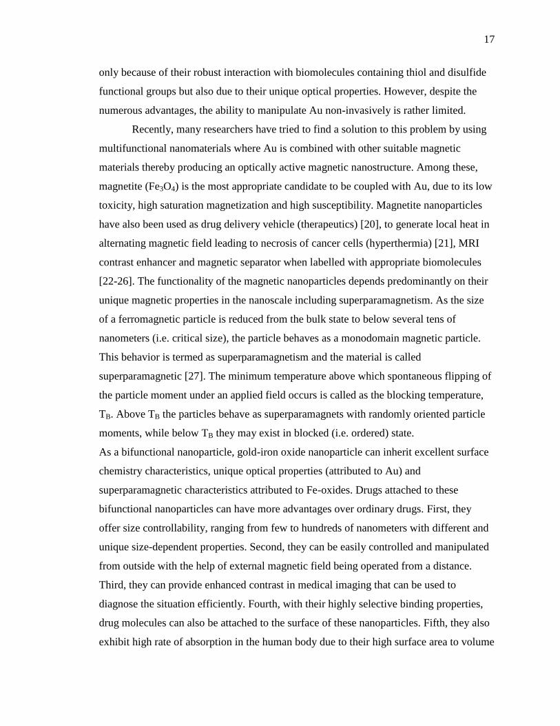
numerous advantages, the ability to manipulate Au non-invasively is rather limited.
Recently, many researchers have tried to find a solution to this problem by using
multifunctional nanomaterials where Au is combined with other suitable magnetic
materials thereby producing an optically active magnetic nanostructure. Among these,
magnetite (Fe3O4) is the most appropriate candidate to be coupled with Au, due to its low
toxicity, high saturation magnetization and high susceptibility. Magnetite nanoparticles
have also been used as drug delivery vehicle (therapeutics) [20], to generate local heat in
alternating magnetic field leading to necrosis of cancer cells (hyperthermia) [21], MRI
contrast enhancer and magnetic separator when labelled with appropriate biomolecules
[22-26]. The functionality of the magnetic nanoparticles depends predominantly on their
unique magnetic properties in the nanoscale including superparamagnetism. As the size
of a ferromagnetic particle is reduced from the bulk state to below several tens of
nanometers (i.e. critical size), the particle behaves as a monodomain magnetic particle.
This behavior is termed as superparamagnetism and the material is called
superparamagnetic [27]. The minimum temperature above which spontaneous flipping of
the particle moment under an applied field occurs is called as the blocking temperature,
TB. Above TB the particles behave as superparamagnets with randomly oriented particle
moments, while below TB they may exist in blocked (i.e. ordered) state.
As a bifunctional nanoparticle, gold-iron oxide nanoparticle can inherit excellent surface
chemistry characteristics, unique optical properties (attributed to Au) and
superparamagnetic characteristics attributed to Fe-oxides. Drugs attached to these
bifunctional nanoparticles can have more advantages over ordinary drugs. First, they
offer size controllability, ranging from few to hundreds of nanometers with different and
unique size-dependent properties. Second, they can be easily controlled and manipulated
from outside with the help of external magnetic field being operated from a distance.
Third, they can provide enhanced contrast in medical imaging that can be used to
diagnose the situation efficiently. Fourth, with their highly selective binding properties,
drug molecules can also be attached to the surface of these nanoparticles. Fifth, they also
exhibit high rate of absorption in the human body due to their high surface area to volume

18
ratio. These characteristics would further enhance and broaden the application of these
nanoparticles for theranostic applications.
Given such importance of the gold-iron oxide nanoparticles in theranostic
applications, there have been considerable efforts directed towards synthesis of these
bifunctional nanoparticles. Reported synthesis protocols for Au-Fe3O4 include
decomposition of iron precursors (e.g. iron acetylacetonate) on gold nanoparticle seeds,
reduction of Au3+
on iron oxide nanoparticles with porous silica shell, by chemical bond
linkage using intermediary molecules to form core-shell, core-hollow shell, and
dumbbell-like nanostructures [28-34]. However, most of these methods were multi-step
processes, which might be detrimental for large scale synthesis of these potentially
transformative nanoparticles. Hence, there is still a need for producing these gold-iron
oxide nanoparticles through simpler reactions involving less number of steps and more
biocompatible precursors.
In this article we report a facile one-pot synthesis of bifunctional Au-Fe3O4
superparamagnetic nanoparticles and their biofunctionalization with simple amino acid
like cysteine. These bifunctional nanoparticles were synthesized in a single step from
reaction between HAuCl4 and Fe(CO)5 in presence of oleic acid and oleylamine.
Magnetic and optical properties of these nanoparticles were studied in details. Cell
viability and toxicity studies were also carried out on these nanoparticles which revealed
that these were not toxic to CHO cells even at moderately high exposure. The
superparamagnetic nature with high saturation magnetization and very low toxicity
makes these Au-Fe3O4 nanoparticles very suitable for theranostic applications.
2. EXPERIMENTAL SECTION
2.1. Materials. Iron pentacarbonyl (Fe(CO)5), oleylamine, ninhydrin,
Dulbecco’s Modified Eagle Medium (DMEM) / Nutrient mixture F-12 Ham culture
media, penicillin streptomycin sterile solution and L-cysteine were purchased from
Sigma Aldrich. Oleic acid, acetic acid and phosphoric acid were purchased from Fischer
Scientific. Triton® X-100 and hydrogen tetrachloroaurate (III) trihydrate
(HAuCl4·3H2O) were purchased from Acros Organics. Fetal bovine serum (FBS) was
purchased from Atlanta Biologicals. MTS assay dye was purchased from Promega.
19
Solutions were prepared according to standard laboratory procedures. All chemicals used
were reagent grade and were used as supplied.
2.2. Synthesis of bifunctional Au-Fe3O4 nanoparticles. The synthesis of
Au-Fe3O4 nanoparticles was carried out in air by simultaneous addition of Fe(CO)5 and
HAuCl4 with excess oleylamine (OLAM) and oleic acid (OLAC). 5 mL Triton® X-100
was added to a three neck round bottom flask equipped with a magnetic stir bar and air
condenser. The solution was heated to 85 ºC. 2.5 mM of Fe(CO)5, 0.25 mM of HAuCl4,
2.5 mM OLAC, and 2.5 mM of OLAM were injected in this hot solution. The
temperature was then ramped to 300 ºC. Upon injection of the Au and Fe-precursors, the
solution turned black with rapid evolution of gases. After 10 min the gases subsided and
the black solution was allowed to reflux for 1 h. After 1 h, heating was stopped and the
reaction was cooled to room temperature.
2.3. Purification of crude product. The product was isolated from the reaction
mixture by washing and centrifugation at least 3-4 times with ethanol using
ultrasonication to remove excess Triton® X-100 and any unreacted precursors. The
powder collected at the bottom of the centrifuge tube was dried in air. The black powder
obtained after drying was characterized further through powder X-ray diffraction
(PXRD), scanning and transmission electron microscopy (SEM and TEM, respectively),
energy dispersive spectroscopy (EDS), UV-Vis, Mössbauer and magnetometry studies.
Millipore water was used throughout to disperse the nanoparticles as needed for
characterization as well as further studies.
2.4. Characterization of bifunctional Au-Fe3O4 nanoparticles.
PXRD: As-synthesized nanoparticles was ground and used for PXRD, which was carried
out on Philips Xpert diffractometer scanning from 5 º to 90 º.
TEM & SEM: TEM images were taken by using Tecnai F20 transmission electron
microscope with FEG at an accelerating voltage of 200kV. A dual beam Helios 600 Nano
was used for SEM and STEM studies. Samples for TEM and STEM studies were made
by dispersing as-synthesized nanoparticles in ethanol by ultrasonication for 2 h and
adding drops from the diluted dispersion on a carbon coated 300 mesh Cu grid followed
by drying in air overnight.

20
Optical studies: The UV-vis absorption studies were carried out with Cary 50 UV-vis
spectrophotometer.
Magnetic characterization: Temperature dependent magnetic moment at constant field
and field dependent isothermal magnetization measurements were performed with
SQUID (Superconducting quantum interference device) magnetometer. The powdered
sample of a known mass (8.8 mg) was loaded in a gel cap, which was inserted into the
magnetometer with the help of standard sample loader. Background signal was collected
from the diamagnetic gel cap separately and subtracted from the sample signal. Zero field
cooled (ZFC) data was collected after cooling down the sample under zero magnetic
field, and them recording magnetization as a function of warming temperature under an
applied field of 100 Oe. The field cooled data (FC) on the other hand, was collected by
cooling down the sample under an applied magnetic field and simultaneously recording
the sample magnetization as a function of decreasing temperature. The isothermal
magnetization at various temperatures (5 K, 100 K and 300 K) was collected by varying
applied magnetic field from -20000 Oe to 20000 Oe and recording the change in sample
magnetization.
Mössbauer spectroscopy: The transmission 57
Fe Mössbauer spectrum of the powdered
sample was recorded at room temperature using a gamma-ray source of 57
Co in a
Rhodium matrix. The spectrum data was fitted using Lorentzian function in RECOIL
software [35]. Isomer shifts and quadruple splitting are given with respect to α-Fe foil at
298 K.
2.5. Preparation of L-cysteine modified bifunctional Au-Fe3O4 nanoparticles.
Cysteine solution of concentration 0.5 mg/mL was prepared by dissolving 50 mg of L-
cysteine in 100 mL Millipore water. 10 mL of this solution and 20mg of as-synthesized
Au-Fe3O4 nanoparticles were taken into a flat-bottomed conical flask with glass stopper.
The flask was sealed with parafilm and placed on an orbital shaker for 24hrs at 150 rpm.
After incubation, the nanoparticles were separated by attraction to a magnet and 0.5 mL
from the supernatant was taken for acid ninhydrin assay [36]. The magnetically separated
nanoparticles were further dried and characterized by FTIR and Raman spectroscopic
studies.

21
2.6. Characterization of L-cysteine modified bifunctional Au-Fe3O4
nanoparticles. IR spectra were acquired with a Thermo Nicolet Nexus 470 FTIR
spectrometer. The functionalized nanoparticle powder was intimately mixed with dry
KBr into a homogenous fine powder with a mortar-pestle. Pellets were made from this
homogenized mixture by compression using Carver Press at 15000 psi. The spectra were
collected over a range of 400 – 4000 cm-1
. Horiba Jobin Yvon Lab Raman ARAMIS
model was used to perform Raman microspectroscopy on the functionalized
nanoparticles. The laser used was He-Ne with a power of about 1.7 mW over a range of
100 – 2000 cm-1
. The spectra were iterated over an average of 25 scans.
2.7. Acid ninhydrin assay. Acid ninhydrin assay was performed according to
the literature as an instant method of L-cysteine detection. The reaction mixture for
standard containing 0.5 mL of L-cysteine solution (0 – 0.5 mg/mL), 0.5mL of acetic acid
and 0.5mL of acid ninhydrin reagent was mixed thoroughly. The test tubes were covered
with caps and heated in boiling water for 10 min. They were rapidly cooled under
running tap water, and the contents were diluted to 5 mL using 95% ethanol. A blank
containing only sample solution from the supernatant without L-cysteine was also
prepared under the same conditions. The absorbance standard plot was calculated at 560
nm with Shimadzu UV 1700 spectrophotometer. Acid ninhydrin reagent was prepared by
dissolving 250 mg of ninhydrin in a mixture of 6 mL of acetic acid and 4 mL of 0.6 M
phosphoric acid. The reagent was stable for 2 weeks at 4 ºC.
2.8. Cell culture. CHO cells were cultured in Dulbecco’s Modified Eagle
Medium (DMEM) / Nutrient mixture F-12 Ham culture media supplemented with 10%
fetal bovine serum (FBS) and 1% penicillin/streptomycin. Cell cultures were incubated at
37 ºC in a humidified atmosphere of 5% CO2 and 95% air.
2.9. Cytotoxicity. Cell viability was assessed by the MTS assay (CellTiter 96®
AQueous One Solution Cell Proliferation Assay), based on the conversion of a
tetrazolium compound, 3-(4,5-dimethylthiazol-2 yl)-5-(3-carboxymethoxyphenyl)-2-(4-
sulfophenyl)-2H tetrazolium (MTS), to a colored formazan product by living cells [37].
Absorbance was read by a microplate reader at 490 nm. The quantity of formazan
product, as measured by the amount of absorbance, was directly proportional to the
number of viable cells in the culture.

22
10,000 CHO cells were seeded in each well of 96-well plates. After 24 h, the
medium was then discarded and replaced with serum free fresh medium containing the
Au-Fe3O4 nanoparticles at different concentrations for 48 hr. CellTiter 96® AQueous One
reagent (20 µl/well) was added to each well and the plate was incubated for 1.5 h at 37 ºC
in a humidified atmosphere of 5% CO2 and 95% air, and then centrifuged to get rid of
nanoparticles. The MTS formazan product was measured by determining the absorbance
of the supernatant (100 µL) at 490 nm using a 96-well plate reader (FLUOstar, BMG
Labtechnologies, Durham, NC, USA). The relative cell viability (%) related to control
wells containing culture medium without nanoparticles was calculated by [A]test/[A]control
× 100.
3. RESULTS AND DISCUSSION
Figure 1. PXRD pattern of Au-Fe3O4 bifunctional nanoparticles synthesized at 300°C
Figure 1 shows the formation of highly crystalline Au-Fe3O4 nanoparticles confirmed by
PXRD pattern, which corroborated very well with the standard patterns of Fe3O4 with
inverse spinel structures(JCPDS 04-007-2718) and Au (JCPDS 00-004-0784). The PXRD
pattern was very clean and did not show any impurity peaks corresponding to other iron
oxide phases like Fe2O3 and FeOOH. From Schrerrer equation the size of these particles
was calculated to be 75 nm with the Fe3O4 region being around 60 nm while the Au part

23
was around 15 nm in average [38]. The size of nanoparticles could be tuned by
controlling the time at which HAuCl4 was injected, presence of OLAM in the reaction
mixture (excess OLAM led to bigger gold particles), reflux time and the HAuCl4/Fe3O4
ratio. Specifically, injecting Fe(CO)5 in large excess compared to HAuCl4 (~20:1 Fe:Au
molar ratio) led to overgrowth of Fe3O4 part while refluxing the reaction mixture for
longer time resulted in agglomeration of Au-Fe3O4 particles. Presence of OLAM helps in
reduction of Au+3
to Au0 onto the Fe3O4 surface. The role of Triton® X-100 was more as
a highly viscous solvent which inhibited overgrowth of the Fe3O4 part and changing the
concentration of Triton® X-100 had very minimal effect on the morphology of the Au-
Fe3O4 nanoparticles.
Figure 2. (A) TEM image of the bifunctional nanoparticles showing decoration of Fe3O4
with Au. (B) HRTEM showing the attachment of Au to Fe3O4 and the crystallinity of the
individual regions. Lattice fringes from the Au and Fe3O4 regions corresponds to <111>
and <311> planes, respectively.
The morphology and composition of the bifunctional nanoparticles were
characterized by transmission electron microscopy (TEM) and scanning electron
microscopy (SEM). Investigations from TEM and SEM showed a high yield of
bifunctional nanoparticles in the product with uniform particle size distribution. Figure
2(A) shows a typical distribution of the Au-Fe3O4 nanoparticles where each Fe3O4 region
is decorated with several Au dots. Typically the Fe3O4 regions (lighter contrast in the

24
images) were 80 nm while the Au part (darker contrast) was approximately 20 nm.
Several wide area SEM images were obtained to calculate the average particles size. The
size of the Au and Fe3O4 particles ranged from 5-20 nm and 50-80 nm, respectively, with
average bifunctional particle size around 80 nm. This kind of morphology is slightly
different than the dumbbell shaped particles and is closer to multifaceted Janus particles
[39, 40]. This kind of morphology (Au-decorated Fe3O4) gives the added advantage that
both Au and Fe3O4 functionalities are available for full utilization of their potential,
while, several Au dots on the Fe3O4 host ensures that there are maximum number of spots
for molecular recognition since Au-terminal acts as anchor for the aptamers used as
targeting agents. Au-Fe3O4 nanoparticles were highly crystalline as revealed by the high
resolution TEM (HRTEM) image shown in figure 2(B). The HRTEM image exhibited
lattice fringes corresponding to (111) planes of Au and (311) lattice planes of Fe3O4.
Through these HRTEM images we also looked in detail at the interfaces between the Au
and Fe3O4 regions. The interface in these nanoparticles was very clean and well-defined
with minimal mixing of the Au and Fe3O4 phases. There was no fuzziness or presence of
any other crystalline phase at the interface. Interfacial surface can be very important in
these superparamagnetic nanoparticles as depending on the composition on both sides of
the interface they might lead to exchange bias magnetic interactions leading to
interparticle interaction and ordering.
Figure 3. Absorption spectra of the bifunctional Au-Fe3O4 nanoparticles and Fe3O4
nanoparticles
25
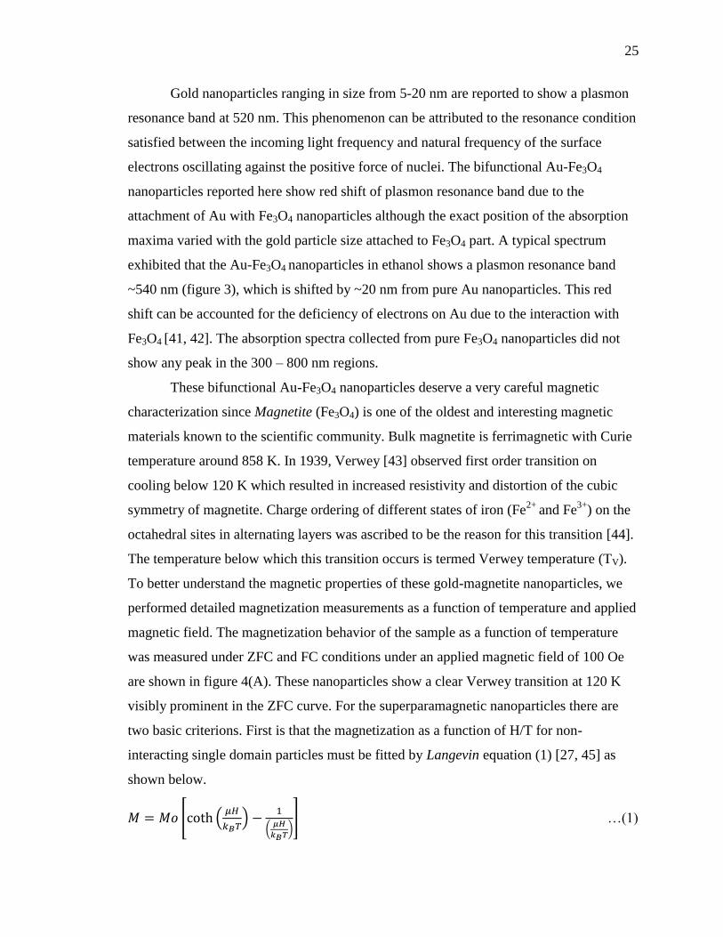
Gold nanoparticles ranging in size from 5-20 nm are reported to show a plasmon
resonance band at 520 nm. This phenomenon can be attributed to the resonance condition
satisfied between the incoming light frequency and natural frequency of the surface
electrons oscillating against the positive force of nuclei. The bifunctional Au-Fe3O4
nanoparticles reported here show red shift of plasmon resonance band due to the
attachment of Au with Fe3O4 nanoparticles although the exact position of the absorption
maxima varied with the gold particle size attached to Fe3O4 part. A typical spectrum
exhibited that the Au-Fe3O4 nanoparticles in ethanol shows a plasmon resonance band
~540 nm (figure 3), which is shifted by ~20 nm from pure Au nanoparticles. This red
shift can be accounted for the deficiency of electrons on Au due to the interaction with
Fe3O4 [41, 42]. The absorption spectra collected from pure Fe3O4 nanoparticles did not
show any peak in the 300 – 800 nm regions.
These bifunctional Au-Fe3O4 nanoparticles deserve a very careful magnetic
characterization since Magnetite (Fe3O4) is one of the oldest and interesting magnetic
materials known to the scientific community. Bulk magnetite is ferrimagnetic with Curie
temperature around 858 K. In 1939, Verwey [43] observed first order transition on
cooling below 120 K which resulted in increased resistivity and distortion of the cubic
symmetry of magnetite. Charge ordering of different states of iron (Fe2+
and Fe3+
) on the
octahedral sites in alternating layers was ascribed to be the reason for this transition [44].
The temperature below which this transition occurs is termed Verwey temperature (TV).
To better understand the magnetic properties of these gold-magnetite nanoparticles, we
performed detailed magnetization measurements as a function of temperature and applied
magnetic field. The magnetization behavior of the sample as a function of temperature
was measured under ZFC and FC conditions under an applied magnetic field of 100 Oe
are shown in figure 4(A). These nanoparticles show a clear Verwey transition at 120 K
visibly prominent in the ZFC curve. For the superparamagnetic nanoparticles there are
two basic criterions. First is that the magnetization as a function of H/T for non-
interacting single domain particles must be fitted by Langevin equation (1) [27, 45] as
shown below.
[ (
)
(
)] …(1)

26
where M = magnetization, M0 = saturation magnetization, H = applied magnetic moment,
µ = magnetic moment, T = temperature and kB = Boltzmann constant. The plot of M
versus H/T at 5 K, 100 K and 300 K converge into a single universal curve and could be
fitted using Langevin equation with R2=0.9981 as shown in figure 4(B). Second
condition for superparamagnetic particles is the anhysteretic nature of isothermal
magnetization against applied magnetic field with zero coercivity, which is also
temperature independent above blocking temperature (TB).
Figure 4. (A) ZFC and FC curves of the Au-Fe3O4 nanoparticles under an applied field of
100 Oe. (B) M vs H plots at 5 K, 100 K and 300 K. (C) The Langevin fit at 300 K. (D) M
vs H plots at 5 and 100 K magnified to show the coercive fields.
Figure 4(C) shows the isothermal magnetization curves as a function of applied
magnetic field at 5 K, 100 K and 300 K for the bifunctional nanoparticles. The presence
of coercivity was very apparent at 5K and 100K. At 300 K on the other hand, the M vs H
was anhysteretic in nature typical for a superparamagnetic nanoparticle, as shown in
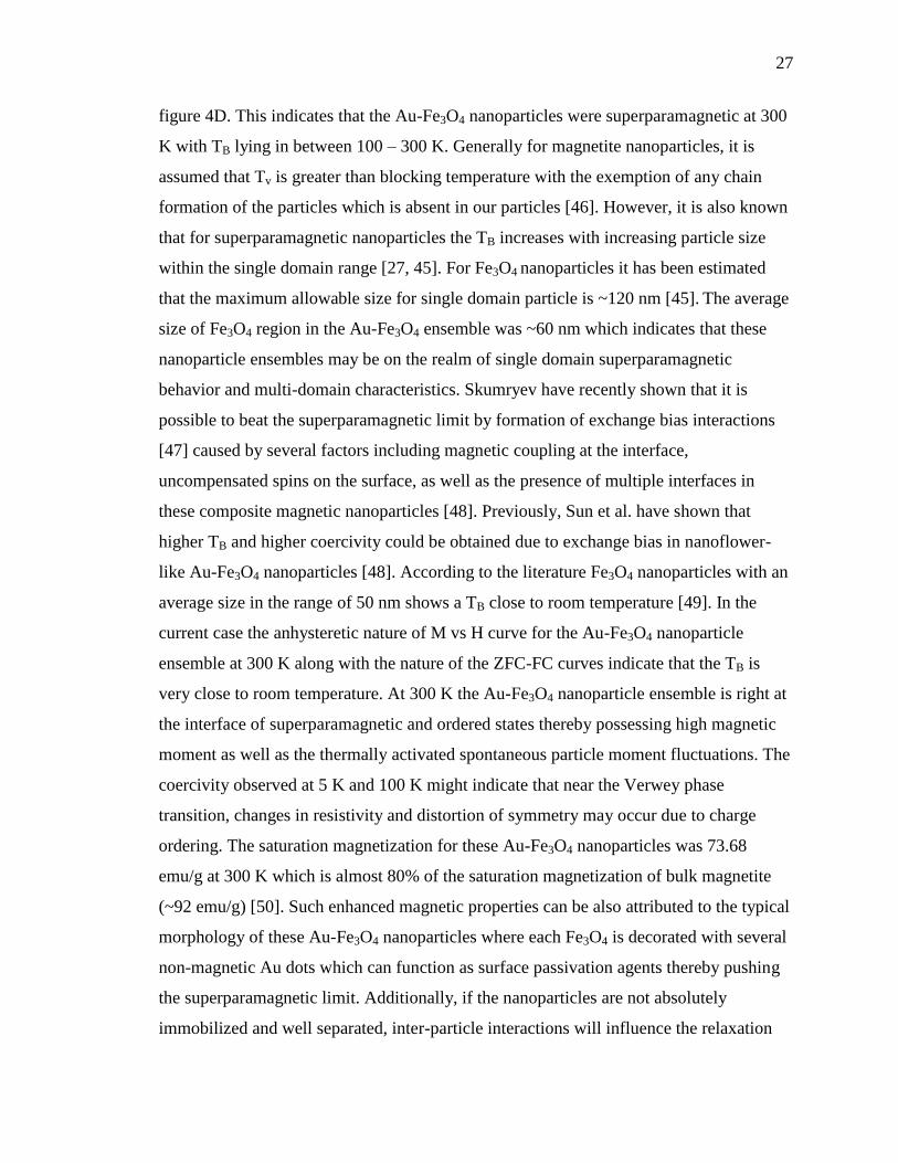
27
figure 4D. This indicates that the Au-Fe3O4 nanoparticles were superparamagnetic at 300
K with TB lying in between 100 – 300 K. Generally for magnetite nanoparticles, it is
assumed that Tv is greater than blocking temperature with the exemption of any chain
formation of the particles which is absent in our particles [46]. However, it is also known
that for superparamagnetic nanoparticles the TB increases with increasing particle size
within the single domain range [27, 45]. For Fe3O4 nanoparticles it has been estimated
that the maximum allowable size for single domain particle is ~120 nm [45]. The average
size of Fe3O4 region in the Au-Fe3O4 ensemble was ~60 nm which indicates that these
nanoparticle ensembles may be on the realm of single domain superparamagnetic
behavior and multi-domain characteristics. Skumryev have recently shown that it is
possible to beat the superparamagnetic limit by formation of exchange bias interactions
[47] caused by several factors including magnetic coupling at the interface,
uncompensated spins on the surface, as well as the presence of multiple interfaces in
these composite magnetic nanoparticles [48]. Previously, Sun et al. have shown that
higher TB and higher coercivity could be obtained due to exchange bias in nanoflower-
like Au-Fe3O4 nanoparticles [48]. According to the literature Fe3O4 nanoparticles with an
average size in the range of 50 nm shows a TB close to room temperature [49]. In the
current case the anhysteretic nature of M vs H curve for the Au-Fe3O4 nanoparticle
ensemble at 300 K along with the nature of the ZFC-FC curves indicate that the TB is
very close to room temperature. At 300 K the Au-Fe3O4 nanoparticle ensemble is right at
the interface of superparamagnetic and ordered states thereby possessing high magnetic
moment as well as the thermally activated spontaneous particle moment fluctuations. The
coercivity observed at 5 K and 100 K might indicate that near the Verwey phase
transition, changes in resistivity and distortion of symmetry may occur due to charge
ordering. The saturation magnetization for these Au-Fe3O4 nanoparticles was 73.68
emu/g at 300 K which is almost 80% of the saturation magnetization of bulk magnetite
(~92 emu/g) [50]. Such enhanced magnetic properties can be also attributed to the typical
morphology of these Au-Fe3O4 nanoparticles where each Fe3O4 is decorated with several
non-magnetic Au dots which can function as surface passivation agents thereby pushing
the superparamagnetic limit. Additionally, if the nanoparticles are not absolutely
immobilized and well separated, inter-particle interactions will influence the relaxation
28
and if the magnetic interaction energy exceeds the thermal energy, it will lead to a degree
of ordering of the particle moments, resulting in behavior typically referred to as
superferromagnetism [51]. These phenomena can give rise to higher saturation
magnetization compared to bare Fe3O4 nanoparticles with smaller sizes.
The average particle volume of superparamagnetic nanoparticles could be also estimated
using the following equations along with the Langevin equation (1).
[ ( )
( )] …(2)
where x = H/T; y = M; a = M0 and b = µ/kB.
Particle moment, µ = MS<V> …(3)
where MS = saturation moment of Fe3O4 nanoparticles; and <V> = average particles
volume.
The observed saturation moment of Fe3O4 is ~74 emu/g from the Langevin fit.
Parameters a and b could be obtained from the Langevin fit of M vs H/T at 300 K and
through proper substitution of these parameters in equation 3, the average particle volume
can be calculated (the density of Fe3O4 was taken to be 5.197 g/cc corresponding to the
literature). Assuming the particles were spherical, the average particle diameter obtained
using this approach was approximately 58 nm. Since this approach only accounts for the
superparamagnetic part in these bifunctional nanoparticles (i.e. Fe3O4 region), the overall
size of the nanoparticles including the average diameter of the Au region (~20 nm) would
be approximately 78 nm, which was close to the average particle size observed through
STEM and TEM investigations (~80 nm).
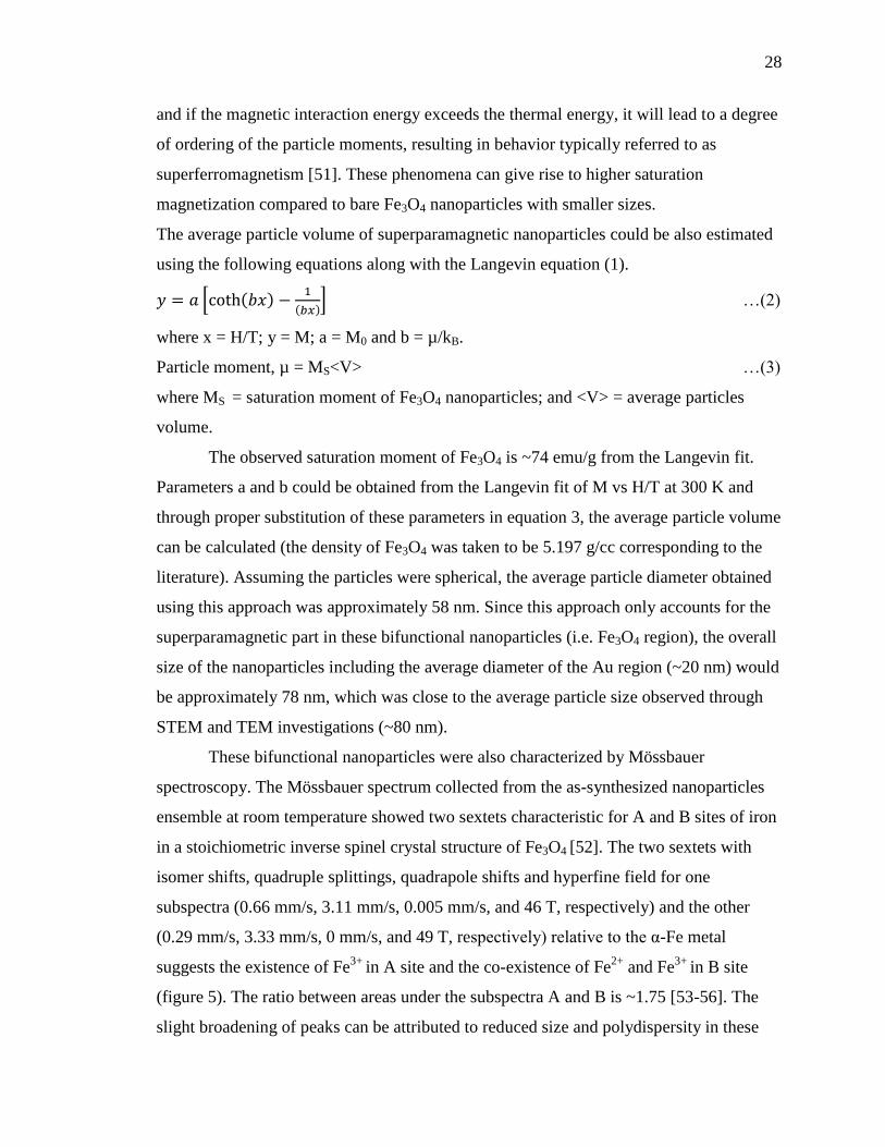
These bifunctional nanoparticles were also characterized by Mössbauer
spectroscopy. The Mössbauer spectrum collected from the as-synthesized nanoparticles
ensemble at room temperature showed two sextets characteristic for A and B sites of iron
in a stoichiometric inverse spinel crystal structure of Fe3O4 [52]. The two sextets with
isomer shifts, quadruple splittings, quadrapole shifts and hyperfine field for one
subspectra (0.66 mm/s, 3.11 mm/s, 0.005 mm/s, and 46 T, respectively) and the other
(0.29 mm/s, 3.33 mm/s, 0 mm/s, and 49 T, respectively) relative to the α-Fe metal
suggests the existence of Fe3+
in A site and the co-existence of Fe2+
and Fe3+
in B site
(figure 5). The ratio between areas under the subspectra A and B is ~1.75 [53-56]. The
slight broadening of peaks can be attributed to reduced size and polydispersity in these

29
nanoparticle ensembles. The observed Mössbauer spectra were very similar to the ones
reported in the literature for Fe3O4 albeit with size ranges close to 50 – 100 nm [50]. It
should be noted that two sets of sextet observed in Mössbauer spectra was more closely
related to the ordered state rather than the superparamagnetic state for Fe3O4
nanoparticles. This also corroborates with our hypothesis that at 300 K the ensemble is on
the cusp of superparamagnetic and ordered states. Typically, TB is very dependent on the
analysis method since the relaxation time for the moment fluctuation (τo) is dependent on
the measurement time of the instrument (τm) as shown below in equation 4 [47].
(
) …(4)
Figure 5. Mössbauer spectrum of Au-Fe3O4 nanoparticles collected at room temperature
with zero magnetic field showing the two characteristic sextets corresponding to Fe3+
and
Fe2+
states.
Since, γ-ray detection in Mössbauer spectroscopy is a comparatively fast technique (τo =
10-7
s), the TB estimated from Mössbauer data is typically higher than that obtained from

30
magnetization studies. In general the TB determined from these two techniques is related
by the following equation 5 [47].
( ̈ )
( ) …(5)
Hence, it can be concluded that at 300 K with respect to Mössbauer studies the
Au-Fe3O4 ensembles still behaves as if it is in the ordered state (i.e. below TB) while in
magnetization studies under an applied magnetic field it behaves closer to a
superparamagnet. The similarity between these spectra with that observed for bulk Fe3O4
also ruled out the presence of Fe in any other oxidation states.
Figure 6. Scheme showing the functionalization of Au-Fe3O4 bifunctional nanoparticles
by attachment of L-cysteine to the Au-terminal.
As mentioned previously, one of the most potentially transformative applications
of these magnetic nanoparticles is in biomedical fields. Hence, to demonstrate the
feasibility of using these bifunctional nanoparticles for biological applications, we
functionalized the nanoparticles taking advantage of Au-thiol facile interactions to make
these composite structures more biocompatible. In this regards, we have successfully
attached L-cysteine to Au terminal of the bifunctional nanoparticles by the process
described in the experimental section. The process for functionalization with L-cysteine
and the expected morphology has been schematically shown in figure 6. Both qualitative
(through FTIR and Raman) and quantitative (through acid ninhydrin assay) analysis of
the L-cysteine-modified Au-Fe3O4 bifunctional nanoparticles were performed. Figure
31
7(A) shows the FTIR spectra of L-cysteine-modified Au-Fe3O4 bifunctional nanoparticles
(curve 2) as compared with that of free L-cysteine (curve 1). While most of the bands
assigned to cysteine were visible for both the samples, the absence of band at ~2551 cm-1
corresponding to the stretching vibration of S-H bond was prominent in the FTIR spectra
of the cysteine functionalized nanoparticles as shown in curve b. This indicates breakage
of the S-H bond on attachment of cysteine to the Au –terminal through S, and has been
accepted as a signatory evidence for successful molecule attachment. [57-59]. Trans C-S
stretching vibrational modes can be assigned to the presence of band at ~637 cm-1
. The
band at ~3422 cm-1
can be assigned to N-H stretching vibrations [59], while bands in the
range 1510 to 1680 cm-1
can be assigned to carbonyl and N-H stretching vibrations [60].
Detailed analysis of the FTIR spectra confirmed the attachment of L-cysteine to the Au-
Fe3O4 bifunctional nanoparticles.
Figure 7. (A) FTIR spectra of (1) pure L-cysteine and (2) L-cysteine modified-
bifunctional Au-Fe3O
4 nanoparticles (B) Raman spectra of (1) bifunctional Au-Fe
3O
4
nanoparticles and (2) L-cysteine modified Au-Fe3O
4 nanoparticles.
From a purely group theory-based analysis, Fe3O4 should exhibit 14 Raman active
modes (3A1+3E+8T2). But, experimentally all modes cannot be detected due to the
mutual exclusion of vibrational modes caused by the presence of inversion center of
symmetry. Additional peaks may be present if the sample has any defects or vacancies.
Group theory analysis doesn’t account for these factors [61]. In our study, we observed
six peaks corresponding to Fe3O4 at 215, 329, 393, 516, 597, 672 cm-1
and two peaks
32
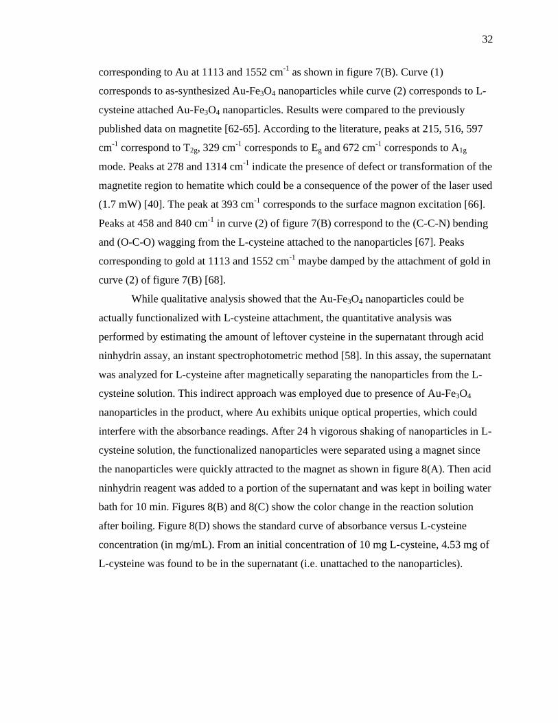
corresponding to Au at 1113 and 1552 cm-1
as shown in figure 7(B). Curve (1)
corresponds to as-synthesized Au-Fe3O4 nanoparticles while curve (2) corresponds to L-
cysteine attached Au-Fe3O4 nanoparticles. Results were compared to the previously
published data on magnetite [62-65]. According to the literature, peaks at 215, 516, 597
cm-1
correspond to T2g, 329 cm-1
corresponds to Eg and 672 cm-1
corresponds to A1g
mode. Peaks at 278 and 1314 cm-1
indicate the presence of defect or transformation of the
magnetite region to hematite which could be a consequence of the power of the laser used
(1.7 mW) [40]. The peak at 393 cm-1
corresponds to the surface magnon excitation [66].
Peaks at 458 and 840 cm-1
in curve (2) of figure 7(B) correspond to the (C-C-N) bending
and (O-C-O) wagging from the L-cysteine attached to the nanoparticles [67]. Peaks
corresponding to gold at 1113 and 1552 cm-1
maybe damped by the attachment of gold in
curve (2) of figure 7(B) [68].
While qualitative analysis showed that the Au-Fe3O4 nanoparticles could be
actually functionalized with L-cysteine attachment, the quantitative analysis was
performed by estimating the amount of leftover cysteine in the supernatant through acid
ninhydrin assay, an instant spectrophotometric method [58]. In this assay, the supernatant
was analyzed for L-cysteine after magnetically separating the nanoparticles from the L-
cysteine solution. This indirect approach was employed due to presence of Au-Fe3O4
nanoparticles in the product, where Au exhibits unique optical properties, which could
interfere with the absorbance readings. After 24 h vigorous shaking of nanoparticles in L-
cysteine solution, the functionalized nanoparticles were separated using a magnet since
the nanoparticles were quickly attracted to the magnet as shown in figure 8(A). Then acid
ninhydrin reagent was added to a portion of the supernatant and was kept in boiling water
bath for 10 min. Figures 8(B) and 8(C) show the color change in the reaction solution
after boiling. Figure 8(D) shows the standard curve of absorbance versus L-cysteine
concentration (in mg/mL). From an initial concentration of 10 mg L-cysteine, 4.53 mg of
L-cysteine was found to be in the supernatant (i.e. unattached to the nanoparticles).

33
Figure 8. (A) Magnetic separation of the nanoparticles from the L-cysteine solution. (B)
Color of supernatant after adding acid ninhydrin and (C) after heating in water bath for
15 minutes. (D) Standard curve of absorbance versus L-cysteine concentration.
Assuming 100% of L-cysteine activity, the loading capacity of the bifunctional
nanoparticles used in the study is approximately 1 mg of L-cysteine per 4 mg of
nanoparticles. We are presently trying to attach various other biomolecules to gain a
better insight into the loading capacity of these bifunctional nanoparticles.
Au-Fe3O4 nanoparticles are intended to be applied for magnetic fluid hyperthermia and
delivery of biomolecules. Hence the cytotoxicity of these nanoparticles was also
evaluated. For this study, the cell viability of CHO cells was evaluated after 48 h
incubation at various increasing concentration of nanoparticles ranging from 0.1 – 1
mg/ml by MTS assay. The nanoparticles didn’t show any sign of cytotoxicity even at 1
mg/mL (figure 9). The results obtained corroborates well with polymer coated magnetite
nanoparticles and other morphologies [69-73].
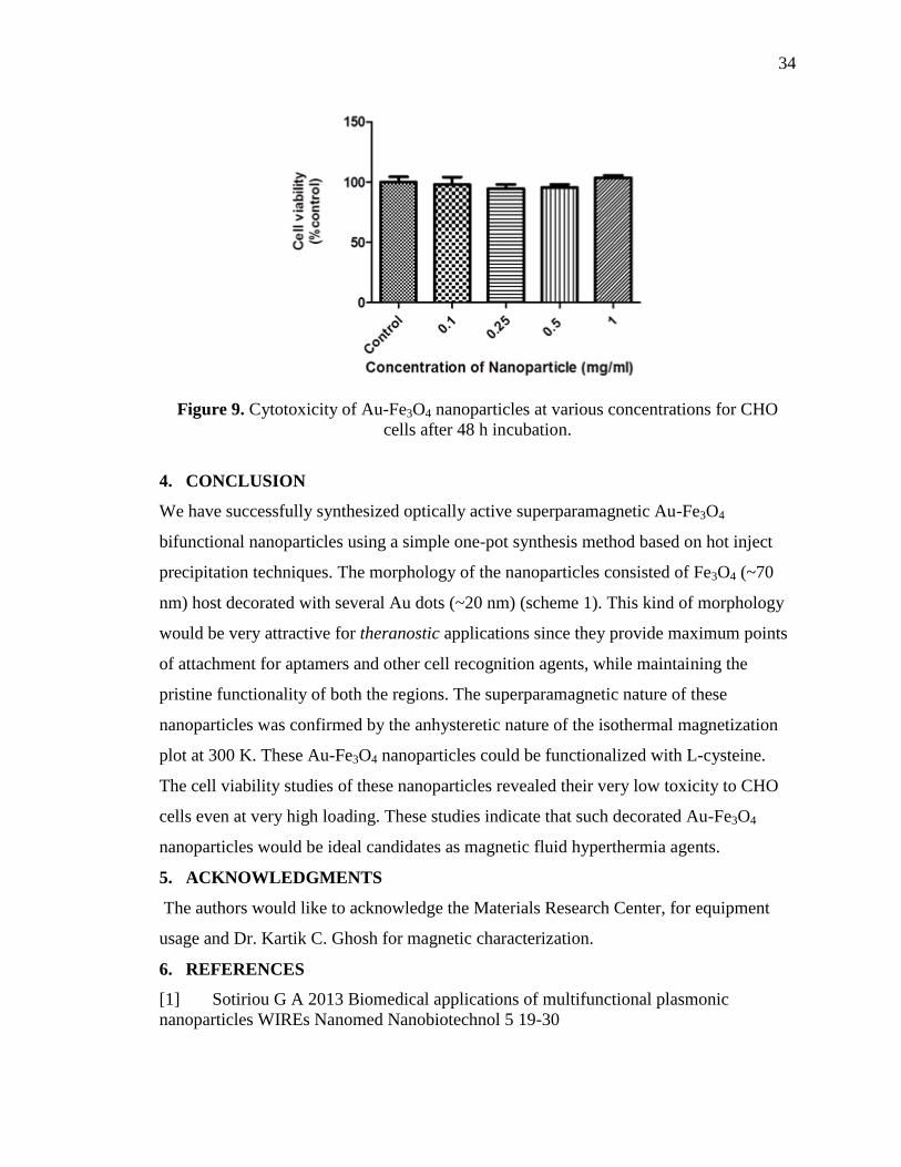
34
Figure 9. Cytotoxicity of Au-Fe3O4 nanoparticles at various concentrations for CHO
cells after 48 h incubation.
4. CONCLUSION
We have successfully synthesized optically active superparamagnetic Au-Fe3O4
bifunctional nanoparticles using a simple one-pot synthesis method based on hot inject
precipitation techniques. The morphology of the nanoparticles consisted of Fe3O4 (~70
nm) host decorated with several Au dots (~20 nm) (scheme 1). This kind of morphology
would be very attractive for theranostic applications since they provide maximum points
of attachment for aptamers and other cell recognition agents, while maintaining the
pristine functionality of both the regions. The superparamagnetic nature of these
nanoparticles was confirmed by the anhysteretic nature of the isothermal magnetization
plot at 300 K. These Au-Fe3O4 nanoparticles could be functionalized with L-cysteine.
The cell viability studies of these nanoparticles revealed their very low toxicity to CHO
cells even at very high loading. These studies indicate that such decorated Au-Fe3O4
nanoparticles would be ideal candidates as magnetic fluid hyperthermia agents.
5. ACKNOWLEDGMENTS
The authors would like to acknowledge the Materials Research Center, for equipment
usage and Dr. Kartik C. Ghosh for magnetic characterization.
6. REFERENCES
[1] Sotiriou G A 2013 Biomedical applications of multifunctional plasmonic
nanoparticles WIREs Nanomed Nanobiotechnol 5 19-30
35
[2] Zeng H and Sun S 2008 Syntheses, Properties, and Potential Applications of
Multicomponent Magnetic Nanoparticles Adv. Funct. Mater. 18 391-400
[3] Kim J, Piao Y and Hyeon T 2009 Multifunctional nanostructured materials for
multimodal imaging, and simultaneous imaging and therapy Chem. Soc. Rev. 38 372-90
[4] Yue X-L, Ma F and Dai Z-F 2014 Multifunctional magnetic nanoparticles for
magnetic resonance image-guided photothermal therapy for cancer Chin. Phys. B 23
044301
[5] Yan K, Li P, Zhu H, Zhou Y, Ding J, Shen J, Li Z, Xu Z and Chu P K 2013
Recent advances in multifunctional magnetic nanoparticles and applications to
biomedical diagnosis and treatment RSC Adv. 3 10598-618
[6] Hao R, Xing R, Xu Z, Hou Y, Gao S and Sun S 2010 Synthesis,
Functionalization, and Biomedical Applications of Multifunctional Magnetic
Nanoparticles Adv. Mater. 22 2729-42
[7] Sotiriou G A, Starsich F, Dasargyri A, Wurnig M C, Krumeich F, Boss A, Leroux
J-C and Pratsinis S E 2014 Photothermal Killing of Cancer Cells by the Controlled
Plasmonic Coupling of Silica-Coated Au/Fe2O3 Nanoaggregates Adv. Funct. Mater. 24
2818-27
[8] Kim J, Kim H S, Lee N, Kim T, Kim H, Yu T, Song I C, Moon W K and Hyeon T
2008 Multifunctional Uniform Nanoparticles Composed of a Magnetite Nanocrystal Core
and a Mesoporous Silica Shell for Magnetic Resonance and Fluorescence Imaging and
for Drug Delivery Angew. Chem. Int. Ed. 47 8438-41
[9] McCarthy J R and Weissleder R 2008 Multifunctional magnetic nanoparticles for
targeted imaging and therapy Adv. Drug Deliv. Rev.60 1241-51
[10] Huh Y-M, Jun Y-w, Song H-T, Kim S, Choi J-s, Lee J-H, Yoon S, Kim K-S, Shin
J-S, Suh J-S and Cheon J 2005 In Vivo Magnetic Resonance Detection of Cancer by
Using Multifunctional Magnetic Nanocrystals J. Am. Chem. Soc. 127 12387-91
[11] Fang C and Zhang M 2009 Multifunctional magnetic nanoparticles for medical
imaging applications J. Mater. Chem. 19 6258-66
[12] Di Corato R, Bigall N C, Ragusa A, Dorfs D, Genovese A, Marotta R, Manna L
and Pellegrino T 2011 Multifunctional Nanobeads Based on Quantum Dots and Magnetic
Nanoparticles: Synthesis and Cancer Cell Targeting and Sorting ACS Nano 5 1109-21

36
[13] Bao G, Mitragotri S and Tong S 2013 Multifunctional Nanoparticles for Drug
Delivery and Molecular Imaging Annu. Rev.Biomed. Eng. 15 253-82
[14] Bhaskar S, Tian F, Stoeger T, Kreyling W, de la Fuente J M, Grazu V, Borm P,
Estrada G, Ntziachristos V and Razansky D 2010 Multifunctional Nanocarriers for
diagnostics, drug delivery and targeted treatment across blood-brain barrier: perspectives
on tracking and neuroimaging Part. Fibre Toxicol. 7 3
[15] Gao J, Gu H and Xu B 2009 Multifunctional magnetic nanoparticles: design,
synthesis, and biomedical applications Acc. Chem. Res. 42 1097-107
[16] Daniel M-C and Astruc D 2004 Gold Nanoparticles: Assembly, Supramolecular
Chemistry, Quantum-Size-Related Properties, and Applications toward
Biology,Catalysis, and Nanotechnology Chem. Rev. 104 293-346
[17] Sperling R A, Gil P R, Zhang F, Zanella M and Parak W J 2008 Biological
applications of gold nanoparticles Chem. Soc. Rev. 37 1896-908
[18] Ghosh P, Han G, De M, Kim C K and Rotello V M 2008 Gold nanoparticles in
delivery applications Adv. Drug Deliv. Rev. 60 1307-15
[19] El-Sayed I H, Huang X and El-Sayed M A 2006 Selective laser photo-thermal
therapy of epithelial carcinoma using anti-EGFR antibody conjugated gold nanoparticles
Cancer Lett. 239 129-35
[20] Gupta A K and Gupta M 2005 Synthesis and surface engineering of iron oxide
nanoparticles for biomedical applications Biomaterials 26 3995-4021
[21] Laurent S, Dutz S, Häfeli U O and Mahmoudi M 2011 Magnetic fluid
hyperthermia: Focus on superparamagnetic iron oxide nanoparticles Adv. Colloid
Interface Sci. 166 8-23
[22] Laurent S, Forge D, Port M, Roch A, Robic C, Vander Elst L and Muller R N
2008 Magnetic Iron Oxide Nanoparticles: Synthesis, Stabilization, Vectorization,
Physicochemical Characterizations, and Biological Applications Chem. Rev. 108 2064-
110
[23] Qiao R, Yang C and Gao M 2009 Superparamagnetic iron oxide nanoparticles:
from preparations to in vivo MRI applications J. Mater. Chem. 19 6274-93
[24] Lewin M, Carlesso N, Tung C-H, Tang X-W, Cory D, Scadden D T and
Weissleder R 2000 Tat peptide-derivatized magnetic nanoparticles allow in vivo tracking
and recovery of progenitor cells Nature Biotechnol. 18 410-4

37
[25] Weissleder R, Moore A, Mahmood U, Bhorade R, Benveniste H, Chiocca E A
and Basilion J P 2000 In vivo magnetic resonance imaging of transgene expression
Nature Med. 6 351-4
[26] Zhao M, Beauregard D A, Loizou L, Daveletov B and Brindle K M 2001 Non-
invasive detection of apoptosis using magnetic resonance imaging and a targeted contrast
agent Nature Med. 7 1241-4
[27] Coey J M D 2010 Magnetism and Magnetic Materials (Cambridge University
Press) p264
[28] Bao J, Chen W, Liu T, Zhu Y, Jin P, Wang L, Liu J, Wei Y and Li Y 2007
Bifunctional Au-Fe3O4 Nanoparticles for Protein Separation ACS Nano 1 293-8
[29] Yu H, Chen M, Rice P M, Wang S X, White R L and Sun S 2005 Dumbbell-like
Bifunctional Au-Fe3O4particles Nano Lett. 5 379-82
[30] Liang C-H, Wang C-C, Lin Y-C, Chen C-H, Wong C-H and Wu C-Y 2009 Iron
Oxide/Gold Core/Shell Nanoparticles for Ultrasensitive Detection of Carbohydrate-
Protein Interactions Anal. Chem. 81 7750-6
[31] Xu Z, Hou Y and Sun S 2007 Magnetic Core/Shell Fe3O4/Au and Fe3O4/Au/Ag
Nanoparticles with Tunable Plasmonic Properties J. Am. Chem. Soc. 129 8698-9
[32] Shevchenko E V, Bodnarchuk M I, Kovalenko M V, Talapin D V, Smith R K,
Aloni S, Heiss W and Alivisatos A P 2008 Gold/Iron Oxide Core/Hollow-Shell
Nanoparticles Adv. Mater. 20 4323-9
[33] Ji X, Shao R, Elliott A M, Stafford R J, Esparza-Coss E, Bankson J A, Liang G,
Luo Z-P, Park K, Markert J T and Li C 2007 Bifunctional Gold Nanoshells with a
Superparamagnetic Iron Oxide-Silica Core Suitable for Both MR Imaging and
Photothermal Therapy J. Phy. Chem. C 111 6245-51
[34] Wang L, Luo J, Maye M M, Fan Q, Rendeng Q, Engelhard M H, Wang C, Lin Y
and Zhong C-J 2005 Iron oxide–gold core–shell nanoparticles and thin film assembly J.
Mater. Chem. 15 1821-32
[35] Vandenberghe R E, Grave E and Bakker P M A 1994 On the methodology of the
analysis of Mössbauer spectra Hyperfine Interact. 83 29-49
[36] Gaitonde M K 1967 A Spectrophotometric Method for the Direct Determination
of Cysteine in the Presence of Other Naturally Occurring Amino Acids Biochem. J. 104
627-33
[37] Cory AH, Owen TC, Barltrop JA, Cory JG 1991 Use of an aqueous soluble
tetrazolium/formazan assay for cell growth assays in culture Cancer Comm. 3 207–12.

38
[38] Patterson A 1939 The Scherrer Formula for X-Ray Particle Size Determination
Phys. Rev. 56 978-82
[39] Lattuada M and Hatton T A 2011 Synthesis, properties and applications of Janus
nanoparticles Nano Today 6 286-308
[40] Perro A, Reculusa S, Ravaine S, Bourgeat-Lami E and Duguet E 2005 Design and
synthesis of Janus micro- and nanoparticles J. Mater. Chem. 15 3745-60
[41] Haiss W, Thanh N T K, Aveyard J and Fernig D G 2007 Determination of Size
and Concentration of Gold Nanoparticles from UV-Vis Spectra Anal. Chem. 79 4215-21
[42] Link S and El-Sayed M A 1999 Size and Temperature Dependence of the
Plasmon Absorption of Colloidal Gold Nanoparticles J. Phy. Chem. B 103 4212-7
[43] Verwey E J W 1939 Electronic conduction of magnetite and its transition point at
low temperatures Nature 144 327-8
[44] Wang J, Chen Q, Li X, Shi L, Peng Z and Zeng C 2004 Disappearing of the
Verwey transition in magnetite nanoparticles synthesized under a magnetic field:
implications for the origin of charge ordering Chem. Phys. Lett. 390 55-8
[46] Prozorov R, Prozorov T, Mallapragada S, Narasimhan B, Williams T and
Bazylinski D 2007 Magnetic irreversibility and the Verwey transition in nanocrystalline
bacterial magnetite Phys. Rev. B 76 054406
[47] Skumryev V, Stoyanov S, Zhang Y, Hadjipanayis G, Givord D and Nogues J
2003 Beating the superparamagnetic limit with exchange bias Nature 423 850-3
[48] Chandra S, Huls N A, Phan M H, Srinath S, Garcia M A, Lee Y, Wang C, Sun S,
Iglesias O and Srikanth H 2014 Exchange bias effect in Au-Fe3O4 nanocomposites
Nanotechnology 25 055702
[49] Goya G F, Berqu T S, Fonseca F C and Morales M P 2003 Static and dynamic
magnetic properties of spherical magnetite nanoparticles J. Appl. Phys. 94 3520-8
[50] Roca A G, Morales M P, O’Grady K and Serna C J 2006 Structural and magnetic
properties of uniform magnetite nanoparticles prepared by high temperature
decomposition of organic precursors Nanotechnology 17 2783-8
[51] Morup, S. Superferromagnetic nanostructures Hyperfine Interactions, 1994, 90,
171-185
[52] O'Neill H S C and Dollase W A 1994 Crystal structures and cation distributions in
simple spinels from powder XRD structural refinements: MgCr2O4, ZnCr2O4, Fe3O4

39
and the temperature dependence of the cation distribution in ZnAl2O4 Phys. Chem.
Minerals 20 541-55
[53] Oh S J, Cook D C and Townsend H E 1998 Characterization of iron oxides
commonly formed as corrosion products of steel Hyperfine Interact. 112 59-66
[54] Woo K, Hong J, Choi S, Lee H-W, Ahn J-P, Kim C S and Lee S W 2004 Easy
Synthesis and Magnetic Properties of Iron Oxide Nanoparticles Chem. Mater. 16 2814-8
[55] Vandenberghe R E, Barrero C A, Costa G M d, Sana E V and Grave E D 2000
M¨ossbauer characterization of iron oxides and (oxy)hydroxides: the present state of the
art Hyperfine Interact. 126 247-59
[56] Kumar R V, Koltypin Y, Xu X N, Yeshurun Y, Gedanken A and Felner I 2001
Fabrication of magnetite nanorods by ultrasound irradiation J. Appl. Phys. 89 6324-8
[57] Gao W, Ji L, Li L, Cui G, Xu K, Li P and Tang B 2012 Bifunctional combined
Au-Fe(2)O(3) nanoparticles for induction of cancer cell-specific apoptosis and real-time
imaging Biomaterials 33 3710-8
[58] Lo C K, Xiao D and Choi M M F 2007 Homocysteine-protected gold-coated
magnetic nanoparticles: synthesis and characterization J. Mater. Chem. 17 2418-27
[59] Kumar A, Mandal S, Selvakannan P R, Pasricha R, Mandale A B and Sastry M
2003 Investigation into the interaction between surface-bound alkylamines and gold
nanoparticles Langmuir 19 6277-82
[60] Shankar S S, Ahmad A, Pasricha R, Khan M I, Kumar R and Sastry M 2004
Immobilization of biogenic gold nanoparticles in thermally evaporated fatty acid and
amine thin films J. Colloid Interface Sci. 274 69-75
[61] Wang Z, Lazor P, Saxena S K and O'Neill H S C 2002 High pressure Raman
spectroscopy of ferrite MgFe2O4 Mater. Res. Bull. 37 1589-602
[62] Mandal M, Kundu S, Ghosh S K, Panigrahi S, Sau T K, Yusuf S M and Pal T
2005 Magnetite nanoparticles with tunable gold or silver shell J. Colloid Interface Sci.
286 187-94
[63] Faria D L A d, Silva S V and Oliveira M T d 1997 Raman Microspectroscopy of
Some Iron Oxides and Oxyhydroxides J. Raman Spectrosc. 28 873-8
[64] Thibeau R J, Brown C W and Heidersbach R H 1978 Raman spectra of possible
corrosion products of iron Appl. Spectrosc. 32 532-5
[65] Dunnwald J and Otta A 1989 An investigation of phase transitions in rust layers
using Raman spectroscopy Corros. Sci. 29 1167-76
40
[66] Whangbo M-H, Koo H-J and Dai D 2003 Spin exchange interactions and
magnetic structures of extended magnetic solids with localized spins: theoretical
descriptions on formal, quantitative and qualitative levels J. Solid State Chem. 176 417-
81
[67] Pawlukojc A, Leciejewicz J, Ramirez-Cuesta A J and Nowicka-Scheibe J 2005 l-
Cysteine: Neutron spectroscopy, Raman, IR and ab initio study Spectrochim. Acta. Part
A 61 2474-81
[68] Park H-Y, Schadt M J, Wang L, Lim I-I S, Njoki P N, Kim S H, Jang M-Y, Luo J
and Zhong C-J 2007 Fabrication of Magnetic Core@Shell Fe Oxide@Au Nanoparticles
for Interfacial Bioactivity and Bio-separation Langmuir 23 9050-6
[69] Salado J, Insausti M, Lezama L, Gil de Muro I, Moros M, Pelaz B, Grazu V, de la
Fuente J M and Rojo T 2012 Functionalized Fe(3)O(4)@Au superparamagnetic
nanoparticles: in vitro bioactivity Nanotechnology 23 315102
[70] Liong M, Lu J, Kovochich M, Xia T, Ruehm S G, Nel A E, Tamanoi F and Zink J
I 2008 Multifunctional inorganic nanoparticles for imaging, targeting, and drug delivery
ACS Nano 2 889-96
[71] Berry C C, Wells S, Charles S, Aitchison G and Curtis A S 2004 Cell response to
dextran-derivatised iron oxide nanoparticles post internalisation Biomaterials 25 5405-13
[72] Yu W W, Chang E, Sayes C M, Drezek R and Colvin V L 2006 Aqueous
dispersion of monodisperse magnetic iron oxide nanocrystals through phase transfer
Nanotechnology 17 4483-7
[73] Fuente J M d l, Berry C C, Riehle M O and Curtis A S G 2006 Nanoparticle
targeting at cells Langmuir 22 3286-93
41
SECTION
2. CONCLUSION AND FUTURE WORK
Optically active superparamagnetic gold-magnetite nanoparticles were
synthesized using one pot hot injection precipitation method. The synthesis involved
using Fe(CO)5 as iron precursor and HAuCl4 as gold precursor in presence of oleylamine
and oleic acid. Oleylamine helps in reducing Au3+ to Au
0 seeds which simultaneously
oxidizes Fe(0) to form Au-Fe3O4 bifunctional nanoparticles. Triton® X-100 was
employed as a highly viscous solvent to prevent agglomeration of Fe3O4 nanoparticles.
The morphology of the nanoparticles consisted of magnetite (~70 nm) part with gold dots
(~20 nm). The superparamagnetic nature was confirmed by the anhysteretic nature of the
isothermal plot at 300 K. The observed magnetization was approximately 74 emu/g
which is significantly higher than previously reported values. By attaining high
magnetization saturation, the patient need not be exposed to high magnetic fields for
treatment. The pXRD pattern also didn’t show any peaks corresponding to maghemite or
iron oxyhydroxide. The toxicity of the nanoparticles was evaluated using MTS assay on
CHO cells. They showed no significant toxic effect on CHO cells up to 48 hours.
L-cysteine was attached to these nanoparticles and was characterized qualitatively
and quantitatively. Results showed that the loading capacity of the nanoparticles was 1
mg of L-cysteine per 4 mg of nanoparticles. The methods adopted in this thesis serve as
facile and feasible method for synthesis of gold nanoparticles and their biomolecule
conjugation through the gold terminal. The L-cysteine attached nanoparticles couldn’t be
dispersed in water. Future steps, would be to conjugate (-)-EGCG (epigallocatechin
gallate) to these nanoparticles for specific binding to tumor cells.
Epigallocatechin gallate is the most abundant catechin in tea and is a potential
antioxidant that has many therapeutic applications. Recent research shows that EGCG
may be useful in preventing or treating various gastrointestinal, prostrate and other
cancers. Initial studies show that the (-)-EGCG conjugated nanoparticles can be water
dispersed. Figure 3.1 illustrate the attachment of EGCG to gold-magnetite nanoparticles.
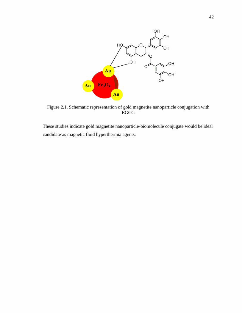
42
Figure 2.1. Schematic representation of gold magnetite nanoparticle conjugation with
EGCG
These studies indicate gold magnetite nanoparticle-biomolecule conjugate would be ideal
candidate as magnetic fluid hyperthermia agents.

43
BIBLIOGRAPHY
1. M-C. Daniel and D. Astruc, “Gold Nanoparticles: Assembly, Supramolecular
Chemistry, Quantum-Size-Related Properties, and Applications toward Biology,
Catalysis, and Nanotechnology,” Chemical Reviews, Vol. 104, No. 1, Pp. 293-346
(2004).
2. A.P. Alivisatos, “Semiconductor Clusters, Nanocrystals, and Quantum Dots,”
Science, Vol. 271, No.5251, Pp. 933-7 (1996).
3. D.L. Fedlheim and C.A. Foss, Metal nanoparticles: synthesis, characterizations, and
applications, CRC Press (2001).
4. K.A. Willets and R.P.Van Duyne, “Localized Surface Plasmon Resonance
Spectroscopy and Sensing,” Annual Reviews of Physical Chemistry, Vol. 58, Pp. 267-
97 (2007).
5. H.C. van de Hulst, Light Scattering by Small Metal Particles, Wiley: New York
(1957).
6. G. Mie, “Beiträge zur Optik Trüber Medien, Speziell Kolloidaler Metallӧsungen,”
Annalen der Physik, Vol. 330, No. 3, Pp. 377-445 (1908).
7. K.L. Kelly, E. Coronado, L.L. Zhao and G.C. Schatz, “The Optical Properties of
Metal Nanoparticles: The Influence of Size, Shape, and Dielectric Environment,” The
Journal of Physical Chemistry B, Vol. 107, No. 3, Pp. 668-77 (2003).
8. S. Eustis and M.A. El-Sayed, “Why gold nanoparticles are more precious than pretty
gold: Noble metal surface plasmon resonance and its enhancement of the radiative
and nonradiative properties of nanocrystals of different shapes,” Chemical Society
Reviews, Vol. 35, No. 3, Pp. 209-17 (2006).
9. G. Schmid, “Large clusters and colloids. Metals in the embryonic state,” Chemical
Reviews, Vol. 92, No. 8, Pp. 1709-27 (1992).
10. M. Brust, M. Walker, D. Bethell, D.J. Schiffrin and R. Whyman, “Synthesis of
thiolderivatized gold nanoparticles in a 2-phase liquid–liquid system,” Journal of the
Chemical Society, Chemical communications, No. 7, Pp. 801–802 (1994).
11. A.C. Templeton, W.P. Wuelfing and R.W. Murray, “Monolayer-Protected Cluster
Molecules,” Accounts of Chemical Research, Vol. 33, No. 1, Pp. 27-36 (2000).
12. J. Turkevich, P.C. Stevenson and J. Hillier, “A study of the nucleation and growth
processes in the synthesis of colloidal gold,” Discussions of Faraday Society, No. 11,
Pp. 55-75 (1951).

44
13. R.A. Sperling, P.R. Gil, F. Zhang, M. Zanella and W.J. Parak, “Biological
applications of gold nanoparticles,” Chemical Society Reviews, Vol. 37, No. 9, Pp.
1896-1908 (2008).
14. P. Ghosh, G. Han, M. De, C.K. Kim and V.M. Rotello, “Gold nanoparticles in
delivery applications,” Advanced Drug Delivery Reviews, Vol. 60, No. 11, Pp. 1307-
1315 (2008).
15. K. Saha, S.S. Agasti, C. Kim, X. Li and V.M. Rotello, “Gold Nanoparticles in
Chemical and Biological Sensing,” Chemical Reviews, Vol. 112, No. 5, Pp. 2739-79
(2012).
16. C.A. Mirkin, R.L. Letsinger, R.C. Mucic and J.J. Storhoff, “A DNA-based method
for rationally assembling nanoparticles into macroscopic materials,” Nature, Vol.
382, Pp. 607–609 (1996).
17. R. Elghanian, J.J. Storhoff, R.C. Mucic, R.L. Letsinger and C.A. Mirkin, “Selective
Colorimetric Detection of Polynucleotides Based on the Distance-Dependent Optical
Properties of Gold Nanoparticles,” Science, Vol. 277, No. 5329, Pp. 1078-81 (1997).
18. M-R. Choi, K.J. Stanton-Maxey, J.K. Stanley, C.S. Levin, R. Bardhan, D. Akin, S.
Badve, J. Sturgis, J.P. Robinson, R. Bashir, N.J. Halas and S.E. Clare, “A Cellular
Trojan Horse for Delivery of Therapeutic Nanoparticles into Tumors,” Nano Letters,
Vol. 7, No. 12, Pp. 3759-65 (2007).
19. T.B. Huff, L. Tong, Y. Zhao, M.N. Hansen, J-X. Cheng and A. Wei, “Hyperthermic
effects of gold nanorods on tumor cells,” Nanomedicine, Vol. 2, No. 1, Pp. 125-32
(2007).
20. X. Huang, P.K. Jain, I.H. El-Sayed and M.A. El-Sayed, “Determination of the
Minimum Temperature Required for Selective Photothermal Destruction of Cancer
Cells with the Use of Immunotargeted Gold Nanoparticles,” Photochemistry and
Photobiology, Vol. 82, No. 2, Pp. 412-7 (2006).
21. D. Kim, S. Park, J.H. Lee, Y.Y. Jeong and S. Jon, “Antibiofouling Polymer-Coated
Gold Nanoparticles as a Contrast Agent for in Vivo X-ray Computed Tomography
Imaging,” Journal of the American Chemical Society, Vol. 129, No. 24, Pp.7661-5
(2007).
22. J.F. Hainfeld, D.N. Slatkin and H.M. Smilowitz, “The use of gold nanoparticles to
enhance radiotherapy in mice,” Physics in Medicine and Biology, Vol. 49, N309
(2004).

45
23. B.D. Chithrani, A.A. Ghazani and W.C.W.Chan, “Determining the Size and Shape
Dependence of Gold Nanoparticle Uptake into Mammalian Cells,” Nano Letters, Vol.
6, No. 4, Pp. 662-8 (2006).
24. B.D. Chithrani and W.C.W. Chan, “Elucidating the Mechanism of Cellular Uptake
and Removal of Protein-Coated Gold Nanoparticles of Different Sizes and Shapes,”
Nano Letters, Vol. 7, No. 6, Pp. 1542-50 (2007).
25. J.M. de la Fuente and C.C. Berry, “Tat Peptide as an Efficient Molecule To
Translocate Gold Nanoparticles into the Cell Nucleus,” Bioconjugate Chemistry, Vol.
16, No. 5, Pp. 1176-80 (2005).
26. M. Lewin, N. Carlesso, C.H. Tung, X.W. Tang, D. Corry, D.T. Scadden and R.
Weissleder, “Tat peptide-derivatized magnetic nanoparticles allow in vivo tracking
and recovery of progenitor cells,” Nature Biotechnology, Vol. 18, Pp. 410–414
(2000).
27. A.K. Salem, P.C. Searson and K.W. Leong, “Multifunctional nanorods for gene
delivery,” Nature Materials, Vol. 2, Pp. 668–671 (2003).
28. S.D. Brown, P. Nativo, J-A. Smith, D. Stirling, P.R. Edwards, B. Venugopal, D.J.
Flint, J.A. Plumb, D. Graham and N.J. Wheate, “Gold Nanoparticles for the Improved
Anticancer Drug Delivery of the Active Component of Oxaliplatin,” Journal of the
American Chemical Society, Vol. 132, No. 13, Pp. 4678-84 (2010).
29. K. Raj, B. Moskowitz and R. Casciari, “Advances in ferrofluid technology,” Journal
of Magnetism and Magnetic Materials, Vol. 149, No. 1-2, Pp. 174-80 (1995).
30. D. Speliotis, “Magnetic recording beyond the first 100 Years,” Journal of Magnetism
and Magnetic Materials, Vol. 193, No. 1-3, Pp. 29-35 (1999).
31. R. Qiao, C. Yang and M. Gao, “Superparamagnetic iron oxide nanoparticles: from
preparations to in vivo MRI applications,” Journal of Materials Chemistry, Vol. 19,
No. 35, Pp. 6274-93 (2009).
32. M. Mahmoudi, S. Sant, B. Wang, S. Laurent and T. Sen, “Superparamagnetic iron
oxide nanoparticles (SPIONs): Development, surface modification and applications in
chemotherapy,” Advanced Drug Delivery Reviews, Vol. 63, No. 1-2, Pp. 24-46
(2011).
33. A.K. Gupta and M. Gupta, “Synthesis and surface engineering of iron oxide
nanoparticles for biomedical applications,” Biomaterials, Vol. 26, No. 18, Pp. 3995-
4021 (2005).
46
34. P. Xu, G.M. Zeng, D.L. Huang, C.L. Feng, S. Hu, M.H. Zhao, C. lai, Z. Wei, C.
Huang, G.X. Xie and Z. H. Liu, “Use of iron oxide nanomaterials in wastewater
treatment: A review,” Science of Total Environment, Vol. 424, Pp. 1-10 (2012).
35. G.F. Goya, T.S. Berquó, F.C. Fonseca and M.P. Morales, “Static and dynamic
magnetic properties of spherical magnetite nanoparticles,” Journal of Applied
Physics, Vol. 94, Pp. 3520-8 (2003).
36. C.P. Bean, “Hysteresis Loops of Mixtures of Ferromagnetic Micropowders,” Journal
of Applied Physics, Vol. 26, Pp. 1381-3 (1955).
37. J. Frenkel and J. Dorfman, “Spontaneous and Induced Magnetisation in
Ferromagnetic Bodies,” Nature, Vol. 126, Pp. 274-5 (1930).
38. C. Kittel, “Theory of the Dispersion of Magnetic Permeability in Ferromagnetic
Materials at Microwave Frequencies,” Physical Review, Vol. 70, No. 5-6, Pp. 281-90
(1946).
39. C.P. Bean and J.D. Livingston, “Superparamagnetism,” Journal of Applied Physics,
Vol. 30, Pp. S120-S9 (1959).
40. G.A. Candela and R.A. Haines, “A method for determining the region of
superparamagnetism,” Applied Physics Letters, Vol. 34, Pp. 868-70 (1979).
41. E. J. W. Verwey, “Electronic conduction of magnetite and its transition point at low
temperatures,” Nature, Vol. 144, Pp. 327-8 (1939).
42. J. Wang, Q. Chen, X. Li, L. Shi, Z. Peng and C. Zeng, “Disappearing of the Verwey
transition in magnetite nanoparticles synthesized under a magnetic field: implications
for the origin of charge ordering,” Chemical Physics Letters, Vol. 390, No. 1-3, Pp.
55-8 (2004).
43. Y. Sahoo, A. Goodarzi, M.T. Swihart, T.Y. Ohulchanskyy, N Kaur, E.P. Furlani and
P.N. Prasad, “Aqueous Ferrofluid of Magnetite Nanoparticles: Fluorescence Labeling
and Magnetophoretic Control,” The Journal of Physical Chemistry B, Vol. 109, No.
9, Pp. 3879-85 (2005).
44. P.S. Stephen, Low viscosity magnetic fluid obtained by the colloidal suspension of
magnetic particles, US Patent 3215572 (1965).
45. Y-G. Zhao, H-Y Shen, S-D Pan and M-Q. Hu, “Synthesis, characterization and
properties of ethylenediamine-functionalized Fe3O4 magnetic polymers for removal
of Cr(VI) in wastewater,” Journal of Hazardous Materials, Vol. 182, No. 1-3, Pp.
295-302 (2010).
47
46. S.R. Chowdhury and E.K. Yanful, “Arsenic and chromium removal by mixed
magnetite–maghemite nanoparticles and the effect of phosphate on removal,” Journal
of Environmental Management, Vol. 91, No. 11, Pp. 2238-47 (2010).
47. J. Hu, G. Chen and I.M.C. Lo, “Removal and recovery of Cr(VI) from wastewater by
maghemite nanoparticles,” Water Research, Vol. 39, No. 18, Pp. 4528-36 (2005).
48. K. J. Widder, A.E. Senyei and D.F. Ranney, “In vitro release of biologically active
adriamycin by magnetically responsive albumin microspheres,” Cancer Research,
Vol. 40, Pp. 3512–7 (1980).
49. J. M. Gallo, C.T. Hung, P.K. Gupta and P.G. Perrier, “Physiological pharmacokinetic
model of adriamycin delivered via magnetic albumin microspheres in the rat,”
Journal of Pharmacokinetics and Biopharmaceutics, Vol. 17, No. 3, Pp. 305–26
(1989).
50. S. Laurent, D. Forge, M. Port, A. Roch, C. Robic, L. Vander Elst and R. N. Muller,
“Magnetic Iron Oxide Nanoparticles: Synthesis, Stabilization, Vectorization,
Physicochemical Characterizations, and Biological Applications,” Chemical Reviews,
Vol. 108, No. 6, Pp. 2064-110 (2008).
51. A-H Lu, E.L. Salabas and F. Schüth, “Magnetic Nanoparticles: Synthesis, Protection,
Functionalization, and Application,” Angewandte Chemie International Edition, Vol.
46, No. 8, Pp. 1222-44 (2007).
52. J-P Jolivet, C. Chaneac and E. Tronc, “Iron oxide chemistry. From molecular clusters
to extended solid networks,” Chemical Communications, No. 5, Pp. 481-3 (2004).
53. N.M. Gribanov, E.E. Bibik, O.V. Buzunov and V.N. Naumov, “Physico-chemical
regularities of obtaining highly dispersed magnetite by the method of chemical
condensation,” Journal of Magnetism and Magnetic Materials, Vol. 85, No. 1-3, Pp.
7-10 (1990).
54. W.C. Elmore, “Ferromagnetic Colloid for Studying Magnetic Structures,” Physical
Review Letters, Vol. 54, Pp. 309-10 (1938).
55. T.J. Daou, G. Pourroy, S. Bégin-Colin, J.M. Grenèche, C. Ulhaq-Bouillet, P. Legaré,
P. Bernhardt, C. Leuvrey and G. Rogez, “Hydrothermal Synthesis of Monodisperse
Magnetite Nanoparticles,” Chemistry of Materials, Vol. 18, No. 18, Pp. 4399-404
(2006).
56. S. Sun S and H. Zeng, “Size-Controlled Synthesis of Magnetite Nanoparticles,”
Journal of the American Chemical Society, Vol. 124, No. 28, Pp. 8204-5 (2002).

48
57. Z. Xu, C. Shen, Y. Hou, H. Gao and S. Sun, “Oleylamine as Both Reducing Agent
and Stabilizer in a Facile Synthesis of Magnetite Nanoparticles,” Chemistry of
Materials, Vol. 21, No. 9, Pp. 1778-80 (2009).
58. H. Zeng and S. Sun, “Syntheses, Properties, and Potential Applications of
Multicomponent Magnetic Nanoparticles,” Advanced Functional Materials, Vol. 18,
No. 3, Pp. 391-400 (2008).
59. N. Lewinski, V. Colvin and R. Drezek, “Cytotoxicity of Nanoparticles,” Small, Vol.
4, No. 1, Pp. 26-49 (2008).
60. G.A. Sotiriou, “Biomedical applications of multifunctional plasmonic nanoparticles,”
Wiley Interdisciplinary Reviews: Nanomedicine Nanobiotechnology, Vol. 5, No. 1,
Pp. 19-30 (2013).
49
VITA
Akshay Pariti was born on January 17, 1991 in Bangalore, India and brought up in
Hyderabad, India. He obtained his undergraduate degree in Biotechnology from Manipal
Institute of Technology (MIT), Manipal, India in 2012. During his senior year, he worked
on the project titled “Treatment of Dairy Wastewater by Trickling Filter”, under the
supervision of Dr. Ramananda Bhat. His work involved designing, fabrication and
evaluation of the trickling filter by chemical analysis of the influent and effluent
wastewater. After his graduation, in Fall 2012 he started graduate studies in Chemical
Engineering at Missouri University of Science and Technology, Rolla, Missouri.
During his course of study, he has worked on synthesis, characterization and
functionalization of superparamagnetic gold-iron oxide nanoparticles for medicinal
applications (specifically, for hyperthermia). He received his M.S. degree in Chemical
Engineering from Missouri University of Science and Technology in August, 2014.

50