Embed Size (px)
Citation preview

1
Physiology of Massive Transfusion
Eric Senaldi, MD
646-539-8988
Why six packs are good for more
than just beer & abs

2
Trauma Stats
•2020 – second leading cause of death globally
•US – 93,000 deaths annually
•50% die before getting to hospital
– 40% die of uncontrolled bleeding
•Make it to hospital
– Median time to death 2 hours
– 90% dead within 12 hours
– 30% die of bleeding

3
Change in Focus
•Change from reactive to proactive,
regarding trauma coagulopathy
•Change from maintaining circulation
first and dealing with coagulopathy
second to the reverse
•Can’t wait for labs, treat empirically

4
Is the ER the mortuary? NO
•8-9% of people ever use a single unit of blood
•72% blood used in 3% of people
•Bulk of transfusions in first day of admission
•Blood use and mortality is correlated
Units used in first hour Survival Rate
1-2 64%
3-4 50%
5-8 25%
>9 <10%

5
Ultramassive Transfusion
•1360 patients, >20 rbcs in 2 consecutive days
– 32% solid organ transplant, 22% cardiac, 17% general surgery, 16% trauma, 9% GI bleed
•Median dose: 35 rbc, 30 plasma, 7 sdp, 1:1:1 ratio
•Five and 30 day survivals
differ among the groups
•30 day survival
– 69% solid organ transplant
– 15% trauma

6
War Causes Change
• Iraq and Afghanistan – change of philosophy
•At the start – IV fluids first then rbcs only
•Better outcomes observed later on with:
– Less use of crystalloids
– Fresh whole blood
– Use of FFP, cryo, and platelets

7
Lab Picture of Patient
•Not universal
•Elevated PT and INR
• 25% patients PT >18, PTT>60, TT>15
• 3-4 fold increase in death
• Independent variable for:
– Increased transfusion, organ injury
– Sepsis, critical care stay
• 30-40% patients on admission have coagulopathy
•Need for speed

8
Phases of Coagulopathy
•Phase 1 – activation of hemostatic pathways
including fibrinolysis
•Phase 2 – dilutional effects of iv fluids and rbcs
only
•Phase 3 – pro-thrombotic state leading to
venous thromboembolism
•Phase 4 – tissue hypoxia and DIC
9
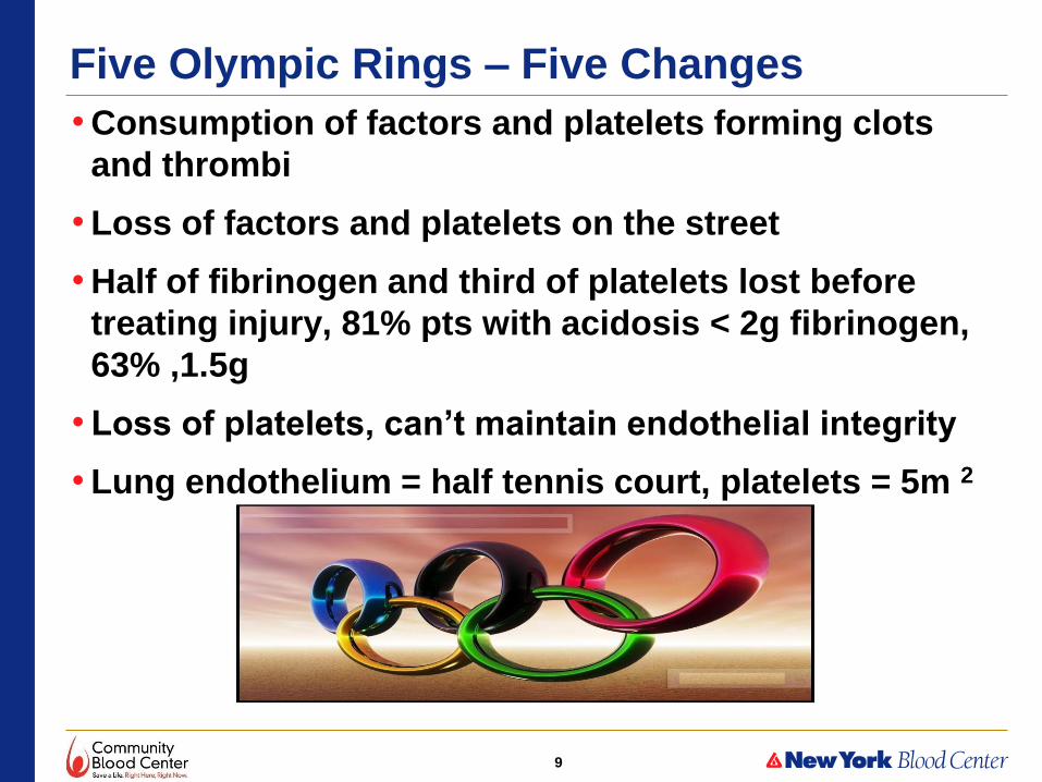
Five Olympic Rings – Five Changes
•Consumption of factors and platelets forming clots
and thrombi
•Loss of factors and platelets on the street
•Half of fibrinogen and third of platelets lost before
treating injury, 81% pts with acidosis < 2g fibrinogen,
63% ,1.5g
•Loss of platelets, can’t maintain endothelial integrity
•Lung endothelium = half tennis court, platelets = 5m 2

10
Loss of RBCs
•Loss of rbcs = loss of axial flow
•Axial flow = rbcs push platelets and factors to
vessel walls
•Hct <30% = increase in bleeding time, a few rbc
units can help coagulation mechanism
•Factor 9 activation thru rbc erythroelastase is
diminshed
•Try to keep hematocrit between 27-30%.

11
Dilution Effects
•Blood pressure drops & fluids move from tissue
to vessel
• Infusion of iv fluids leads to dilution
•Change by limiting fluids and tolerating lower
blood pressure on resuscitation
Pre-Hospital Fluids % with coagulopathy
2 liters 40%
3 liters 50%
4 liters 70%

12
What’s in that Blood?
•RBC transfusion alone leads to dilution
•1:1:1 ratio of rbc:FFP:plt = Hct. of 29%,
Plt count of 80,000, Coag factors at 65%
•Worse if you factor in storage
– Hct 26%, Plt ct. 55,000, INR of 1.4
•60% reduction in coag factors leads to
300% increase in thrombin generation,
short period of time due to lack of
antithrombin

13
Tissue Injury = Hormones & Cytokines
•Epinephrine, Vasopressin = increase thrombin
•Plasminogen activated = fibrinolysis
•Protein C activated inhibits plasminogen inactivator
•Endothelial cells activate and change from anti thrombotic to pro thrombotic = more clots
•Clots are malformed due to lower fibrinogen, more prone to lysis
• Increase permeability of cell walls and lose fluid into tissue
•Brain injury, fat or amniotic fluid embolism activate extrinsic arm of coagulation through tissue thromboplastin

14
Multiple Factors Lead to Coagulopathy

15
Triple H
• Hypoxia, low pH, Hypothermia
• Impair platelets and activator enzymes of coag factors so increase bleeding due to poor performance of coagulation system, PT and INR up
• Hypothermia – loss of body heat, cold fluids, cold rbcs
• 30C body temp – plts inactivated GP Ib, F9, vWf complex does not activate platelet
• Low pH – acidosis due to low O2, hypoperfusion, accumulation of acids – affects Vit K factors need negative charged surfaces to activate
• Long storage rbc = more acidotic due to lactate release

16
Immune System
•Platelets release CD40
•Stimulates immune system then complement
system
•Breakdown of proteins
•High level of oxidation

17
Can We Predict Coagulopathy?
•Various algorithms – none catches all at risk
•Assume more than a few rbcs = potential for coagulopathy
•Need MTP not only for ER but rest of hospital
•Standard PT, INR, PTT, Fib not designed for multiple factor deficiencies also don’t account for platelets or fibrinolysis
•Most bleeding occurs 1.4-1.7x normal range for tests
•TEG can be used for fibrinolysis
•Underestimates deficiency related to hypothermia
•Advocated to guide therapy but multiple reviews do not show decreased mortality

18
Pathophysiology
• Massive thrombin production, consumption of platelets and fibrinogen, increase fibrinolysis
• Tissue damage exposes tissue factor – local thrombin and fibrin generation
• Exposed collagen binds plt GPVI and GPIb binds vWf activating platelets
• Activated plts amplify thrombin generation
• At first, good stable clot, even hypercoagulability
• Consumption coagulopathy – reduced factors and plts with high thrombin production
• Fibrinogen and Factor V are most consumed
• Clot strength weakens
• Fibrinogen below 225 increases mortality
19
Fibrinolytic Side
•Thrombin stimulates Protein C – cleaves Factors V and VIII, consumes plasminogen activator inhibitor-1
•Leads to high levels of t-PA activity
•High thrombin stimulates endothelial t-PA, tissue plasminogen activator
•Activates plasminogen to break down clots, increased D- dimer levels
•Fibrinolysis is part of coagulopathy, see on TEG
•Use TXA, tranexamic acid to inhibit activation of plaminogen
•Lowers risk of death by 1/3
•Protein C then is consumed and hypo becomes hyper coagulable state

20
Endothelial Cell Activation
•Pro thrombotic environment feeding into consumptive coagulopathy
•Net production of plasminogen activator inhibitor overcomes plasminogen activator
•Fibrinolysis slows leading to more thrombosis
•TXA will not help in this phase
•Degradation of glycocalyx
– can cause autoheparinization
– measured by syndecan-1
– activates Protein C

21
Platelets
• Start as amplifiers of coagulation
• Shock decreases response to agonists, collagen, ADP, arachidonic acid = “exhaustion”
• Adequate number may be present but are non-functional
• Transfusion of platelets may improve response to agonists for a period of time
• Transfused platelets have Factor 5 and PAI-1 to replace
• Presence of microparticles – procoagulant – increase clot strength
• Lower plt counts on admission predict higher mortality

22
Confused???
•Multiple competing processes all using up hemostatic resources
•TEG – shows different phases of coagulopathy
• ISS 5-15 moderate severity - TEG shows hypercoagulable state
• ISS 20-40 severe pt. - TEG shows hypocoagulable state, consumption of factors and platelets, activation Protein C
• ISS 40-75 most severe pt. - TEG shows hyperfibrinolysis which correlates with mortality
23
Logistics
•Scoop and run – large majority of Americans live within 60 min of Level 1 or 2 trauma center
• IV fluids en-route, get control of airway
•No POC testing, ultrasound, transfusion, or administration of prothrombotic or antifibrinolytic drugs
•Hospital – rapid ID and treatment of active hemorrhage
•Physical exam, initial fluid bolus, rapid ultrasound and CT of chest and abdomen

24
Philosophy of Treatment
•Damage control surgery
•Rapid surgical or angiographic control of
hemorrhage
•Don’t fix bowel or orthopedics or close abdomen
•Establish normal physiology first
• 1-2 days later come back and fix anatomy
•Limit surgical time initially to 40 minutes – prevent
hypothermia, = 1 degree lost in OR

25
Initial Fluid Bolus
• Important to observe response
•BP up and stays up – no ongoing hemorrhage
•BP up and drops – active bleeder
•BP no change – terminal bleeding or non
bleeding reason for hypotension – tamponade,
tension pneumothorax or high spinal injury
•Next is transfusion – products for speed
uncrossmatched rbcs, liquid plasma, platelets

26
Differences in Protocols
•Europe uses TEG and replaces individual factors, US recreates whole blood
•US - synergistic balance of procoagulants and other factors and mediators
•Fresh whole blood is best – military experience
•Maintain hemostasis and vascular integrity – preserve endothelial glycocalyx
•Europe uses much more TXA – antifibrinolytic
•US uses but not as part of routine protocol

27
Beyond Hemostasis
• Follow acid base status, serum lactate
• Delayed clearance – more fluids
• Monitor cardiac output via echocardiography
• Use fewer rbcs in non-bleeding pt., limit iv fluids if possible, prevent dilution
• Tolerate lower Hcts and BP – keep at mean pressure 80mm
• Watch for deep vein thrombosis – hypercoagulable state
• 60% patients develop asymptomatic clots
• Patients with replacement parts are at high risk
• Use compression on legs
• May start LMWH for anticoagulation as soon as 48 hours after stable

28
Platelets
•Low plt counts first few days
•No increase after 4 days = increase risk of
death
•Sepsis, bone marrow suppression, PTP, HIT
•25% of patients, 2 weeks out, thrombocytosis
but no risk

29
MTP at St. Luke’s
•Start MTP after exam in ER by anyone on trauma team
•Call BB to initiate
• Indications to start MTP, two of the following:
– Pulse >120
– Systolic blood pressure <90
– Positive FAST – ultrasound, CT of chest & abdomen
– Penetrating wound
– Blood loss >150ml/hr.

30
MTP - BB
•6 packed rbcs
•6 FFP
•1 platelet apheresis
•Next pack – when ready or within 30 minutes
•Continues until ER, OR, or ICU calls it off
•Draw two purple tops for type and cross

31
What’s Going on in ER?
•Rapid infuser blood warmers, 16 gauge or better
•Air blanket keep patient warm
•Draw labs pre-MTP and then hourly
•Shock profile – ABG, CBC, PT, PTT, INR, fibrinogen, chemistries – Na, K, Cl, glucose, lactate, ionized Ca
•Other products available for order – cryo, TXA
•Keep fibrinogen above 200
•OB MTP – watch fibrinogen, keep >400 to prevent postpartum hemorrhage

32
Goals
•Maintain 1:1:1 ratio
•HgB – 8
• INR <1.5
•Platelets 100,000
•Stabilized hemostatic picture
•Factor 7 – tried but proved to be of no benefit,
fewer rbcs used but no better survivability

33
O+ vs O- RBCs
• Problem – not enough O negs, 6.5% in normal population vs. hospital usage of >10%
• Beth Israel Deaconess - Boston
– 268 patients in 10 year retrospective review of MTPs
– 63% male, 23% female >50, 12%<50, 86% of all patients Rh +
– 50% mortality male, 34% female in 7 days
– 18 of 39 Rh neg received Rh + blood, median 10 u, avg 12.5 u
– 8 of 18 lived > 7 days, antibody screens done, 1 of 8 had D ab
– Rate of anti D formation, 12.5% of Rh neg getting Rh pos
– Previous papers showed Anti D formation 22% not in trauma pts
• Yazer Transfusion 2007 47:2197-201 Frohn Transfusion 2003 43:893-8
• 88% of MTPs could get O+,
• Females Rh neg <50 were 1.5% of patients, received only 12% of O negs used
• Implemented O pos for all except Rh neg females < 50
Lynne Uhl Transfusion 2015 55:791-795

34
Plasma Ratios 1:1 vs 1:2 vs ? • PROMMTT
• RCT 1245 highest level trauma patients – 10 Level 1 trauma centers
• Real time data collection on infusions and interventions until resuscitation ended, also tracked in-hospital mortality, complications, subsequent treatments until death or discharge
• Increased plasma:rbc ratio associated with lower 6 hour mortality but not 24 hour or 30 day mortality
• Ratio less than 1:2 = 3-4 fold increase in risk of dying36
• 2nd analysis – time of transfusion vs ratio of plasma:rbc
• Early plasma transfusion <2.5 hours had half the mortality risk in 6hr, 24 hr and 30 day periods compared to no plasma or plasma >2.5 hours after admission
• Fewer rbc’s used in early plasma transfusion group
• Speed to plasma transfusion more important than ratio of plasma:rbc37
• PROPPR – 1:1 more patients achieve hemostasis, reduced hemorrhagic mortality at 3 hours and fewer died by exsanguination at 24 hours vs 1:2 but no differences in complications or mortality at 24 hours or 30 days38
• 86% of 177 major trauma units in TQIP use 1:1:1
36. Holcomb, JB et al. 2, 2013, JAMA Surg, Vol. 148, pp. 127-36.
37. del Junco, DJ et al. 2013, J Trauma Acute Care Surg, Vol. 75, pp. s24-30.
38. Holcomb, JB et al. 2015 , JAMA Vol. 313, pp. 471-82

35
A Plasma
• AB is universal plasma, problem is 4% of population is AB, not enough to keep thawed at all Level 1 trauma
• A plasma is the answer, compatible with 85% of population
• PROPPR trial1
– 3 of 12 sites used type A thawed
– 2 of 3 untitered, 1 titered 1:25
– 141 A units transfused to AB or B patients – no evidence of hemolysis
– 2 of 12 hospitals had 25% wastage of AB plasma
• Mayo Clinic Retrospective review 2
– 254 patients – 35 incompatible – 14%
– No difference in clinical outcomes across wide variety of indicators – Safe to use Group A, they do not titer
– Reduce AB plasma 96%
• You only need use it for as long as it takes to type the patient and thaw ABO identical plasma
• Early in resuscitation, most of the patient’s rbc will be the O rbcs you have transfused
1. Holcomb, JB et al. 2015 , JAMA Vol. 313, pp. 471-82 2. Zielinski et al J Trauma Acute Care Surg 2013 74(1) 69-74

36
Thawed vs. Liquid Never Frozen
• Thawed plasma 5 day limit after thawing
• Liquid plasma – plasma which has been refrigerated but never frozen
• Expiration is 5 days after expiration of wb anticoagulant
– cpd/additive – 26 days, cpda-1 - 40 days
• FDA licensed, available since 1940’s
• Used in Sweden interchangeably for over 30 years, storage to 14 days only, roughly 1/3 liquid, 2/3 FFP
• 10 yr observational study 1
• 90k pts, 350k units – no difference in clinical outcomes between FFP and liquid regardless of age of liquid even beyond 15 days
1. Norda et al J Trauma 2012 72(4) 954-961

37
Liquid Plasma Profile
• 0-30 days factor activity, At least 50% or more activity in all factors at day 15
• Minimal changes in FII, FX, FXIII
• FBG, FIX, FXI – no change to day 5, significant reduction after day 20
• FXII – no change to day 5 then increase afterward similar pattern in FVII – cold activation
• vWF, FV, FVII, FVIII – no change to day 5, significant difference by day 15, 30% decline vs. day 1, still at 50% or better at day 15
• No change in AT, PLG, PC but significance drop in PS but remained at 53% level
• Increase in PT and aPTT by 2 sec, over 30 days, significance reached at day 15
• Recommendation – limit use to less than 15 days of age, use with FFP where feasible in MTP
Gosselin Transfusion 2013 53(3) 579-590

38
Liquid vs. Thawed
• Compare thrombin generation and clot kinetics liquid plasma and FFP at day 0 and storage limit
• Thrombogram at day 0 showed liquid plasma higher than thawed FFP in endogenous thrombin potential
• Higher performance continued until day 26 when liquid plasma equals thawed plasma on day 0
• Liquid retained 86% of day 0 potential at day 26
• TEG – Liquid had higher MA, G and TTG values at day 0 than thawed plasma
• At end of storage both were equal
• PT increased 2.2 seconds at day 26, aPTT increased 3.1 seconds at day 26 for liquid plasma
• All Factors on day 26 at 88% of day 0 except FV and FVIII, 39% and 60% resp.
• All inhibitors stable at day 26 except PS, 29%
• Initial hemostatic profile better in liquid than in thawed at day 0
• Residual platelet count 1.5x higher in liquid than thawed – better initial clot formation
• As platelets age, release vWf and microparticles, aiding thrombin formation
• Freezing plasma destroys platelets
• Explains why TEG and thrombin generation are better in liquid plasma
• Done by the trauma center in Houston which uses liquid plasma instead of thawed FFP
• What do you prefer, better coag factor percentages or better thrombin and clots?????
Matijevic J Trauma Acute Care Surg 74(1) 84-91

39
Recommendation
• O+ rbc – all males, females >50
• Group A plasma
• Speed to transfusion most important
• Try to maintain 1:1:1 ratio
• Liquid plasma, limit to 14 day storage
– Nothing wrong with thawed plasma but requires more inventory management
• MTP is a relay race. O+ rbc, Group A plasma, liquid plasma get you off to a fast start when time is blood lost.
• Pass the baton to ABO identical rbcs and fresh thawed FFP as the finishers.

40
What’s Better?
•6 pack MTP or 6 pack beer and 6 pack abs
•Depends on side of the needle you’re on
•Patient likes 6 pack MTP
•Doctor likes 6 pack beer after working out 6 pack
abs
•But remember: