Embed Size (px)
Citation preview

PHOTOTHERAPY OF GINGIVITIS: PILOTCLINICAL STUDY
ELINA A. GENINA*,¶, VLADIMIR A. TITORENKO†,VALERY V. TUCHIN*,‡, GEORGY V. SIMONENKO*,ALEXEY N. BASHKATOV*, GENNADY M. SHUB†,
ALEXANDER V. LEPILIN†, ILYA V. YAROSLAVSKY§
and GREGORY B. ALTSHULER§
*Saratov State University, Saratov, 410012, Russia
†Saratov State Medical University, Saratov, 410012, Russia
‡Institute of Precise Mechanics and Control of RASSaratov, 410028, Russia
§Palomar Medical Technology Inc.Burlington, MA 01803, USA
Accepted 18 July 2011
The goal of this work was to evaluate the safety and e±cacy of the Red Light Emitted Tooth-brush (R-LETB) emitting at wavelength of 663 nm with power density of 3.3mW/cm2 incombination with 0.1%-methylene blue (MB) solution for the reduction of plaque and treatmentof gingivitis. A microbiological in vitro study and a pilot clinical study were conducted. Themicrobiological study has shown total suppression of pathogenic °ora after a 3-min exposure tothe dye solution followed by a 20-sec treatment with the R-LETB. For the clinical study, 37subjects of both sexes with gingivitis were enrolled and randomly assigned to one of two groups.Subjects in the ¯rst (treatment) group were instructed to rinse their mouth with MB solutionprovided for 1min and then brush the teeth with the R-LETB and standardized toothpaste. Thesecond (control) group used only the toothpaste and a regular toothbrush. Subjects in bothgroups followed their respective procedures 2 times a day (morning and evening) for 30 days.Indices of plaque, gingival bleeding, and in°ammation were evaluated at 14-day and 30-daytimepoints. In the both groups, all indices improved in comparison with baseline. However, thetreatment group demonstrated more pronounced improvement of the studied indices that wasattributed to additional anti-microbial action of red light and MB on gum tissue. Thus, the use ofR-LETB with MB appears to have a multifactor therapeutic action on oral pathologicalmicro°ora: mechanical removal of the bacteria and suppressing action on microorganisms due tophotodynamic reaction.
Keywords: Tooth brushing; methylene blue; photodynamic therapy; pathogenic bacteria.
¶Corresponding author. Optics and Biophotonics Department, Saratov State University, 83, Astrakhanskaya str., Saratov, 410012,Russia. E-mail: [email protected]
Journal of Innovative Optical Health SciencesVol. 4, No. 4 (2011) 437�446#.c World Scienti¯c Publishing CompanyDOI: 10.1142/S1793545811001745
437

1. Introduction
Periodontal disease is an acute medical problem.According to WHO statistics, about 95% of adultsand 80% of children worldwide su®er from someform of periodontal disease. Severe periodontaldisease, which may result in tooth loss, is found in5%�20% of middle-aged adults.1 The main cause ofthe periodontal disease is vital activity of con-ditionally pathogenic micro°ora of oral cavity.2,3
Currently, the main method of both prophylaxisand treatment of catarrhal gingivitis is a rigorousoral hygiene regime with the use of toothbrush andtoothpaste.4,5 However, more e®ective aids of indi-vidual hygiene are desirable.
Antibiotic therapy is sometimes used to controlperiodontal disease.6,7 However, medicamentaltherapy is insu±ciently e®ective and not alwaysjusti¯ed. This is caused by the following factors: highfrequency of allergic reactions, contra-indicationsand side e®ects to prescription drugs; resistance ofmicro°ora to the widely used antibiotics and anti-septics; and adverse e®ects on benign micro°ora oforal cavity.8,9
Antibacterial e®ects of light of di®erent wave-lengths used to illuminate mucosa have beenreported in the literature.10�13 Violet and blueportions of the spectrum are the most e®ective vis-ible wavelengths for photoactivation of the majorendogenous porphyrins of bacteria but have poorpenetration depth into tissue.14 Light of infraredwavelengths is absorbed by cellular water and may
lead to overheating and even fragmentation of cel-lular structure.12,13 The adverse e®ects associatedwith overheating include thermal damage of rootsurfaces and production of toxic by-products.13
The red light penetrates deeper but is less e±-cient in photoactivating of endogenous porphyrins.However, in combination with an exogenous sensi-tizer such as methylene blue (MB) red light maycontrollably produce free radicals for soft photo-dynamic therapy,15�19 including gingival treatments.Antimicrobial e±cacy of combined application ofHe-Ne laser radiation (wavelength 632.8 nm) and0.1%MB solution to elective anaerobic component oftotal micro°ora of periodontal pockets has beenreported in Ref. 20.
Absorption bands of MB solution are centered atthe wavelengths of 610 and 664 nm (see Fig. 1),therefore MB can be e±ciently excited by 663 nmemission of LEDs. This red light penetrates wellinto tissue, much deeper than shorter visible andUV wavelengths.15 Red light may also have ad-ditional photobiomodulating e®ect through in°u-encing release of proin°ammatory cytokines frommacrophages, which stimulate ¯broblast prolifer-ation and production of growth factors.21 Use of MBis bene¯cial due to its low toxicity,15 low cost, andavailability.
We propose the combined use of low-intensity redLED toothbrush emitting at wavelength of 663 nmand 0.1% MB solution as a novel tool of individualoral hygiene. Brushing of teeth in combination with
(a) (b)
Fig. 1. R-LETB in a charger (a) and the emission spectrum of the R-LETB (b). Insert shows absorption spectrum of MB rinsesolution.
438 E. A. Genina et al.

phototherapy can provide a multifactor therapeuticaction. The goal of this pilot study was to evaluatesafety and e±cacy of the treatment for reduction oftooth plaque and improvement of gingivitis.
2. Materials and Methods
2.1. Light-emitting toothbrush
For microbiological and clinical study, Red LightEmitting Toothbrushes (R-LETBs) with 10 opticalbristle bundles and photon-recycling mirror wereused. The central wavelength was 663 nm; thespectral width was about 30 nm. The power densityof the R-LETBs was 3.3mW/cm2. The photographand the emission spectrum of the R-LETB arepresented in Fig. 1. R-LETBs were designed andmanufactured by the Laser Center of St. PetersburgState University of Information Technologies,Mechanics and Optics (St. Petersburg, Russia) incooperation with Palomar Medical TechnologiesInc. (Burlington, MA, USA).
2.2. Microbiological study
Samples of subgingival plaque have been obtainedclinically by standard dental applicator and storedin 1ml of isotonic solution. About 0.1ml ofobtained substance has been diluted in 0.9ml ofsugar substrate. The 0.05ml of the suspension hasbeen seeded on Petri dish with sugar substrate andplaced into thermostat with temperature 37�C for24 h. Multisectional plate for irradiation of sus-pensions of cultures was used (volume of each cellwas 0.2ml). Three bacterial strains were studied:Micrococcus, Stomatococcus, and Staphylococcuslugdensis.
For the study of MB action on the pathogenicsubgingival micro°ora, 0.1 ml of MB has been putinto base substrate to obtain ¯nal concentrations ofthe dye at 0.001%, 0.01%, and 0.1%. Time of MBaction was 3min.
To ¯nd the appropriate staining and radiationparameters for photodynamic inactivation ofpathogenic subgingival micro°ora using R-LETBirradiation of bacteria photosensitized by MB, threesampleswere used. The surfaces of three cover glasseshave been covered by the bacterial suspension(0.05ml on each glass). The glasses with suspensionhave been put into thermostat with temperature
37�C for 30min. The ¯rst sample served as a control,the second one was used for mechanical treatment,and the third one was subjected to both mechanicaland photodynamic treatments.
The ¯rst glass with dried suspension has been putinto bath with 2ml of isotonic solution. About0.1ml of the obtained substance has been thendiluted in 0.9ml of sugar substrate. The secondsample has been treated by toothbrush with tooth-paste (six circular motions). The glass with sus-pension has been put into bath with isotonicsolution. The third glass with the dried suspensionhas been treated by 0.1%-MB solution during 3min.The suspension has been treated by R-LETB andtoothpaste (six circular motions). Then the glasswith emulsion has been put into bath with isotonicsolution.
After the treatments, 10X consecutive dilutions(from 10�2 to 10�7) of all samples have been pre-pared. Test tubes with dilutions have been put intothermostat with temperature 37�C for 24 h.
2.3. Clinical study
A total of 37 subjects of both genders with catarrhalgingivitis were randomly divided into 2 groups. The¯rst (treatment) group included 7 volunteers(3 females and 4 males) who were treated withR-LETB and MB; and the remaining 30 subjects(16 females and 14 males) formed the second (con-trol) group, which used standard toothbrushes\Braun Oral-B" (Procter & Gamble, Cincinnati,OH, USA). The average age of volunteers was21 years. The severity of gingivitis was moderate(35% of volunteers) or light (65%). People wereexcluded if they had diseases of internal organs ororthodontic pathology. In total, 320 persons werescreened for the study; frequency of catarrhal gingi-vitis was found to be about 9%. All subjects gavetheir informed consent for participation in the study.
This study was designed as a split-mouth, single-blind, randomized, prospective clinical study. Priorto the start of the study, all subjects received the R-LETB or the \Braun Oral-B" toothbrush accordingto their assigned group and standard toothpaste\Blend-a-Med cavity protection mineral action"(Procter & Gamble, Cincinnati, OH, USA). Allsubjects from the ¯rst group were instructed how touse the R-LETB. Instructions were given verballyand followed by a demonstration.
Phototherapy of Gingivitis: Pilot Clinical Study 439

For both groups, the method of the brushing wassimilar. Subjects were instructed to brush two timesa day: in the morning and in the evening, withtoothpaste \Blend-a-Med cavity protection mineralaction" for 3min. The ¯rst group of the volunteersused the paste with the prior rinsing of oral cavityfor one minute by 0.1% MB solution. All clinicalexaminations were performed by the same examinerusing the same dental units and operating lamp. Atthe time of examination, the examiner was unawareof the subject's group assignment. Records of earlierexaminations were not available to the examiner atthe time of re-examination. Treatment e®ects weredetermined using the comparison of the patient'sscores from each follow-up visit to the baselinescores, which were also documented using NikonCoolpix 990 (Japan) digital camera.
Clinical evaluation of changes in gingivitis com-pared with the baseline was visually assessed usingoriginal augmented Approximate Hygiene Index(AHI)22 and standard indices complying with theAmerican Dental Association Acceptance ProgramGuidelines23: Gingival Bleeding IndexADA (GBI),24
and Gingivitis Index PMA.25 Gingiva papilla is themain part of a gum where in°ammation sets in,which then extends on gingiva marginalis and gin-giva alveolaris. Besides, plaque retention in aninterdental space promotes the development ofapproximal caries. Used hygienic indices allowquantitative valuation of tooth plaque on bothfacial and lingual surfaces of teeth26,27 as well asqualitative evaluation of the presence of plaque on acontact surface.28
We have used augmented AHI22 based on themethod of evaluation of Turesky modi¯cation of theQuigley-Hein plaque index,27 but instead of evalu-ation of facial and lingual surface, medial and distaltooth surfaces were evaluated. Tooth staining with1% rosein solution was used to visualize the plaque.To evaluate AHI, a score of 0 to 5 was assigned toeach medial and distal surface of all the teeth exceptthird molars, as follows: 0, no plaque; 1, separate°ecks of plaque at the cervical margin of the tooth;2, a thin continuous band of plaque (up to one mm)at the cervical margin of the tooth; 3, a band ofplaque wider than one mm but covering less thanone-third of the crown of the tooth; 4, plaque cov-ering at least one-third but less than two-thirds ofthe crown of the tooth; 5, plaque covering two-thirds or more of the crown of the tooth. An index
for the entire mouth was determined by dividing thetotal cumulative score by the number of surfacesexamined:
Index ¼ Total score
the number of surfaces examined: ð1Þ
Evaluation of gingival bleeding degree by theGingival Bleeding Index (GBI) suggested by Cowellet al.24 was carried out in area of 12, 16, 24,32, 36, and 44 teeth on both vestibular and oralsurfaces by button or special dulled probe. Thetip of the probe was pressed against the wall ofthe sulcus and passed slowly from medial to distalside of the tooth. The state of the gingival bleedingwas graded on a scale from 0 to 3: 0, gingivalbleeding was absent; 1, gingival bleeding appearednot earlier than in 30 sec; 2, gingival bleedingappeared right away or during 30 sec; and 3, gin-gival bleeding appeared during food intake or toothbrushing.
To evaluate the Gingival Index PMA, the gin-giva was divided in three regions: papilla inter-dentalis (P), gingiva marginalis (M), and gingivaalveolaris (A). A Shiller�Pisarev probe was used forgum staining (solution of 1 g crystal iodine, 2 gpotassium iodide, and 40ml distillate water). Gumcolor changes depending on the in°ammatory state:for a healthy gum, mucosa is straw-yellow colored,at chronic in°ammation due to glycogen store it isbrown colored. By intensity of staining stages ofin°ammation were di®erentiated: negative, straw-yellow colored; light-positive, light-brown colored;positive, brown colored; In°ammation of the papillawas evaluated as Grade 1, in°ammation of marginalgingiva as Grade 2, and in°ammation of alveolargingiva was evaluated as Grade 3. All highestgrades for each tooth were summarized25:
PMA ¼ ½ð� GradesÞ � 100%�ð3� number of teethÞ : ð2Þ
The value of the index up to 30% correspondedto gingivitis of mild degree; 30%�60% to gingivitisof moderate degree, and more than 60% to gingivitisof severe degree.
Subjects completed a ¯nal self-assessment ques-tionnaire based on the overall product e®ectiveness.Subjects were asked to compare the state of theirteeth's and gingiva's health at the end of thetreatment period to that at the baseline.
440 E. A. Genina et al.

3. Results and Discussion
3.1. Treatments of dental plaque
samples
Figure 2 shows action of MB solution on micro°oraof tooth plaque. It can be clearly seen that 3-minstaining of the samples with di®erent solutions ofMB did not in°uence by itself (in darkness) thenumber of microbial colonies. The control numberof the colonies was 7:6� 107. After staining by MBsolutions with concentrations of 0.001%, 0.01%, and0.1%, the average number of the colonies was about6:4� 107, 8:2� 107, and 7:6� 107, respectively.Thus, the staining alone at the used concentrationof MB did not suppress the micro°ora of plaques.
Average values of bacterial seeding rate of toothplaques for di®erent treatments are shown in Fig. 3.The ¯rst column corresponds to the control groupwithout any treatment. The result of mechanicalaction by the standard toothbrush alone is pre-sented by the second column. The third columnshows combined e®ect of MB exposure in 3min,mechanical action and red light irradiation in15�20 sec on dental plaque bacteria. The mechan-ical treatment decreased the number of the coloniesup to 91.2% in comparison with the control sampledue to the mechanical removal of the bacteria.However, the staining and irradiation with simul-taneous mechanical action decreased the total
number of colonies up to 99.9%. Thus, practicallytotal suppression of the pathological °ora wasobserved.
It was previously reported in the literature thatMB with red light had a high photobactericidalactivity against S. aureus, S. epidermidis, S. pyo-genes,16,17,29P. aeruginosa,30 and other pathogens.Proteins and lipids forming the cell membranes areeasy to be photooxidized. This process leads to theloss or change of the functions of photodamagedbiological molecules, which causes morphologicalchanges in the cell. Thus, MB sensitizes damage ofthe membrane under the action of red light in thevicinity of the MB molecule localization. Thedamage of the membrane may cause the photo-sensitizer molecule to move away from the place ofits localization, its di®usion inside and redistribu-tion within the cell, and the damage of secondarytargets such as mitochondria and the Golgi complexof the cell.31,32
The use of R-LETB combined with MB resultedin multifactor therapeutic action on oral patho-logical micro°ora: besides mechanical removal ofthe bacteria as in ordinary tooth-brushing pro-cedure, it has shown additional suppression actionon microorganisms due to photodynamic reaction.
3.2. Treatments of subjects
No adverse side e®ects were observed. Figure 4presents the dynamics of the average value of plaque
Fig. 2. Average value of in vitro measured bacterial colonyforming rate (number of the colonies) of bacteria taken fromtooth plaque after exposure to MB solution. The ¯rst columncorresponds to control sample (without staining); the second,third and fourth columns to 3-min duration of 0.1%, 0.01%, and0.001% MB solution action, respectively.
Fig. 3. Average value of in vitro measured bacterial colonyforming rate (number of the colonies) of bacteria taken fromtooth plaque: without treatment (control), after mechanicaland photodynamic treatments.
Phototherapy of Gingivitis: Pilot Clinical Study 441

index AHI (Approximate Hygiene Index). The indexcharacterizes the area of the tooth covering by pla-ques. It is seen well that due to treatment the indexdecreased for both groups of the subjects. The mostsigni¯cant reduction was observed on the 14th day ofthe treatment (in about 2 times for the tested groupand 1.2 times for the control group). At the 30 daystimepoint, the decrease of the index values was only1.6 times for the tested group and 1.1 times for thecontrol group. The improvement tooth status (I )was calculated with the formula:
I ¼Baseline index value
� Current index value
� �
Baseline index value; ð3Þ
where Current index values were scored for the 14thand the 30th days.
The improvement of the tooth status for thepatients from the tested group on the 14th and 30thdays in comparison with the baseline was 35% and16% for AHI, respectively. For comparison, theimprovement of the patients from the control groupwas 18% and 11% only. The dynamics of the plaqueindex behavior in both groups can be explained by\learning e®ect." It has been discussed earlier33 thatprofessional instructions regarding correct tooth-brushing procedure promoted better results in pla-que removal. Thus, in the course of the presentstudy, a decrease in the plaque index in the controlgroup was also observed. By the end of the study
the values of the index started to go up, i.e., thetooth plaque increased. Apparently, it was theresult of the partial loss of mechanical properties ofthe brushes. When the subjects used their toothbrushes for a month (to the end of the study), theybecame softer, bristle bundles of the brushes dis-oriented and caused less gingival abrasion as aresult of brushing. Besides, it is possible that in thebeginning of the study the subjects brushed theirteeth carefully and ful¯lled all requirements of theinvestigator, but by the end of the treatment periodthey reverted to their usual manner of toothbrushing. Nevertheless, in spite of the partialincrease of the plaque for the tested group, the toothstatus in that group was better than that of thecontrol group (even though standard toothbrusheskept their high quality during whole study to ahigher degree than R-LETB). It also should benoted that R-LETB construction does not allow oneto provide the °exibility and good mechanical con-tact with oral cavity tissues at brushing as standardtoothbrush did.
Gingival bleeding index has shown more signi¯-cant di®erences in two studied groups (Fig. 5). Theaverage value of the GBI in the tested groupdecreased more than 2 times in 14 days and morethan 3 times in 30 days, whereas in the controlgroup the decrease was about 1.6 and 1.5 times,respectively, at 14 and 30 days timepoints. Thus,after 14 days the improvement of bleeding index
Fig. 4. Dynamics of AHI (Approximate Hygiene Index). Lightgrey and dark grey columns correspond to the results of toothbrushing by R-LETB with methylene blue prior rinsing andstandard toothbrush, respectively. Bars show standard deviation.
Fig. 5. Dynamics of GBI (Gingival Bleeding Index). Lightgrey and dark grey columns correspond to the results of toothbrushing by R-LETB with methylene blue prior rinsing andstandard toothbrush, respectively. Bars show standard deviation.
442 E. A. Genina et al.

evaluated with the help of the formula (3) was 52%in the tested group and 37% in the control one.After a month the improvement of GBI increasedup to 68%, and decreased to 35% in comparisonwith baseline in the tested and control groups,respectively.
This correlates with dynamics of gingivalin°ammation development presented in Fig. 6. Theaverage value of the index PMA in the tested groupdecreased more than 4 times in 14 days and 6.6times in 30 days, whereas in the control group thedecrease was 2 and 3.4 times respectively in 14 and30 days. As a result of the brushing by the R-LETB,the improvement of gum in°ammation was 76% in14 days and 84% in 30 days of the treatment.Control group has shown improvement by 52% and70% in 14 and 30 days, respectively.
In°ammatory periodontal disease is caused bydental plaque, which is a bio¯lm. Bacterial endo-toxins, cytotoxins, and other pathogenic substancesare released from the bio¯lm and di®use into theadjacent soft tissues where they elicit an in°am-matory response that results in tissue disruptionand degradation.34�36 Therefore, removal of plaqueand plaque-derived products has been a key to thetreatment of periodontal disease.36,37 Result of theproper tooth brushing was the decreasing in°am-mation in both groups of the volunteers. Since thebleeding is caused by gingival in°ammation, the
decreasing of in°ammation process leads to signi¯-cant improvement of the status of gums. However,in the 30th day of the study, the di®erences inthe GBI between the patients from the ¯rst and thesecond groups increased. It can be related to theadditional anti-in°ammatory action of red light andMB on gum tissue. It was found that bleeding ofgums on probing is the most sensitive method todetect di®erences in the development of gingivitisbetween experimental groups.38 Our study hasshown that e®ectiveness of photodynamic therapyof gingivitis can be evaluated by both GBI andPMA indices.
Figure 7 illustrates the improvement of the sta-tus of tooth plaque and gingival in°ammationduring the combined treatment by R-LETB andMB. AHI allows for evaluating the size of toothplaque. It can be seen that at the baseline plaquecovered the main part of tooth, and the color of theplaques is saturated brown. During the treatment,
Fig. 6. Dynamics of Gingival Index PMA (P, papilla inter-dentalis; M, gingiva marginalis; A, gingiva alveolaris). Lightgrey and dark grey columns correspond to the results of toothbrushing by R-LETB with methylene blue prior rinsing andstandard toothbrush, respectively. Bars show standarddeviation.
(a)
(b)
Fig. 7. Images of a subject's teeth before the treatment byR-LETB: (a) 14 days into the study, (b) 30 days into thestudy, (c) arrows mark areas of gum stained by Shiller�Pisarevprobe. Intensity of brown color shows degree of in°ammationprocess.
Phototherapy of Gingivitis: Pilot Clinical Study 443
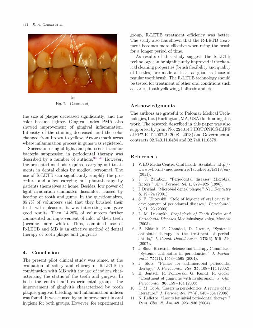
the size of plaque decreased signi¯cantly, and thecolor became lighter. Gingival Index PMA alsoshowed improvement of gingival in°ammation.Intensity of the staining decreased, and the colorchanged from brown to yellow. Arrows mark areaswhere in°ammation process in gums was registered.
Successful using of light and photosensitizers forbacteria suppression in periodontal therapy wasdescribed by a number of authors.39�42 However,the presented methods required carrying out treat-ments in dental clinics by medical personnel. Theuse of R-LETB can signi¯cantly simplify the pro-cedure and allow carrying out phototherapy bypatients themselves at home. Besides, low power oflight irradiation eliminates discomfort caused byheating of tooth and gums. In the questionnaires,85.7% of volunteers said that they brushed theirteeth with pleasure, it was interesting and gavegood results. Then 14.28% of volunteers furthercommented on improvement of color of their teeth(became more white). Thus, combined use ofR-LETB and MB is an e®ective method of dentaltherapy of tooth plaque and gingivitis.
4. Conclusion
The present pilot clinical study was aimed at theevaluation of safety and e±cacy of R-LETB incombination with MB with the use of indices char-acterizing the status of the teeth and gingiva. Inboth the control and experimental groups, theimprovement of gingivitis characterized by toothplaque, gingival bleeding, and in°ammation indiceswas found. It was caused by an improvement in oralhygiene for both groups. However, for experimental
group, R-LETB treatment e±ciency was better.The study also has shown that the R-LETB treat-ment becomes more e®ective when using the brushfor a longer period of time.
As results of this study suggest, the R-LETBtechnology can be signi¯cantly improved if mechan-ical cleaning properties (brush °exibility and qualityof bristles) are made at least as good as those ofregular toothbrush. The R-LETB technology shouldbe tested for treatment of other oral conditions suchas caries, tooth yellowing, halitosis and etc.
Acknowledgments
The authors are grateful to Palomar Medical Tech-nologies, Inc. (Burlington,MA,USA) for funding thiswork. The research described in this paper was alsosupported by grant No. 224014 PHOTONICS4LIFEof FP7-ICT-2007-2 (2008�2013) and Governmentalcontracts 02.740.11.0484 and 02.740.11.0879.
References
1. WHO Media Centre, Oral health. Available: http://www.who.int/mediacentre/factsheets/fs318/en/(2011).
2. J. J. Zambon, \Periodontal diseases: Microbialfactors," Ann. Periodontol. 1, 879�925 (1996).
3. I. Drizhal, \Microbial dental plaque," New Dentistry8, 19�24 (2001).
4. S. B. Ulitovskii, \Role of hygiene of oral cavity indevelopment of periodontal diseases," Periodontol.3, 21�23 (2000).
5. L. M. Lukinykh, Prophylaxis of Tooth Caries andPeriodontal Diseases, Meditsinskaya kniga, Moscow(2003).
6. P. Bidault, F. Chandad, D. Grenier, \Systemicantibiotic therapy in the treatment of period-ontitis," J. Canad. Dental Assoc. 173(6), 515�520(2007).
7. J. Slots, Research, Science and Therapy Committee,\Systemic antibiotics in periodontics," J. Period-ontol. 75(11), 1553�1565 (2004).
8. J. Slots, \Primer for antimicrobial periodontaltherapy," J. Periodontol. Res. 35, 108�114 (2002).
9. H. Jentsch, R. Pomowski, G. Kundt, R. G€ocke,\Treatment of gingivitis with hyaluronan," J. Clin.Periodontol. 30, 159�164 (2003).
10. C. M. Cobb, \Lasers in periodontics: A review of theliterature," J. Periodontol. 77(4), 545�564 (2006).
11. N. Ra®etto, \Lasers for initial periodontal therapy,"Dent. Clin. N. Am. 48, 923�936 (2004).
(c)
Fig. 7. (Continued)
444 E. A. Genina et al.

12. S. Parker, \Lasers and soft tissue: Periodontaltherapy," Brit. Dental J. 202(6), 309�315 (2007).
13. R. Chanthaboury, T. Irinakis, \The use of lasers forperiodontal debridement: Marketing tool or proventherapy?" J. Can. Dent. Assoc. 71(9), 653�658(2005).
14. V. V. Tuchin, Tissue Optics: Light ScatteringMethods and Instruments for Medical Diagnosis,SPIE Press, Bellingham (2007).
15. A. Ruck, K. Heckelsmiller, N. Akgun, G. Beck,K. Kunzi-Rapp, E. Schick, R. Steiner, \Nonlineardynamics of intracellular methylene blue duringlight activation of cell cultures," Photochem. Pho-tobiol. 66(6), 838�841 (1997).
16. M. N. Usacheva, M. C. Teichert, M. A. Biel,\Comparison of the methylene blue and toluidineblue photobactericidal e±cacy against gram-posi-tive and gram-negative microorganisms," LasersSurg. Med. 29, 165�173 (2001).
17. E. A. Genina, A. N. Bashkatov, E. E. Chikina, A. B.Knyazev, O. V. Mareev, V. V. Tuchin, \Methyleneblue mediated laser therapy of maxillary sinusitis,"Laser Physics 16(7), 1128�1133 (2006).
18. F. Aghahosseini, F. Arbabi-Kalati, L. A. Fashtami,G. E. Djavid, M. Fateh, J. M. Beitollahi,\Methylene blue-mediated photodynamic therapy:A possible alternative treatment for oral lichenplanus," Lasers Surg. Med. 38, 33�38 (2006).
19. S. George, A. Kishen, \Photophysical, photo-chemical, and photobiological characterization ofmethylene blue formulations for light-activated rootcanal disinfection," J. Biomed. Opt. 12(3), 034029(2007).
20. V. A. Titorenko, Antimicrobial Action of Helium-Neon Laser Irradiation on Periodontal PocketMicro°ora Sensitized by Methylene Blue, Ph.D.thesis, Saratov (2002).
21. S. Yong, P. Bolton, M. Dyson, W. Harvey, C.Diamantopoulos, \Macrophage responsiveness tolight therapy," Lasers Surg. Med. 9, 497�505 (1989).
22. A. V. Lepilin, V. A. Titorenko, \Augmented Ap-proximate Hygiene Index. New technologies indentistry and implantology," Proc. 8th All-RussianConference, May 23�24, 2006, Saratov StateTechnical University (2006) 243�244.
23. http://www.ada.org/prof/resources/positions/standards/denmat.asp#ada
24. C. R. Cowell, C. A. Saxton, A. Sheiham, B. J.Wagg, \Testing therapeutic measures for control-ling chronic gingivitis in man: A suggested proto-col," J. Clin. Periodontol. 2, 231�240 (1975).
25. I. Schour, M. Massler, \Gingival disease in postwarItaly (1945). I. Prevalence of gingivitis in variousage groups," J. Am. Dental Assoc. 35, 475�482(1947).
26. J. Silness, H. L€oe, \Periodontal disease in preg-nancy. II. Correlation between oral hygiene andperiodontal condition," Acta Odontol. Scand. 22,121�135 (1964).
27. S. Turesky, N. D. Gilmore, I. Glickman, \Reducedplaque formation by chloromethyl analogue of vic-tamine C," J. Periodontol. 41, 41�43 (1970).
28. E. M. Rateitschak, \Gingivectomy (GV) and Gin-givoplasty (GP)," in Color Atlas of Dental Medicine1-Periodontology, p. 288, Thieme Medical Publish-ers, Inc., New York (1989).
29. B. Zeina, J. Greenman, D. Corry, W. M. Purcell,\Cytotoxic e®ect of antimicrobial photodynamictherapy on keratinocytes in vitro," Br. J. Dermatol.146, 568�573 (2002).
30. M. N. Usacheva, M. C. Teichert, Y. M. Usachev,C. E. Sievert, M. A. Biel, \Interaction of the pho-tobactericides methylene blue and toluidine bluewith a °uorophore in Pseudomonas aeruginosacells," Lasers Surg. Med. 40, 55�61 (2008).
31. A. A. Krasnovsky, Jr. \Photodynamic actionand singlet oxygen," Biophysics 49(2), 305�321(2004).
32. A. S. Sobolev, A. A. Rozenkranz, D. G. Gilyazova,\Approaches to directed intracellular delivering ofphotosensitizers for increase of their e±cacy andcell speci¯city," Biophysics 49(2), 351�379 (2004).
33. G. A. van der Weijden, M. F. Timmerman, M.Piscaer, Y. Ijzerman, U. van der Velden,\Oscillating/rotating electric toothbrushes com-pared: Plaque removal and gingival abrasion,"J. Clin. Periodontol. 28, 536�543 (2001).
34. R. C. Page, S. O®enbacher, H. E. Schroeder, G. J.Seymour, K. S. Kornman, \Advances in the patho-genesis of periodontitis: Summary of developments,clinical implications and future directions," Period-ontol. 2000 14, 216�248 (1997).
35. K. S. Kornman, R. C. Page, M. S. Tonetti, \Thehost response to the microbial challenge in period-ontitis: Assembling the players," Periodontol. 200014, 33�53 (1997).
36. J. M. de Almeida, L. H. Theodoro, A. F. Bosco,M. J. H. Nagata, M. Oshiiwa, V. G. Garcia, \In vivoe®ect of photodynamic therapy on periodontal boneloss in dental furcations," J. Periodontol. 79(6),1081�1087 (2008).
37. W. A. Jones, T. J. O'Leary, \The e®ectiveness ofin vivo root planning in removing bacterial endo-toxin from the roots of periodontally involvedteeth," J. Periodontol. 49, 337�342 (1978).
38. D. S. Barendregt, M. F. Timmerman, U. van derVelden, G. A. van der Weijden, \Comparison of thebleeding onmarginal probing index and the Eastmaninterdental bleeding index as indicators of gingivitis,"J. Clin. Periodontol. 29, 195�200 (2002).
Phototherapy of Gingivitis: Pilot Clinical Study 445

39. S. Sarkar, M. Wilson, \Lethal photosensitization ofbacteria in subgingival plaque from patients withchronic periodontitis," J. Periodontal. Res. 28,204�210 (1993).
40. M. Bhatti, A. MacRobert, S. Meghji, B. Henderson,M. Wilson, \E®ect of dosimetric and physiologicalfactors on the lethal photosensitization of Porphyr-omonas gingivalis in vitro," Photochem. Photobiol.65, 1026�1031 (1997).
41. N. K€omerik, \In vitro killing of Porphyromonasgingivalis by toluidine blue-mediated photo-sensitization in an animal model," Antimicrob.Agents Chemother. 47, 932�940 (2003).
42. E. Bornstein, \Method and dosimetry for thermo-lysis and removal of bio¯lm in the periodontalpocket with near infrared diode lasers: A casereport," Dent. Today 24(4), 64�70 (2005).
446 E. A. Genina et al.