Embed Size (px)
Citation preview

LYMPHO
LOG
YPHLEBON°44
ISSN 1286-0107
The role of matrix . . . . . PAGE 231metalloproteinases (MMPs) and their inhibitors in venous leg ulcer healing
Y. HEROUY (FREIBURG, GERMANY)
Classification of primary varicose veins . . . . . PAGE 244of the lower extremities: a consensus
statement from Latin AmericaR. SIMKIN (BUENOS AIRES, ARGENTINA),
J. ULLOA (SANTA FE DE BOGOTA, COLOMBIA)
Lymphatic function in severe . . . . . PAGE 253chronic venous insufficiency
I. C. PEARSON, P. S. MORTIMER (LONDON, UK)
1st Latin American Consensus . . . . . PAGE 258 on the management of lymphedema
J. L. CIUCCI (BUENOS AIRES, ARGENTINA)
2 0 0 4 • N o 4 4 • p . 2 3 1 - 2 6 7

Advisory boardAIMS AND SCOPE
H. PARTSCH, MDPast President of the Union Internationale de PhlébologieBaumeistergasse 85, A 1160 Vienna, Austria
C. ALLEGRA, MDHead, Dept of AngiologyPresident of the Union Internationale de PhlébologieHospital S. Giovanni Via S. Giovanni Laterano, 155 - 00184, Rome, Italy
P. COLERIDGE SMITH, MDSenior Lecturer and Consultant Surgeon, University College London Medical SchoolThe Middlesex Hospital Mortimer Street - London W1N 8AA, UK
M. COSPITE, MDHead, Dept of AngiologyUniversity Clinic, Palermo, Italy
G. JANTET, MDConsultant Vascular SurgeonPast President of the Union Internationale de Phlébologie14, rue Duroc, 75007 Paris, France
P. S. MORTIMER, MDConsultant Skin Physician & Senior Lecturer in Medicine (Dermatology)St George’s Hospital - Blackshaw Road, London SW17 OQT, UK
A. N. NICOLAIDES, MDInstitute of Neurology and Genetics6, International Airport Avenue - Ayios Dhom CtiosP. O. Box 3462 CY 1683 Nicosia - Cyprus
M. PERRIN, MDVascular SurgeonPast President of the Société de Chirurgie Vasculaire de Langue FrançaisePast President of the Société Française de PhlébologiePast President of the European Venous Forum26, Chemin de Décines - 69680 Chassieu, France
L. THIERY, MDAngiologist & SurgeonConsultant, University Hospital Gent - Korte Meer 12, 900 Gent, Belgium
V. WIENERT, MDHead, Dept of PhlebologyUniversity Clinic - Pauwelstrasse, 51000 Aachen, Germany
MEMBERS
PRESIDENT
Phlebolymphology is an internationalscientific journal entirely devoted tovenous disease.
The aim of Phlebolymphology is to provide doctors with updated and interesting information on phlebologyand lymphology written by well-known specialists from differentcountries worldwide.
Phlebolymphology is scientifically supported by a prestigious editorialboard.
Phlebolymphology has been publish-ed four times per year since 1994,and, thanks to its high scientific level,was included in the EMBASE data-base in 1998.
Phlebolymphology is made up of several sections: editorial, articleson phlebology and lymphology, news, review, and congress calendar.
© 2004 Les Laboratoires Servier - All rights reserved throughout the world and in all languages.
CITED/ABSTRACTED IN EMBASE/Excerpta Medica

CONTENTS
EDITORIAL
H. PARTSCH (Vienna, Austria)
PHLEBOLOGY
The role of matrix metalloproteinases (MMPs) Page 231
and their inhibitors in venous leg ulcer healingY. HEROUY (Freiburg, Germany)
Classification of primary varicose veins Page 244
of the lower extremities: a consensus statementfron Latin America
R. SIMKIN (Argentina), J. ULLOA (Colombia)
ABSTRACT
Relationship between signs, symptoms, Page 249
reflux, and quality of life in patients with chronic venous desease
M. PERRIN, B. ARNOULD (France)
REVIEW
Symptoms and quality of life: Page 250
a serious and significant relationship
LYMPHOLOGY
Lymphatic function in severe venous insufficiency Page 253
I. C. PEARSON (London, UK), P. S. MORTIMER (London, UK)
1st Latin American consensus Page 258
on the management of lymphedemaJ. L. CIUCCI (Buenos Aires, Argentina)
NEWS
Congress and calendar Page 265
LYMPH
OLO
GY
PHLEBOCited/Abstracted in Embase/Excerpta Medica

EDITORIAL
The articles in this issue of Phlebolymphology deal with very interesting areas of
phlebolymphology.
A special mention is to be given to the Latin American consensus on the management of
lymphedema which held its prior conference under the chairmanship of Dr Ciucci. We do welcome
this excellent initiative.
New proposals for a classification of primary varicose veins of the lower extremities have
come from a Latin American consensus group under the leadership of Roberto Simkin and
Jorge Ulloa. The authors state that their intention is not to replace the CEAP classification, but
to simplify and to refine the clinical segment of the “C.” This initiative contributes to the ongoing
discussion on making the CEAP more user-friendly.
During recent years basic research has provided fascinating new insights into the pathophysiology
of venous ulceration. Dr Herouy from the Dermatological University clinic in Freiburg,
Germany, gives a stimulating review on the role of matrix metalloproteinases and their inhibitors
in venous leg ulcer healing.
Many years ago, van der Molen had already proposed the term “chronic venolymphatic
insufficiency” as being more appropriate than the commonly used “chronic venous insufficiency”
(CVI). However, up to now the role of the lymphatic drainage in any form of CVI is still highly
neglected. Pearson and Mortimer present a convincing survey on the close relationship between
venous and lymphatic anatomy and pathophysiology. The main targets of any kind of treatment
in patients with CVI are:
1. A decrease in capillary filtration, and
2. An improvement in lymphatic function.
Encouraging data show that these two effects may also be obtained by medications containing
micronized purified flavonoid fraction (MPFF), so such drugs may offer a useful adjunctive
treatment modality.
Prof Dr Hugo Partsch
PHLEBOLOGY
PHLEBOLYMPHOLOGY 2004;44:231-243 231
The role of matrix metalloproteinases(MMPs) and their inhibitors in venous legulcer healing
SUMMARY
Different hypotheses have been postulated to describe the cause of venous leg ulceration including the “fibrin cuff hypothesis” with pressure-damagedcapillary vessels and the “white cell trapping hypothesis” based on accumulatedleukocytes. To date, several studies have revealed the important role of matrixmetalloproteinases (MMPs) in the process of venous leg ulcer formation andcontributed significantly to better understanding of the pathogenesis of venousleg ulcers. MMPs, also known as matrixins, hydrolyze components of the extra-cellular matrix. Currently over 20 MMP genes have been identified in humans,and most are multidomain proteins. These enzymes had been shown togenerate an epidermal and dermal skin defect in lipodermatosclerosis, the stagepreceding venous leg ulcers. In addition, misregulation of MMP activity andTIMP-mediated counterregulation in the wound fluid and wound bedcontribute to impaired and prolonged wound healing of venous leg ulcers.However, there is also accumulating evidence indicating that several distinctmatrixins are also repair-associated and not only prolonging factors. Therefore,the role of matrixins during leg ulcer healing has to be regarded in a differen-tiated way, and has to consider spatial and temporal factors. This reviewdescribes different members of the matrixin family and discusses substratespecificity, domain structure and function, the activation of proMMPs, theregulation of matrixin activity by tissue inhibitors of metalloproteinases(TIMPs), and their pathophysiological implication in severe stages of chronicvenous insufficiency.
INTRODUCTION
Proteolytic degradation of the extracellular matrix (ECM) is an importantprocess during venous ulcer formation and healing. Several investigations haveprovided evidence that matrix metalloproteinases (MMPs), collectively calledmatrixins, participate strongly in different stages of the ulcerative process, fromtheir formation with the initial epithelial defect until ulcer resolution and repair.The ECM is important for creating the cellular environments required duringmorphogenesis. MMPs are proteinases that participate in ECM degradation.1,2
Yared HEROUY
From the Department of Dermatology, University Hospital, Freiburg, Germany
Keywords:Proteinases, metalloproteinase, chronic venous insufficiency, lipodermatosclerosis, varicose veins,venous leg ulcers

PHLEBOLOGY
232 PHLEBOLYMPHOLOGY 2004;44:231-243
Yared HEROUY
Under physiological conditions, the activities of MMPs areprecisely regulated at the level of transcription, activationof the precursor zymogens, interaction with specific ECMcomponents, and inhibition by endogenous inhibitors.1,2 Itwas postulated that a loss of activity control may result indifferent diseases such as arthritis, atherosclerosis,nephritis, aneurysms, fibrosis, and cancer.3 Specificinhibitors of matrixins are tissue inhibitors of metallopro-teinases (TIMPs) that participate in controlling the localactivities of MMPs.4,5 The pathological effects of MMPs inchronic venous insufficiency that involve vascular remod-eling, and skin tissue instability, as well as inflammatoryprocesses, are of major interest. In the following review,we give an overview of the structure, function, andbiochemistry of MMPs and their inhibitors, and provideinsight into the pathogenic role of MMPs in lipoder-matosclerosis, during the wound healing process ofvenous leg ulcers, and into modalities to influence theirproteolytic properties.
Causes of venous leg ulcers – different hypotheses
Chronic venous insufficiency is associated with venoushypertension, leading, in complicated cases, to venous legulceration. The main histological features of advancedstages of chronic venous insufficiency such as lipoder-matosclerosis are loss of papillary structures in the dermal-epidermal junction zone, dermal pericapillary fibrin cuffs,and fibrosis of the reticular dermis, whereas venous ulcersare characterized by total loss of epidermal and partlydermal cellular and matrix tissue components.6 In the“fibrin cuff hypothesis”, pressure-damaged capillaryvessels are made responsible for leakage of fibrinogen.7 Incontrast, the “white cell trapping hypothesis” is based onthe presence of toxic metabolites and proteolytic enzymescaused by accumulated leukocytes damaging capillaryvessels.8 Finally, cytokines released by leukocytes havebeen suggested to stimulate endothelial cells and fibrob-lasts to synthesize pericapillary fibrin cuff-formingmolecules.6 Impairment of gas and nutrient exchangebetween plasma and dermis had been suggested ascommon features, resulting in ulcer formation. Althoughdifferent hypotheses had suggested underlying factors inthe pathophysiology of ulcer formation, there had beengreat controversy as to how increased tissue pressure mayresult in such drastic cellular and matrix changes of venousulcerations. Pericapillary fibrin cuffs are complex struc-tures containing whorls of molecules such as laminin,fibronectin, tenascin, and type I and III collagens.9 Theymay be synthesized in response to increased venous
pressure to protect the vessels and enable them towithstand the higher pressures. Such cuffs may inhibitcapillary sprouting, so limiting angiogenesis and neo-vascularization, as well as acting as a barrier to gases andnutrients. Degradation of the cuffs by proteases mayrelease endothelial cells and pericytes from adhesiveenvironments and so facilitates sprouting and angiogenesis,as well as improving tissue nutrition and oxygenation,thereby supporting wound healing of venous ulcers.However, these alterations depend on the dynamics ofmatrix turnover, due to proteolytic properties of specificmatrix-degrading endopeptidases in advanced stages ofvenous insufficiency.
Extracellular matrix and its turnover
The ECM is often viewed as the “scaffolding” that supportsthe cellular components of various tissues. It is now recog-nized that in addition to their structural functions, thecomponents of the ECM serve as modulators of cell growthand tissue differentiation. The latter results in changingdynamics between the cellular and stromal matrixelements. Changes in matrix composition require theremoval of the previous extracellular components. This isaccomplished through the action of proteases, whichselectively degrade matrix macromolecules and may altercell-matrix attachments.10 Matrix removal and synthesisoccur simultaneously in an orderly and progressivefashion. The end stage of differentiation is a homeostasisachieved between new matrix formation and matrixturnover. Turnover of the ECM is a unique biologicalproblem because of the high collagen content of most ECM structures and the resistance of these triple helicalmacromolecules to cleavage of most proteases. It is wellrecognized that connective tissue remodeling, eitherphysiological or pathological, is in most cases a highlyorganized process that involves the selective action ofMMPs, that collectively degrade major components of theECM.11
The family of matrix metalloproteinases
MMPs play an important role in the remodeling of theECM. Recent studies have increased the list of biologicalprocesses in which MMPs appear to be involved, and inseveral cases pointed to processes that do directly involvematrix remodeling. These enzymes constitute a family ofseveral zinc-dependent endopeptidases which areexpressed at low levels in normal adult tissues. They areupregulated during different normal and pathological

PHLEBOLOGY
PHLEBOLYMPHOLOGY 2004;44:231-243 233
Matrixins in venous leg ulcers
remodeling processes such as embryonic development,tissue repair, inflammation, tumor invasion, and metas-tasis.12-14 MMPs are known to be proteases that can cleave
collagen macromolecules, which are of significant impor-tance in maintaining the architecture and integrity of skin.MMPs belong to a growing family of soluble and
Figure 1. The matrixins displays majordomain structures.Each matrix metalloproteinase(MMP) subtypeconsists of a propeptide, a catalyticdomain containingthe zinc-binding site,and a hinge regionconnected to fourpexin-like domains.Gelatinases containfibronectin type IIrepeats. Membrane-type(MT)-MMPs have atransmembranedomain.

PHLEBOLOGY
234 PHLEBOLYMPHOLOGY 2004;44:231-243
Yared HEROUY
membrane-bound endopeptidases which degrade import-ant structural proteins. MMPs generally consist of aprodomain, a catalytic domain, a hinge region, and ahemopexin domain (Figure 1). The catalytic domain, whichcontains the active Zn2+ and stabilizing Ca2+-binding site, isrequired for proteolytic activity and for membranebinding.15 Proteolytic properties of these enzymes arecontrolled by transcriptionally regulated protein synthesisas well as by posttranslational modification of the synthe-sized proteins. Most MMPs are constitutively expressed invitro at low levels by different cell types, such askeratinocytes, fibroblasts, macrophages, endothelial cells,mast cells, eosinophils, and neutrophils.16,17
The first MMP discovered was a collagenase in the tail of atadpole undergoing metamorphosis. To date, differentvertebrate MMPs have been identified, of which 23 arefound in humans. MMPs are also found in sea urchin,18
Hydra,19 and Arabidopsis.20 The sequence homology withcollagenase 1 (MMP-1), the zinc-binding motifHEXGHXXGXXH in the catalytic domain, and the cysteineswitch motif PRCGXPD in the propeptide that maintainsMMPs in their zymogen form (proMMP) are the signa-tures used to assign proteinases to this family. The onlyexception is MMP-23, which lacks the cysteine switchmotif. However, the amino acid sequence of the catalyticdomain is related strongly to MMP-1. To date, the MMPfamily consists of several structurally related members,which can be classified according to the primary structureand substrate specificity into distinct subgroups of collage-nases, gelatinases, stromelysins, and membrane-typematrix metalloproteinase (MT-MMP).12 On the basis ofsubstrate specificity, sequence similarity, and domainorganization, vertebrate MMPs can be divided into sixgroups (Table I). MMP-1, MMP-8, MMP-13, and MMP-18(Xenopus) belong to the group of collagenases. The keyfeature of these enzymes is their ability to cleave intersti-tial collagens I, II, and III at a specific site three-fourthsfrom the N-terminus. Collagenases can also degrade anumber of other ECM and non-ECM molecules. Gelatinase A (MMP-2) and gelatinase B (MMP-9) belongto a further group named gelatinases. Gelatinases containthree repeats of type II fibronectin domain, which areinserted in the catalytic domain and bind to gelatin, collagens,as well as laminin.21 They readily degrade the denaturedcollagens and gelatins. MMP-2, but not MMP-9, digeststype I, II, and III collagens.22,23 Although MMP-2 null micedevelop without any apparent abnormality,24 mutations inhuman MMP-2 resulting in the absence of active enzymeare linked with an autosomal recessive form of multicentricosteolysis. This disease belongs to a rare genetic disorder
that causes destruction and resorption of the affectedbones.25 Stromelysin 1 (MMP-3) and stromelysin 2 (MMP-10)both have similar substrate specificities, but MMP-3 has aproteolytic efficiency higher than that of MMP-10. Besidesdigesting ECM components, MMP-3 activates a number ofproMMPs. The action of MMP-3 on a partially processedproMMP-1 is critical for the generation of fully activeMMP-1.26 MMP-11 is called stromelysin 3. The matrilysinsare characterized by the lack of a hemopexin domain.Matrilysin 1 (MMP-7) and matrilysin 2 (MMP-26),27 alsocalled endometase,28 belong in this group. Apart from ECMcomponents, MMP-7 digests cell surface molecules such aspro-defensin, Fas-ligand, and E-cadherin. Matrilysin 2(MMP-26) also degrades a number of ECM components.Membrane-type (MT)-MMPs have a transmembranedomain, which is constituted by a transmembranesequence in the carboxy-terminal. They have shortspanning domains and a cytoplasmic tail downstream ofthe hemopexin domain. To date six membrane-typeMMPs (MT-MMPs) have been identified. Four membrane-type MMPs are type I transmembrane proteins (MMP-14,MMP-15, MMP-16, and MMP-24), and two are glyco-sylphosphatidylinositol (GPI) anchored proteins (MMP-17and MMP-25). With the exception of MT4-MMP, they areall able to activate proMMP-2. These enzymes can alsodegrade a number of ECM molecules, and MT1-MMPshows collagenolytic activity on type I, II, and III collagens.29
During postnatal development, MT1-MMP null miceexhibit skeletal abnormalities, since these mice probablylack collagenolytic activity.30 MT1-MMP plays an essentialrole in angiogenesis.31 MT5-MMP is brain-specific and ismainly expressed in the cerebellum.32 MT6-MMP (MMP-25)is expressed almost exclusively in peripheral blood leuko-cytes and in anaplastic astrocytomas and glioblastomas,but not in meningiomas.33,34 Seven MMPs are not classifiedin the above categories. Metalloelastase (MMP-12) ismainly expressed in macrophages 35 and is essential formacrophage migration.36 Besides elastin, it degrades anumber of other proteins. MMP-19 was identified by cDNA cloning from liver37 andas a T-cell–derived autoantigen from patients withrheumatoid arthritis.38 Enamelysin (MMP-20), whichdegrades amelogenin, is located within newly formedtooth enamel. MMP-22 was first cloned from chickenfibroblasts,39 and a human homologue has been identifiedon the basis of EST sequences. However, the function ofthis enzyme is still unclear. The so-called cysteine array MMP (MMP-23) is mainlyexpressed in reproductive tissues.40 This enzyme lacks thecysteine switch motif in the prodomain and the

Subtype of MMPs MMP No. Substrates and function
TABLE I
PHLEBOLOGY
PHLEBOLYMPHOLOGY 2004;44:231-243 235
Matrixins in venous leg ulcers
Gelatinases
Gelatinase A MMP-2 type I, IV, V, VII, X, and XI collagens, fibronectin, laminin, aggrekan, elastin, tenascin C, and vitronektin
Gelatinase B MMP-9 type IV, V, XIV collagens, aggrecan, elastin, entactin and vitronectin
Other MMPs
Matrilysin-1 MMP-7 aggrecan, fibronectin, laminin, type IV collagen, elastin, entactin, tenascin, and vitronectin
Matrilysin-2 MMP-26 type IV collagen, fibronectin, fibrinogen, and gelatin
Stomelysins
Stromelysin-1 MMP-3 aggrecan, fibronectin, laminin, type III, IV, IX, and X collagens, tenascin C, and vitronectin
Stromelysin-2 MMP-10 aggrecan, fibronectin, and type IV collagen
Stromelysin-3 MMP-11 fibronectin, laminin, type IV collagen, aggrecan
Membrane-type MMPs
MT1-MMP MMP-14 activator of proMMP-2, type I, II, IIIcollagens, fibronectin, laminin-1, and vitronectin
MT2-MMP MMP-15 activator of proMMP-2, fibronectin, tenascin, aggrecan
MT3-MMP MMP-16 activator of proMMP-2, type III collagen, fibronectin
MT4-MMP MMP-17 unknown
MT5-MMP MMP-24 activator of proMMP-2
MT6-MMP MMP-25 type IV collagen, gelatin, fibronectin, fibrin
Collagenase
Interstitial collagenase MMP-1 type I, II, III, VI, and X collagens, entactin, and aggrecan
Neutrophil-collagenase MMP-8 type I, II, and III collagens, aggrecan
Collagenase-3 MMP-13 type I, II, and III collagens
Metalloelastase
Metalloelastase MMP-12 elastin, type IV collagen, fibronectin, laminin, vitronectin, proteoglycan
Enamelysin MMP-20 aggrecan, cartilage oligomeric matrix protein (COMP)
Epilysin MMP-28 unknown
No trivial name MMP-19 aggrecan, cartilage oligomeric matrix protein (COMP)
Table I. Selected substrates of different matrix metalloproteinases (MMPs). Interstitial collagenases cleave preferentially different interstitialcollagen subtypes. Stromelysins digest basement membrane proteins as substrates, whereas gelatinases process primarily cleaved matrixproteins into smaller fragments.

PHLEBOLOGY
236 PHLEBOLYMPHOLOGY 2004;44:231-243
Yared HEROUY
hemopexin domain. Instead, it has a cysteine-rich domainfollowed by an immunoglobulin-like domain. Epilysin(MMP-28) is the latest addition to the MMP family, whichmainly is expressed in keratinocytes.41,42 Expressionpatterns in intact and damaged skin suggest that MMP-28might function in tissue hemostasis and wound repair.41-43
MMPs are induced at transcriptional level by a variety ofmediators such as interleukin-1 and – 6 (IL-1 and IL-6),tumor necrosis factor α(TNF-α), epidermal growth factor(EGF), platelet-derived growth factor (PDGF), fibroblastgrowth factor (FGF), and transforming growth factor-β(TGF-β).44
Stepwise proteolytic activation
Proteolytic activation of MMPs is stepwise and it may haveevolved to accommodate finer regulatory mechanisms tocontrol destructive enzymes.45 MMPs are secreted incatalytically latent forms which are subsequently activatedin the pericellular and extracellular environment. MMPscan be activated by proteinases or in vitro by chemicalagents,45 such as thiol-modifying agents (HgCl2, 4-aminophenylmercuric acetate, and N-ethylmaleimide),oxidized glutathione, chaotropic agents, SDS, and reactiveoxygens. There is accumulating evidence indicating themajor activation step to be based on the formulated"cysteine switch hypothesis."45 Upon activation, the Zn2+-binding site is converted to a catalytic propetide. Prote-olytic attack on the propeptide domain destabilizes theCys-Zn2+ bond and leads to a spontaneous opening of theswitch. The stepwise process of MMP activation by loss ofthe propeptide region may be initiated by exogeneousproteolytic cleavage.46 Activation of interstitial collagenases(MMP-1), stromelysin-1 and -2 (MMP-3 and MMP-10),matrilysin (MMP-7), and gelatinase B (MMP-9) can beinitiated by plasmin, kallikreins, cathepsins B or G, or byan autocatalytic process.47 Investigations of proMMP-3activation with a mercurial compound have indicated thatthe initial cleavage occurs within the propeptide. Thisreaction seems to be intramolecular rather than inter-molecular.48 The subsequent removal of the rest of thepropeptide is due to intermolecular reaction of the generatedintermediates.48,49 Importantly, activation of specific MMPsalso occurs by the plasminogen activation system in vivo.50
Plasmin is generated from plasminogen by tissueplasminogen activator bound to fibrin and urokinaseplasminogen activator bound to a specific cell surfacereceptor. Both plasminogen and urokinase plasminogenactivator are membrane-associated, thereby creating localizedproMMP activation and subsequent ECM turnover.
Plasmin has been reported to activate proMMP-1,proMMP-3, proMMP-7, proMMP-9, proMMP-10, andproMMP-13.51
ProMMP-2 is not readily activated by proteinases. Activa-tion of proMMP-2 and proMMP-13 can be initiated by acell-bound MT-MMP-complex,52-54 which includes MT1-MMP, MT2-MMP,55 MT3-MMP,56 MT5-MMP,57,58 and MT6-MMP.33 MT4-MMP does not activate proMMP-2. The mainactivation of proMMP-2 takes place on the cell surface andis mediated by MT-MMPs.59 The unique aspect of thisactivation step is that it requires the assistance of theinhibitor TIMP-2.52,60,61 ProMMP-2 forms a tight complexwith TIMP-2 through their C-terminal domains, thereforepermitting the N-terminal inhibitory domain of TIMP-2 inthe complex to bind to MT1-MMP on the cell surface. Thecell surface–bound proMMP-2 is then activated by anMT1-MMP that is free of TIMP-2.
Inhibition of matrix metalloproteinases
Natural inhibitors of MMPs are multifunctional proteinswith both MMP inhibitor activity and cell growthmodulating properties. This group of related, endogenousinhibitors are known as tissue inhibitors of metallopro-teinases (TIMPs). Distinct TIMP molecules have beenisolated, cloned, sequenced, and characterized fromseveral species.62-65 TIMPs are specific inhibitors that bindMMPs in a 1:1 stoichiometry, and MMPs form predomi-nantly binary, inhibited non-covalent complexes withtheir TIMPs.66 At present, four TIMPs (TIMP-1, TIMP-2,TIMP-3, and TIMP-4) have been identified in vertebrates.5
TIMPs have 12 identically conserved cysteine residuesforming six disulfide bonds which confer marked stabilityto the molecules. TIMPs (ranging from 21 to 29 kDa) havean N-and C-terminal domain.67 The N-terminal domainfolds as a separate unit and is capable of inhibiting MMPs.68
A two-domain structure of three loops each can be delin-eated, and it could be shown that the N-terminal threeloops can fold independently and function as an efficientinhibitor of most MMPs. Under the pathological conditionsassociated with unbalanced MMP activities, changes inTIMP levels are considered to be important because theydirectly affect the level of MMP activity. TIMPs inhibit allMMPs tested so far, except that TIMP-1 fails to inhibitMT1-MMP.69 The inhibitory property of TIMP-3 is differentfrom the rest, inasmuch as it inhibits A disintegrin andmetalloproteinase (ADAM 17) (TNF-α converting enzyme,TACE),70 ADAM-10,71 ADAM-12,72 and the aggrecanases(ADAMTS-4 and ADAMTS-5).73 Another unique featureof TIMP-3 is that it binds tightly to sulfated glycosamino-
PHLEBOLOGY
PHLEBOLYMPHOLOGY 2004;44:231-243 237
Matrixins in venous leg ulcers
glycans.74 A possible role for TIMP-3 heart failure wasobserved with a reduction in the levels of TIMP-3, corres-ponding with adverse matrix remodeling in a cardiomyo-pathic hamster model and in the failing human heart.75
Venous ulcer formation
Lipodermatosclerosis is associated with patients sufferingfrom venous circulatory disorders of the lower limbs.Lipodermatosclerosis is the preceding stage of venous legulcers, and is associated with a combination of skinchanges seen in venous hypertension.76,77 Long-terminflammation results in scarring and induration of the skin,which resembles scleroderma. The woody hardening ofthe skin is located on the medial aspect of the leg. Withinlipodermatosclerosis there can furthermore be areas ofatrophie blanche, which are ivory-white sclerotic plaques.Liposclerotic lesions can surround venous ulcerations,thereby displaying a close relationship between bothclinical entities. Characteristically ongoing subcutaneousfibrosis and sclerosis in lipodermatosclerosis lead to aconstriction of the mid-portion of the leg. Extensivesclerosis can finally end in the affected leg resembling aninverted bottle, in contrast to lipodermatosclerosis, whichshows specific histologic alterations of the epidermal anddermal layer. The main histological features of lipoder-matosclerosis are loss of papillary structures in the dermal-epidermal junction zone, dermal pericapillary fibrin cuffs,and fibrosis of the reticular epidermal and partly cellularand matrix tissue components.6 Venous leg ulcers, incontrast, are characterized by total loss of epidermal andpartly dermal cellular matrix tissue components. In spite ofmany attempts to understand the underlying pathophy-siology of venous ulceration, there has been no clearexplanation for these major and drastic progressive tissuechanges. Analysis of venous ulcer exudates revealedelevated expression and activation of different subtypes ofMMPs.17,78,79 Through their ability to degrade importantmatrix proteins, MMPS have been considered having amajor implication in maintaining venous ulceration byimpairing wound healing.78,46 We showed that differentsubtypes of MMPs are expressed and activated in lesionalskin of lipodermatosclerosis.80 Our investigations provideevidence of these enzymes having important pathogenicimplications in forming venous leg ulcers. Despite collagenbundles being packed densely in lipodermatosclerosis,there is evidence of ongoing collagen digestion by prote-olytic active MMP-1 and gelatinase A (MMP-2). Elevatedexpression on mRNA- and protein levels of MMP-1, MMP-2,and TIMP-1 can be proven in liposclerotic tissue. TIMP,
which inhibits preferentially MMP-2 does not showelevated expression in comparison with controls.Currently, the mechanisms initiating the imbalancebetween MMP-2 and TIMP-2 in lesional skin of lipoder-matosclerosis are not known. Enhanced proteolytic MMP-2activity can be demonstrated in liposclerotic lesions.Obviously, the clinical impression and the histologicalfindings of skin hardening with elevated collagen deposi-tion as described previously stand in sharp contrast to thedynamic picture presented in the fibrotic stage of lipoder-matosclerosis.Consistently ongoing proteolytic processes in lipoder-matosclerosis can be demonstrated by activated compo-nents of the plasminogen activation system. Earlier therehad already been evidence of lipodermatosclerosis beinglinked to fibrinolytic abnormalities. Jarret et al showed,that patients with lipodermatosclerosis revealed increasedplasma levels of D-dimer, D-monomer, and fibrinmonomer.81 Apart from fibrosis of the reticular dermis,important histopathological characteristics of lipoder-matosclerosis are dermal pericapillary fibrin cuffs. In spiteof their well-organized structures containing (apart fromfibrin) fibronectin type I and type III collagens,6 they hadoften been accused of being responsible for impaired gasand nutrient exchange between capillary vessels anddermis. Recently, we found that the urokinase-typeplasminogen activator (uPA) and its receptor (uPAR) werehighly expressed in fibrin cuffed vessels of patients withlipodermatosclerosis and with venous ulcerations. Bindingof the uPA to its receptor (uPAR) accelerates plasminogenactivation.82 The fibrinolysin plasmin itself degrades avariety of ECM components, which constitute perivascularfibrin cuffs. In addition, plasmin is known to be an impor-tant activator of several MMPs. Mazzieri and coworkersprovided evidence of MMP-2 activity being controlled bycomponents of the plasminogen activation system.50
Therefore, it is tempting to speculate that enhanced prote-olytic MMP-2 expression is based on plasminogen activa-tion in lipodermatosclerosis. Plasminogen activation couldbe interpreted as beneficial, since its target seems to consistof restoring nutrient and oxygen exchange, which isheavily impaired by pericapillary cuffs built up of matrixsubstrates for fibrinolysin plasmin.6 Uncontrolled activa-tion of MMPs due to preceding activation of theplasminogen activation system could have implications onthe dynamic balance of matrix synthesis and breakdown.Ulcer formation, therefore may be favored by enhancedturnover of the ECM mediated by unrestrained activity ofspecific MMPs.

Yared HEROUYPHLEBOLOGY
238 PHLEBOLYMPHOLOGY 2004;44:231-243
Matrixins - repair and prolonged venous ulcer healing
Healing of venous leg ulcers requires properly controlledreepithelialization, angiogenesis, and matrix deposition.Delayed reepithelialization and persistant epithelial defectsare typical features of chronic venous ulcers, which barelyheal. Failure of reepithelialization appears to be due not toinadequate cell proliferation, but to the impaired capacityof keratinocytes to migrate across the wound bed and formstable attachments to the underlying stromal layer totissue.46 There is mounting evidence that misregulation ofMMP activity and TIMP-mediated counterregulationcontributes to impaired and prolonged wound healing ofvenous ulcers. However, the pattern of expression ofMMPs during venous ulcer healing has to be observed in amore detailed manner. Spatially and temporally controlledexpression of several distinct MMPs appears to be asso-ciated with different repair phases of venous ulcers. Inter-stitial collagenase (MMP-1), MMP-2, and MMP-10 areconsistently expressed by migrating keratinocytes thatmove off the basement membrane (Figure 2). Synthesis ofthese MMPs by migrating keratinocytes may help in disso-ciating the cell from the collagenous dermal matrix andpromote efficient locomotion over dermal and provisionalmatrices. Keratinocytes use MMP-1 to cleave collagen togelatin, thereby providing a substrate that is moreconducive to migration.83 Several investigations havedemonstrated a key role for altered cell-matrix interac-tions, particularly contact with type I collagen, in initiatingkeratinocyte MMP-1 synthesis.84 The expression of MMP-1is diminished if reepithelialization is completed. Bymodulating the amount of intracellular calcium, theMMP-1 expression can be blocked in keratinocytes whichhave migrated away from the basal membrane.85
Furthermore, MMP-3 and MMP-10 are secreted by basalkeratinocytes in chronic venous ulcers.17 However, only adistinct population of cells is able to express these specificmetalloproteinases. MMP-3 is expressed at a distance fromthe wound edge and is probably not required for theprocess of reepithelialization, but for restructuring thenewly formed basement membrane. Since MMP-3-positive keratinocytes reside on an intact basementmembrane, the primary stimulus for production may besoluble mediators such as IL-1, TNF-α, EGF, PDGF, or TGF-β.16
MMP-10 displays a coexpressional pattern with MMP-2 atthe epithelial tip bordering venous leg ulcers. This endopep-tidase may be involved in the activation step of the inter-stitial collagenase (MMP-1). MMP-2 may hereby facilitatekeratinocyte migration by degrading noncollagenousmatrix molecules.17 During wound repair, both MMP-1and MMP-3, but not MMP-10, are expressed in dermalfibroblasts and participate in the formation and removal ofgranulation tissue and resolution of scar tissue. Thenumber of cells expressing MMP-1 and -3 in the stromaltissue is greater than the number of positive cells innormally healing wounds. Collagenase-3 (MMP-13) is notexpressed by keratinocytes bordering normally healingwounds. In fact, MMP-2 and MMP-13 are abundantlyexpressed by stromal cells in chronic wounds distinct fromareas of stromal MMP-1 expression. Human skin fibrob-lasts cultured in a three-dimensional collagen gel alsoexpress MMP-13. In contrast, dermal fibroblasts culturedon tissue culture plastic do not, indicating an importantrole for cell-matrix interactions in the control of MMP-13expression in fibroblasts. While MMP-1 is critical forreepithelialization, MMP-13 is involved in the degradationof type I and III collagens and their cleavage products inthe chronic venous leg ulcer bed may therefore play a
Figure 2. Pattern of matrix metalloproteinase (MMP)
expression in wounded dermisof venous leg ulcers. Interstitial
collagenase-1 (MMP-1),MMP-2, and MMP-10 are prominently expressed by
migrating basal keratinocytes.Furthermore, MMP-3 is
expressed at sites of disruptedbasement membrane of basal
keratinocytes at a distancefrom the wound edge. Fibro-
blasts in dermis also secrete different subtypes of
MMPs: MMP-1, MMP-2, and MMP-13.
Matrixins in venous leg ulcers PHLEBOLOGY
PHLEBOLYMPHOLOGY 2004;44:231-243 239
pivotal role in the pathogenesis of chronic ulcers.46
Recently, we provided evidence of intense staining forEMMPRIN, MT1-MMP and MT2-MMP in dermal struc-tures of venous leg ulcers, whereas only EMMPRIN isexpressed in perivascular regions. Our findings indicatethat venous leg ulcers are characterized by elevatedexpression of EMMPRIN, MT1-MMP, and MT2-MMP. Theimmunohistological findings of skin alterations alsoindirectly reflects the dynamic process of activation ofsoluble and membrane-bound MMPs, which may behighly induced by EMMPRIN in venous leg ulcers.86
Inhibitors of metalloproteinases play a crucial role in theremodeling process of venous ulceration. A sudden disruptionof the balance between proteinase activity and inhibitorlevel may deteriorate wound healing. TIMP-1 is spatiallyand temporally regulated during venous ulcer healing. Incomparison with acute wounds, nonhealing venous ulcerscontain high levels of activated gelatinases and low levelsof TIMP-1.78,79 Lack of TIMP-1 expression in keratinocytesof chronic ulcers in comparison with normally healingwounds suggests that excessive proteolysis retards thehealing of venous ulcers.65 Epidermal TIMP-1 may inhibitproteolytic active metalloproteinases from degrading theepidermal basement membrane, thereby representing aprotecting role.
Among other proteases, there had been evidence of theneutrophil elastase being upregulated during acute woundhealing and that an abnormality in downregulation of thisprotease could be partially responsible for the transition tochronic wound healing states in the aged.87 In light of theseobservations of elevated protease activity of chronicwound fluid, there had been also evaluations with regardto the stability of growth factors and their receptors inchronic wound fluids.88 Specific growth factors had beenshown to be destroyed by fluid samples from chronicwounds. Furthermore, there had been accumulatingevidence that in wound fluids of venous leg ulcers MMP-1and MMP-2 are strongly activated. These proteases arealso expressed and activated in stromal tissue of thewound bed in leg ulcers. These data indicated that MMPsplay a dual role in chronic venous leg ulcers. On the onehand, they are essential components during the phase ofreepithelialization, and on the other hand elevated degra-dation due to activated matrixins in the wound fluid, aswell as in the wound bed, significantly prolong woundhealing in venous leg ulcers. Therefore, antagonizingmatrixins in venous leg ulcers must take into account thefact that these proteinases are distinctly spatially andtemporally expressed and activated.
Future treatment modalities
Pharmaceutical research into antagonizing MMPsprogressed in the past few years. Inhibition analysis ofMMPs using MMP inhibitors has been performed indifferent diseases such as neurodegenerative and cardio-vascular diseases, corneal ulcers, osteoporosis, rheumatoidarthritis, dysfunctional uterine bleeding, T-cell-mediatedtissue injuries, inflammatory bowel diseases, Crohn´sdisease, and host-versus-graft disease.47 In spite of effortsin the development of MMP inhibitors, limited bioavail-ability and a lack of enzyme selectivity have hindered aquicker progression. Earlier inhibitors were designedrelying upon sites of substrate cleavage. They werepeptidomimetic, with the hydroxamic acid moietyreplacing the terminal carboxylic acid of the correspondingpeptide cleavage product. Unfortunately, these inhibitorslacked good bioavailability, and displayed little specificityfor individual MMPs. Hydroxamic acid inhibitors arerapidly metabolized by the liver and require frequentdosing to maintain therapeutic plasma drug levels. In themean time, progression in pharmacokinetic profiles hasallowed the design of nonpeptidic inhibitors.89-94
Application of TIMPs as a therapeutic tool for cardiovas-cular disease and cancer through gene therapy or directprotein application is still in the early phase of develop-ment.95 There is a clear potential for the application ofTIMPs as endogenous inhibitors, especially because theresults of clinical trials with small molecule inhibitors havebeen disappointing.96 Adenoviral overexpression of TIMP-1in a mouse model of atherosclerosis showed a reduction inthe lesion.97 Local expression of TIMP-1 in a rat model ofaneurysm prevented aneurysm degradation and rupture.98
However, expressing wild-type TIMPs could havedrawbacks because multiple MMPs may be inhibited, andin the case of TIMP-3, ADAMs and ADAMTSs may beinhibited as well. Probably the best way to achieve successwill be the development of engineered TIMPs with alteredspecificity, to allow targeting of specific proteinases. Todate, there have been no studies analyzing the effect ofspecific inhibitors of metalloproteinases in venous legulcers. Reasons limiting the general application may relyupon its poor bioavailability. It is conceivable that localapplication of selective compounds of MMP inhibitors mayhave a beneficial effect on the early phase of woundhealing in venous ulcers. Success in the clinical applicationof MMP inhibitors in the near future might also challengeclinical investigations in the field of wound healing.

Yared HEROUYPHLEBOLOGY
240 PHLEBOLYMPHOLOGY 2004;44:231-243
CONCLUSIONS
MMPs are important enzymes in many biological andpathological processes, due to their ability to degrade ECMcomponents. This review provides new insight into theinterplay between cells, the ECM, and its catabolism invenous leg ulcers. Considerable progress has been made in theunderstanding of biochemical aspects of MMPs in the last decade, including their activation and catalytic mechanisms, substrate specificity, and the mechanism ofinhibition by TIMPs. However, there are importantquestions that remain unresolved. In spite of the fact thatcollagenase was the first member of the family to bediscovered, the mechanism by which collagenases cleavetriple-helical collagens is not fully understood. An explan-ation as to how TIMP-3 inhibits metalloproteinases of theADAM family awaits future experimental studies. Progressin structural analyses has led to the design of syntheticmetalloproteinase inhibitors, some of which have exhibitedefficacy in animal models. In contrast, clinical trials haveunfortunately often shown no significant benefit inhumans. Such discrepancies may be due to the fact thatthose trials were conducted in patients at advanced stagesof the disease. Another possibility is that the inhibitorconcentration reached in vivo was insufficient to inhibittarget enzymes in the tissue, or that nontarget enzymeswere inhibited. However, it is conceivable that these trialsdid not take into account the fact that matrixins aredistinctly spatially and temporally expressed and activated.The design of specific inhibitors of matrixins is an importantfuture challenge. Such inhibitors are useful not only forgaining insights into the biological roles of MMPs, but alsofor the development of therapeutic interventions fordiseases associated with unbalanced ECM degradation. An outstanding example of such a disease is venous legulceration. In this review, different investigations,including our own experiments, were discussed, whichsuggest a pivotal role of matrixins in the continuum ofprocesses linking ulceration and repair. What remains tobe learned is the exact role of these proteinases in differentcompartments of tissue. It might be that genetics will helpto provide answers to these questions - specifically trans-genic mouse technology. Mice that inappropriately expressMMPs which have been engineered to eliminate matrixinsboth help in this regard. Transgenic mice are complexsystems; however, it can be difficult to fully understand themolecular mechanism leading to a phenotype, despite thefact that alteration was made in only a single gene. There-fore, it will be necessary to combine different geneticstudies. A number of pharmaceutical companies are
working in the field of inhibitor development, theinhibitor being administered systemically. Approaches toinhibit MMP activity, besides direct targeting of theenzyme´s active site, still seem important to consider forthe healing of venous leg ulcers. One likely possibilitycould be inhibiting MMP activity indirectly by targetingMMP synthesis. Understanding the molecular pathway ofmatrix degradation by proteolytic enzymes of MMPs mayfacilitate finding potential therapeutic strategies in managingpatients with advanced complications of chronic venousinsufficiency.
ACKNOWLEDGMENT
This manuscript is dedicated to the memory of our dearfriend and colleague Prof Dr José Antonio Jiménez Cossío.
Address for correspondence
Priv-Doz. Dr med Yared Herouy
Department of Dermatology
University Medical SchoolHauptstr. 7D-79104 Freiburg - Germany
Tel: +49-761-270-6710Fax: +49-761-270-6720e-mail: [email protected]

Matrixins in venous leg ulcers PHLEBOLOGY
PHLEBOLYMPHOLOGY 2004;44:231-243 241
REFERENCES
18. Lepage T, Gache C. Early expression of acollagenase-like hatching enzyme gene in thesea urchin embryo. EMBO J. 1990;9:3003-3012.
19. Leontovich AA, Zhang J, Shimokawa K,et al. A novel hydra matrix metalloproteinase(HMMP) functions in extracellular matrixdegradation, morphogenesis and themaintenance of differentiated cells in the footprocess. Development. 2000;127:907-920.
20. Maidment JM, Moore D, Murphy GP, etal. Matrix metalloproteinase homologuesfrom Arabidopsis thaliana: expression andactivity. J Biol Chem. 1999;274:34706-34710.
21. Allan JA, Docherty AJ, Barker PJ, et al.Binding of gelatinases A and B to type-Icollagen and other matrix components.Biochem J. 1995;309:299-306.
22. Aimes RT, Quigley JP. Matrixmetalloproteinase-2 is an interstitialcollagenase: inhibitor-free enzyme catalyzesthe cleavage of collagen fibrils and solublenative type I collagen generating the lengthfragments. J Biol Chem. 1995;270:5872-5876.
23. Patterson ML, Atkinson SJ, Knäuper V,et al. Specific collagenolysis by gelatinase A,MMP-2, is determined by the hemopexindomain and not the fibronectin-like domain.FEBS Lett. 2001;503:158-162.
24. Itoh T, Ikeda T, Gomi H, et al. Unalteredsecretion of β-amyloid precursor protein ingelatinase A (matrix metalloproteinase 2)-deficient mice. J Biol Chem. 1997;272:22389-22392.
25. Martignetti JA, Aqeel AA, Sewairi WA,et al. Mutation of the matrixmetalloproteinase 2 gene (MMP2) causes amulticentric osteolysis and arthritis syndrome.Nat Genet. 2001;28:261-265.
26. Suzuki K, Enghild JJ, Morodomi T, et al.Mechanisms of activation of tissueprocollagenase by matrix metalloproteinase 3(stromelysin). Biochemistry. 1990;29:10261-10270.
27. Uria JA, López-Otín C. Matrilysin-2, a newmatrix metalloproteinase expressed in humantumors and showing the minimal domainorganization required for secretion, latency,and activity. Cancer Res. 2000;60:4745-4751.
28. Park HI, Ni J, Gerkema FE, et al.Identification and characterization of humanendometase (matrix metalloproteinase-26)from endometrial tumor. J Biol Chem.2000;275:20540-20544.
29. Ohuchi E, Imai K, Fujii Y, et al. Membranetype 1 matrix metalloproteinase digestsinterstitial collagens and other extracellularmatrix macromolecules. J Biol Chem.1997;272:2446-2451.
30. Holmbeck K, Bianco P, Caterina J, et al.MT1-MMP-deficient mice develop dwarfism,osteopenia, arthritis, and connective tissuedisease due to inadequate collagen turnover.Cell. 1999;99:81-92.
31. Pepper MS. Extracellular proteolysis andangiogenesis. Thromb Haemost.2001;86:346-355.
32. Sekine-Aizawa Y, Hama E, Watanabe K,et al. Matrix metalloproteinase (MMP)system in brain: identification andcharacterization of brain-specific MMP highlyexpressed in cerebellum. Eur J Neurosci.2001;13:935-948.
33. Velasco G, Cal S, Merlos-Suárez A, et al.Human MT6-matrix metalloproteinase:identification, progelatinase A activation, andexpression in brain tumors. Cancer Res.2000;60:877-882.
34. Pei D. Leukolysin/MMP25/MT6-MMP: anovel matrix metalloproteinase specificallyexpressed in the leukocyte lineage. Cell Res.1999;9:291-303.
35. Shapiro SD, Kobayashi DK, Ley TJ.Cloning and characterization of a uniqueelastolytic metalloproteinase produced byhuman alveolar macrophages. J Biol Chem.1993;268:23824-23829.
36. Shipley JM, Wesselschmidt RL,Kobayashi DK, et al. Metalloelastase isrequired for macrophage-mediated proteolysisand matrix invasion in mice. Proc Natl Acad SciU S A. 1996;93:3942-3946.
37. Péndas AM, Knäuper V, Puente XS, et al.Identification and characterization of a novelhuman matrix metalloproteinase with uniquestructural characteristics, chromosomallocation, and tissue distribution. J Biol Chem.1997;272:4281-4286.
38. Kolb C, Mauch S, Peter HH, et al. Thematrix metalloproteinase RASI-1 is expressedin synovial blood vessels of a rheumatoidarthritis patient. Immunol Lett. 1997;57:83-88.
39. Yang M, Kurkinen M. Cloning andcharacterization of a novel matrixmetalloproteinase (MMP), CMMP, fromchicken embryo fibroblasts: CMMP, XenopusXMMP, and human MMP19 have a conservedunique cysteine in the catalytic domain. J BiolChem. 1998;273:7893-17900.
40. Velasco G, Pendas AM, Fueyo A, et al.Cloning and characterization of human MMP-23, a new matrix metalloproteinasepredominantly expressed in reproductivetissues and lacking conserved domains inother family members. J Biol Chem.1999;274:4570-4576.
41. Marchenko GN, Strongin AY. MMP-28, anew human matrix metalloproteinase with anunusual cysteine-switch sequence is widelyexpressed in tumors. Gene. 2001;265:87-93.
42. Lohi J, Wilson CL, Roby JD, et al. Epilysin,a novel human matrix metalloproteinase(MMP-28) expressed in testis andkeratinocytes and in response to injury. J BiolChem. 2001; 276:10134-10144.
43. Saarialho-Kere U, Kerkela E, Jahkola T,et al. Epilysin (MMP-28) expression isassociated with cell proliferation duringepithelial repair. J Invest Dermatol.2002;119:14-21.
44. Esteve PO, Tremblay P, Houde M, et al. Invitro expression of MMP-2 and MMP-9 inglioma cells following exposure toinflammatory mediators. Biochim Biophys Acta.1998;1403:85-96.
1. Nagase H, Woessner JF Jr. Matrixmetalloproteinases. J Biol Chem.1999;274:21491-21494.
2. Sternlicht MD, Werb Z. How matrixmetalloproteinases regulate cell behavior.Annu Rev Cell Dev Biol. 2001;17:463-516.
3. Woessner JF. The matrix metalloproteinasefamily. In: Parks WC, Mecham RP, eds. MatrixMetalloproteinases. San Diego, Calif: AcademicPress;1998:1-13.
4. Gomez DE, Alonso DF, Yoshiji H, et al.Tissue inhibitors of metalloproteinases:structure, regulation and biological functions.Eur J Cell Biol. 1997;74:111-122.
5. Brew K, Dinakarpandian D, Nagase H.Tissue inhibitors of metalloproteinases:evolution, structure and function. BiochimBiophys Acta. 2000;1477:267-283.
6. Herrick SE, Sloan P, McGurk M, et al.Sequential changes in histologic pattern andextracellular matrix deposition during thehealing of chronic venous ulcers. Am J Pathol.1992;141:1085-1095.
7. Browse NL, Burnand KG. The cause ofvenous ulceration. Lancet. 1982;2:243-245.
8. Coleridge Smith PD, Thomas P, Acurr JHet al. Causes of venous ulceration: A newhypothesis. BMJ. 1988;296:1726-1727.
9. Neumann HA, Van den Broek MJ.Increased collagen IV layer in the basalmembrane area of the capillaries in severechronic venous insufficiency. Vasa.1991;20:26-29.
10. Murphy G, Gavrilovic J. Proteolysis and cellmigration: creating a path? Curr Opin Cell Biol.1999;11:614-621.
11. Raghow R. The role of extracellular matrixin postinflammatory wound healing andfibrosis. FASEB J. 1994;8:823-831.
12. Shapiro SD. Matrix metalloproteinasedegradation of extracellular matrix: biologicalconsequences. Curr Opin Cell Biol. 1998;10:602-608.
13. Birkedal-Hansen H. Proteolytic remodelingof extracellular matrix. Curr Opin Cell Biol.1995;7:728-735.
14. Powell WC, Matrisan LM. Complex roles ofmatrix metalloproteinases in tumor progression.Attempts to understand metastasis formation.In: Gunther U, Schlag PM, Birchmeier W, eds.Metastasis Related Molecules. Heidelberg,Germany:Springer-Verlag;1996:1-21.
15. Seltzer JL, Jeffrey JJ, Eisen AZ. Evidencefor mammalian collagenase as zinc ionmetalloenzymes. Biochim Biophys Acta.1977;485:179-187.
16. Goetzl EJ, Banda MJ, Leppert D. Matrixmetalloproteinases in immunity. J Immunol.1996;156:1-4.
17. Saarialho-Kere UK, Pentland AP,Birkedal-Hansen H, et al. Distinctpopulations of basal keratinocytes expressstromelysin-1 and stromelysin-2 in chronicwounds. J Clin Invest. 1994;94:79-88.

Yared HEROUYPHLEBOLOGY
242 PHLEBOLYMPHOLOGY 2004;44:231-243
45. Nagase H. Activation mechanisms of matrixmetalloproteinases. Biol Chem. 1997;378:151-160
46. Vaalomo M, Mattila L, Johansson N, etal. Distinct populations of stromal cellsexpress collagenase-3 (MMP-13) andcollagenase-1 (MMP-1) in chronic ulcers butnot in normally healing wounds. J InvestDermatol. 1997;109:96-101.
47. Johnson LL, Dyer R, Hupe DJ. Matrixmetalloproteinases. Curr Opin Chem Biol.1998;466-471.
48. Okada Y, Harris ED, Jr, Nagase H. Theprecursor of a metalloendopeptidase fromhuman rheumatoid synovial fibroblasts:purification and mechanisms of activation byendopeptidases and 4-aminophenylmercuricacetate. Biochem J. 1988;254:731-741.
49. Nagase H, Enghild JJ, Suzuki K, et al.Stepwise activation mechanisms of theprecursor of matrix metalloproteinase 3(stromelysin) by proteinases and (4-aminophenyl)mercuric acetate. Biochemistry.1990;29:5783-5789.
50. Mazzieri R, Masiero L, Zanetta L, et al.Control of type IV collagenase activity bycomponents of the urokinase-plasmin system:a regulatory mechanism with cell-boundreactants. EMBO J. 1997;16:2319-2332.
51. Lijnen HR. Plasmin and matrixmetalloproteinases in vascular remodeling.Thromb Haemost. 2001;86:324-333.
52. Strongin AY, Collier I, Bannikov G, et al.Mechanism of cell surface activation of 72kDa type IV collagenase. Isolation of theactivated form of the membranemetalloproteinase. J Biol Chem.1995;270:5331-5338.
53. Cao J, Sato H, Takino T, Seiki M. The C-terminal region of membrane type matrixmetalloproteinase is a functionaltransmembrane domain required for pro-gelatinase A activation. J Biol Chem.1995;270:801-805.
54. Murphy G, Stanton H, Cowell S, et al.Mechanisms for pro matrix metalloproteinaseactivation. APMIS. 1999;107:38-44.
55. Butler GS, Will H, Atkinson SJ, et al.Membrane-type-2 matrix metalloproteinasecan initiate the processing of progelatinase Aand is regulated by the tissue inhibitors ofmetalloproteinases. Eur J Biochem.1997;244:653-657.
56. Takino T, Sato H, Shinagawa A, et al.Identification of the second membrane-typematrix metalloproteinase (MT-MMP-2) genefrom a human placenta cDNA library: MT-MMPs form a unique membrane-typesubclass in the MMP family. J Biol Chem.1995;270:23013-23020.
57. Llano E, Pendas AM, Freije JP, et al.Identification and characterization of humanMT5-MMP, a new membrane-bound activatorof progelatinase A overexpressed in braintumors. Cancer Res. 1999;59:2570-2576.
58. Pei D. Identification and characterization ofthe fifth membrane-type matrixmetalloproteinase MT5-MMP. J Biol Chem.1999;274:8925-8932.
59. English WR, Puente XS, Freije JM, et al.Membrane type 4 matrix metalloproteinase(MMP17) has tumor necrosis factorconvertase activity but does not activate pro-MMP2. J Biol Chem. 2000;275:14046-14055.
60. Butler GS, Butler MJ, Atkinson SJ, et al.The TIMP2 membrane type 1metalloproteinase "receptor" regulates theconcentration and efficient activation ofprogelatinase A: a kinetic study. J Biol Chem.1998;273:871-880.
61. Wang Z, Juttermann R, Soloway PD.TIMP-2 is required for efficient activation ofproMMP-2 in vivo. J Biol Chem. 2000;275:26411-26415.
62. Ward RV, Hembry RM, Reynolds JJ, et al.The purification of tissue inhibitor ofmetalloproteinase-2 form ist 72 kDaprogelatinase complex. Demonstration of thebiochemical similarities of tissue inhibitor ofmetalloproteinase-2 and tissue inhibitor ofmetalloproteinse-1. Biochem J. 1991;278:179-187.
63. Wilde CG, Hawkins PR, Coleman RT, etal. Cloning and characterization of humantissue inhibitor of metalloproteinases-3. DNACell Biol. 1994;13:711-718.
64. Leco KJ, Apte SS, Taniguchi GT, et al.Murine tissue inhibitor of metalloproteinases-4 (TIMP-4) cDNA isolation and expression inadult mouse tissues. FEBS Lett. 1997;401:213-217.
65. Vaalamo M, Leivo T, Saarialho-Kere U.Differential expression of tissue inhibitors ofmetalloproteinases (TIMP-1, -2,-3 and -4) innormal and aberrant wound healing. HumPathol. 1999;30:795-802.
66. Howard EW, Bullen EC, Banda MJ.Preferential inhibition of 72- and 92-kDagelatinases by tissue inhibitors ofmetalloproteinase-2. J Biol Chem.1991;266:13070-13075.
67. Williamson RA, Martorell G, Carr MD, etal. Solution structure of the active domain oftissue inhibitor of metalloproteinases-2: a newmember of the OB fold protein family.Biochemistry. 1994;33:11745-11759.
68. Murphy G, Houbrechts A, Cockett MI, etal. The N-terminal domain of tissue inhibitorof metalloproteinases retainsmetalloproteinase inhibitory activity.Biochemistry. 1991;30: 8097-8102
69. Will H, Atkinson SJ, Butler GS, et al. Thesoluble catalytic domain of membrane type 1matrix metalloproteinase cleaves thepropeptide of progelatinase A and initiatesautoproteolytic activation: regulation byTIMP-2 and TIMP-3. J Biol Chem.1996;271:17119–17123.
70. Amour A, Slocombe PM, Webster A, etal. TNF-alpha converting enzyme (TACE) isinhibited by TIMP-3. FEBS Lett. 1998;435:39-44.
71. Amour A, Knight CG, Webster A, et al.The in vitro activity of ADAM-10 is inhibitedby TIMP-1 and TIMP-3. FEBS Lett.2000;473:275-279.
72. Loechel F, Fox JW, Murphy G, et al.ADAM 12-S cleaves IGFBP-3 and IGFBP-5and is inhibited by TIMP-3. Biochem BiophysRes Commun. 2000;278:511-515.
73. Kashiwagi M, Tortorella M, Nagase H, etal. TIMP-3 is a potent inhibitor of aggrecanase1 (ADAM-TS4) and aggrecanase 2 (ADAM-TS5). J Biol Chem. 2001;276:12501-12504.
74. Yu WH, Yu S, Meng Q, et al. TIMP-3 bindsto sulfated glycosaminoglycans of theextracellular matrix. J Biol Chem.2000;275:31226-31232.
75. Fedak PW, Altamentova SM, Weisel RD,et al. Matrix remodeling in experimental andhuman heart failure: a possible regulatoryrole for TIMP-3. Am J Physiol. 2003;284:H626-H634.
76. Vowden K. Lipodermatosclerosis andatrophie blanche. J Wound Care. 1998;7:441-443.
77. Herouy Y, Schöpf E, Vanscheidt W.Lipodermatosclerosis. Phleb Digest. 1998;3:6-8.
78. Weckroth M, Vaheri A, Lauharanta J, etal. Matrix metalloproteinases, gelatinase andcollagenase in chronic leg ulcers. J InvestDermatol 1996;106:1119-1124.
79. Wysocki AB, Staiano-Coico L, Grinnell F.Wound fluid from chronic leg ulcers containselevated levels of metalloproteinases MMP-2and MMP-9. J Invest Dermatol. 1993;101:64-68.
80. Herouy Y, May AE, Pornschlegel G, et al.Lipodermatosclerosis is characterized byelevated expression and activation of matrixmetalloproteinases. Implications for venousulcer formation. J Invest Dermatol.1998;111:822-827.
81. Jarret PE, Burnand KG, Morland M, et al.Proceedings: Fibrinolysis and fat necrosis inthe lower leg. Br J Surg. 1976;63:157-160.
82. Herouy Y, Nockowski P, Schöpf E, et al.Lipodermatosclerosis and the significance ofproteolytic remodeling in the pathogenesis ofvenous ulceration. Int J Mol Med. 1999;3:511-515.
83. Pilcher BK, Dumin JA, Sudbeck BD, etal. The activity of collagenase-1 is required forkeratinocyte migration on a type I collagenmatrix. Cell Biol. 1997;137:1445-1457.
84. Sudbeck BS, Parks WC, Welgus HG, et al.Collagen-stimulated induction of keratinocytecollagenase is mediated via tyrosine kinaseand protein kinase C activities. J Biol Chem.1994;269:30022-30029.
85. Sudbeck BD, Pilcher BK, Pentland AP, etal. Modulation of intracellular calcium levelsinhibits secretion of collagenase-1 bymigrating keratinocytes. Mol Biol Cell.1997;8:811-824.
REFERENCES
Matrixins in venous leg ulcers PHLEBOLOGY
PHLEBOLYMPHOLOGY 2004;44:231-243 243
86. Norgauer J, Hildenbrand T, Idzko M, etal. Elevated expression of extracellular matrixmetalloproteinase inducer (CD147) andmembrane-type matrix metalloproteinases invenous leg ulcers. Br J Dermatol.2002;147:1180-1186.
87. Herrick S, Ashcroft G, Ireland G, et al.Upregulation of elastase in acute wounds ofhealthy aged humans and chronic venous legulcers are associated with matrix degradation.Lab Invest. 1997;77:281-288.
88. Tarnuzzer RW, Schultz GS. Biochemicalanalysis of acute and chronic woundenvironement. Wound Rep Reg. 1996;4:321-325.
89. Levy D, Lapierre F, Liang W, et al. Matrixmetalloproteinase inhibitors: a structureactivity study. J Med Chem. 1998;41:199-223.
90. Gomis-Ruth F, Maskos K, Betz M, et al.Mechanism of inhibition of human matrixmetalloproteinase stromelysin-1 by TIMP-1.Nature. 1997;389:77-81.
91. Dhanaraj V, Ye QZ, Johnson L, et al. X-raystructure of a hydroxamate inhibitor complexof stromelysin catalytic domain and itscomparison with members of the zincmetalloproteinase superfamily. Structure.1996;4:375-386.
92. Ye QZ, Hupe D, Johnson L. Catalyticdomains of matrix metalloproteinases: amolecular biology approach to drug discovery.Curr Med Chem. 1996,3:407-418.
93. Beckett R, Whittaker M. Matrixmetalloproteinase inhibitors. Exp Opin TherPatents. 1998;8:259-282.
94. Levy D, Ezrin A. Matrix metalloproteinaseinhibitor drugs. Emerg Drugs. 1997;2:205-230.
95. Baker AH, Edwards DR, Murphy G.Metalloproteinase inhibitors: biologicalactions and therapeutic opportunities. J CellSci. 2002;115:3719-3727.
96. Zucker S, Cao J, Chen WT. Critical appraisalof the use of matrix metalloproteinaseinhibitors in cancer treatment. Oncogene.2000;19:6642-6650.
97. Rouis M, Adamy C, Duverger N, et al.Adenovirus-mediated overexpression of tissueinhibitor of metalloproteinase-1 reducesatherosclerotic lesions in apolipoprotein E-deficient mice. Circulation. 1999;100:533-540.
98. Allaire E, Forough R, Clowes M, et al.Local overexpression of TIMP-1 preventsaortic aneurysm degeneration and rupture ina rat model. J Clin Invest. 1998;102:1413-1420.
REFERENCES

PHLEBOLOGY
244 PHLEBOLYMPHOLOGY 2004;44:244-248
Classification of primary varicose veins of the lower extremities: a consensus statement from Latin America
Roberto SIMKIN (Argentina) Jorge ULLOA (Colombia) Co-Chairmen
Michel Loustric (France) Coordinator
FACULTY
Hector CALDEVILLA (Argentina)Mario H. GENERO (Argentina)Newton de BARROS, Jr. (Brazil)Almeida R. HAMILTON (Brazil)R. MOURA (Brazil)Raul POBLETE (Chile)Jorge FERNANDEZ-DIEZ (Mexico)Marcelo PARAMO DIAZ (Mexico)Luis SIGLER (Mexico)J. Leonel VILLAVICENCIO (Mexico-USA)
Cuernavaca, Mexico, February 1998Miami, Florida. January 1999
Supported by an unrestricted educational grant by the Institut de Recherches Internationales Servier
INTRODUCTION
This document is the result of two consensus conferences organized with thecollaboration of a group of specialists representing several Latin Americancountries, who met with the purpose of establishing a classification of primaryvaricose veins. The Latin American Consensus Committee (LACC) met for thefirst time in Cuernavaca, Mexico, in February 1998 and almost a year later inMiami, Florida, in January 1999. Among the participants were members of thePan-American Society of Phlebology and Lymphology, International Union ofPhlebology, World College of Vascular Diseases, the American Venous Forum,and the Latin American Venous Forum (LAVF). The conferences weresupported by an educational grant from the Institut de Recherches Interna-tionales Servier. Disciplines involved in the study of venous diseases and itscomplications were well represented among the members of the Faculty:Epidemiology, Pharmacology, Genetics, Dermato-pathology, Physiology,Angiology, and Vascular Surgery. The different aspects and characteristics of primary varicose veins were thoroughly discussed. The present report represents the consensus of the participants at the meetings.
THE PROBLEM
Venous diseases are one of the most common problems encountered in medicalpractice throughout the world. The members of the LACC specifically focusedon obtaining information about the prevalence of chronic venous insufficiency(CVI) in Latin America, and in developing a simple, easy-to-understand, andeasy-to-use classification of primary varicose veins.The scarce regional reports available at the present time1,2 suggest that similar towhat occurs in Europe and other countries, CVI in Latin America has a highprevalence in individuals of both sexes, and produces a range of clinicalmanifestations that may lead to different degrees of disability.3 Varicose veinsoccur in about 30% of females and 15% of adult males.4-17
In an epidemiologic study of varicose veins, Brand reported an incidence ofvaricose veins per year of 1.9% in men and 2.6% in women.15 The prevalenceof venous diseases has stimulated various organizations to form consensusgroups to study, in a systematic manner, the impact and consequences of thedisease, not only on the quality of life of the patient, but also on the high cost tosociety caused by the loss of work and disability. Hume, in his PresidentialAddress to the American Venous Forum, reported that the annual cost of CVI

TABLE
PHLEBOLOGY
PHLEBOLYMPHOLOGY 2004;44:244-248 245
Classification of primary varicose veins
in the United States is approximately 1 billion dollars.16
Like the care provided for leg ulcers, varicose veins aretreated predominantly by physicians. However, due totheir prevalence, there is a current trend among severalmedical specialists, such as dermatologists, phlebologists,and angiologists, to include varicose veins within the scopeof their specialty. The care of varicose veins represents aconsiderable overload in surgical care in a hospital. Thishad led to the development of outpatient clinics wherepatients can be operated on and discharged a few hoursafter the procedure. Ambulatory venous surgery has beenperformed for many years in a safe and effective manner.18-20
These procedures have lowered the cost of varicose veinsurgery and decreased the surgical caseload of the generalsurgical training programs.From the outset, the goal of the LACC was to develop aclassification of primary veins of the lower extremity. Whyshould we add another classification to the ones currentlyknown? All the current classifications have advantagesand disadvantages. Some of them are too simple andothers are too complicated. There is no unanimous accept-ance of any classification. We reviewed all the publishedclassifications, including the most recent.21,22 The mostthorough classification of chronic venous disease was theresult of the Consensus Committee of the AmericanVenous Forum that met in Maui, Hawaii, in February1994. The deliberations of the Committee produced theClinical, Etiologic, Anatomic and Pathophysiologic (CEAP)Classification. The latter was prepared with the aim “topresent a more precise classification of chronic venousdysfunction which is simple enough to encourage itsuniversal acceptance.” The classification of venous disor-ders of the extremity was based on their clinical, etiologic,anatomic, and pathophysiologic (CEAP) characteristics.Despite its thoroughness and objectivity, this classificationhas often been the subject of criticism for its complexity,which prevents its application in clinical practice andultimately limits its usefulness to clinical investigationreports. The CEAP classification allows description of CVIonly and not grading of the disease. Therefore, gradingscales have been further set up.23,24 The latest one has beenrecently validated.25 In this classification, clinical descrip-tion of CVI has been divided into 7 classes, going from 0 to 6.Varicose veins of the lower extremities are classified asclinical “C” classes 1 or 2. Within the Anatomic classifica-tion, telangiectasias are defined as intradermal veins witha diameter of 1 mm or less, reticular veins as intradermalveins measuring 4 mm or less, and truncular veins assubdermal veins greater than 4 mm in diameter. Due tothe complexity of the CEAP classification, which at the
present time is going through a thorough process ofrevision, the LACC met in two conse- cutive years todefine the impact that CVI produced in Latin America andpropose a simple, practical classification of primaryvaricose veins that does not claim to substitute the CEAP.It is of interest to note that, since 1999, the date of theLACC consensus, several committees have independentlymet in Europe and in the United States to discuss refine-ments of the current CEAP classification.26 It is hoped thatthe efforts of the LACC be considered as a step in the rightdirection trying to simplify and refine the clinical segmentof C of the CEAP. To achieve this objective, each of theparticipants was assigned to discuss a particular subjectranging from epidemiology, pharmacology, genetics,dermato-pathology, clinical manifestations, and patho-physiology. These subjects were extensively discussed bythe members of the Committee.
CLINICAL CLASSIFICATION OF PRIMARY VARICOSE VEINS
DEFINITION OF TERMS
• Primary varicose veins. Primary varicose veins of thelower extremities are dilated and tortuous veins whichfunction abnormally and whose etiology is largelyunknown.22-34 From the anatomical point of view, thevarices may be superficial or deep. Varicose veins may be secondary to insufficiency of the superficial system or to insufficiency of the deep system and some large
A. Telangiectasias:
• Localized• Diffuse
B. Reticular veins
• Localized• Diffuse
C. Truncular
• Secondary to insufficiency of:• Great saphenous vein and tributaries• Small saphenous vein and tributaries24
• Perforating veins• Other venous systems: gastrocnemius
SolearPelvic
PHLEBOLOGY
246 PHLEBOLYMPHOLOGY 2004;44:244-248
Roberto SIMKIN, Jorge ULLOA
perforators. It is recognized that the deep system may be the subject of primary valvular incompetence but thesuperficial system is the most frequently affected.
• Telangiectasias are dilatations of intradermic veinsmeasuring 1 mm in diameter or less. The dimension is 1 mm. They may be localized or diffuse and may be red,purplish, or blue. They are localized most commonly inthe thighs, but may affect any other area of theextremity. Often they are present in limbs sufferingconcomitantly from other types of varicose veins such asreticular or truncular varicosities.
• Reticular varices are dilated veins of the dermic 26
plexus measuring from 1 to 4 mm. Depending on theirlocation, their color varies from purplish to blue. Theymay be localized or disseminated and may be associatedwith other stages of varicose veins in different territoriesof the extremity.
• Truncular varicose veins are varicosities distributedalong the great or small saphenous veins and their tributaries. Their diameter is 4 mm or larger and may beassociated with varicose veins of other venous territoriessuch as pelvis and gastrocnemius and soleus muscles.They may be the result of isolated incompetent perfora-tors.
CLINICAL STAGES
Primary varicose veins of the lower extremities can beclassified into four clinical stages:
• Stage 1. Asymptomatic.
• Stage 2. Symptomatic. Edema, heaviness, tiredness,burning sensation, cramps, itching, and pain are themost common symptoms.
• Stage 3. Skin Changes I. In this stage, pigmentation,eczema, scaling, and cellulitis are common. Thesemanifestations are usually localized to the distal third ofthe leg and predominantly on the medial aspect.
• Stage 4. Skin changes II. Lipodermatosclerosis andulceration. Lipodermatosclerosis is an advanced stage ofchronic venous insufficiency. It may be present as aresult of superficial venous incompetence, either aloneor in combination with deep venous insufficiency. Thelatter may be the result of reflux, obstruction, or acombination of both. The microcirculatory changes andthe chronic inflammatory process produce fibrosis andscar tissue which involve skin, subcutaneous tissue, and
sometimes fascia and periosteum.
• Ulceration is the final stage of either superficial or deepCVI. It is localized preferentially on the medial aspect,distal third of the extremity, even though less frequentlyit may occur on the lateral aspect of the leg and dorsumof the foot.
COMPLICATIONS
1.Varicose hemorrahageThis is a frequent acute complication in those who sufferfrom varicose veins. Bleeding may be external or subcu-taneous. The hemorrhage may be alarming.
2.VaricophlebitisAlso known as varicothrombosis, it is characterized bythe presence of a thrombus in varicose veins, whichprovokes acute inflammatory clinical manifestationssuch as pain, redness, swelling, and induration. It generallyoccurs in the truncular veins.
CONCLUSION
A simple classification enables us to have a commonlanguage, and allows us to guide the therapeutic stages ina simple manner. It is simple for the specialist, and under-standable for the nonspecialist. Differing from others, thisclassification, based on descriptive clinical observations,allows us to observe the existence and magnitude of CVI.Using appropriate noninvasive diagnostic techniques, it ispossible to confirm the diagnosis and determine the degreeof severity of the disease, which is essential for its propermanagement. The proposed classification is simple andeasy to use. It will guide the physician to select the neces-sary noninvasive diagnostic methods to arrive at a workingdiagnosis and proper management.

PHLEBOLYMPHOLOGY 2004;44:244-248 247
Classification of primary varicose veins PHLEBOLOGY
1. Cossio JA. The epidemiology of varicosedisease. Phlebolymphology. 1993;1:3-8.
2. Maffei FHA, Magaldi C, Pinho SZ,Lastoria S, Pinho W. Varicose veins andchronic venous insufficiency in Brazil:prevalence among 1755 inhabitants of acountry town. Int J Epidemiol.1986;15:210-217.
3. Kurtz X, Kahn SR, Abenhaim L, et al.Chronic venous disorders of the leg:epidemiology, outcomes, diagnosis andmanagement - summary of an evidence-based report of the VEINES Task Force. Int Angiol. 1999;18:83-102.
4. Weddell JM. Varicose veins pilot study. Br J Prev Soc Med. 1966;23:179-186.
5. Meddy S, Schilling RSF, Walford J.Varicose veins in women cotton workers.An epidemiological study in England andEgypt. Br Med J. 1969;2:591-595.
6. Coon MM, Willis PW, Keller JB. Venousthromboembolism and other venousdisease in the Tecumseh community healthstudy. Circulation. 1973;48:839-846.
7. Gruberan W, Widmer LK, Glaus L, etal. Causative factors of varicose veins:myths and facts. VASA. 1973;2:115-120.
8. DaSilva A, Widmer LK, Martin H, MallTH. Varicose veins and chronic venousinsufficiency-prevalence and risk factors in4,376 subjects in the Basle Study II. VASA.1974;3:118-125.
9. Beaglehole R, Prior IAM, Salmond CE,Davidson F. Varicose veins in the SouthPacific. Int J Epidemiol. 1975;4:295-299.
10. Widmer LK. Peripheral Venous Disorders.Prevalence and socio-medical importance.Observations in 4,529 apparently healthypersons. Basle III Study. Bern, Switzerland:Hans Hüber;1978:1-90.
11. Abramson JH, Hopp C, Epstein LM.The epidemiology of varicose veins -asurvey of Western Jerusalem. J EpidermiolComm Health. 1981;35:213-217.
12. Strano A, Novo S, Avellone G, AdamoL. Alaimo G. Prévalence des varicesprimitives des membres inférieurs dans unepopulation randomisée de la Sicileoccidentale. Arteres Veines. 1984;2:167-171.
13. Novo S, Avellone G, Pinto A, et al.Prevalence of primitive varicose veins oflower limbs in a random population sampleof western Sicily. Int Angiol. 1988;7:176-181.
14. Stvrtinová V, Kolesar J, Wimmer G.Prevalence of varicose veins of the lowerlimbs in the women working at adepartment store. Int Angiol. 1991;10:2-5.
15. Brand FN, Dannenberg AL, AbbottRD, Kannel WB. The epidemiology ofvaricose veins: the Framingham Study. Am J Prev Med. 1988;4:96-101.
16. Hume M. A venous renaissance?Presidential Address. J Vasc Surg.1992;15:947-951.
17. Bradbury A, Evans C, Allan P, Lee A,Ruckley CV, Fowkes FG. What are thesymptoms of varicose veins? Edinburghvein study cross sectional populationsurvey. BMJ. 1999. 6;318:353-356.
18. DeTakats G. Ambulatory ligation of thesaphenous vein. JAMA. 1930;94:1194.
19. DeTakats G, Quillin L. Ligation of thesaphenous vein. A report of 200ambulatory operations. Arch Surg.1933;26:72-88.
20. Nabatoff RA, Starck DCC. Completestripping of varicose veins with the patienton an ambulatory basis. Am J Surg.1972;124:634-636.
21. Porter JP, Rutherford RB, Clagett JP,Raju S, et al. Reporting standards invenous disease. J Vasc Surg. 1988;8:172-179.
22. Porter JM, Moneta GL and aninternational consensus committee onchronic venous disease. Reportingstandards in venous disease: an update. J Vasc Surg. 1995;21:635-645.
23. Nicolaides A, Elkof B, Kistner RL, etal. Classification and grading of chronicvenous disease in the lower limbs: aconsensus statement. Phlebology.1995;10:42-45.
24. Caggiati A, Bergan J, Gloviczki P et al.Nomenclature of the veins of the lowerlimbs: An international interdisciplinaryconsensus statement. J Vasc Surg. 2002;36:416-422.
25. Meissmer MH, Natiello C, Michiells SC.Performance characteristics of the venousclinical severity score. J Vasc Surg.2002;36:889-895.
26 Allegra C, Antignani PL, Bergan J, etal. The “C” of CEAP: suggested definitionsand refinements: an international Union ofPhlebology conference of experts. J VascSurg. 2003;37:129-131.
FURTHER READING
Andreoti L, Cammelli D, Bianchi G,Guarneri M, Serantoni C. Collagen, elastinand sugar content in primary varicose veins.Ric Clin Lab. 1978;8:273-285.
Haardt B. Histochemistry of normal andvaricose veins. Phlebology. 1987; 2:135-158.
Obitsu Y, Ishimaru S, Furukawa K,Yoshihama I. Histopathological studies ofvaricose veins. Phlebology. 1990;5:245-254.
Sverdlova AM, Budnova NA,Baranovskaya SS, Vasina VI, Avitisjan AOSchwartz EI. Prevalence of themethylenetetrahydrofolate reductase (MTHFR)mutation in patients with varicose veins of thelower limbs. Mol Genet Metabo. 1998;3:35-36.
Saddick NS. Predisposing factors of varicoseveins and telangiectatic leg veins. J DermatolSurg Oncol. 1992;18:883-86.
Barber DA, Wong X, Gloviczki P.Characterization of endothelin receptors inhuman varicose veins. J Vasc Surg . 1997;26:61-69.
Rose SS, Ahmed A. Some thoughts on theetiology of varicose veins. J Cardiovasc Surg.1986;27:534-543.
Lowell RC, Gloviczki P, Miller VM. In vitroevaluation of endothelial and smooth musclefunction of primary varicose veins. J Vasc Surg.1992;16:679-686.
Gandhi RH, Irizarry A. Analysis of theconnective tissue matrix and proteolyticactivity of primary varicose veins. J Vasc Surg.1993;18:814-820.
Leu HJ, Vogt M, Pfrunder H. Morphologicalalterations of varicose and non-varicose veins.Basic Res Cardiol. 1979;74:435-444.
Baron HC, Cassaro S. The role of arterio-venous shunts in the pathogenesis of varicoseveins. J Vasc Surg. 1986;4:124-128.
Chello M. An analysis of collagen and elastincontent in primary varicose veins. J Vasc Surg.1994;20:490.
Gooley NA, Sumner DS. Relationship ofvenous reflux to the site of venous valvularincompetence: implications for venousreconstructive surgery. J Vasc Surg. 1988;7:50-59.
Hanrahan LM, Araki CT, Rodriguez AA,Kechejian GL, LaMorte WW, MenzoianJO. Distribution of valvular incompetence inpatients with venous stasis ulceration. J VascSurg. 1991;13:805-812.
Address for correspondence
Jorge Ulloa, Jr, MD
Fundacion Colombiana de EnfermedadesVasculares
Santa Fe de Bogata
Colombia
Tel: +57-1320-2511Fax: +57-1288-7559e-mail: [email protected]
REFERENCES
Address for correspondence
Roberto Simkin
Republica Arabe Siria 31669th floor1425 Buenos AiresArgentina
Fax: +54 11 4805 4774e-mail: [email protected]

PHLEBOLOGY
248 PHLEBOLYMPHOLOGY 2004;44:244-248
Szendro G, Nicolaides AN, Zukowski AJ.Duplex scanning in the assessment of deepvenous incompetence. J Vasc Surg.1986;4:237-242.
Van Bemmelen PS, Bedford J, Beach K,Strandness DE. Quantitative segmentalevaluation of venous valvular reflux withduplex ultrasound scanning. J Vasc Surg.1989;10:425-431.
Lees TA, Lambert D. Patterns of venousreflux in limbs with skin changes associatedwith chronic venous insufficiency. Br J Surg.1993;80:725-728.
Van Bemmelen PS, Bedford G, Beach K,Strandness DE. Status of the valves in thesuperficial and deep venous system in chronicvenous disease. Surgery. 1991;109:730-734.
Wokalek H, Vanscheidt W, Martay K, et al.Morphology and localization of sunburstvaricosities: an electron microscopic andmorphometric study. J Dermatol Surg Oncol.1989;15:149-154.
De Faria JL, Moraes IN. Histopathology ofthe telangiectasia associated with varicoseveins. Dermatologica. 1963;127:321-329.
Goldman MP, Bennett RG. Treatment oftelangiectasia: a review. J Am Acad Dermatol.1987;17:167-182.
Weiss RA, Weiss MA. Resolution of painassociated with varicose and telangiectatic legveins after compression sclerotherapy. J Dermatol Surg Oncol. 1989;16:333-336.
Merlen JF. Red telangiectasia, bluetelangiectasias. Soc Franc Phlebol. 1970;22:167.
Shelley WB. Essential progressivetelangiectasia. JAMA. 1971;210:1343-1344.
Duffy DM. Small vessel sclerotherapy: anoverview. Adv Dermatol. 1988;3:221-242.
Redisch W, Pelzer RH. Localized vasculardilatations of the human skin: capillarymicroscopy and related studies. Am Heart J.1949;37:106.
Raju S, Villavicencio JL. In: Raju S,Villavicencio JL, eds. Surgical Management ofVenous Disease. Baltimore, Md: Williams &Wilkins; 1997.
Simkin R. Varices, Ulceras y Angiodisplasias.(Varicose Veins, Ulcers, and Angiodisplasies)Buenos Aires, Argentina: Lopez Libreros(Editores);1991.
Franceschi C. Theory and practice of theconservative haemodynamic cure ofincompetent varicose veins in ambulatorypatients. El Armacon;1993.
Enrici y Caldevilla. Insuficiencia venosa cronicade los miembros inferiores.(Chronic venousinsufficiency of the lower extremities). BuenosAires, Argentina:E. Celsius;1992.
Bernik B. La varicose tronculare precede-ellel’ insuffisance des veines perforantes.Phlebologie. 1983;36:45-51.
Michiels C. La development des veinsvaricouses. Phlebologie. 1995; 48:203-206.
Blanchemaison P. Atlas of Anatomy of theSuperficial Veins of theLower Limbs. Paris,France:Servier;1998,Vol 2.
Gillot C. Atlas anatomique des dispositifs veineuxsuperficiels du membre inferieur. Paris, France:Ed. Phlébologiques Francaises;1998.
Odisio A. Las varices de los miembros inferiores(The varicose veins of the lower limbs).Buenos Aires: E Akadia;1979.
DeTakats G. Venous insufficiency of thelower extremities. In:Vascular Surgery.Philadelphia, Pa:WB Saunders; 1959:338.
Pietravallo A. Flebopatias superficiales yprofundas. (Deep and superficial phlebopaties)Buenos Aires, Argentina: Cuadernillos de Ciba;1985.
Pietravallo A. Venas Perforantes. (Perforatorveins). (Ed), Buenos Aires, Argentina;1999.
Pozzi J. Varices Recidivadas.(Recurrent,varicoseveins)Univ. De Rosario. Rosario,abril 2001Argentina: Rosario.
Mariani F, Binaghi F, Pitzus F.Scleroterapia.TestoAtlante.Edicione. Turin,Italy: Minerva Medica;1996.
Ulloa J. Escleroterapia.(Sclerotherapy). Santa Fede Bogota, Colombia: J.Ulloa;1999.
Altman Canestri E, Colab. Lecciones de cirugíavenosa (Lessons of venous surgery). BuenosAires, Argentina: Puma;1999
Sánchez C. Manual de escleroterapia (Manual ofscleroteraphy). Buenos Aires, Argentina:CesarVallory;1988.
Lacour R. Insuficiencia venosa de los miembrosinferiores (venous insufficiency of the lowerextremities). Buenos Aires, Argentina:ElAteneo;1981.
Rutherford RB, Padbug F, Comerota AJ,et al. Venous severity scoring: an adjunct tovenous outcome assessment. J Vasc Surg. 2000;31:1307-1312.
FURTHER READING

ABSTRACT
PHLEBOLYMPHOLOGY 2004;44:249-252 249
Relationship between signs, symptoms,reflux, and quality of life in patients withchronic venous disease Michel PERRIN, Benoît ARNOULD*
Background: Quality of life (QoL) of patients suffering from chronic venous disease(CVD) has been shown to be globally impaired. Little is known about the individualimpact of clinical parameters on the QoL of patients with CVD.
Objective: To assess the impact of demographic data, clinical data, signs, andsymptoms on the QoL of patients with CVD.
Methods: Patients: Symptomatic patients, diagnosed as CEAP clinical class 0 to 4,aged over 18 years, male or female, of any race, wearing compression stockings or not,were enrolled in the study.
Reflux assessment: Detection by means of pocket Doppler, confirmed by photo-plethysmography.
Symptom assessment: Sensation of swelling, cramps, and leg heaviness using a 4-point scale (0 = absent to 3 = severe), pain using a 10-cm visual analog scale (VAS).Edema: By measuring leg circumference.Quality of Life: Using the Global Index (from 0= bad QoL to 100= excellent QoL) ofthe CIVIQ questionnaire (20 severity items).
Statistical analysis: A multivariate linear regression model was used to assess thespecific contribution of each factor to the level of QoL.
Results: The database included 3516 observations for which the clinical and the QoLdata were completed. Patients were mostly Caucasian (77.9%), female (81.1%), withmean age 45.5±12.3 years, mean BMI 26.1±6.47, mean duration of CVD 12.4±9.8years. Venous reflux was found in 46.6% and CEAP distribution C0s-C1 in 21.1%, C2in 40.5%, and C3-4 in 36%.Age, female gender, BMI, ankle perimeter, presence of reflux, and assignment to asevere CEAP clinical class had a slight impact on QoL, with minor change in the CIVIQglobal score. In contrast, symptoms, particularly heaviness and pain, were found toseriously affect patients’ QoL. (Table I)
Conclusion: This study demonstrates that symptoms usually attributed to CVD arethe factors with the most negative influence on the patients’ QoL. This suggests thata treatment reducing symptoms might improve the QoL of symptomatic patients,even in the absence of detectable reflux or signs.
*Perrin M, Arnould B. Relationship between signs,symptoms, reflux, and quality of life in patientswith chronic venousdisease. Paper presentedat the American VenousForum Congress.

REVIEW
250 PHLEBOLYMPHOLOGY 2004;44:249-252
I - EARLIEST SYMPTOMS OF CVI: A VERY DISABLING QUALITY OF LIFE
Chronic venous insufficiency (CVI) is a serious clinical condition affecting alarge number of people, and is important both from an epidemiological point ofview, and on account of its socioeconomic repercussions.1 In the Western world,the consequences of the high prevalence of CVI are well known, as are the costsof diagnostic procedures and treatment programs as well as the significantamount of working hours lost and the repercussions on quality of life.2-4
A specific questionnaire for CVI (CIVIQ) has been in use since 1992, withsurprising results for a disease that has hitherto been so severely underesti-mated.5 CVI has a profoundly negative effect on patients, daily life and theresults illustrate the impact of CVI on morbidity. In addition, in the Frenchsurvey by Levy in 2002, 53% of CVI patients considered that venous symptomsseverely impaired their quality of life.6
CVI leads to serious phlebological repercussions and also physical and socialconsequences in the daily life of CVI patients. Pain and leg heaviness are causesof sensation of fatigue.5
* CIVIQ score
Symptoms and quality of life: a serious and significant relationship
Table I. CVI symptoms have the most negative influence on quality of lifeResults of multivariate analyses
Independent factor Attributable in the P valuequalify of life global score*
Heaviness (severe/absent) -19.75 <0.0001
Pain (3.69 cm on VAS) -10.0 <0.0001
Sensation of swelling (severe/absent) -9.92 <0.0001
Nocturnal cramps (severe/absent) -8.96 <0.0001
Sex (female compared with male) -5.6 <0.0001
Reflux (presence compared with absence) -2.27 <0.0001
CEAP (assignment to C4
compared with assignment to C0) -0.83 <0.0005
Ankle circumference (every additional cm) -0.14 0.0042
Age (every additional year) -0.12 <0.0001
BMI (every kg/m2) -0.08 0.037

Symptoms and quality of life: a serious and significant relationship REVIEW
PHLEBOLYMPHOLOGY 2004;44:249-252 251
The patients feel very uncomfortable, which can makeeveryday life more or less difficult: for instance, patientsare troubled, and almost limited, while standing for a longtime, climbing stairs, crouching, or kneeling. They are alsohandicapped in their daily life activities, such as working inthe kitchen, carrying a child, ironing, cleaning floors orfurniture, or doing jobs around the house. They alsocomplain of problems with sporting activities and physicallystrenuous efforts.
II - QUALITY OF LIFE SIGNIFICANTLY DEPENDS ON SYMPTOMS7
(see abstract above)As demonstrated by this new study which was presentedin Orlando at the last American Venous Forum Congress,quality of life is significantly linked to the presence of symptoms (Table I). Surprisingly, the presence of refluxat assignment to a high clinical CEAP class had a slightinfluence on the quality of life of CVD patients, as fordemography (sex, age, ethnic origin). This strongly
suggests that a treatment reducing symptoms mightimprove the quality of life of symptomatic patients, evenin the absence of severe signs or reflux.
III - DAFLON 500mg IMPROVES THE QUALITY OF LIFE OF CVI PATIENTS
The new results presented at Orlando come from a newstatistical analysis of the RELIEF study.8 The primary aim ofthe RELIEF study was to internationally validate the CIVIQquestionnaire wich appears to be a reliable, sensitive, andclinically valid tool.The RELIEF study also confirmed the efficacy of Daflon 500 mgin improving quality of life of CVI patients. A significantimprovement in quality of life scores was obtainedaccording to dimension and global index in both groups ofpatients (with and without venous reflux). Psychological,social, physical, and pain dimensions significantlyimproved with Daflon 500 mg treatment. A sustainedimprovement was observed from the beginning of thetreatment to the end of the study (Figure 1).
Figure 1. Daflon 500 mg significantly improves global index of quality of life score
40
50
60
70
80
90
100
Quality of life score *
100 = Optimal quality of life score * = CIVIQ score
P= 0.0001
D0 D60 D120 D180
65
73*78*
82*
Figure 1. Jantet G, and the RELIEF Study Group. Chronic venous insufficiency: worldwide results of theRELIEF study. Angiology. 2002;53:245-256.
REVIEW
252 PHLEBOLYMPHOLOGY 2004;44:249-252
1. Agus GB, Allegra C, Arpaia G, et al.Guidelines for the diagnosis and treatmentof chronic venous insufficiency. Italian College of Phlebology. IntAngiol.2001;20(suppl 2):1-73.
2. Consensus Statement: The investigationof chronic venous insufficiency. Circulation.2000;102:E126-E163.
3. International Task Force. The manage-ment of chronic venous disorders of the leg:an evidence-based report. Phlebology.1999;(suppl 1):14.
4. Franks PJ. Quality of life for leg ulcerpatients. In: Ruckley CV, Fowkes FGR, Bradbury A, eds. Venous Disease. London,UK: Springer; 1999:51-70.
5. Launois R, Reboul-Marty J, Henry B.Construction and validation of a quality oflife questionnaire in chronic lower limbvenous insufficiency (CIVIQ). Qual Life Res.1996;5:539-554.
6. Levy E, Levy P, Los F, Chevallier H.Patient profile of the French active popula-tion treated by phlebotropic drugs dispensedon prescription. Phlebology. 2002;17:121-125.
7. Gloviczki P, Yao JST. Handbook of VenousDisorders. Guidelines of the American VenousForum. 2nd ed. London, UK: Arnold;2001:309-321.
8. Jantet G, and the RELIEF study group.Chronic venous insufficiency: worldwideresults of the RELIEF study. Angiology.2002;53:245-256.
REFERENCES
As regards the global index of CIVIQ, the main improvementwas noted after the first 2 months of treatment, andfurther improvements were observed during each visituntil the end of the study (Figure 1). The evolution of theglobal index of the CIVIQ significantly differed betweenthe two groups (P=0.0001), the group without refluxbeing less impaired (of 5 points in GIS) than the grouppresenting reflux.
CONCLUSION
Quality of life of CVI patients has been evidenced to bevery impaired due to symptoms such as pain, leg heaviness,
itching, and sensation of swelling. The main drive for apatient with CVD to consult a doctor might be the presenceof symptoms, even in the absence of reflux or severe signs.Evidence from the RELIEF study indicates that CVI, even“modest” cannot be limited to cosmetic problems.Symptoms that are very often associated with CVI, rightfrom the beginning of the disease, immediately translatesinto limitations in daily activities.The quality of life results of the worldwide RELIEF studyshowed a significant improvement in the patients’ QoLwith a Daflon 500 mg treatment. This improvement inQoL scores is the logical result of the improvement in allclinical manifestations of CVI by Daflon 500 mg.

LYMPHOLOGY
PHLEBOLYMPHOLOGY 2004;44:253-257 253
Lymphatic function in severe chronic venousinsufficiency
Peter S. MORTIMER,Ian C. PEARSON
St George’s Hospital Medical School,
London, UK
SUMMARY
Clinical and laboratory evidence confirm that in chronic venous insufficiency(CVI) there is microangiopathy of the lymphatic network. The resulting lym-phatic dysfunction leads to chronic edema which complicates the clinical pictureof severe CVI. Optimum management requires measures to improve lymphaticdrainage as well as attending to the venous disease. Exercise, elevation, andcompression are principles of treatment effective in both pathologies.
ABSTRACT
The veins and lymphatics have a similar embryological origin and anatomy.When pathological changes occur in the venous system, there are microangio-pathic changes of both the vascular and lymphatic networks. The loss of lymphatic function allows the development of edema, which is commonly seenin severe chronic venous insufficiency. The edema complicates management,particularly if ulceration has occurred. Management of venous disease andlymphatic dysfunction is based on similar principles of exercise, elevation, andcompression.
INTRODUCTION
The lymphatic system is organized into a one-way drainage system to allow re-circulation of tissue fluid, macromolecules and immunologically active cellsback to the vascular compartment. If there is excessive demand on the lym-phatics, or a congenital or acquired abnormality of the lymphatics, there is a lossof tissue fluid homeostasis. This allows edema to develop, which can be recog-nized clinically and is a feature of severe chronic venous insufficiency (CVI).
ANATOMY AND PHYSIOLOGY OF NORMAL LYMPHATICS
Evidence exists that the venous and lymphatic systems share a similar embryo-logical origin. The first lymphatics to appear are lymph sacs which arise fromnearby major veins. The VEGFR3 gene is found expressed in venous and lymphatic (not arterial) endothelia at 12.5 days of development, but is subse-quently only expressed on developing lymphatic vessels, and in the adult onlymphatic endothelia and high endothelial venules.1
The lymphatic system is composed of an intradermal network of initial

Peter S. MORTIMER, Ian C. PEARSONLYMPHOLOGY
254 PHLEBOLYMPHOLOGY 2004;44:253-257
lymphatics which drain into precollecting lymphatic vessels. The initial lymphatics are blind-ending endothelialtubes with open intercellular junctions, which allow theentry of fluid if the interstitial pressure is greater than theintraluminal pressure. However, these intercellular junc-tions close if intraluminal pressure exceeds interstitialpressure, avoiding the egress of collected lymph. At thislevel, fluid movement is effected by local pressure gradients and mechanical forces, such as massage andintermittent contraction of skeletal muscle.2
The precollecting vessels drain into muscular collectingvessels running up the limbs in close association with themajor blood vessels. These lymphatic vessels are lined bysmooth muscle and are intrinsically contractile, in contrastto veins. At this level, the contractility acts as the majorsource of lymph propulsion.3 All lymphatic vessels largerthan the initial lymphatics possess valves which act to prevent reflux of fluid.The anatomy of the lymphatic system sets up a one-waydrainage system to allow the return of fluid and macromo-lecules from the interstitium to the circulation to maintaintissue homeostasis. In effect, this is a safety valve preventinginterstitial edema. The lymph also contains immunologicallyactive cells such as lymphocytes and macrophages, andantigenic organic material such as bacteria. The recirculationand presentation to lymph nodes of these componentsmeans the lymphatic sytem also has a vital immune sur-veillance role. In the leg, there are superficial and deeplymphatic systems divided by the deep fascia (in parallel tothe venous system). The superficial compartment drainsto the groin following the course of the long saphenousvein. One to two lymphatic trunks will arise from the heeland follow the course of the short saphenous vein to thepopliteal fossa. The deep system drains the muscles, bones,joints, and fascia by trunks running in close associationwith the deep veins.
THE ROLE OF THE LYMPHATICS IN NORMAL TISSUEFLUID HOMEOSTASIS
The traditional view is that most of the filtrate provided bythe arterial capillaries is continuously reabsorbed by thedownstream venous microvessels where pressure is nor-mally low, leaving behind a small concentrated fraction ofthe original filtrate to drain away as lymph. There is nowconsiderable evidence against this concept. In the steadystate, combined interstitial hydrostatic and osmotic pres-sures measured directly in the human arm never reachedlevels sufficient to overcome filtration.4 These results arejust one of many data sets obtained from a variety of
tissues and species in which summation of the Starlingforces (see below) reveals no net reabsorption in venouscapillaries.5 In most tissues, lymph is produced by a net filtration force that dwindles along the longitudinal axis of the blood capillary. Tissue fluid balance, therefore,depends crucially on lymph flow. In circumstances wherecapillary pressure is raised, eg, venous hypertension, therole of the lymphatics is even more important.
THE PATHOPHYSIOLOGY OF EDEMA AND VENOUSINSUFFICIENCY
Edema occurs when the rate of production of interstitialfluid exceeds the rate of removal by the lymphatics. Inprinciple:
dv / dt = JV – JL
dv / dt = the rate of swelling
JV = net capillary filtration rate
JL = lymph flow
An increase in capillary filtration will therefore need aconcomitant increase in lymph flow to avoid tissue swel-ling. Most causes of edema are due to the lymph flowbeing unable to compensate for this increased demand. Inprimary lymphedema, net capillary filtration is unchangedbut lymph flow is compromised.In venous disease, there is an increase in capillary filtra-tion. The following equation governs capillary filtration:
JV = Lp . S . ( (Pc – Pi) – o. (πc – πi) )
where Lp = hydraulic conductance of capillary wall
S = surface area for exchange
Pc – Pi = difference between capillary pressure and interstitial pressure
O.
= reflection coefficient for plasmaproteins
πc – πi = difference between plasma and interstitial osmotic pressures
In venous disease there will be an increase in capillarypressure (Pc) initially. This will be counteracted by:a) A rise in interstitial pressure due to an increased inter-stitial fluid volume.b) A fall in interstitial colloid osmotic pressure due to a dilu-
Lymphatic function in severe chronic venous insufficiency LYMPHOLOGY
PHLEBOLYMPHOLOGY 2004;44:253-257 255
tion effect, increasing the osmotic effect of plasma proteinsin the blood compartment.
Balance is lost if venous insufficiency leads to an over-whelming increase in the capillary pressure. If inflammationoccurs concurrently with the venous insufficiency, eg,cellulitis, stasis dermatitis, lipodermatosclerosis, this maylead to an increase in capillary permeability to water (Lp)and proteins (o.) and increased blood flow (further increa-sing capillary pressure).When there is a net rise in capillary filtration, the lymphaticsystem “safety valve” must respond with an increase inlymphatic flow to avoid edema. In venous insufficiency,this has been illustrated by both lymphography and lym-phoscintigraphy.6,7 This is likely to be the main bufferingsystem against edema formation. An anatomical me-chanism already present to buffer gravitational fluid loadingis the increased lymphatic density and lymphatic transportcapacity of the leg in comparison with the arm in healthysubjects.8
LYMPHATIC STRUCTURE AND FUNCTION IN SEVERECHRONIC VENOUS INSUFFICIENCY
In a clinical study of 56 patients with venous ulceration inboth community and hospital settings, edema was presentin 55% of ulcerated legs. The clinical changes of lymphaticinsufficiency, principally an inability to pinch a fold of skinat the base of the second toe (Stemmer’s sign), hyperkera-tosis, and increased skin creases, were present in 22% ofulcerated legs.9 This provides clinical evidence that insevere CVI with ulceration, the lymphatics become com-promised and edema, eventually with skin changes,supervenes.Fluorescence microlymphography undertaken in patientswith severe CVI compared with healthy controls demons-trated obliteration of parts of the lymphatic superficialcapillary network, cutaneous reflux of lymph from deep tosuperficial channels, and increased lymphatic capillarypermeability.10,11 This is evidence that in CVI there is lym-phatic microangiopathy in association with the microan-giopathy of blood capillaries.Quantitative lymphoscintigraphy, which provides a mea-sure of lymphatic function, has been studied in patientswith chronic venous leg ulceration and compared withhealthy controls.12 This study showed a significantdecrease in lymphatic function in ulcerated and nonulcer-ated limbs of patients under 65 years old compared withhealthy control limbs. The study also demonstrated asignificant reduction in lymphatic function with age. This
may explain the increased morbidity from venous ulcer-ation in elderly patients.Morphological studies on skin biopsies taken from patientswith CVI demonstrate structural changes in dermal lym-phatic vessels. There is collapse of lumina of intradermallymphatics, loss of the open intercellular junctions, anddamage to the anchoring filaments which maintain thelymphatic vessel open.13 As described above, these featureslead to the loss of the normal one-way transport mecha-nism of the initial lymphatics and consequent impairmentof lymphatic function. In severe CVI, lipodermatosclerosismay occur with ulceration. A morphological study in lipo-dermatosclerosis has shown a complete absence of lym-phatics in the ulcer bed and a marked decrease in thenumber of lymphatics surrounding the ulcer. In associa-tion, there was destruction of the endothelium and musclelining of lymphatics present.14
Therefore, there is clinical and laboratory evidence thatwith the pathological changes in blood vessels due to CVI,there is concomitant pathology in lymphatics which leadto a deterioration in lymphatic function.In addition, lymphatic function may be compromised dueto congenital or acquired factors. A primary reduction inthe number of lymphatics, similar to an elderly patient,may compromise lymphatic function and allow edema toappear more quickly in CVI. Operative procedures, forexample varicose vein stripping, may also lead to lympha-tic obliteration and functional compromise. Cellulitis,which may cause lymphangitis, can similarly disrupt lym-phatics, probably by lymphatic vessel occlusion. Any pro-cess leading to lymphatic dilatation or luminal destructioncan create lymphatic valvular insufficiency and lymphreflux, analogous to the loss of venous valve function inCVI. The intrinsic contractility of lymphatics is vital tolymph flow, particularly with lower limb immobility andloss of this contractile mechanism is likely to play a majorrole in lymphatic failure and tissue edema.15
In summary, a number of associated factors may occurtogether with the pathological lymphatic changes associ-ated with CVI to explain the common occurence of edemain CVI.
THE IMPLICATIONS OF LYMPHATIC DYSFUNCTION
The presence of edema complicates the management ofCVI and particularly venous ulceration. Edematous tissuewill lead to increased exudation from wounds which willmacerate and damage the skin surrounding ulcers, impai-ring wound healing. The presence of edema is associatedwith longer duration of ulcer.9 Dressings will need to be

Peter S. MORTIMER, Ian C. PEARSONLYMPHOLOGY
256 PHLEBOLYMPHOLOGY 2004;44:253-257
changed more often, necessitating an increase inresources.Moist dressings and macerated tissue are also an idealenvironment for proliferation of microbes, which maylead to infection and the concomitant inhibition of woundhealing. The inflammation in association with infectionwill increase capillary permeability and produce tissue fac-tors, which further increase the load on the lymphatics,setting up a vicious circle.In the setting of chronic edema, hyperkeratosis, papillo-matosis, and eventually fibrosis (elephantiasis) occur. Thisincreases the debris from the legs and creates an increasedsurface area with sanctuary sites for microbes, furtherincreasing the risk of superinfection.The lymphatic dysfunction means a loss of the immunesurveillance role in the affected limb. This sets up a localizedimmunodeficiency and is the likely reason for an increasein cellulitis in patients with lymphedema but not venousdisease. As described, cellulitis can lead to lymphatic des-truction, and further compromise lymphatic function.Squamous cell carcinoma (SCC) is a recognized complica-tion of chronic venous ulceration, although it is unclearwhether this is due to chronic ulceration or lymphaticinsufficiency. SCC occurs in the setting of generalizedimmunodeficiency, for example organ transplant reci-pients, and once again lymphatic dysfunction and conco-mitant immunocompromise may be implicated.The practical result of an increase in tissue fluid in thelower limbs of patients with CVI is “heavy legs” leading toa loss of mobility and increased likelihood of leaving thelower limbs dependent. The consequent decreased venousreturn and worsening of tissue edema will further per-petuate the problem.The principles of treatment of venous disease and lymphaticdysfunction are therefore very similar. The importance ofthe calf pump is well recognized in improving venousreturn, and this will also have an impact on relievingedema. Elevation will similarly make an impact on bothproblems, and is vital during rest or in the setting of lower-limb muscle paralysis.Compression therapy in the form of four-layer bandagingis the same in principle as multilayer bandaging for lym-phedema. The resultant increase in venous return andincrease in stiffness of the tissues will decrease edema for-mation and improve lymphatic function. Once bandaginghas had the desired effect, prophylactic compressionhosiery will perform the same function.The skin changes of severe CVI and the resultant chronicedema need to be avoided. Whilst attending to the pri-mary pathology, it is important to use adequate emollients
to maintain skin hydration, topical corticosteroids if requiredfor eczematous changes, and possibly keratolytic agentsfor troublesome hyperkeratosis.In the future, surgery may have a role to play particularlyin the treatment of the postthrombotic limb. However, it isvital that no further damage is inflicted on lymphaticsduring surgical procedures.16
The ideal medical treatment for CVI would achieve both a decrease in capillary filtration and an improvement in lymphatic function. There is clinical and laboratory evidence that Daflon, a micronized purified flavonoidfraction, has these properties and is a useful adjunctivetreatment in severe CVI.17
Address for correspondence
Peter S. MORTIMER
St George’s Hospital Medical School, London SW17 0QT
e-mail:[email protected]
Address for correspondence
Ian C. PEARSON
St George’s Hospital Medical School, London SW17 0QT
e-mail: [email protected]
Lymphatic function in severe chronic venous insufficiency LYMPHOLOGY
PHLEBOLYMPHOLOGY 2004;44:253-257 257
1. Kaipanen A, Korhonen J, Mustonen T,et al. Expression of the fms-like tyrosinekinase 4 gene becomes restricted tolymphatic endothelium duringdevelopment. Proc Natl Acad Sci USA.1995;92:3566-3570.
2. Roddie IC. Lymph transport mechanismsin peripheral lymphatics. News Physiol Sci.1990;5:85-89.
3. McHale N, Thornbury K. Theimportance of lymphatic pumping in thegenesis of lymphedema. Scope PhlebolLymphol. 1997;4:21-23
4. Bates DO, Levick JR, Mortimer PS.Starling pressures in the human arm andtheir alteration in post-mastectomy edema.J Physiol. 1994;477:355-363.
5. Levick JR. Capillary filtration-absorptionbalance reconsidered in light of dynamicextravascular factors. Exp Physiol.1991;76:825-857.
6. Stewart G, Gaunt JI, Croft DN, BrowseNL. Isotope lymphography: a new methodof investigating the role of the lymphaticsin chronic limb edema. Br J Surg.1985;72:906-909.
7. Brautigam P, Vanscheidt W, Foldi E,Krause T, Moser E. Involvement of thelymphatic system in primary non-lymphogenic edema of the leg. Studieswith 2-compartment lymphoscintigraphy.Hautarzt. 1997;48:556-567.
8. AWB Stanton, Patel HS, Levick SR,Mortimer PS. Increased dermal lymphaticdensity in the human leg compared withthe forearm. Microvasc Res. 1999;57:320-328.
9. Prasad A, Ali-Khan A, Mortimer PS.Leg ulcers and edema: a study exploringthe prevalence, aetiology, and possiblesignificance of edema in venous ulcers.Phlebology. 1990;5:181-187.
10. Bollinger A, Isenring G, Franzeck UK.Lymphatic microangiopathy: acomplication of severe chronic venousincompetence. Lymphology. 1982;15:60-65.
11. Bollinger A, Pfister G, Hoffman U,Franzech UK. Fluorescencemicrolymphography in chronic venousincompetence. Int Angiol. 1989;(4 suppl):23-26.
12. Mortimer PS. Evaluation of lymphaticfunction: abnormal lymph drainage invenous disease. Int Angiology.1995;14(suppl 1,no 3):32-35.
13. Scelsi R et al. Morphological changes ofdermal blood and lymphatic vessels inchronic venous insufficiency of the leg. Int Angiol. 1994;13(4):308-311.
14. Eliska O, Eliskova M. Morphology oflymphatics in human venous crural ulcerswith lipodermatosclerosis. Lymphology.2001;34:11-123.
15. Olszewski WL. Contractility patterns ofnormal and pathologically changed humanlymphatics. Ann NY Acad Sci. 2002;979:52-63.
16. Brunner U. Possibilities for vascularreconstruction in the post-thromboticsyndrome. Langenbecks Arch Chir.1986;369:609-614.
17. Ramelet AA. Clinical benefits of Daflon500 mg in the most severe stages of chronicvenous insufficiency. Angiology. 2001;52(suppl 1):S49-56.
REFERENCES

LYMPHOLOGY
1st Latin American Consensus on themanagement of lymphedema
José L. CIUCCIand the Consensus Group*
Central Military Hospital, Buenos Aires, Argentina
PHLEBOLYMPHOLOGY 2004;44:258-264258
SUMMARY
From 21 to 23 March 2003, the First Latin American Consensus Conference onthe management of lymphedema was held in Buenos Aires, Argentina. Thepanel was composed of distinguished Latin American experts* in the area oflymphology, who discussed the following topics: definition and classification oflymphedema; diagnostic methods including lymphoscintigraphy, colour Dop-pler ultrasonography, direct or contrast capillaroscopy, and magnetic resonanceimaging. Treatments: manual lymphatic drainage, sequential pneumatic com-pression, elastic compression, and benzopyrones. Multidisciplinary management:the nervous system and lymphedema, lymphedema and pain, the musculoske-letal system and lymphedema, cutaneous manifestations associated with lymphedema, lymph flow-enhancing exercises, nutrition and lymphedema,psychotherapy,and preventive recommendations. Lymphangitis: erysipelatoidlymphangitis, prophylactic treatment of lymphangitis, and immunotherapy inthe treatment of lymphangitis. Surgical treatment: indications and advances inthe surgical treatment of lymphedema, lymphatic-vein anastomosis with zerovenous pressure, resection procedures, complications of surgery and their treat-ment, and informed consent.
INTRODUCTION
The desirability of a consensus conference on lymphedema had long been recognized when the Argentinian School of Lymphology was finally able tobring this project to fruition. From March 21 to 23, 2003, the First Latin AmericanConsensus Conference on the management of lymphedema was held, in theprovince of Buenos Aires, Argentina. With the participation of distinguishedlymphologists from across Latin America, the conference reviewed topics rangingfrom the classification of lymphedema to various aspects of the diagnosis andtreatment. The conference proceedings will be reported in a special publicationwithin the course of the year. The importance of this event stems, among otherthings, from the fact that for the first time the most renowned experts fromLatin America were brought together to express their opinions, describe theirpractice, and present their ideas, the ultimate goal being to allow patients with

lymphedema to achieve reintegration within their com-munities. Experts from many countries on our continenthonored us with their presence, representing Brazil,Colombia, Cuba, Ecuador, Mexico, Uruguay, Venezuela,and various provinces of Argentina.
DEFINITION AND CLASSIFICATION OF LYMPHEDEMA
The definition of lymphedema was discussed and deter-mined by consensus among the panel members as follows:lymphedema is an accumulation of water, salt, electrolytes,high-molecular-weight proteins, and other compoundswithin the interstitial compartment as a result of a dynamicor mechanical disturbance in the lymphatic system, theconsequence being gradual and progressive enlargementof the affected extremity or other region of the body,accompanied with declines in functional and immuno-logical capabilities, increased weight, and morphologicalchanges.
A staging and classification scheme for lymphedema wasdeveloped on the basis of correlations linking pathophys-iology, imaging study findings, and the morphology of theaffected extremities. Using these correlations, variousstages were defined for the classification of lymphedema.This classification system will serve as a tool for drawing adetailed picture of the condition, its natural history, and itscourse, while at the same time allowing physicians todetermine the stage in each individual patient and, there-fore, to select the appropriate treatment.
DIAGNOSTIC METHODS
The importance of lymphoscintigraphy in the diagnosis oflymphedema was discussed. This minimally invasivemethod has replaced, in some cases, contrast lymphogra-phy and can detect functional changes in the lymphaticsystem. The main advantage of lymphoscintigraphy is itsability to visualize the lymphatic flow without requiringcatheterization of a lymphatic vessel, thereby eliminatingthe risk of injury to the lymphatic system. Although theanatomic resolution is low, new radiopharmaceuticalsprovide excellent contrast between the lymphatic systemand the surrounding tissues. This reproducible diagnosticinvestigation carries no risk of allergic reactions or adverseeffects. Lymphoscintigraphy is ascending and indirect; itprovides information on both the transport function of thelymphatics (dynamic lymphography) and the function oflymph nodes (static lymphography). Acquisition can be
static, dynamic, or by whole body scanning. The lymphaticsystem can be stimulated by active or passive mobilizationof the limb under investigation. Examination of both sidesfor comparative purposes and comparison of imagesbefore and after treatment are recommended.
The role for ultrasonography and Doppler studies in thediagnosis of edema and lymphedema was discussed.When evaluating edema of the extremities, color ultraso-nography is particularly useful, as it is both cost-effectiveand noninvasive. However, this technique provides onlylimited information on the anatomy and physiology of thelymphatic system and may be viewed as an ancillarymethod for the differential diagnosis of edema of theextremities and, in some specific cases, of lymphedema.The diagnosis of lymphedema is one of exclusion: othercauses of edema of the extremities must be ruled out. Theclinical picture and a number of ultrasonography findingsassist in the diagnosis. *Lymph nodes may be visible whenthey are hypertrophic. *Obstructive lymphedema: mattedmetastatic nodes characterized by increased cortical echo-genicity and unevenness of the capsule contours. Withvenous Doppler, compression may result in absence ofcolor and loss of the spontaneous phasic flow. In primarylymphedema, distal ectasia of the vessels is visible, as wellas absence of collecting lymphatics.
The panel agreed that capillaroscopy, whether direct orwith fluorescein as a contrast agent, is not reliable for eva-luating the lymphatic capillaries. Fluorescent agents suchas Dextran 150 000 labeled with fl Na must be used toassess the lymphatic capillary networks, collecting ducts,and precollecting ducts. The fluorescent agent is injectedunder the dermis. Microscopic examination using acontrast capillaroscope can show normal images, withintact networks seen as ring-shaped spots and subepi-dermal spots. Abnormal aspects include blockage of thecontrast agent, absence of spot development, dilation orunevenness of the rings, and leakage of the contrast agentthrough breached capillaries. One of the most importantcontributions of capillaroscopy to the diagnosis of lymph-edema is dermal backflow after injection of the contrastagent. This has not been observed in normal patients.Today, microlymphography is extremely useful both forestablishing the diagnosis and for classifying lymphedema.
Magnetic resonance imaging (MRI) is a noninvasive diagnostic method that is sensitive for differentiatinglymphedema from other types of edema. In a few cases,MRI can indicate the cause of the lymphedema.
1st Latin American Consensus on the management of lymphedema LYMPHOLOGY
PHLEBOLYMPHOLOGY 2004;44:258-264 259
TREATMENT
Manual lymphatic drainageManual lymphatic drainage (MLD) is a treatment modalityinvolving gentle maneuvers with pressures that do notexceed 40 mm Hg. MLD promotes the penetration ofhigh-molecular-weight proteins into the initial lymphaticcapillary and simultaneously stimulates contraction of thelymphatic vessel, thereby increasing the flow of lymphthrough the subdermal plexus and through the collateralsof the affected limbs. The patient is recumbent and theskin uncovered, with no substances interposed betweenthe hands of the therapist and the patient. The manoeuvers performed during MLD follow the directionof the lymph flow. The rhythm should be slow, about 10 to 12 per minute. The duration of each session variesfrom 15 to 50 minutes.
Contraindications include acute infections, superficial anddeep venous thrombosis, active neoplastic disease, anddecompensated systemic conditions.
Sequential pneumatic compressionSequential pneumatic compression is among the compo-nents of the conservative treatment of lymphedema. Currentdata indicate that 90% of cases of lymphedema respondfavorably to conservative treatment. Sequential intermittentpneumatic compression (SIPC) should always be perfor-med after a session of MLD. It increases the interstitialpressure, thereby promoting drainage of the fluid trappedin the interstitial compartment and promoting reductionof the edema. SIPC relies on pneumatic pumps comprisingmultiple chambers, which fill and empty independentlyfrom one another. The chambers work intermittently andsequentially in the distal-to-proximal direction.
The latest-generation pumps have 10 to 12 overlappingchambers designed to fragment the sequential pressures.
The pressure applied to the limb varies from 20 to 40 mm Hgin order to avoid injury to the lymphatic system; each session may last from 30 minutes to several hours. Beforeapplying the pump, MLD must be performed to relieveobstruction in the body sectors proximal to the site ofaction of the pump. SIPC is contraindicated in patientswith active erysipeloid lymphangitis, thrombophlebitis,heart failure, pulmonary edema, or venous thrombosis.
Compression with elastic bandagesElastic bandage compression is at the core of every treat-
ment program for lymphedema of the limbs. This modalityshould be used during the first treatment phase. Multi-layered bandages ensure a high level of compression bycombining a passive-protective layer and several activelayers of very low-or low-stretch bandages, applied in asuperimposed or overlapping arrangement, with sufficienttension to fit snugly around the limb. These bandageshave a low resting pressure, of 10 mm Hg, contrasting witha high working pressure ranging from 25 to 50 mm Hg.Their effects are ascribable to reabsorption of protein andwater; they increase the effectiveness of the soleus-gastrocnemius pump and protect the diseased limb.Contraindications include local infection, significant arterialdisease, and in situ neoplastic lesions. The orthopedicindustry has developed a series of graduated elastic com-pression garments; thus, for upper-limb lymphedema,elastic sleeves with or without gloves are available, whe-reas for the lower limbs hosiery reaching up to the knee,thigh, or waist can be used. These garments apply graduatedcompression that decreases from the distal to the proximallimb, usually from 20 to 40 mm Hg, and exhibit mediumto high elasticity. These orthopedic devices should bemade of hypoallergenic material, easy to put on and takeoff, well tolerated, and durable. Elastic garments are indi-cated when the patient moves from the induction oraggressive treatment phase to the maintenance phase of physical therapy. Given the high resting pressure, they should be put on when the patient awakens in themorning and kept on until bedtime or until signs of intolerance develop.
PharmacotherapyIt has been demonstrated that benzopyrones act via severaldistinct mechanisms and are extremely effective in thetreatment of edema characterized by a high concentrationof proteins, in particular primary and secondary lymph-edema. The most widely used are the gamma benzopy-rones, and among these micronized diosmin/hesperidin,which is indicated at individually tailored dosages. Thedosage should be titrated, starting with 1500 mg andincreasing the amount each week, to no more than 3000 mg.These agents are extremely effective and have few sideeffects. In follow-up studies, no evidence of nephrotoxicityor hepatotoxicity developed, even with prolonged treat-ment at effective therapeutic dosages.
José L. CIUCCILYMPHOLOGY
260 PHLEBOLYMPHOLOGY 2004;44:258-264

1st Latin American Consensus on the management of lymphedema LYMPHOLOGY
PHLEBOLYMPHOLOGY 2004;44:258-264 261
MULTIDISCIPLINARY TREATMENT
Dr José Luis Ciucci presented an introduction on the mul-tidisciplinary treatment of lymphedema, pointing out thatclose collaboration among team members working in asingle facility plays a key role in reintegrating lymph-edema patients in the community. The composition of theteam, as well as the role of each team member, was discussed.
A number of disease states, including lymphedema, affectthe nervous system. The clinical presentation is variable;stretching of the nerves manifests mainly as motor deficitsfollowed by sensory disturbances, whereas compressionpredominantly induces sensory deficits. A less commonpresentation is hypoesthesia/dysesthesia without motorloss after radiation therapy to the axillary region. A varietyof diagnostic investigations should be used to determinethe severity of the nerve damage. The treatment variesaccording to the severity and nature of the nerve lesions;the diagnosis should be established as early as possible toensure optimal treatment results.
Lymphedema and painAnother topic discussed at the conference was lymph-edema and pain. Special attention was directed to pain caused by lymphedema after treatment for breast cancer.The pain may be somatic or neuropathic. The first-linetreatment of somatic pain is nonsteroidal anti-inflamma-tory drug (NSAID) therapy, with or without adjuvantmedications. Weak opiates are used as second-line therapy; when this fails, strong opiates are given in com-bination with NSAID therapy, with or without adjuvantmedications. Neuropathic pain requires treatment withadjuvant analgesic agents such as anticonvulsant medica-tions, antidepressants, topical anesthetics, etc, eitheralone or in combination with other analgesics. It is worthpointing out that psychotherapeutic support should beprovided concomitantly.
Functional impairmentThe presentation on the musculoskeletal system and lymphedema highlighted the problems raised by func-tional impairment in limbs affected with lymphedema.Alterations in the epifascial structures are the main causeof functional impairment, rather than alterations in thesubfascial structures. Computed tomography (CT) andMRI studies of the extremities have shown changes in thecellular subcutaneous tissue caused by fluid accumulationor fibrosis, with the characteristic honeycomb appearance
and skin thickening and fibrosis; the bone and musclestructures, in contrast, remain normal.
Cutaneous manifestations and postmastectomy lymphedemaThe cutaneous manifestations associated with postmastec-tomy lymphedema were discussed. The clinical manifes-tations depend on the number of lymph nodes removedand on the effects of radiation therapy. The nails, as well asthe hair, are highly sensitive to various stimuli. Theseinsults can produce an abrupt arrest in nail and hairgrowth. Thus, in patients with lymphedema, the nailsmay become yellow, opaque, and dystrophic. *Infectiousskin diseases: lymphangitis, erysipelas, interdigital mycoticinfections. *Traumatic lesions: excoriations, manicure-induced lesions. *Paraneoplastic skin diseases: malignantneoplasms may be associated with dermatological lesionsthat have no malignant characteristics per se but aredirectly related to the presence of the tumor. Thus, theircourse runs parallel to that of the malignancy: theyresolve after surgical removal of the tumor and recur if thetumor recurs. Examples include paraneoplastic acrokera-tosis (Bazex syndrome), erythema gyratum repens, acquiredacanthosis nigricans, and paraneoplastic pemphigus(Cowden syndrome). *Skin metastases: a variety of clinicalpatterns exist. Carapace cancer: tumor spread to the sub-epidermal lymphatic spaces leads to the development ofnodules in an erythematous-edematous area, usuallylocated over the anterior chest wall in patients with breastcancer. Malignant erythema (Hutchinson syndrome) related to tumor cell spread via the deep dermal veins.Telengiectatic carcinoma produced by dissemination oftumour cells through small and superficial vessels. Stewart Treves syndrome (lymphangiosarcoma), in whichnodular metastases develop on slightly erythematous andpainful skin.
Lymph flow-enhancing exercises rely on muscle contrac-tions to improve the flow of lymph; they constitute a training program that provides the patient with the gainsneeded to achieve reintegration into the community.When designing a training program, the location of thelymphedema and the extent of damage to muscle massesor nerves should be taken into consideration. All exercisesthat cause pain should be eliminated: thus, pain serves as alimiting signal. Exercises requiring maximum force shouldalso be avoided. The focus should be on improving flexibi-lity of the muscles and dermis, in order to optimize drainagethrough the veins and lymphatics. Integrated activity is ofthe utmost importance and should always be conductedunder continuous medical supervision.

José L. CIUCCILYMPHOLOGY
262 PHLEBOLYMPHOLOGY 2004;44:258-264
Nutrition and lymphedemaLinks between nutrition and lymphedema were discussed.Lymphedema influences the nutritional status via a numberof mechanisms. The chronic nature of lymphedemainduces an emotional imbalance that modifies thepatient’s quantitative and qualitative choice of foods, there-by directly influencing the nutritional status, causingeither overnutrition or undernutrition. These nutritionaldisorders alter the immune responses, increase the existing edema by inducing hypoproteinemia, induce further trophic abnormalities in the skin and appendages,and hamper the healing of wounds, should any occur. Failure to recover a satisfactory nutritional balance leadsto increased emotional distress, generating a vicious circlethat can have devastating effects. Conversely, a satis-factory nutritional status increases the chances of a favorable outcome and diminishes the risk of intercurrentdiseases.
For each individual patient, a nutritional program shouldbe designed according to the underlying comorbidities andto their negative effects. To this end, after an evaluation ofthe patient’s nutritional status, the indications for nutritionaltherapy should be determined. Immune status improvementshould be achieved by supplementation with arginine,glutamine, omega n-3 fatty acids, and antioxidants, as theunderlying diseases in these patients generate an increasedsusceptibility to infections. Lymphedema may be associatedwith nutritional disorders such as overweight, obesity,dyslipidemia, protein-calorie malnutrition, and anemia;thus, special care should be taken when determining theappropriate nutritional program to ensure that there willbe no negative impact on concomitant diseases.
Psychotherapy in lymphedemaPsychotherapy was incorporated into treatment programsfor lymphedema when lymphedema specialists becameaware of a need among their patients to discuss topics thatfell outside the province of lymphology and phlebologybut were within the realm of ethical and humanisticconcerns shared by all physicians. Psychotherapy providesthe patient with an opportunity to express unmet needs,pain, anxiety, etc. In addition, psychotherapists develop aclose relationship with the patients in order to determinethe meaning of the disease, which is not immediatelyobvious. The psychotherapist attempts to give this meaninga place in the life history of the patient, going beyond theeffect of the condition on the body. Psychotherapists colla-borate directly with the patient, striving to work throughthe psychological aspects present in the cause of the
disease and evident in the physical deformity produced bythe lymphedema. They work in collaboration with theother members of the management team in an effort tobroaden the vision of the healthcare professionals involvedin treating the patient, by integrating the role of psycho-logical factors into the explanation of the symptom and its time course, as well as by analyzing distubances in thepatient-physician or patient-health care team relation-ships.
The family members are also included, most notably whenthe patient is a child. Taking these aspects into accountfacilitates the work of all the team members. By offeringthe patients an opportunity to speak about their expe-rience at the same time as they receive medical and surgi-cal treatment, medications, physiotherapy, and nutritionaltherapy, psychotherapy maximizes the efficacy of each ofthese components of the management program.
Preventive measures for patients with lymphatic systemdisorders: patients with latent or clinically patent lymphe-dema, as well as their family, must receive information onthe importance of preventing further damage to their lym-phatic system, which might promote the development oflymphedema or worsen existing lymphedema. Effectiveprevention involves maintaining the trophism and humi-dity of the skin and appendages; avoiding injuries, such ascuts or contact with skin irritants that might cause contactdermatitis; early treatment of skin lesions such as eczema,folliculitis, or mycotic infections; elimination of intradermal,subcutaneous, and intramuscular injections; avoidingburns related to hot water or exposure to sunlight; andavoidance of muscular effort, carrying heavy loads, andwearing tight garments.
LYMPHANGITIS
The panel considered a protocol for the treatment of erysi-pelatoid lymphangitis designed to optimize outcomeswhile shortening hospital stay times and expediting thereturn to work. The initial induction phase involves anti-biotic therapy, anti-inflammatory agents, analgesics,micronized diosmin/hesperidin, and lymphokineticagents, selected based on the severity of the condition.Once the infectious process is under control, the secondphase consists in initiation of multidisciplinary treatmentcombining drainage, pump therapy, lymph flow-enhancingexercises, skin care, etc, in conjunction with psychothe-rapy. Finally, the third phase consists of supportive therapy
1st Latin American Consensus on the management of lymphedema LYMPHOLOGY
PHLEBOLYMPHOLOGY 2004;44:258-264 263
in those patients whose lymphedema fails to respond satis-factorily to the previous treatments. The drainage andpump therapy are continued, and specialists ensure directcontrol of comorbidities.
Another focus of discussion was the prophylactic treat-ment of lymphangitis: a number of prophylactic measuresshould be recommended in every patient, including properhygiene and nutrition and avoidance of contact with pointedor cutting instruments. An additional measure is adminis-tration of a vaccine consisting of a suspension of dead cells,called Inmunoparvum, manufactured by the pharmaceu-tical company of Pernambuco state (LAFEPE). This vaccinehas produced good results.
Another topic discussed at the conference was immuno-therapy in the treatment of lymphedema. The panel examined the results of sublingual or subcutaneousimmunotherapy in the prevention of recurrent lymphan-gitis. A comparison of the sublingual and subcutaneousvaccines showed that the sublingual route had no disad-vantages as compared to the subcutaneous route. Sublingualimmunotherapy with bacterial extracts (streptococci andstaphylococci) was deemed a valid alternative in the treat-ment of recurrent lymphangitis. Results in the patientsproved very good, as 80% had good outcomes and 20%fair outcomes. The antistreptolysin O test detected theproduction of antibacterial antibodies that may be stimu-lated by the vaccine. Although no relation has been foundbetween antistreptolysin O test results and outcomes, theresults after 3 years of treatment indicate that vaccinationmay have ensured the development of defense mecha-nisms against these organisms, which are susceptible tothe effects of antibodies.
SURGICAL TREATMENT
The indications and limitations of surgical treatment forlymphedema were discussed. A surgical procedure performedappropriately optimizes the benefits produced by medicaland physiotherapeutic means or allows these means tobecome effective by restoring drainage of the lymphthrough anatomically and physiologically intact lymphaticvessels; in this situation, lymphatic-venous anastomosesmay be beneficial. In patients with chronic lymphedema,the goal is initial or residual reduction of the existing “lym-phedema mass” or resection of the irreducible collagenmass (which may promote recurrences of infectious
episodes), thereby improving the course of the disease.
A technical innovation in the area of lymphatic venousanastomoses was presented, namely, lymphatic-venousanastomosis with zero venous pressure in patients withlower limb lymphedema and an intact greater saphenousvein. The greater saphenous vein, whose ostial valve iscompetent, is converted to a cavity with no venous flowvia ligation of the opening of the anastomosis and sectionof all the collaterals; this creates a substitute for the thoraciccanal. The lymphatic-venous anastomoses are then per-formed with this nonfunctional venous segment.
The panel agreed that the resection surgical proceduresused today in the management of lymphedema include:dermolipectomy, which consists merely in focal excision ofthe excess skin and subcutaneous cellular tissue followedby edge-to-edge suture and closed suction drainage. Lipo-suction: used mainly in postmastectomy lymphedema.Penoscrotal reconstructive surgery: using the Cordeirotechnique. If the lymphedema is secondary to chylousreflux due to incompetence of the valves in the ileolumbarlymphatic vessels, the ileolumbar lymphatic chains shouldbe ligated before the penoscrotal reconstructive proce-dure. Amputation is used in patients with malignant lymphedema and extensive lymph node destruction. Anumber of complications can occur. After bypass surgical
procedures: damage to afferent lymphatic vessels, develop-ment of clots, suture dehiscence, absence or loss of unionamong endothelia, surgical infection, lymphocele, loss ofcontractility of the lymphatic vessel, and fibrotic changesin the distal lymphatics and in the lymph nodes. After surgical
resection procedures: hemorrhage, surgical wound infection,skin necrosis, nerve lesions, venous thrombosis, and lym-phorrhea. After surgery for penoscrotal lymphedema: infection,tissue necrosis, nerve lesions, abnormal scar formation,skin implant rejection, and impotence. Given the hugerisk of complications, caution is required, and care shouldbe taken to select the appropriate treatment; should complications occur, they must be treated as promptly aspossible.
The physician should always be the coauthor of the informedconsent document signed with the patient, failing whichthe physician is vulnerable to malpractice litigation.Consequently, the vital importance of this document as a medical act should be borne in mind, as well as the specific limitations and circumstances that make this actcomplex and difficult.

José L. CIUCCILYMPHOLOGY
264 PHLEBOLYMPHOLOGY 2004;44:258-264
Address for correspondence
Prof José Luis Ciucci, MD, PhD
Head of Department of Phlebology andLymphology, Central Military Hospital,Buenos Aires, Argentina
Monte de Oca 7891er Piso1270 Buenos AiresArgentina
Tel: + 54 (11) 43 03 28 29e-mail: [email protected]
Ciucci JL. Linfología. 1st Latin American Consensus on the management of lymphedema. Buenos Aires: Escuela Argentina de Linfología;2003.
FURTHER READING
Consensus delegate members
Dr Solange Do Camo Neto GomesDr Delia Ela Charles Edoward OtranteDr. Delia Esther Mena FLorDr Henrique Jorge Guedes NetoDr Alejandro Latorre ParraDr Salvador NietoDr María Esther Mancebo VasDr Cleusa Ema Quilici BelczakDr Osvaldo CurtoDr Mauro AndradeDr Raúl Ángel BeltraminoDr Luis RossiDr Ángel Esteban GuzmánDr Luis Felipe Gomez IsazaDr Mario BravoDr Elena Ana Schrott de VidelaDr Julio Alfonso Ayguavella Dr Jorge Enrique SoraccoDr Juan Carlos KrappDr Eugenio Oscar BrizzioDr Luis Fernando Flota CerveraDr Luis Daniel MarcovecchioDr Anacleto Carvalho
Guest experts
Dr Héctor CaldevillaDr Gonzalo Javier BonillaDr Ana Cristina ZarlengaDr Beatriz AfonsoProf María Alesandra PancheriDr José Luis CovelliDr Raúl Antonio ZócoliDr Claudia SimeoneLic Mirta Critelli de IsolaLic Inés Lamuedra
**

NEWS
PHLEBOLYMPHOLOGY 2004;44:265-267 265
Congress and conference calendar
ANNUAL MEETING OF THE AUSTRIANSOCIETY FOR PHLEBOLOGY
This congress will be held in Oberlech am Arlberg(Austria) from March 13 to 20, 2004.
• For further information, please contact:
President: Prof Dr W. JureckaMFC – Masi Fuchs CongressPostfach 61106, Vienna, Austria
Tel: + 43 1 602 25 48Fax: + 43 1 602 25 48e-mail: [email protected]
VIIIth INTERNATIONAL MEETING OF COLOPROCTOLOGY
This congress will be held in Saint Vincent (Italy)from March 29 to April 1, 2004.
• For further information, please contact:
President: Dr Ezio Ganio
Tel: + 39 0125 612 400Fax: + 39 0125 414 567e-mail: ganio.ezioponet.it
IInd EADV INTERNATIONAL SPRING SYMPOSIUM
This congress will be held in Budapest (Hungary)from April 29 to May 1st, 2004.
• For further information, please contact:
President: Attila Horvath
MOTESZ (Congress and Travel Agency)Nádor u.36.H-1051 Budapest, Hungary
Tel: + 39 1 311 66 87Fax: + 39 1 383 79 18
e-mail: [email protected]@motesz.hu
Website: www.eadvbudapest2004.com
ANGIOLOGICAL DAYS 2004This congress will be held in Prague (Czech Republic) from February 19 to 21, 2004.
• For further information, please contact:
President: Karel RoztocilMasarykova kolejThákurova 1160 41 Prague 6, Czech Republic
Tel: + 420 233 051 226Fax: + 420 233 051 155e-mail: [email protected]
[email protected]@medicon.cz
Website: www.angiologie.cz
VIth ANNUAL MEETING OF THE AUSTRIAN SOCIETY FOR WOUND HEALING(AWA)
This congress will be held in Graz (Austria) fromFebruary 27 to 28, 2004.
• For further information, please contact:
AWA SekretariatPostfach 651095 Vienna, Austria
Tel: + 43 1 879 03 79Fax: + 43 1 879 04 90e-mail: [email protected]

NEWS
266 PHLEBOLYMPHOLOGY 2004;44:265-267
Vth MEETING OF EUROPEAN VENOUS FORUM
This congress will be held in Warsaw (Poland) from June 25 to 27, 2004.
• For further information, please contact:
President: Prof Arcadiusz JawienKatedra I Klinikahirurgii OgólnejUl. Ujejskiego 7585-168 Bydgoszcz, Polande-mail: [email protected]
XIIth UNITED EUROPEANGASTROENTEROLOGY FEDERATION (UEGF)
This congress will be held in Madrid (Spain) from Septembre 25 to 30, 2004
INTERNATIONAL UNION OF PHLEBOLOGY(UIP) XVth WORLD CONGRESS
This congress will be held in Brazil (Rio de Janeiro)from October 2nd to 7th, 2005.
• For further information, please contact:
President: Angelo Scuderi, MD
Universita di Ferrara – Chirurgia VascolareRio UIP 2005Rue Sancta Clara, 494Sorocaba - SP - 18035 - 421Brazil
Tel: + 55 15 231 6619Fax: + 55 15 221 4074
e-mail: [email protected]@flebologiabrasil.com.br
Website: www. flebologiabrasil.com.br
XXIst WORLD CONGRESS OF THE INTERNATIONAL UNION OF ANGIOLOGY
This congress will be held in Rome (Italy) fromMay 22 to 26, 2004.
• For further information, please contact:
President: Prof S. NovoVia Sardegna, 7690144 Palermo, Italy
Tel: + 39 91 511 375Fax: + 39 91 526269
e-mail: [email protected]
VIIIth ANGIOLOGICAL SYMPOSIUMThis congress will be held held in (Ostrava) fromJune 16 to 18, 2004.
• For further information, please contact:
CVUT - SÚZ Kongresové oddelení
Masarykova kolejThákurova 1160 41 Prague 6, Czech Republic
Tel: + 420 233 051 226Fax: + 420 233 051 155
e-mail: [email protected]
[email protected]: www. angiologie.cz
Karel Roztocil, M.D.
IKEM
Department of peripheral circulationVídenská 1958140 21 Prague, Czech Republic
e-mail: [email protected]
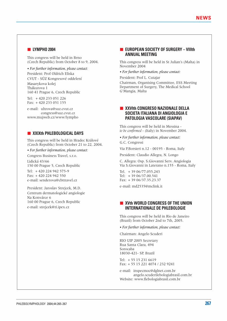
NEWS
PHLEBOLYMPHOLOGY 2004;44:265-267 267
EUROPEAN SOCIETY OF SURGERY – VIIIthANNUAL MEETING
This congress will be held in St Julian’s (Malta) inNovember 2004
• For further information, please contact:
President: Prof L. CutajarChairman, Organising Committee, ESS MeetingDepartment of Surgery, The Medical SchoolG’Mangia, Malta
XXVIth CONGRESO NAZIONALE DELLASOCIETA ITALIANA DI ANGIOLOGIA E PATOLOGIA VASCOLARE (SIAPAV)
This congress will be held in Messina - to be confirmed - (Italy) in November 2004.
• For further information, please contact:G.C. Congressi
Via P.Borsieri n.12 - 00195 - Roma, Italy
President: Claudio Allegra, N. Longo
C. Allegra: Osp. S.Giovanni Serv. Angiologia Via S.Giovanni in Laterano n.155 - Roma, Italy
Tel. + 39 06/77.055.243 Tel: + 39 06/37.00.541Fax: + 39 06/37.35.23.37
e-mail: [email protected]
XVth WORLD CONGRESS OF THE UNIONINTERNATIONALE DE PHLEBOLOGIE
This congress will be held in Rio de Janeiro (Brazil) from October 2nd to 7th, 2005.
• For further information, please contact:
Chairman: Angelo Scuderi
RIO UIP 2005 SecretaryRua Santa Clara, 494Sorocaba18030-421- SP, Brazil
Tel: + 55 15 231 6619Fax: + 55 15 221 4074 / 232 9241
e-mail: [email protected]
Website: www.flebologiabrasil.com.br
LYMPHO 2004
This congress will be held in Brno (Czech Republic) from October 8 to 9, 2004.
• For further information, please contact:President: Prof Oldrich EliskaCVUT - SÚZ Kongresové oddeleníMasarykova kolejThákurova 1160 41 Prague 6, Czech Republic
Tel: + 420 233 051 226Fax: + 420 233 051 155
e-mail: [email protected]@suz.cvut.cz
www.mujweb.cz/www/lympho
XXIXth PHLEBOLOGICAL DAYSThis congress will be held in Hradec Králové(Czech Republic) from October 21 to 22, 2004.
• For further information, please contact:
Congress Business Travel, s.r.o.
Lidická 43/66150 00 Prague 5, Czech Republic
Tel: + 420 224 942 575-9Fax: + 420 224 942 550e-mail: [email protected]
President: Jaroslav Strejcek, M.D.Centrum dermatologické angiologie Na Konvárce 6160 00 Prague 6, Czech Republic
e-mail: [email protected]
The Research Fund of the Union Internationale de Phlébologie (UIP) isproud to announce the fourth Servier Research Fellowship. It will providea 30 000 USD grant for 2 years’ work on a research project in the field ofphlebolymphology selected through a highly competitive peer-reviewprocedure.
The competition is open to young candidates who have a specific interest in the field of phlebolymphology and are a member of a national society inthis field. The projects must consist of original clinical or basic researchin an area of phlebolymphology, such as anatomy, physiology, patho-physiology, diagnostic methods, or clinical research. The fellowship will bemade available from 1 November 2005 and end in October 2007.
The review of the submitted projects and the selection of the best candidatewill be made by a committee of recognized worldwide specialists in thefield of phlebolymphology.
Send us your project at the following address:
Jean-Jérôme GUEX
Fonds de Recherche de l’UIP
32, boulevard Dubouchage
06000 Nice, France
The last research fellowship winner, Dr Maria Gemma PASCUALGONZALEZ (Vascular Biology, Spain) was awarded the grant at the 2003 UIP Chapter in San Diego for her project “Elastin dysregulation in varicoseveins”.
Candidates must submit a synopsis of 8 to 10 pages, double-spaced,typewritten in English, in the form of 5 original printed copies. The synopsisshould clearly present the objectives, methodology, planning, andreferences of the projected work. Candidates must also submit acurriculum vitae and a letter from a referee supporting the project andconfirming its backup, together with details of how the fellowship moneywill be spent. After the first year of work, the award-winner must submit tothe committee a synopsis of progress so far. The project’s results will have to appear in a form suitable for publication in an international journal.
30 000 grant
Submission deadline:
30 April 2005
Next winner awarded at theUIP World Congress
in Rio de Janeiro (October 2005)
Invitation to the 2007 UIP Chapter
to present the project’s results
SERVIER RESEARCH FELLOWSHIP
2005 2007
awarded by the
RESEARCH FUND OF THE
UNION INTERNATIONALE DE PHLEBOLOGIE
For further information or subscription online :www.servier.com / www.uip-phlebologyonline.org

04 D
N 0
25 B
A
At the forefront of research and education in phlebology
Correspondent:
Servier International - 22, rue Garnier, 92578 Neuilly-sur-Seine Cedex - France
Website: www.servier.com
![RESEARCH ARTICLE Open Access Interleukins, laminin ......metastasis in NPC by inducing matrix metalloproteinases (MMPs) in degrading ECM proteins [5]. MMPs have the ability to digest](https://img.dokumen.tips/doc/110x75/60d54502b6fa494b9302f93f/research-article-open-access-interleukins-laminin-metastasis-in-npc-by.jpg)