Embed Size (px)
Citation preview

Journal of Health, Medicine and Nursing www.iiste.org
An Open Access Journal, Vol. 6, 2014
73
Phenomenon of Biological Adaptation to Heavy Water
Oleg Mosin1
Ignat Ignatov2*
1. PhD (Chemistry), Biotechnology Department, Moscow State University of Applied Biotechnology,
Talalikhina Street, 33, Moscow 109316, Russian Federation
2. DSc, Professor, Scientific Research Center of Medical Biophysics (SRCMB),
N. Kopernik Street, 32, Sofia 1111, Bulgaria
* E-mail of the corresponding author: [email protected]
Abstract
Biological influence of deuterium oxide to cells of various taxonomic groups of prokaryotic and eucaryotic
microorganisms realizing methylotrophic, chemoheterotrophic, photo-organotrophic, and photosynthetic ways
of assimilation of carbon substrates (methylotrophic bacteria Brevibacterium methylicum, chemoheterotrophic
bacteria Bacillus subtilis, photo-organotrophic halobacteria Halobacterium halobium, and single sell green
algae Chlorella vulgaris) was investigated at the growth on media with 2H2O. For investigated
microorganisms are submitted the data on growth and adaptation on the growth media containing as sources
of deuterated substrates 2H2O, [
2H]methanol and hydrolisates of deutero-biomass of methylotrophic bacteria
B. methylicum, obtained after multistage adaptation to 2H2O. The qualitative and quantitative composition of
intra- and endocellular amino acids, proteins, carbohydrates and fatty acids in conditions of adaptation to 2H2O is investigated. It is demonstrated that the effects observed at adaptation to
2H2O, possess a complex
multifactorial character and connected to cytological, morphological and physiological changes – the
magnitude of the lag- period, time of cellular generation, output of biomass, a parity ratio of synthesized
amino acids, proteins, carbohydrates and lipids, and also with an evolutionary level of the organization of the
investigated object and the pathways of assimilation of carbon substrates as well. These data suggest that
adaptation to deuterium oxide is a multifactorial phenomenon, affecting many cellular systems, as
biosynthesis of macromolecules, metabolism, transport functions of cells.
Key words: deuterium, deuterium oxide, adaptation, isotopic effects, bacteria, micro algae.
Introduction
The most interesting biological phenomenon is the ability of some microorganisms to grow on heavy water (2H2O)
media in which all hydrogen atoms are replaced with deuterium (Ignatov & Mosin, 2013a; Ignatov & Mosin, 2013b). 2H2O has high environmental potential in biomedical studies due to the absence of radioactivity and poccebility
of detecting the deuterium label in the molecule by high-resolution methods as NMR, IR, and mass
spectrometry that facilitates its use as a tracer in biochemical and biomedical research (Kushner et al., 1999).

Journal of Health, Medicine and Nursing www.iiste.org
An Open Access Journal, Vol. 6, 2014
74
The average ratio of 1H/
2H in nature makes up approximately 1:5700 (Lis et al., 2008). In natural waters, the
deuterium is distributed irregularly: from 0.02–0.03 mol.% for river water and sea water, to 0.015 mol.% for
water of Antarctic ice – the most purified from deuterium natural water containing in 1.5 times less deuterium
than that of seawater. According to the international SMOW standard isotopic shifts for 2H and
18O in sea water:
2H/
1H = (155.76±0.05)
.10
-6 (155.76 ppm) and
18O/
16O = (2005.20±0.45)
.10
-6 (2005 ppm). For SLAP standard
isotopic shifts for 2H and
18O in seawater make up
2H/
1H = 89
.10
-6 (89 ppm) and for a pair of
18O/
16O =
1894.10
-6 (1894 ppm). In surface waters, the ratio
2H/
1H = (1.32–1.51)
.10
-4, while in the coastal seawater –
(1.55–1.56).10
-4. The natural waters of CIS countries are characterized by negative deviations from SMOW
standard to (1.0–1.5).10
-5, in some places up to (6.0–6.7)
.10
-5, but however there are also observed positive
deviations at 2.0.10
-5.
The chemical structure of 2H2O molecule is analogous to that one for Н2O, with small differences in the length
of the covalent H–O-bonds and the angles between them. The molecular mass of 2H2O exceeds on 10% that one
for Н2O. The difference in nuclear masses stipulates the isotopic effects, which may be sufficiently essential for
the 1H/
2H pair (Lobishev & Kalinichenko, 1978). As a result, physical-chemical properties of
2H2O differ from
H2O: 2H2O boils at 101.44
0С, freezes at 3.82
0С, has maximal density at 11.2
0С (1.106 g/cm
3) (Vertes, 2004).
In mixtures of 2H2O with Н2O the isotopic exchange occurs with high speed with the formation of semi-heavy
water (1H
2HO):
2H2O + H2O =
1H
2HO. For this reason deuterium presents in smaller content in aqueous
solutions in form of 1Н
2HO, while in the higher content – in form of
2H2O. The chemical reactions in
2H2O are
somehow slower compared to Н2O. 2H2O is less ionized, the dissociation constant of
2H2O is smaller, and the
solubility of the organic and inorganic substances in 2H2O is smaller compared to these ones in Н2О (Mosin,
1996a). Due to isotopic effects the hydrogen bonds with the participation of deuterium are slightly stronger than
those ones formed of hydrogen.
For a long time it was considered that heavy water was incompatible with life. Experiments with the growing of
cells of different organisms in 2H2O show toxic influence of deuterium. The high concentrations of
2H2O lead to
the slowing down the cellular metabolism, mitotic inhibition of the prophase and in some cases – somatic
mutations (Den’ko, 1970). This is observed even while using natural water with an increased content of 2H2O or
1H
2HO (Stom et al., 2006). Bacteria can endure up to 90 % (v/v)
2H2O, plant cells can develop normally in up
to 75 % (v/v) 2H2O, while animal cells – up to not more than 30 % (v/v)
2H2O (Mosin & Ignatov, 2012a).
Further increase in the concentration of 2H2O for these groups of organisms leads to the cellular death (Katz,
1960; Thomson, 1960), although isolated cell’s cultures suspended in pure 2H2O exert a strong radioprotective
effect in 2H2O-solutions towards –radiation (Michel et al., 1988; Laeng et al., 1991). On the contrary,
deuterium depleted water with decreased deuterium content has benefitial effects on organism and stimulates
the cellular metabolism (Somlyai, 2001; Sinyak et al., 2003).
With the development of new microbiological approaches, there appears an opportunity to use adapted to
deuterium cells for preparation of deuterated natural compounds (Mosin et al., 2013a; Mosin et al., 2013b;
Mosin et al., 2013c). The traditional method for production of deuterium labelled compounds consists in the
growth on media containing maximal concentrations of 2H2O and deuterated substrates as [
2H]methanol,
[2H]glucose etc. (Mosin & Ignatov, 2012b; Mosin et al., 2014). During growth of cells on
2H2O are synthesized
molecules of biologically important natural compounds (DNA, proteins, amino acids, nucleosides,

Journal of Health, Medicine and Nursing www.iiste.org
An Open Access Journal, Vol. 6, 2014
75
carbohydrates, fatty acids), which hydrogen atoms at the carbon backbones are completely substituted with
deuterium. They are isolated from deuterated biomass obtained on growth media with high 2H2O content and
deuterated substrates with using a combination of physico-chemical methods of separation – hydrolysis,
precipitation, extraction with organic solvents and chromatographic purification by column chromatography on
different adsorbents. These deuterated molecules evidently undergo structural adaptational modifications
necessary for the normal functioning in 2H2O.
The adaptation to 2H2O is interested not only from scientific point, but allows to obtain the unique biological
material for the studying of molecular structure by 1H-NMR (Crespi, 1989). Trend towards the use of deuterium
as an isotopic label are stipulated by the absence of radioactivity and possebility of determination the deuterium
localization in the molecule by high resolution NMR spectroscopy (LeMaster, 1990), IR spectroscopy
(MacCarthy, 1985) and mass spectrometry (Mosin et al., 1996a). The recent advances in technical and
computing capabilities of these analytical methods have allowed to considerable increase the efficiency of de
novo biological studies, as well as to carry out structural-functional biophysical studies with deuterated
molecules on a molecular level.
This study is a continuation of our research into the adaptation to deuterium of various procaryotic and
eucaryotic organisms. The purpose of our research was studying the influence of deuterium on the cells of
different taxonomic groups of microorganisms and microalgae realizing methylotrophic, chemoheterotrophic,
photo-organotrophic and photosynthetic pathways of carbon assimilation.
2. Material and Methods
2.1. Biological Objects
The objects of the study were various microorganisms, realizing methylotrophic, chemoheterotrophic,
photo-organotrophic, and photosynthetic ways of assimilation of carbon substrates. The initial strains were
obtained from the State Research Institute of Genetics and Selection of Industrial Microorganisms (Moscow,
Russia):
1. Brevibacterium methylicum B-5652, a leucine auxotroph Gram-positive strain of facultative methylotrophic
bacterium, L-phenylalanine producer, assimilating methanol via the NAD+ dependent methanol dehydrogenase
variant of ribulose-5-monophosphate cycle (RuMP) of carbon fixation.
2. Bacillus subtilis B-3157, a polyauxotrophic for histidine, tyrosine, adenine, and uracil spore-forming aerobic
Gram-positive chemoheterotrophic bacterium, inosine producer, realizing hexose-6-mono-phosphate (GMP)
cycle of carbohydrates assimilation.
3. Halobacterium halobium ET-1001, photo-organotrophic carotenoid-containing strain of extreme halobacteria,
synthesizing the photochrome transmembrane protein bacteriorhodopsin.
4. Chlorella vulgaris B-8765, photosynthesizing single-cell blue-green algae.
2.2. Chemicals
For preparation of growth media was used 2H2O (99.9 atom.%),
2HСl (95.5 аtom.%) and [
2H]methanol (97.5
atom% 2H), purchased from the “Isotope” Russian Research Centre (St. Petersburg, Russian Federation).
Journal of Health, Medicine and Nursing www.iiste.org
An Open Access Journal, Vol. 6, 2014
76
Inorganic salts and D- and L-glucose (“Reanal”, Hungary) were preliminary crystallized in 2H2O and dried in
vacuum before using. 2H2O was distilled over KMnO4 with the subsequent control of isotope enrichment by
1H-NMR-spectroscopy on a Brucker WM-250 device (“Brucker”, Germany) (working frequency: 70 MHz,
internal standard: Me4Si). According to 1H-NMR, the level of isotopic purity of growth media usually was by
8–10 atom% lower than the isotope purity of the initial 2Н2О.
2.3. Adaptation Technique
The initial microorganisms were modified by adaptation to deuterium by plating individual colonies onto 2%
(w/v) agarose growth media with stepwise increasing gradient of 2Н2О concentration and subsequent selection
of individual cell colonies stable to the action of 2Н2О. As a source of deuterated growth substrates for the
growth of chemoheterotrophic bacteria and chemoorganoheterotrophic bacteria was used the deuterated
biomass of facultative methylotrophic bacterium B. methylicum, obtained via a multi-stage adaptation on solid
2% (w/v) agarose M9 media with an increasing gradient of 2Н2О (from 0, 24.5, 49.0, 73.5 up to 98% (v/v)
2Н2О). Raw deuterated biomass (output, 100 gram of wet weight per 1 liter of liquid culture) was suspended in
100 ml of 0.5 N 2HCl (in
2H2O) and autoclaved for 30–40 min at 0.8 atm. The suspension was neutralized with
0.2 N KOH (in 2H2O) to pH = 7.0 and used as a source of growth substrates while adaptation and growing the
chemoheterotrophic bacterium B. sublilis and the photo-organotrophic halobacterium H. halobium.
2.4. Growth Media
The following growth media were used (concentratioin of components are given in g/l):
1. Minimal salt medium M9 for growth of the facultative methanol assimilating methylotrophic bacterium B.
methylicum B-5662, supplemented with 2% (v/v) [2Н]methanol and increasing gradient of
2Н2О concentration
from 0; 24.5; 49.0; 73.5 up to 98 % (v/v) 2Н2О: KH2PO4 – 3; Na2HPO4 – 6; NaCl – 0.5; NH4Cl – 1; L-leucine –
0.01.
2. Hydrolysated medium HM1 for growth of the aerobic Gram-positive chemoheterotrophic bacterium B.
subtilis B-3157, based on 2Н2О (89–90 atom%
2H) and 2% (w/v) hydrolysate of deuterated biomass of B.
methylicum B-5662 as a source of 2H-labeled growth substrates: L-glucose –120; hydrolysate of deuterated
biomass of B. methylicum – 20, NH4NO3 – 20; MgSO4.7H2O – 10; СаСО3 – 20; adenine, and uracil – 0.01. As a
control was used protonated medium containing 2% (w/v) yeast protein–vitamin concentrate (PVC).
3. Hydrolysated medium HM2 for the growth of the extreme aerobic halobacterium Halobacterium halobium
ET-1001 (based on 99.9 atom% 2H2O): NaCl - 250; MgSO4
.7H2O - 20; KCl - 2; CaCl2
. 6H2O – 0.065; sodium
citrate - 0.5; hydrolyzate of deuterated biomass of B. methylicum B-5662 – 20; biotin – 1.10
-4; folic acid –
1.5.10
-4, vitamin B12 – 2
.10
-5).
4. Tamiya medium for the growth of the photosynthetic green microalgae C. vulgaris B-8765 (based on 99.9
atom% 2H2O): KNO3 – 5.0; MgSO4
.7H2O – 2.5; KH2PO4 – 1.25; FeSO4 – 0.003; MnSO4
.2H2O – 3
.10
-4;
CaCl2.6H2O – 0.065; ZnSO4
.7H2O – 4
.10
-5; CuSO4
.5H2O – 5
.10
-5, CoCl2
.6H2O – 5
.10
-6).
2.5. Growth Conditions
The cells were grown in 500 ml Erlenmeyer flasks containing 100 ml of the growth medium at 32–34 0С and

Journal of Health, Medicine and Nursing www.iiste.org
An Open Access Journal, Vol. 6, 2014
77
vigorously aerated on an orbital shaker Biorad (“Biorad Labs”, Poland). Photo-organotrophic bacteria and
blue-green algae were grown at illumination by fluorescent monochromatic lamps LDS-40-2 (40 W)
("Alfa-Electro", Russia). Growing of microalgae C. vulgaris was carried out at 32 0C in a photoreactor with
CO2 bubbling. The bacterial growth was monitored on the ability to form individual colonies on the surface of
solid 2 % (w/v) agarose media, as well as on the optical density of the cell suspension measured on a Beckman
DU-6 spectrophotometer (“Beckman Coulter”, USA) at λ = 620 nm. After 67 days the cells were harvested
and separated by centrifugation (10000 g, 20 min) on T-24 centrifuge ("Heracules", Germany). The biomass
was washed with 2H2O and extracted with a mixure of organic solvents: chloroformmethanolacetone =
2:1:1, % (v/v) for isolating lipids and pigments. The resulting precipitate (1012 mg) was dried in vacuum and
used as a protein fraction, while the liquid extract as a lipid fraction. The exogenious deuterated amino acids
and ribonucleosides were isolated from culture liquids (CL) of appropriate strain-producers. Inosine was
isolated from the CL of B. subtilis by adsorption/desorption on activated carbon as adsorbent with following
extraction with 0.3 M NH4-formate buffer (pH = 8.9), subsequent crystallization in 80 % (v/v) ethanol, and ion
exchange chromatography (IEC) on a column with cation exchange resin AG50WX 4 equilibrated with 0.3 M
NH4-formate buffer and 0.045 M NH4Cl (output, 3.1 g/l (80 %); []D20
= 1.61 (ethanol)). Bacteriorhodopsin was
isolated from the purple membranes of photo-organotrophic halobacterium H. halobium by the method of D.
Osterhelt, modificated by the authors, with using SDS as a detergent (Mosin et al., 1999a).
2.6. Protein Hydrolysis
Dry biomass (10 g) was treated with a chloroform–methanol–acetone mixture (2:1:1, % (v/v)) and
supplemented with 5 ml of 6 N 2HCl (in
2H2О). The ampules were kept at 110
0С for ~24 h. Then the reaction
mixture was suspended in hot 2H2О and filtered. The hydrolysate was evaporated at 10 mm Hg. Residual
2HCl
was removed in an exsiccator over solid NaOH.
2.7. Hydrolysis of Intracellular Policarbohydrates
Dry biomass (50 mg) was placed into a 250 ml round bottomed flask, supplemented with 50 ml distilled 2H2О
and 1.6 ml of 25% (v/v) H2SO4 (in 2H2О), and boiled in a reflux water evaporator for ~90 min. After cooling,
the reaction mixture was suspended in one volume of hot distilled 2H2О and neutralized with 1 N Ba(ОН)2 (in
2H2О) to pH = 7.0. BaSO4 was separated by centrifugation (1500 g, 5 min); the supernatant was decanted and
evaporated at 10 mm Hg.
2.8. Amino Acid Analysis
The amino acids of the hydrolyzed biomass were analyzed on a Biotronic LC-5001 (2303.2) column
(“Eppendorf–Nethleler–Hinz”, Germany) with a UR-30 sulfonated styrene resin (“Beckman–Spinco”, USA) as
a stationary phase; the temperature – 2025 0C;
the mobile phase – 0.2 N sodium–citrate buffer (pH = 2.5); the
granule diameter – 25 μm; working pressure – 50–60 atm; the eluent input rate – 18.5 ml/h; the ninhydrin input
rate – 9.25 ml/h; detection at λ = 570 and λ = 440 nm (for proline).

Journal of Health, Medicine and Nursing www.iiste.org
An Open Access Journal, Vol. 6, 2014
78
2.9. Analysis of Carbohydrates
Carbohydrates were analyzed on a Knauer Smartline chromatograph (“Knauer”, Germany) equipped with a
Gilson pump (“Gilson Inc.”, USA) and a Waters K 401 refractometer (”Water Associates”, USA) using
Ultrasorb CN column (25010 mm) as a stationary phase; the temperature – 2025 0C; the mobile phase –
acetonitrile–water (75:25, % (w/w); the granule diameter – 10 μm; the input rate – 0.6 ml/min.
2.10. Analysis of Fatty Acids
Fatty acids were analyzed on a Beckman Gold System (USA) chromatograph (2504.6 mm), equiped with
Model 126 UV-Detector (USA), 2025 0C . Stationary phase – Ultrasphere ODS 5 m; mobile phase – linear
gradient of 5 mM KH2PO4–acetonitrile; elution rate – 0.5 ml/min, detection at λ = 210 nm.
2.11. Mass Spectrometry
For evaluation of deuterium enrichment levels EI and FAB mass spectrometry was used. EI mass spectra were
recorded on MB-80A device (“Hitachi”, Japan) with double focusing (the energy of ionizing electrons – 70 eV;
the accelerating voltage – 8 kV; the cathode temperature – 180–200 0С) after amino acid modification into
methyl esters of N-5-dimethylamino(naphthalene)-1-sulfonyl (dansyl) amino acid derivatives according to an
earlier elaborated protocol (Mosin et al., 1998). FAB-mass spectra were recorded on a VG-70 SEQ
chromatograph (“Fisons VG Analytical”, USA) equipped with a cesium Cs+
source on a glycerol matrix with
accelerating voltage 5 kV and ion current 0.6–0.8 mA. Calculation of deuterium enrichment of the molecules
was carried out using the ratio of contributions of molecular ion peaks of deuterated compounds extracted on
D2O-media relative to the control obtained on H2O.
2.6. Scanning Electrom Microscopy (SEM)
SEM was carried out on JSM 35 CF (JEOL Ltd., Korea) device, equiped with SE detector, thermomolecular
pump, and tungsten electron gun (Harpin type W filament, DC heating); working pressure – 10-4
Pa (10-6
Torr);
magnification – 150.000, resolution – 3.0 nm, accelerating voltage – 1–30 kV; sample size – 60–130 mm.
2.12. IR-spectroscopy
IR-spectroscopy was performed on Brucker Vertex spectrometer (“Brucker”, Germany) (spectral range: average
IR – 370–7800 cm-1
; visible – 2500–8000 cm-1
; the permission – 0.5 cm-1
; accuracy of wave number – 0.1 cm-1
on 2000 cm-1
).
3. Results
3.1. Adaptation to deuterium the methylotrophic bacterium B. methylicum
Numerous studies carried out by us with various biological objects in 2H2O, proved that when biological objects
being exposed to water with different deuterium content, their reaction varies depending on the isotopic
composition of water (the content of deuterium in water) and magnitude of isotope effects determined by the
difference of constants of chemical reactions rates kH/kD in H2O and 2H2O. The maximum kinetic isotopic effect

Journal of Health, Medicine and Nursing www.iiste.org
An Open Access Journal, Vol. 6, 2014
79
observed at ordinary temperatures in chemical reactions leading to rupture of bonds involving hydrogen and
deuterium atoms lies in the range kH/kD = 5–8 for C–H versus C–2H, N–
2H versus N–
2H, and O–
2H versus
O–2H-bonds (Mosin, 1996; Mosin & Ignatov, 2012a; Mosin & Ignatov, 2012b). Isotopic effects have an impact
not only on the physical and chemical properties of deuterated macromolecules in which H atoms are
substituted with 2H atoms, but also on the biological behaviour of biological objects in
2H2O. Experiments with
2H2O (Table 1) have shown, that green-blue algae is capable to grow on 70 % (v/v)
2H2O, methylotrophic
bacteria – 75 % (v/v) 2H2O, chemoheterotrophic bacteria – 82 % (v/v)
2H2O, and photo-organotrophic
halobacteria – 95 % (v/v) 2H2O.
0
10
20
30
40
50
60
70
80
90
100
Halobacteria
Halobacterium
halobium
Chemoheterotrophic
bacteria Bacillus
subtilis
Methylotrophic
bacteria
Brevibacterium
methylicum
Blue-green algae
Chlorella vulgaris
Deu
teri
um
co
nte
nts
in
wate
r, v
ol.
%
Figure 1. Cell survival of various microorganisms in water with different deuterium content (%, v/v)
In the course of the experiment were obtained adapted to the maximum concentration of 2H2O cells
belonging to different taxonomic groups of microorganisms, realizing methylotrophic, chemoheterotrophic,
photo-organotrophic and photosynthetic pathways of assimilation of carbon substrata, as facultative
methylotrophic bacterium B. methylicum, chemoheterotrophic bacterium B. subtilis, halobacterium H.
halobium and blue-green algae C. vulgaris.
Selection of methanol-assimilating facultative methylotrophic bacterium B. methylicum was connected with
the development of new microbiological strategies for preparation of deuterated biomass via bioconversion
of [2H]methanol and
2H2O and its further use as a source of deuterated growth substrates for the growing
other strains-producers in 2H2O.
Choosing of photo photo-organotrophic halobacterium H. halobium was stipulated by the prospects of
further isolation of retinal containing transmembrane protein bacteriorhodopsin (BR) – chromoprotein of
248 amino acid residues, containing as a chromophore an equimolar mixture of 13-cis-and 13-trans C20
carotenoid associated with a protein part of the molecule via a Lys-216 residue (Mosin & Ignatov, 2014).
BR performs in the cells of halobacteria the role of ATP-dependent translocase, which creates an
electrochemical gradient of H+
on the surface of the cell membrane, which energy is used by the cell for the
Journal of Health, Medicine and Nursing www.iiste.org
An Open Access Journal, Vol. 6, 2014
80
synthesis of ATP in the anaerobic photosynthetic phosphorylation.
Using chemoheterotrophic bacterium B. subtilis was determined by preparative isolation produced by this
bacterium deuterated ribonucleoside – inosine (total deuteration level 65.5 atom.% 2H) for biomedical use
(Mosin & Ignatov, 2013d), and the use of photosynthetic blue-green C. vulgaris was stipulated by the study
of biosynthesis of deuterated chlorophyll and carotenoid pigments (deuteration level 95–97 atom.% 2H) on
growth media with high 2H2O-content (Mosin & Ignatov, 2012b).
We used stepwise increasing gradient concentration of 2H2O in growth media, because it was assumed that
the gradual accustoming of micorganisms to deuterium would have a beneficial effect upon the growth and
physiological parameters. The strategy of adaptation to 2H2O is shown in Table. 1 on an example of
methylotrophic bacterium B. methylicum, which deuterated biomass was used in further experiments as a
source of deuterated growth substrates for growing of chemoheterotrophic and photo-organotrophic
bacteria. For this, deuterium enrichment technique was applied via plating cell colonies on 2% (w/v)
agarose M9 media supplemented with 2% (v/v) [U-2H]MeOH with an increase in the
2H2O content from 0;
24.5; 49.0; 73.5 up to 98% (v/v) 2H2O, combined with subsequent selection of cell colonies which were
resistant to deuterium. The degree of cell survive on maximum deuterated medium was approx. 40%. The
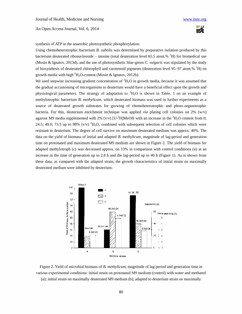
data on the yield of biomass of initial and adapted B. methylicum, magnitude of lag-period and generation
time on protonated and maximum deuterated M9 medium are shown in Figure 2. The yield of biomass for
adapted methylotroph (c) was decreased approx. on 13% in comparison with control conditions (a) at an
increase in the time of generation up to 2.8 h and the lag-period up to 40 h (Figure 1). As is shown from
these data, as compared with the adapted strain, the growth characteristics of initial strain on maximally
deuterated medium were inhibited by deuterium.
Figure 2. Yield of microbial biomass of B. methylicum, magnitude of lag-period and generation time in
various experimental conditions: initial strain on protonated М9 medium (control) with water and methanol
(a); initial strain on maximally deuterated M9 medium (b); adapted to deuterium strain on maximally
Journal of Health, Medicine and Nursing www.iiste.org
An Open Access Journal, Vol. 6, 2014
81
deuterated M9 medium (c): 1 – yield of biomass, % from the control; 2 – duration of lag-period, h; 3 –
generation time, h.
Experimental conditions are given in Table 1 (expts. 1–10) relative to the control (expt. 1) on protonated
medium M9 and to the adapted bacterium (expt. 10’). Various compositions of [U-2H]MeOH and
2H2O
were added to growth media M9 as hydrogen/deuterium atoms could be assimilated both from MeOH and
H2O. The maximum deuterium content was under conditions (10) and (10’) in which we used 98% (v/v) 2H2O and 2% (v/v) [U-
2H]MeOH. The even numbers of experiment (Table 1, expts. 2, 4, 6, 8, 10) were
chosen to investigate whether the replacement of MeOH by its deuterated analogue affected growth
characteristics in presence of 2H2O. That caused small alterations in growth characteristics (Table 1, expts.
2, 4, 6, 8, 10) relative to experiments, where we used protonated methanol (Table 1, expts. 3, 5, 7, 9). The
gradual increment in the concentration of 2H2O into growth medium caused the proportional increase in
lag-period and yields of microbial biomass in all isotopic experiments. Thus, in the control (Table 1, expt.
1), the duration of lag-period did not exceed 20.2 h, the yield of microbial biomass (wet weight) and
production of phenylalanine were 200.2 and 0.95 gram per 1 liter of growth medium. The adding gradually
increasing concentrations of 2H2O into growth media caused the proportional increasing of lag-period and
yield of microbial biomass in all isotopic experiments. The results suggested, that below 49% (v/v) 2H2O
(Table 1, expts. 2–4) there was a small inhibition of bacterial growth compared with the control (Table 1,
expt. 1). However, above 49% (v/v) 2H2O (Table 1, expts. 5–8), growth was markedly reduced, while at the
upper content of 2H2O (Table 1, expts. 9–10) growth got 3.3-fold reduced. With increasing content of
2H2O
in growth media there was a simultaneous increase both of lag-period and generation time. Thus, on
maximally deuterated growth medium (Table 1, expt. 10) with 98% (v/v) 2H2O and 2% (v/v) [U-
2H]MeOH,
lag-period was 3 fold higher with an increased generation time to 2.2 fold as compared to protonated
growth medium with protonated water and methanol which serve as control (Table 1, expt. 1). While on
comparing adapted bacterium on maximally deuterated growth medium (Table 1, expt. 10’) containing 98%
(v/v) 2H2O and 2% (v/v) [U-
2H]MeOH with non adapted bacterium at similar concentration showed 2.10
and 2.89 fold increase in terms of phenylalanine production and biomass yield due to deuterium enrichment
technique, while, the lag phase as well as generation time also got reduced to 1.5 fold and 1.75 fold in case
of adapted bacterium.
The adapted bacterium of B. methylicum eventually came back to normal growth at placing over in
protonated growth medium after some lag-period that proves phenotypical nature of a phenomenon of
adaptation that was observed for others adapted by us strains of methylotrophic bacteria and representatives
of other taxonomic groups of microorganisms [Mosin & Ignatov, 2012a; Mosin & Ignatov, 2013a, Ignatov
& Mosin, 2013b]. Literature reports clearly reveal that the transfer of deuterated cells to protonated medium
M9 eventually after some lag period results in normal growth that could be due to the phenomenon of
adaptation wherein phenotypic variation was observed by the strain of methylotrophic bacteria (Mosin &
Ignatov, 2013b; Mosin et al., 2013). The improved growth characteristics of adapted methylotroph
essentially simplify the scheme of obtaining the deutero-biomass which optimum conditions are М9 growth
medium with 98% 2Н2О and 2% [
2Н]methanol with incubation period 3–4 days at temperature 35
0С.

Journal of Health, Medicine and Nursing www.iiste.org
An Open Access Journal, Vol. 6, 2014
82
Table 1. Effect of variation in isotopic content (0–98% 2H2O, v/v) in growth media M9 on bacterial growth
of B. methylicum and phenylalanine production
Bacterial
strains
Exp.
numb
er
Media components, % (v/v)
Lag-period
(h)
Yield in terms
of wet biomass
(g/l)
Generat
ion time
(h)
Phenylala
nine
productio
n (g/l)
H2O 2H2O MeOH [U-
2H]
MeOH
Non
adapted
1(con
trol)
98.0 0 2 0 20.2±1.40 200.2±3.20 2.2±0.2 0.95±0.12
Non
adapted
2 98.0 0 0 2 20.3±1.44 184.6±2.78 2.4±0.2 0.92±0.10
Non
adapted
3 73.5 24.5 2 0 20.5±0.91 181.2±1.89 2.4±0.2 0.90±0.10
Non
adapted
4 73.5 24.5 0 2 34.6±0.89 171.8±1.81 2.6±0.2 0.90±0.08
Non
adapted
5 49.0 49.0 2 0 40.1±0.90 140.2±1.96 3.0±0.3 0.86±0.10
Non
adapted
6 49.0 49.0 0 2 44.2±1.38 121.0±1.83 3.2±0.3 0.81±0.09
Non
adapted
7 24.5 73.5 2 0 45.4±1.41 112.8±1.19 3.5±0.2 0.69±0.08
Non
adapted
8 24.5 73.5 0 2 49.3±0.91 94.4±1.74 3.8±0.2 0.67±0.08
Non
adapted
9 98.0 0 2 0 58.5±1.94 65.8±1.13 4.4±0.7 0.37±0.06
Non
adapted
10 98.0 0 0 2 60.1±2.01 60.2±1.44 4.9±0.7 0.39±0.05
Adapted 10’ 98.0 0 0 2 40.2±0.88 174.0±1.83 2.8±0.3 0.82±0.08
Notes:
* The date in expts. 1–10 described the growth characteristics for non-adapted bacteria in growth media,
containing 2 % (v/v) MeOH/[U-2H]MeOH and specified amounts (%, v/v)
2Н2О.
** The date in expt. 10’ described the growth characteristics for bacteria adapted to maximum content of
deuterium in growth medium.
***As the control used exprt. 1 where used ordinary protonated water and methanol

Journal of Health, Medicine and Nursing www.iiste.org
An Open Access Journal, Vol. 6, 2014
83
Adaptation, which conditions are shown in experiment 10’ (Table 1) was observed by investigation of
growth dynamics (expts. 1a, 1b, 1c) and accumulation of L-phenylalanine into growth media (expts. 2a, 2b,
2c) by initial (a) and adapted to deuterium (c) strain B. methylicum in maximum deuterated growth medium
М9 (Figure 3, the control (b) is obtained on protonated growth medium M9). In the present study, the
production of phenylalanine (Figure 2, expts. 1b, 2b, 3b) was studied and was found to show a close linear
extrapolation with respect to the time up to exponential growth dynamics (Figure 3, expts. 1a, 2a, 3a). The
level of phenylalanine production for non-adapted bacterium on maximally deuterated medium M9 was
0.39 g/liter after 80 hours of growth (Figure 2, expt. 2b). The level of phenylalanine production by adapted
bacterium under those growth conditions was 0.82 g/liter (Figure 3, expt. 3b). Unlike to the adapted strain
the growth of initial strain and production of phenylalanine in maximum deuterated growth medium were
inhibited. The important feature of adapted to 2Н2О strain В. methylicum was that it has kept its ability to
synthesize and exogenously produce L-phenylalanine into growth medium. Thus, the use of the adapted
bacterium enabled to improve the level of phenylalanine production on maximally deuterated medium by
2.1 times with the reduction in the lag phase up to 20 h. This is an essential achievement for this strain of
methylotrophic bacteria, because up till today there have not been any reports about production of
phenylalanine by leucine auxotrophic methylotrophs with the NAD+
dependent methanol dehydrogenase
(EC 1.6.99.3) variant of the RuMP cycle of carbon assimilation. This makes this isolated strain unique for
production of deuterated phenylalanine and other metabolically related amino acids.
Figure 3. Growth dynamics of B. methylicum (1a, 2a, 3a) and production of phenylalanine (1b, 2b, 3b) on
media M9 with various isotopic content: 1a, 1b – non-adapted bacterium on protonated medium (Table 1,
expt. 1); 2a, 2b – non-adapted bacterium on maximally deuterated medium (Table 1, expt. 10); 3a, 3b –
adapted bacterium on maximally deuterated medium (Table 1, expt. 10’)
The general feature of phenylalanine biosynthesis in Н2О/2H2O-media was increase of its production at
early exponential phase of growth when outputs of a microbial biomass were insignificant (Figure 3). In all
Journal of Health, Medicine and Nursing www.iiste.org
An Open Access Journal, Vol. 6, 2014
84
the experiments it was observed that there was a decrease in phenylalanine accumulation in growth media
at the late exponential phase of growth. Microscopic research of growing population of microorganisms
showed that the character of phenylalanine accumulation in growth media did not correlate with
morphological changes at various stages of the cellular growth. Most likely that phenylalanine,
accumulated in growth media, inhibited enzymes of its biosynthetic pathways, or it later may be
transformed into intermediate compounds of its biosynthesis, e.g. phenylpyruvate (Maksimova et al., 1990;
Skladnev et al., 1996). It is necessary to notice, that phenylalanine is synthesised in cells of microorganisms
from prephenic acid, which through a formation stage of phenylpiruvate turns into phenylalanine under the
influence of cellular transaminases. However, phenylalanine was not the only product of biosynthesis; other
metabolically related amino acids (alanine, valine, and leucine/isoleucine) were also produced and
accumulated into growth media in amounts of 5–6 mol in addition to phenylalanine. This fact required
isolation of [2H]phenylalanine from growth medium, which was carried out by extraction of lyophilized LC
with iso-PrOH and the subsequent crystallization in EtOH. Analytical separation of [2H]phenylalanine and
metabolically related [2H]amino acids was performed using a reversed-phase HPLC method on Separon
SGX C18 Column, developed for methyl esters of N-DNS-[2H]amino acids with chromatographic purity of
96–98% and yield of 67–89%.
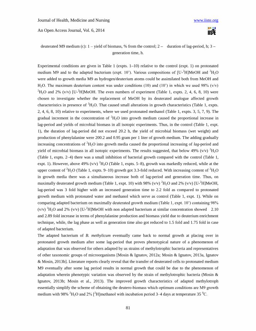
Table 2. Effect of deuterium enrichment levels (atom.%) in the molecules of [2Н]amino acids excreted by B.
methylicum*
[2Н]amino acid
Concentration of 2Н2О in growth media, % (v/v)**
24.5 49.0 73.5 98.0
Alanine 24.0±0.70 50.0±0.89 50.0±0.83 50.0±1.13
Valine 20.0±0.72 50.0±0.88 50.0±0.72 62.5±1.40
Leucine/isoleucine 20.0±0.90 50.0±1.38 50.0±1.37 50.0±1.25
Phenylalanine 17.0±1.13 27.5±0.88 50.0±1.12 75.0±1.40
Notes:
* At calculation of enrichment levels protons (deuterons) at COOH- and NH2-groups of amino acids were
not considered because of dissociation in Н2О (2Н2О).
** The data on enrichment levels described bacteria grown on minimal growth media M9 containing 2%
(v/v) [U-2Н]MeOH and specified amounts (%, v/v)
2Н2О.
With increasing 2H2O content in growth media, the levels of deuterium enrichment in exogenous [
2H]amino
acids (phenylalanine, alanine, valine, and leucine/isoleucine), secreted by B. methylicum, were varied
proportionally. The similar result on proportional specific increase of levels of deuterium enrichment into
[2Н]phenylalanine and other metabolically related [
2Н]amino acids was observed in all isotopic
experiments where used increasing concentration 2Н2О in growth media (Table 2). Predictably, enrichment
levels of [2Н]phenylalanine related to the family of aromatic amino acids synthesised from shikimic acid
and metabolically related [2Н]amino acids of pyruvic acid family – alanine, valine and leucine at identical

Journal of Health, Medicine and Nursing www.iiste.org
An Open Access Journal, Vol. 6, 2014
85
2Н2О concentration in growth media are correlated among themselves. Such result is fixed in all isotope
experiments with 2Н2О (Table 2). Unlike [
2Н]phenylalanine, deuterium enrichment levels in accompanying
[2Н]amino acids – Ala, Val and Leu/Ile keep a stable constancy within a wide interval of
2Н2О
concentration: from 49% (v/v) to 98% (v/v) 2Н2О (Table 2). Summarizing these data, it is possible to draw
a conclusion on preservation of minor pathways of the metabolism connected with biosynthesis of leucine
and metabolic related amino acids of pyruvic acid family – alanine and valine, which enrichment levels
were in correlation within identical concentration of Н2О in growth media (phenylalanine is related to the
family of aromatic amino acids synthesized from shikimic acid). Since leucine was added into growth
media in protonated form, another explanation of this effect, taking into consideration the various
biosynthetic pathways of Leu and Ileu (Ileu belongs to the family of aspartic acid, while Leu belongs to the
pyruvic acid family), could be cell assimilation of protonated leucine from growth media. Since Leu and
Ileu could not be clearly estimated by EI MS method, nothing could be said about possible biosynthesis of
[2H]isoleucine. Evidently, higher levels of deuterium enrichment can be achieved by replacement of
protonated leucine on its deuterated analogue, which may be isolated from hydrolysates of deuterated
biomass of this methylotrophic bacterium.
It should be noted that the yields of biomass on deuterated growth media were varried 85-90% for different
taxonomic groups of microorganisms. All adapted microorganisms had a slightly reduced levels of
microbial biomass accumulation and increased cell generation times on maximal deuterated media.
3.2. Adaptation to deuterium the chemoheterotrophic bacterium B. subtillis
The result obtained in experiments on the adaptation of methylotrophic bacterium B. methylicum to 2Н2О
allowed to use hydrolysates of biomass of this bacterium obtained in the process of multi-stage adaptation
to 2Н2О, as a source of deuterated growth substrates for the growing of the chemoheterotrophic bacterium B.
subtillis and the photoorganotrophic halobacterium H. halobium.
The assimilation rate of methylotrophic biomass by protozoa and eukaryotic cells amounts to 85–98%,
while the productivity calculated on the level of methanol bioconversion into cell components makes up
50–60% (Mosin et al., 1999a; Mosin et al., 1999b). While developing the composition of growth media on
the basis of deutereted biomass of methylotrophic bacteria B. methylicum it was taken into account the
ability of methylotrophic bacteria to synthesize large amounts of protein (output, 50% (w/w) of dry weight),
15–17% (w/w) of polysaccharides, 10–12% (w/w) of lipids (mainly, phospholipids), and 18% (w/w) of ash
(Mosin & Ignatov, 2013b). To provide high outputs of these compounds and minimize the isotopic
exchange (1Н–
2Н) in amino acid residues of protein molecules, the biomass was hydrolyzed by autoclaving
in 0.5 N 2НCl (in
2H2О) and used for the growing of chemoheterotrophic bacterium B. subtillis and
photoorganoheterotrophic halobacteria H. halobium.
The methylotrophic hydrolysate, obtained on the maximally deuterated medium M9 with 98% (v/v) 2H2О
and 2% (v/v) [2H]methanol, contains 15 identified amino acids (except for proline detected at λ = 440 nm)
with tyrosine and histidine content per 1 gram of dry methylotrophic hydrolysate 1.82% and 3.72% (w/w),
thereby satisfying the auxotrophic requirements of the inosine producer strain for these amino acids (Table
3). The content of other amino acids in the hydrolysate is also comparable with the needs of the strain in

Journal of Health, Medicine and Nursing www.iiste.org
An Open Access Journal, Vol. 6, 2014
86
sources of carbon and amine nitrogen. The indicator determining the high efficiency of deuterium
incorporation into the synthesized product is high levels of deuterium enrichment of amino acid molecules,
varied from 50 atom% 2H
for leucine/isoleucine to 98.5 atom%
2H for alanine (Table 3).
Table 3. Amino acid composition of hydrolyzed biomass of the facultative methylotrophic bacterium B.
methylicum obtained on maximally deuterated M9 medium with 98% (v/v) 2H2O and 2% (v/v)
[2H]methanol and levels of deuterium enrichment*
Amino acid Yield, % (w/w) dry
weight per 1 gram of
biomass
Number of deuterium
atoms incorporated into
the carbon backbone of
a molecule**
Level of deuterium
enrichment of
molecules, % of the total
number of hydrogen
atoms***
Glycine 9.55 2 92.5±1.86
Alanine 13.30 4 98.5±1.96
Valine 4.21 4 52.2±1.60
Leucine 8.52 5 50.0±1.52
Isoleucine 4.01 5 50.0±1.55
Phenylalanine 3.89 8 96.0±1.85
Tyrosine 2.10 7 95.5±1.82
Serine 3.60 3 86.7±1.55
Threonine 4.89
Methionine 2.62
Asparagine 10.02 2 68.5±1.62
Glutamic acid 10.31 4 70.0±1.65
Lysine 3.53 5 59.0±1.60
Arginine 4.65
Histidine 3.98
Keys: * The data were obtained for methyl esters of N-5-dimethylamino(naphthalene)-1-sulfonyl (dansyl)
chloride amino acid derivatives.
** At calculation the level of deuterium enrichment, the protons (deuterons) at COOH- and NH2- groups of
amino acid molecules were not taken into account because of the dissociation in H2O/2H2O.
*** A dash denotes the absence of data.
Taking into account the pathways of assimilation of carbon substrates, the adaptation of
chemoheterotrophic bacterium B. subtilis was carried out via plating of initial cells to separate colonies on
solid 2% (w/v) agarose HM1 media based on 99,9 atom% 2Н2О and deuterated hydrolyzate biomass of B.
methylicum, with the following subsequent selection of the colonies resistant to 2Н2О. On contrary to
2Н2О,
Journal of Health, Medicine and Nursing www.iiste.org
An Open Access Journal, Vol. 6, 2014
87
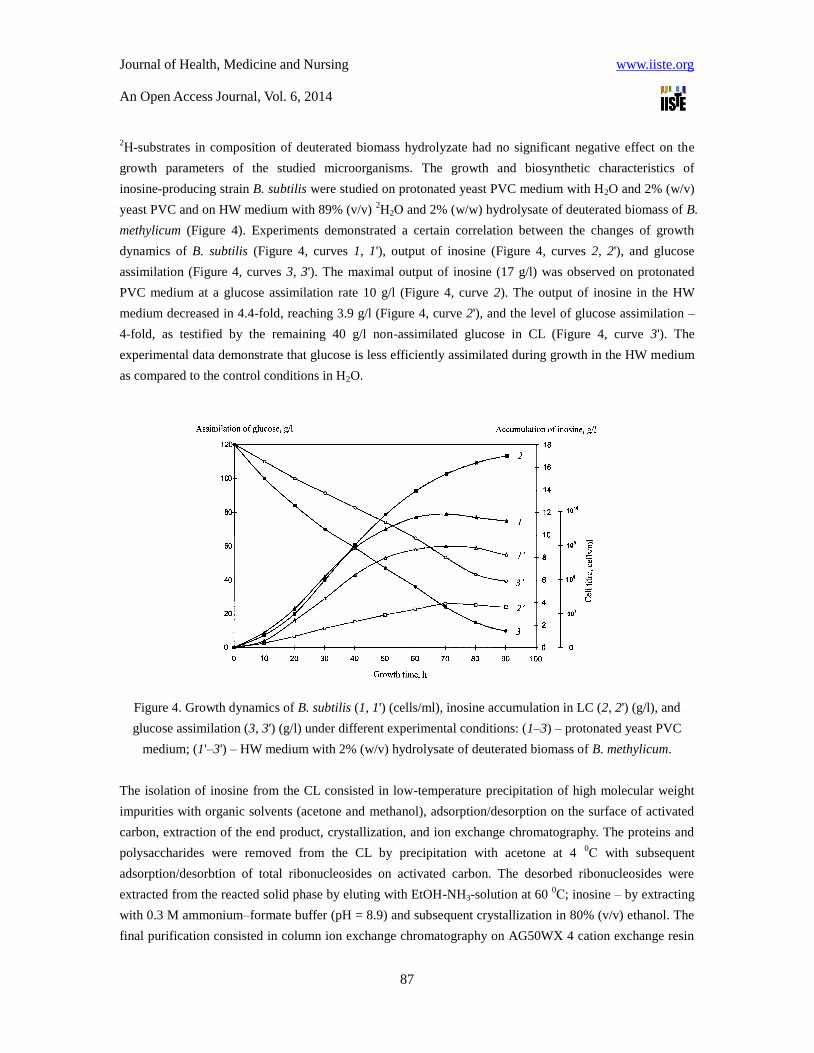
2Н-substrates in composition of deuterated biomass hydrolyzate had no significant negative effect on the
growth parameters of the studied microorganisms. The growth and biosynthetic characteristics of
inosine-producing strain B. subtilis were studied on protonated yeast PVC medium with H2O and 2% (w/v)
yeast PVC and on HW medium with 89% (v/v) 2H2О and 2% (w/w) hydrolysate of deuterated biomass of B.
methylicum (Figure 4). Experiments demonstrated a certain correlation between the changes of growth
dynamics of B. subtilis (Figure 4, curves 1, 1'), output of inosine (Figure 4, curves 2, 2'), and glucose
assimilation (Figure 4, curves 3, 3'). The maximal output of inosine (17 g/l) was observed on protonated
PVC medium at a glucose assimilation rate 10 g/l (Figure 4, curve 2). The output of inosine in the HW
medium decreased in 4.4-fold, reaching 3.9 g/l (Figure 4, curve 2'), and the level of glucose assimilation –
4-fold, as testified by the remaining 40 g/l non-assimilated glucose in CL (Figure 4, curve 3'). The
experimental data demonstrate that glucose is less efficiently assimilated during growth in the HW medium
as compared to the control conditions in H2O.
Figure 4. Growth dynamics of B. subtilis (1, 1') (cells/ml), inosine accumulation in LC (2, 2') (g/l), and
glucose assimilation (3, 3') (g/l) under different experimental conditions: (1–3) – protonated yeast PVC
medium; (1'–3') – HW medium with 2% (w/v) hydrolysate of deuterated biomass of B. methylicum.
The isolation of inosine from the CL consisted in low-temperature precipitation of high molecular weight
impurities with organic solvents (acetone and methanol), adsorption/desorption on the surface of activated
carbon, extraction of the end product, crystallization, and ion exchange chromatography. The proteins and
polysaccharides were removed from the CL by precipitation with acetone at 4 0С with subsequent
adsorption/desorbtion of total ribonucleosides on activated carbon. The desorbed ribonucleosides were
extracted from the reacted solid phase by eluting with EtOH-NH3-solution at 60 0С; inosine – by extracting
with 0.3 M ammonium–formate buffer (pH = 8.9) and subsequent crystallization in 80% (v/v) ethanol. The
final purification consisted in column ion exchange chromatography on AG50WX 4 cation exchange resin

Journal of Health, Medicine and Nursing www.iiste.org
An Open Access Journal, Vol. 6, 2014
88
equilibrated with 0.3 M ammonium–formate buffer containing 0.045 M NH4Cl with collection of fractions
at Rf = 0.5. The curves 1–3 in Figure 5 shows UV-absorption spectra of inosine isolated from the CL at
various stages of isolation procedure. The presence of major absorbance band I, corresponding to natural
inosine (λmax = 249 nm, ε249 = 7100 M-1
cm-1
), and the absence of secondary metabolites II and III in the
analyzed sample (Figure 5, curve 3), demonstrates the homogeneity of the isolated product and the
efficiency of the isolation method.
Figure 5. UV-absorption spectra of inosine (0.1 N HCl): (1) – initial LC after the growth of B. subtilis on
HW medium; (2) – natural inosine, and (3) – inosine extracted from the LC. Natural inosine (2) was used as
a control: (I) – inosine, (II, III) – secondary metabolites.
The level of deuterium enrichment of [2H]inosine was determined by FAB mass spectrometry, the high
sensitivity of which allows to detect 10-8
to 10-10
moles of a substance in a sample (Caprioli, 1990). The
formation of a molecular ion peak for inosine in FAB mass spectrometry was accompanied by the
migration of H+. Biosynthetically
2H-labeled inosine, which FAB mass-spectrum represented in Figure 6b
regarding the control (natural protonated inosine, Figure 6a), represented a mixture of isotope-substituted
molecules with different numbers of hydrogen atoms replaced by deuterium. Correspondingly, the
molecular ion peak of inosine [M+H]+, was polymorphically splintered into individual clusters with
admixtures of molecules with statistical set of mass numbers m/z and different contributions to the total
Journal of Health, Medicine and Nursing www.iiste.org
An Open Access Journal, Vol. 6, 2014
89
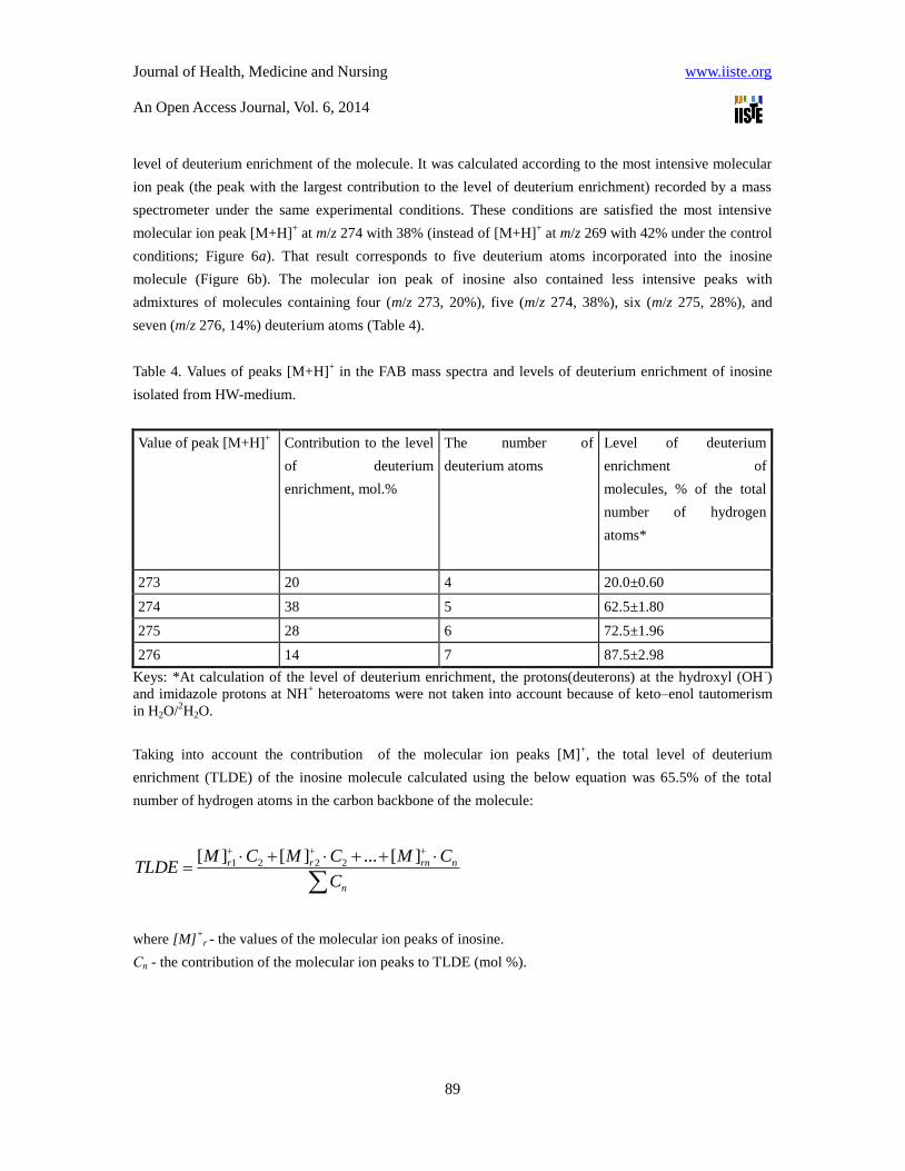
level of deuterium enrichment of the molecule. It was calculated according to the most intensive molecular
ion peak (the peak with the largest contribution to the level of deuterium enrichment) recorded by a mass
spectrometer under the same experimental conditions. These conditions are satisfied the most intensive
molecular ion peak [М+Н]+ at m/z 274 with 38% (instead of [М+Н]
+ at m/z 269 with 42% under the control
conditions; Figure 6a). That result corresponds to five deuterium atoms incorporated into the inosine
molecule (Figure 6b). The molecular ion peak of inosine also contained less intensive peaks with
admixtures of molecules containing four (m/z 273, 20%), five (m/z 274, 38%), six (m/z 275, 28%), and
seven (m/z 276, 14%) deuterium atoms (Table 4).
Table 4. Values of peaks [M+H]+ in the FAB mass spectra and levels of deuterium enrichment of inosine
isolated from HW-medium.
Value of peak [М+Н]+
Contribution to the level
of deuterium
enrichment, mol.%
The number of
deuterium atoms
Level of deuterium
enrichment of
molecules, % of the total
number of hydrogen
atoms*
273 20 4 20.0±0.60
274 38 5 62.5±1.80
275 28 6 72.5±1.96
276 14 7 87.5±2.98
Keys: *At calculation of the level of deuterium enrichment, the protons(deuterons) at the hydroxyl (OH-)
and imidazole protons at NH+ heteroatoms were not taken into account because of keto–enol tautomerism
in H2O/2H2O.
Taking into account the contribution of the molecular ion peaks [M]+, the total level of deuterium
enrichment (TLDE) of the inosine molecule calculated using the below equation was 65.5% of the total
number of hydrogen atoms in the carbon backbone of the molecule:
1 2 2 2[ ] [ ] ... [ ]r r rn n
n
M C M C M CTLDE
C
where [M]+
r - the values of the molecular ion peaks of inosine.
Сn - the contribution of the molecular ion peaks to TLDE (mol %).

Journal of Health, Medicine and Nursing www.iiste.org
An Open Access Journal, Vol. 6, 2014
90
Figure 6. FAB mass spectra of inosine (glycerol as a matrix) under different experimental conditions: (a) –
natural inosine; (b) – [2H]inosine isolated from HW medium (scanning interval at m/z 50–350; major peaks
with a relative intensity of 100% at m/z 52 and m/z 54; ionization conditions: cesium source; accelerating
voltage, 5 kV; ion current, 0.6–0.8 mA; resolution, 7500 arbitrary units): I – relative intensity of peaks (%);
(I) – inosine; (II) – ribose fragment; (III) – hypoxanthine fragment.
The fragmentation of the inosine molecule, shown in Figure 7, gives more precise information on the
deuterium distribution in the molecule. The FAB fragmentation pathways of the inosine molecule (I) lead to
formation of ribose (C5H9O4)+
fragment (II) at m/z 133 and hypoxanthine (C5H4ON4)+ fragment (III) at m/z
136 (their fragmentation is accompanied by the migration of Н+), which in turn, later disintegrated into
several low-molecular-weight splinter fragments at m/z 109, 108, 82, 81, and 54 due to HCN and CO
elimination from hypoxanthine (Figure 7). Consequently, the presence of two “heavy” fragments of ribose

Journal of Health, Medicine and Nursing www.iiste.org
An Open Access Journal, Vol. 6, 2014
91
II (C5H9O4)+
at m/z 136 (46%) (instead of m/z 133 (41%) in the control) and hypoxanthine III (C5H4ON4)+
at m/z 138 (55%) (instead of m/z 136 (48%) in the control), as well as the peaks of low molecular weight
splinter fragments formed from FAB-decomposition of hypoxanthine fragment at m/z 111 (49%) (instead of
m/z 109 (45%) in the control) and m/z 84 (43%) (instead of 82 (41%) in the control) suggests that three
deuterium atoms are incorporated into the ribose residue, and two other deuterium atoms – into the
hypoxanthine residue of the inosine molecule (Figure 7). Such selective character of the deuterium
inclusion into the inosine molecule on specific locations of the molecule was confirmed by the presence of
deuterium in the smaller fission fragments.
Figure 7. The fragmentation pathways of the inosine molecule leading to formation of smaller fragments by
the FAB-method
When analyzing the level of deuterium enrichment of the inosine molecule we took into account the fact
that the character of deuterium incorporation into the molecule is determined by the pathways of carbon
assimilation. The carbon source was glucose as a main substrate and a mixture of deuterated amino acids
from deuterated hydrolizate of methylotrophic bacterium B. methylicum as a source of deuterated substrated
and amine nitrogen. Since the protons (deuterons) at positions of the ribose residue in the inosine molecule
could have been originated from glucose, the character of deuterium inclusion into the ribose residue is
mainly determined by hexose monophosphate (HMP) shunt, associated with the assimilation of glucose and
other carbohydrates. HMP shunt is a complex of 12 reversible enzymatic reactions resulting in the
oxidation of glucose to CO2 to form reduced NADPH, and H+, and the synthesis of phosphorylated sugars
containing from 3 to 7 carbon atoms. Since glucose in our experiments was used in a protonated form, its
contribution to the level of deuterium enrichment of the ribose residue was neglected. However, as the
investigation of deuterium incorporation into the molecule by FAB method showed that deuterium was
incorporated into the ribose residue of the inosine molecule owing to the preservation in this bacterium the
minor pathways of de novo glucose biosynthesis in 2H2O-medium. Evidently the cell uses its own resources
for intracellular biosynthesis of glucose from intracellular precursors. It should be noted that numerous
isotopic 1Н–
2Н exchange processes could also have led to specific incorporation of deuterium atoms at
certain positions in the inosine molecule. Such accessible positions in the inosine molecule are hydroxyl

Journal of Health, Medicine and Nursing www.iiste.org
An Open Access Journal, Vol. 6, 2014
92
(OH-)- and imidazole protons at NH
+ heteroatoms, which can be easily exchanged on deuterium in
2Н2О via
keto–enol tautomerism. Three non-exchangeable deuterium atoms in the ribose residue of inosine are
synthesized de novo and could have been originated from HMP shunt reactions, while two other deuterium
atoms at C2,C8-positions in the hypoxanthine residue could be synthesized de novo at the expense of
[2H]amino acids, primarily glutamine and glycine, that originated from deuterated hydrolysate of the
methylotrophic bacterium B. methylicum obtained on 98 % of 2H2O medium. In particular, the glycoside
proton at -N9-glycosidic bond could be replaced with deuterium via the reaction of СО2 elimination at the
stage of ribulose-5-monophosphate formation from 3-keto-6-phosphogluconic acid with subsequent proton
(deuteron) attachment at the С1-position of ribulose-5-monophosphate. Two other protons at C2(C3) and
C4 positions in ribose residue could be replaced with deuterium via further enzimatic isomerization of
ribulose-5-monophosphate into ribose-5-monophosphate (Figure 8). In general, our studies confirmed this
scheme (Ignatov & Mosin, 2013b). However, it should be noted that the level of deuterium enrichment of
inosine molecule is determined by isotopic purity of 2H2O and deuterated substrates and, therefore, for the
total administration of the deuterium label into the inosine molecule instead of protonated glucoce it must
be used its deuterated analogue. Deuterated glucose may be isolated in gram-scale quntities from deuterated
biomass of the methylotrophic bacterium B. methylicum.
Figure 8. Scheme of biosynthesis of IMP by microbial cell (adapted from Bohinski, 1987)

Journal of Health, Medicine and Nursing www.iiste.org
An Open Access Journal, Vol. 6, 2014
93
3.3. Adaptation to deuterium the microalgae C. vulgaris
For adaptation of microalgae C. vulgaris was used Tamiya liquid mineral medium containing 25, 50, 75,
and 98% (v/v) 2Н2О (Mosin & Ignatov, 2012a). The levels of deuterium enrichment of carotenoids were In
the case of C. vulgaris and H. halobium used fluorescent illumination, as both microorganisms grown in the
presence of light. Individual colonies of cells of these microorganisms resistant to 2Н2О, allocated by
selection were grown on liquid growth media of the same composition with 99.9 atom% 2Н2О for
producing the deuterated biomass.
3.4. Adaptation to deuterium of photoorganotrophic halobacterium H. halobium
The cell membrane of extreme aerobic photo-organotrophic halobacterium Halobacterium halobium
contains a chromoprotein trans-membrane protein - bacteriorhodopsin (BR) with the molecular weight
26.5 kDa, determining the purple-red culour of halophilic bacteria. BR contains as chromophore group an
equimolar mixture of 13-cis- and 13-trans-retinol C20 carotenoid, bound by aldemine bond schiff base (as
in the visual animal pigments) with Lys-216 residue of the protein. In halobacteria BR functions as a
light-driven transmembrane proton pump pumping a proton accros the membrane. Along with the BR the
cell membrane of halobacteria contains a small amount of other related carotenoid pigments, the main of
which bakterioruberin determining the stability of halobacteria to solar radiation (Oesterhelt & Stoeckenius,
1971; Oesterhelt, 1988).
The adaptation of photo-organotrophic halobacterium Halobacterium halobium was carried out via plating
of initial cells to separate colonies on solid 2% (w/v) agarose HM2 media based on 99,9 atom% 2Н2О and
deuterated hydrolyzate biomass of B. methylicum, with the following subsequent selection of the colonies
resistant to 2Н2О. The growing of halobacteria was carried out under illumination by light fluorescent
lamps LDS-40-2 (40 W) with monochromatic light with λ = 560 nm for 4–5 days at 35 0C as swoun in
Figure 9. While growing of H. halobium on HM2 growth medium cells synthesized the purple carotenoid
pigment, identified as a native BR on the the spectral ratio of protein and chromophore fragments in the
molecule (D280/D568 = 1.5:1.0). The growth of this bacterium on 2Н2О medium was slightly inhibited as
compared with the control on protonated growth medium that simplifies the optimization of conditions
for the production of microbial biomass, which consists in the growing of the halobacterium on deuterated
growth medium with 2% (w/v) of deuterated biomass hydrolyzate of B. methylicum, cell disintegration and
lysis; isolation of purple membrane (PM) fraction; purification of PM from the low and high-molecular
weight impurities, cellular RNA, carotenoids and phospholipids; solubilization of PM in 0.5% (w/v)
solution of ionic detergent SDS–Na to form a microemulsion; fractionation of solubilized BR by MeOH;
gel permeation chromatography (GPC) on Sephadex G-200 and electrophoresis in 12.5% (w/v) PAAG in
0.1% (w/v) SDS –Na (Mosin & Ignatov, 2014).

Journal of Health, Medicine and Nursing www.iiste.org
An Open Access Journal, Vol. 6, 2014
94
Figure 9. Growth dynamics of H. halobium under various experimental conditions: a) – HW2-medium; b) –
peptone medium. Growing conditions: the incubation period: 4–5 days, temperature: 35 0C, illumination
under monochrome light at λ = 560 nm
In an attempt to remove a large fraction of the carotenoids and phospholipids from the membrane by
column GPC, PM fraction was washed by 50% (v/v) of EtOH before stabilization by SDS-Na. Removing
of carotenoids, consisting in repeated treatment of PM with 50% (v/v) EtOH at 0 0C, was a routine but
necessary step, in spite of the significant loss of the chromoprotein. It was used five treatments by 50% (v/v)
EtOH to obtain the absorption spectrum of PM suspension purified from carotenoids (4) and (5) (degree of
chromatographic purity of 80-85%), as shown in Figure 9 at various processing stages (b) and (c) relative to
the native BR (a). Figure 10 shows a dark-adapted absorption maximum at = 548 nm. Formation of
retinal-protein complex in the BR molecule leads to a bathochromic shift in the absorption spectrum of PM
(Figure 10c) - the main bandwith (1) with the absorption maximum at = 568 nm caused by the light
isomerization of the chromophore by the C13=C14 bond is determined by the presence of 13-trans-retinal
residue in BR568; additional low-intensity bandwith (2) at = 412 nm characterizes a minor impurity of a
spectral form of meta-bacteriorhodopsin M412 (formed in the light) with deprotonated aldimine bond
between 13-trans-retinal residue and protein; the total bandwith (3) with = 280 nm is determined by the
absorption of aromatic amino acids in the polypeptide chain of the protein (for native BR D280/D568 =
1.5:1.0). Upon light absorption, the maximum absorbance of PM shifts to = 556 nm with 6-8% increase
in extinction. The 280/568 nm absorbance ratio of BR is directly related to the ratio of total protein (native
BR) and is a convenient indicator for BR stability and integrity. Identical absorbance ratios are monitored
using the conventional optics on a Beckman DU-6 spectrophotometer (“Beckman Coulter”, USA) for
detergent-solubilized BR or purified BR-solubilized in detergent.

Journal of Health, Medicine and Nursing www.iiste.org
An Open Access Journal, Vol. 6, 2014
95
Figure 10. The absorption spectra of PM (50% (v/v) EtOH) at various stages of processing: (a) – natural
BR; (b) – PM after intermediate treatment; (c) – PM purified from carotenoids. The bandwith (1) is the
spectral form of BR568, (2) – impurity of spectral form of meta-bacteriorhodopsin M412, (3) – the total
absorption bandwith of aromatic amino acids, (4) and (5) – extraneous carotenoids. As a control used the
native BR.
The final stage of purification involved the crystallization of the solubilized in 0.5% (w/v) SDS-Na solution
protein by MeOH and further separation of the protein from low-molecular-weight impurities by GPC. For
this purpose the fractions containing BR were passed twice through a column with dextran Sephadex G-200
balanced with 0.09 M Tris buffer (pH = 8.35) containing 0.1% (w/v) SDS-Na and 2.5 mM EDTA.
The homogeneity of isolated BR satisfies to the requirements for reconstruction of native membranes, and
was confirmed by electrophoresis in 12.5% (w/v) PAAG with 0.1% (w/v) SDS-Na and in vitro
regeneration of AP with 13-trans-retinal. The degree of regeneration of PM was determined by the ratio:
Dnat.280.Dnat..568/Dreg..280
.Dreg.568 (D280 and D568 the absorbance of a suspension of native and regenerated PM
at λ = 280 and λ = 568 nm) was 65 mol.%. Output of crystalline protein makes up approximately 5 mg. The
total level of deuterium enrichment of the BR molecule, calculated on deuterium enrichment levels of
amino acids of the protein hydrolyzate was 95.7 atom% 2Н.
4. Discussion
Our studies indicated that the ability of adaptation to 2Н2О for different taxonomic groups of
Journal of Health, Medicine and Nursing www.iiste.org
An Open Access Journal, Vol. 6, 2014
96
microorganisms is different, and stipulated by taxonomic affiliation, metabolic characteristics, pathways of
assimilation of substrates, as well as by evolutionary niche occupied by the object. Thus, the lower the level
of evolutionary organization of the organism, the easier it adapted to the presence of deuterium in growth
media. Thus, most primitive in evolutionary terms (cell membrane structure, cell organization, resistance to
environmental factors) of the studied objects are photo-organotrophic halobacteria related to archaebacteria,
standing apart from both prokaryotic and eukaryotic microorganisms, exhibiting increased resistance to 2Н2О and practically needed no adaptation to
2Н2О, contrary to blue-green algae, which, being eukaryotes,
are the more difficult adapted to 2Н2О and, therefore, exhibit inhibition of growth at 70–75 % (v/v) D2О.
The composition of growth media evidently also plays an important role in process of adaptation to 2Н2О,
because the reason of inhibition of cell growth and cell death can be changes of the parity ratio of
synthesized metabolites in 2Н2О-media: amino acids, proteins and carbohydrates. It is noted that adaptation
to 2Н2О occures easier on complex growth media than on the minimal growth media with full substrates at
a gradual increasing of deuterium content in the growth media, as the sensitivity to 2Н2О of different vital
systems is different. As a rule, even highly deuterated growth media contain remaining protons 0,2–10
atom.%. These remaining protons facilitate the restructuring to the changed conditions during the
adaptation to 2Н2О, presumably integrating into those sites, which are the most sensitive to the replacement
of hydrogen by deuterium. The evidence has been obtained that cells evidently are able to regulate the 2Н/
1H ratios, while its changes trigger distinct molecular processes. One possibility to modify intracellular
2Н/
1H ratios is the activation of the H
+-transport system, which preferentially eliminates H
+, resulting in
increased 2Н/
1H ratios within cells (Somlyai et al., 2012). Furthermore deuterium induces physiological,
morphological and cytological alterations on the cell. There were marked the significant differences in the
morphology of the protonated and deuterated cells of blue-green algae C. vulgaris. Cells grown on 2Н2О-media were 2–3 times larger in size and had thicker cell walls, than the control cells grown on a
conventional protonated growth media with ordinary water, the distribution of DNA in them was
non-uniform. In some cases on on the surface of cell membranes may be observed areas consisting of
tightly packed pleats of a cytoplasmic membrane resembling mezosoms – intracytoplasmic bacterial
membrane of vesicular structure and tubular form formed by the invasion of cytoplasmic membrane into
the cytoplasm (Figure 11). It is assumed that mezosoms involved in the formation of cell walls, replication
and segregation of DNA, nucleotides and other processes. There is also evidence that the majority number
of mezosoms being absent in normal cells is formed by a chemical action of some external factors – low
and high temperatures, fluctuation of pH and and other factors. Furthermore, deuterated cells of C. vulgaris
were also characterized by a drastic change in cell form and direction of their division. The observed cell
division cytodieresis did not end by the usual divergence of the daughter cells, but led to the formation of
abnormal cells, as described by other authors (Eryomin et al., 1978). The observed morphological changes
associated with the inhibition of growth of deuterated cells were stipulated by the cell restructuring during
the process of adaptation to 2Н2О. The fact that the deuterated cells are larger in size (apparent size was of
2–4 times larger than the size of the protonated cells), apparently is a general biological phenomenn
proved by growing a number of other adapted to 2Н2О prokaryotic and eukaryotic cells (Mosin & Ignatov,
2012a; Mosin & Ignatov, 2012b; Mosin & Ignatov, 2014).

Journal of Health, Medicine and Nursing www.iiste.org
An Open Access Journal, Vol. 6, 2014
97
Figure 11. Electron micrographs of Micrococcus lysodeikticus cells obtained by SEM method: a) –
protonated cells obtained on H2O-medium; b) – deuterated cells obtained on 2Н2О-medium. The arrows
indicate the tightly-packed portions of the membranes
Our data generally confirm a stable notion that adaptation to 2Н2О is a phenotypic phenomenon as the
adapted cells eventually return back to the normal growth after some lag-period after their replacement
back onto H2O-medium. However, the effect of reversion of growth on H2O/2Н2О media does not exclude
an opportunity that a certain genotype determines the manifistation of the same phenotypic attribute in 2Н2О-media with high deuterium content. At placing a cell onto
2Н2О-media lacking protons, not only
2Н2О
is removed from a cell due to isotopic (1H–
2Н) exchange, but also there are occurred a rapid isotopic
(1H–
2Н) exchange in hydroxyl (-OH), sulfohydryl (-SH) and amino (-NH2) groups in all molecules of
organic substances, including proteins, nucleic acids, carbohydrates and lipids. It is known, that in these
conditions only covalent C–H bond is not exposed to isotopic (1H–
2Н) exchange and, thereof only
molecules with bonds such as C–2Н can be synthesized de novo (Mosin et al., 1996b; Mosin & Ignatov,
2012a). Depending on the position of the deuterium atom in the molecule, there are distinguished primary
and secondary isotopic effects mediated by intermolecular interactions. In this aspect, the most important
for the structure of macromolecules are dynamic short-lived hydrogen (deuterium) bonds formed between
the electron deficient 1H(
2Н) atoms and adjacent electronegative O, C, N, S- heteroatoms in the molecules,
acting as acceptors of H-bond (Ignatov & Mosin, 2013c). The hydrogen bond, based on weak electrostatic

Journal of Health, Medicine and Nursing www.iiste.org
An Open Access Journal, Vol. 6, 2014
98
forces, donor-acceptor interactions with charge-transfer and intermolecular van der Waals forces, is of the
vital importance in the chemistry of intermolecular interactions and maintaining the spatial structure of
macromolecules in aqueous solutions (Ignatov & Mosin, 2013d). Another important property is defined by
the three-dimensional structure of 2Н2О molecule having the tendency to pull together hydrophobic groups
of macromolecules to minimize their disruptive effect on the hydrogen (deuterium)-bonded network in 2Н2О. This leads to stabilization of the structure of protein and nucleic acid macromolecules in the presence
of 2Н2О. That is why, the structure of macromolecules of proteins and nucleic acids in the presence of
2Н2О
is somehow stabilized (Cioni & Strambini, 2002).
Evidently the cell implements a special adaptive mechanisms promoting the functional reorganization of
vital systems in 2Н2О. Thus, for the normal synthesis and function in D2О of such vital compounds as
nucleic acids and proteins contributes to the maintenance of their structure by forming hydrogen
(deuterium) bonds in the molecules. The bonds formed by deuterium atoms are differed in strength and
energy from similar bonds formed by hydrogen. Somewhat greater strength of 2Н–O bond compared to
1H–O bond causes the differences in the kinetics of reactions in H2O and
2Н2О. Thus, according to the
theory of a chemical bond the breaking up of сovalent 1H–C bonds can occur faster than C–
2Н bonds, the
mobility of 2Н3O
+ ion is lower on 28.5 % than Н3O
+ ion, and О
2Н
- ion is lower on 39.8 % than OH
- ion, the
constant of ionization of 2Н2О is less than that of H2O (Mosin et al., 1999b). These chemical-physical
factors lead to slowing down in the rates of enzymatic reactions in D2О (Cleland, 1976). However, there are
also such reactions which rates in 2Н2О are higher than in H2O. In general these reactions are catalyzed by
2Н3O
+ or H3O
+ ions or O
2Н
- and OH
- ions. The substitution of
1H with
2Н affects the stability and
geometry of hydrogen bonds in an apparently rather complex way and may, through the changes in the
hydrogen bond zero-point vibration energies, alter the conformational dynamics of hydrogen
(deuterium)-bonded structures of DNA and proteins in 2Н2О. It may cause disturbances in the
DNA-synthesis during mitosis, leading to permanent changes on DNA structure and consequently on cell
genotype (Lamprecht et al., 1989). Isotopic effects of deuterium, which would occur in macromolecules of
even a small difference between hydrogen and deuterium, would certainly have the effect upon the
structure. The sensitivity of enzyme function to the structure and the sensitivity of nucleic acid function
(genetic and mitotic) would lead to a noticeable effect on the metabolic pathways and reproductive
behaviour of an organism in the presence of 2Н2О (Török et al., 2010). And next, the changes in
dissociation constants of DNA and protein ionizable groups when transferring the macromolecule from
H2O into 2Н2О may perturb the charge state of the DNA and protein molecules. All this can cause variations
in nucleic acid synthesis, which can lead to structural changes and functional differences in the cell and its
organelles. Hence, the structural and dynamic properties of the cell membrane, which depends on
qualitative and quantitative composition of membrane’s fatty acids, can also be modified in the presence of 2Н2О. The cellular membrane is one of the most important organelles in the bacteria for metabolic
regulation, combining apparatus of biosynthesis of polysaccharides, transformation of energy, supplying
cells with nutrients and involvement in the biosynthesis of proteins, nucleic acids and fatty acids.
Obviously, the cell membrane plays an important role in the adaptation to 2Н2О. But it has been not clearly
known what occurs with the membranes how they react to the replacement of protium to deuterium and
Journal of Health, Medicine and Nursing www.iiste.org
An Open Access Journal, Vol. 6, 2014
99
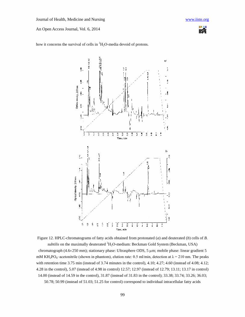
how it concerns the survival of cells in 2Н2О-media devoid of protons.
Figure 12. HPLC-chromatograms of fatty acids obtained from protonated (a) and deuterated (b) cells of B.
subtilis on the maximally deuterated 2Н2О-medium: Beckman Gold System (Beckman, USA)
chromatograph (4.6250 mm); stationary phase: Ultrasphere ODS, 5 m; mobile phase: linear gradient 5
mM KH2PO4–acetonitrile (shown in phantom), elution rate: 0.5 ml/min, detection at λ = 210 nm. The peaks
with retention time 3.75 min (instead of 3.74 minutes in the control), 4.10; 4.27; 4.60 (instead of 4.08; 4.12;
4.28 in the control), 5.07 (instead of 4.98 in control) 12.57; 12.97 (instead of 12.79; 13.11; 13.17 in control)
14.00 (instead of 14.59 in the control), 31.87 (instead of 31.83 in the control); 33.38; 33.74; 33.26; 36.03;
50.78; 50.99 (instead of 51.03; 51.25 for control) correspond to individual intracellular fatty acids

Journal of Health, Medicine and Nursing www.iiste.org
An Open Access Journal, Vol. 6, 2014
100
Figure 13. HPLC-chromatograms of amino acids obtained from hydrolizates of protonated (a) and
deuterated (b) cells of B. subtilis on the maximally deuterated D2O-medium: Biotronic LC-5001 (2303.2
mm) column (“Eppendorf–Nethleler–Hinz”, Germany); stationary phase: UR-30 sulfonated styrene resin
(“Beckman–Spinco”, USA); 25 μm; 50–60 atm; mobile phase: 0.2 N sodium–citrate buffer (pH = 2.5); the
eluent input rate: 18.5 ml/h; the ninhydrin input rate: 9.25 ml/h; detection at λ = 570 and λ = 440 nm (for
proline).
Comparative analysis of the fatty acid composition of deuterated cells of chemoheterotrophic bacteria B.
subtilis, obtained on the maximum deuterated medium with 99.9 atom.% 2Н2О (Figure 12b), carried out by
HPLC method, revealed significant quantitative differences in the fatty acid composition compared to the
control obtained in ordinary water (Figure 12a). Characteristically, in a deuterated sample fatty acids having

Journal of Health, Medicine and Nursing www.iiste.org
An Open Access Journal, Vol. 6, 2014
101
retention times at 33.38; 33.74; 33.26 and 36.03 min are not detected in HPLC-chromatogram (Fig. 12b).
This result is apparently due to the fact that the cell membrane is one of the first cell organelles, sensitive to
the effects of 2Н2О, and thus compensates the changes in rheological properties of a membrane (viscosity,
fluidity, structuredness) not only by quantitative but also by qualitative composition of membrane fatty
acids. Similar situation was observed with the separation of other natural compounds (proteins, amino
acids, carbohydrates) extracted from deutero-biomass obtained from maximally deuterated 2Н2О-medium.
Table 5. Amino acid composition of the protein hydrolysates of B. subtilis, obtained on the maximum
deuterated medium and levels of deuterium enrichment of molecules*
Amino acid Yield, % (w/w) dry weight per 1
gram of biomass
Number of
deuterium
atoms
incorporated into
the carbon
backbone of a
molecule**
Level of deuterium
enrichment of
molecules, % of the
total number of
hydrogen atoms***
Protonated
sample (control)
The sample
obtained in 99.9
atom.% 2Н2О
Glycine 8.03 9.69 2 90.0
Alanine 12.95 13.98 4 97.5
Valine 3.54 3.74 4 50.0
Leucine 8.62 7.33 5 50.0
Isoleucine 4.14 3.64 5 50.0
Phenylalanine 3.88 3.94 8 95.0
Tyrosine 1.56 1.83 7 92.8
Serine 4.18 4.90 3 86.6
Threonine 4.81 5.51
Methionine 4.94 2.25
Asparagine 7.88 9.59 2 66.6
Glutamic acid 11.68 10.38 4 70.0
Lysine 4.34 3.98 5 58.9
Arginine 4.63 5.28
Histidine 3.43 3.73
Notes:
* The data obtained by mass spectrometry for the methyl esters of N-5-(dimethylamino)
naphthalene-1-sulfonyl chloride (dansyl) amino acid derivatives.
** While calculating the level of deuterium enrichment protons (deuterons) at the carboxyl (COOH-) and
NH2-groups of amino acid molecules are not taken into account because of their easy dissociation in
H2O/2Н2О
*** A dash means absence of data.

Journal of Health, Medicine and Nursing www.iiste.org
An Open Access Journal, Vol. 6, 2014
102
Amino acid analysis of protein hydrolysates isolated from deuterated cells of B. subtilis, also revealed the
differences in quantitative composition of amino acids synthesized in 2Н2О-medium (Figure 13). Protein
hydrolyzates contains fifteen identified amino acids (except proline, which was detected at λ = 440 nm)
(Table 5). An indicator that determines a high efficiency of deuterium inclusion into amino acid molecules of
protein hydrolyzates are high levels of deuterium enrichment of amino acid molecules, which are varied from
50 atom.% for leucine/isoleucine to 97.5 atom.% for alanine.
Qualitative and quantitative composition of the intracellular carbohydrates of B. subtilis obtained on
maximally deuterated 2Н2О-medium is shown in Table. 6 (the numbering is given to the sequence of their
elution from the column) contained monosaccharides (glucose, fructose, rhamnose, arabinose),
disaccharides (maltose, sucrose), and four other unidentified carbohydrates with retention time 3.08 min
(15.63 %); 4.26 min (7.46 %); 7.23 min (11.72 %) and 9.14 min (7.95 %) (not shown) (Figure 14). Yield of
glucose in deuterated sample makes up 21.4 % by dry weight, i.e. higher than for fructose (6.82 %),
rhamnose (3.47 %), arabinose (3.69 %), and maltose (11.62 %). Their outputs are not significantly different
from the control in H2O except for sucrose in deuterated sample that was not detected (Table 6). The
deuterium enrichment levels of carbohydrates were varied from 90.7 atom.% for arabinose to 80.6 atom.%
for glucose.
Table 6. Qualitative and quantitative composition of intracellular carbohydrates of B. subtilis obtained on
the maximally deuterated medium and levels of deuterium enrichment of molecules*
Carbohydrate Content in the biomass, % of the dry weight of 1 g
biomass
Level of deuterium
enrichment, % of the total
number of hydrogen
atoms***
Protonated sample
(control)
The sample obtained in
99.9 atom.% 2Н2О**
Glucose 20.01 21.40 80.6
Fructose 6.12 6.82 85.5
Rhamnose 2.91 3.47 90.3
Arabinose 3.26 3.69 90.7
Maltose 15.30 11.62
Sucrose 8.62 ND
Notes:
* The data were obtained by IR-spectroscopy.
** ND not detected
** A dash means the absence of data.

Journal of Health, Medicine and Nursing www.iiste.org
An Open Access Journal, Vol. 6, 2014
103
Figure 14. HPLC-chromatograms of intracellular carbohydrates obtained from protonated (a) and
deuterated (b) cells of B. subtilis on the maximally deuterated 2Н2О-medium: Knauer Smartline
chromatograph (25010 mm) (“Knauer”, Germany); stationary phase: Ultrasorb CN; 10 μm; mobile phase:
acetonitrile–water (75:25, % (w/w); the input rate: 0.6 ml/min
In conclusion it should be noted that comparative analysis of IR-spectra of H2O solutions and its deuterated
analogues (2Н2О, H
2НO) is of considerable interest for biophysical studies, because at changing of the atomic
mass of hydrogen by deuterium atoms in H2O molecule their interaction will also change, although the
electronic structure of the molecule and its ability to form H-bonds, however, remains the same. The local
maximums in IR-spectra reflect vibrational-rotational transitions in the ground electronic state; the
substitution with deuterium changes the vibrational-rotational transitions in H2O molecule, that is why it
appear other local maximums in IR-spectra. In the water vapor state, the vibrations involve combinations of
symmetric stretch (v1), asymmetric stretch (v3) and bending (v2) of the covalent bonds with absorption
intensity (H2O) v1;v2;v3 = 2671; 1178.4; 2787.7 cm-1
. For liquid water absorption bands are observed in other
regions of the IR-spectrum, the most intense of which are located at 2100, cm-1
and 710-645 cm-1
. For 2Н2О
molecule these ratio compiles 2723.7, 1403.5 and 3707.5 cm-1
, while for H2НО molecule – 2671.6, 1178.4
and 2787.7 cm-1
. H2НO (50 mole% H2O + 50 mole%
2Н2О; ~50 % H
2НO, ~25 % H2O, ~25 %
2Н2О) has local
maxima in IR-spectra at 3415 cm-1
, 2495 cm-1
1850 cm-1
and 1450 cm-1
assigned to OH- -stretch, O
2Н
-
-stretch, as well as combination of bending and libration and H2НO bending respectively.

Journal of Health, Medicine and Nursing www.iiste.org
An Open Access Journal, Vol. 6, 2014
104
In the IR-spectrum of liquid water absorbance band considerably broadened and shifted relative to the
corresponding bands in the spectrum of water vapor. Their position depends on the temperature (Ignatov &
Mosin, 2013b). The temperature dependence of individual spectral bands of liquid water is very complex
(Zelsmann, 1995). Furthermore, the complexity of the IR-spectrum in the area of OH- stretching vibration
can be explained by the existence of different types of H2O associations, manifestation of overtones and
composite frequencies of OH- groups in the hydrogen bonds, and the tunneling effect of the proton (for relay
mechanism) (Yukhnevitch, 1973). Such complexity makes it difficult to interpret the spectrum and partly
explains the discrepancy in the literature available on this subject.
In liquid water and ice the IR-spectra are far more complex than those ones of the vapor due to vibrational
overtones and combinations with librations (restricted rotations, i.g. rocking motions). These librations are
due to the restrictions imposed by hydrogen bonding (minor L1 band at 395.5 cm-1
; major L2 band at 686.3
cm-1
; for liquid water at 0 0C, the absorbance of L1 increasing with increasing temperature, while L2
absorbance decreases but broadens with reduced wave number with increasing temperature (Brubach et al.,
2005). The IR spectra of liquid water usually contain three absorbance bands, which can be identified on
absorption band of the stretching vibration of OH- group; absorption band of the first overtone of the bending
vibration of the molecule H2НO and absorption band of stretching vibration of O
2Н
- group (Max &
Chapados, 2009). Hydroxyl group OH- is able to absorb much infrared radiation in the infrared region of the
IR-spectrum. Because of its polarity, these groups typically react with each other or with other polar groups to
form intra-and intermolecular hydrogen bonds. The hydroxyl groups, which are not involved in formation of
hydrogen bonds usually produce the narrow bands in IR spectrum, while the associated groups – broad
intense absorbance bands at lower frequencies. The magnitude of the frequency shift is determined by the
strength of the hydrogen bond. Complication of the IR spectrum in the area of OH- stretching vibrations can
be explained by the existence of different types of associations of H2O molecules, a manifestation of
overtones and combination frequencies of OH- groups in hydrogen bonding, as well as the proton tunneling
effect (on the relay mechanism).
Assignment of main absorbtion bands in the IR-spectrum of liquid water is given in the Table. 7. The IR
spectrum of H2O molecule was examined in detail from the microwave till the middle (4–17500 cm-1
) visible
region and the ultraviolet region – from 200 nm-1
to ionization limit at 98 nm-1
(Walrafen, 1972). In the
middle visible region at 4–7500 cm-1
are located rotational spectrum and the bands corresponding to the
vibrational-rotational transitions in the ground electronic state. In the ultraviolet region (200 to 98 nm-1
) are
located bands corresponding to transitions from the excited electronic states close to the ionization limit in
the electronic ground state. The intermediate region of the IR-spectrum – from 570 nm to 200 nm
corresponds to transitions to higher vibrational levels of the ground electronic state.
At the transition from H2O monomers to H2O dimer and H3O trimer absorption maximum of valent stretching
vibrations of the O-H bond is shifted toward lower frequencies (v3 = 3490 cm-1
and v1 = 3280 cm-1
)
(Eisenberg & Kauzmann, 1969) and the bending frequency increased (v2 = 1644 cm-1
) because of hydrogen
bonding. The increased strength of hydrogen bonding typically shifts the stretch vibration to lower
frequencies (red-shift) with greatly increased intensity in the infrared due to the increased dipoles. In contrast,

Journal of Health, Medicine and Nursing www.iiste.org
An Open Access Journal, Vol. 6, 2014
105
for the deformation vibrations of the H-O-H, it is observed a shift towards higher frequencies. Absorption
bands at 3546 and 3691 cm-1
were attributed to the stretching modes of the dimer [(H2O)2]. These frequencies
are significantly lower than the valence modes of ν1 and ν3 vibrations of isolated H2O molecules at 3657 and
3756 cm-1
respectively). The absorbtion band at 3250 cm-1
represents overtones of deformation vibrations.
Among frequencies between 3250 and 3420 cm-1
is possible Fermi resonance (this resonance is a single
substitution of intensity of one fluctuations by another fluctuation when they accidentally overlap each
other). The absorption band at 1620 cm-1
is attributed to the deformation mode of the dimer. This frequency is
slightly higher than the deformation mode of the isolated H2O molecule (1596 cm-1
). A shift of the band of
deformation vibration of water in the direction of high frequencies at the transition from a liquid to a solid
atate is attributed by the appearance of additional force, preventing O-H bond bending. Deformation
absorption band in IR-spectum of water has a frequency at 1645 cm-1
and a very weak temperature
dependence. It changes little in the transition to the individual H2O molecule at a frequency of 1595 cm-1
.
This frequency is found to be sufficiently stable, while all other frequencies are greatly affected by
temperature changes, the dissolution of the salts and phase transitions. It is believed that the persistence of
deformation oscillations is stipulated by processes of intermolecular interactions, i.g. by the change in bond
angle as a result of interaction of H2O molecules with each other, as well as with cations and anions.
Table 7. The assignment of main frequencies in IR-spectra of liquid water H2O and 2Н2О
Main vibrations of liquid H2O and 2Н2О
Vibration(s) H2O (t = 25 0C)
2Н2О (t = 25
0C)
v, cm-1
E0, M-1
cm-1
v, cm-1
E0, M-1
cm-1
Spinning ν1 +
deformation ν 2
780-1645 21.65 1210 17.10
Composite ν1 + ν2 2150 3.46 1555 1.88
Valence symmetrical ν1,
valence asymmetrical
ν3, and overtone 2ν2
3290-3450 100.65 2510 69.70
Thus the study of the characteristics of the IR spectrum of water allows to answer the question not only on the
physical parameters of the molecule and the covalent bonds at isotopic substitution with deuterium, but also
to make a certain conclusion on associative environment in water. The latter fact is important in the study of
structural and functional properties of water assotiates and its isotopomers at the isotopic substitution with
deuterium.
Conclusions
The experimental data demonstrated that the effects observed at the cellular growth on 2Н2О-media possess a
complex multifactor character stipulated by changes of morpholodical, cytological and physiological
parameters – magnitude of the lag-period, time of cellular generation, outputs of biomass, a ratio of amino

Journal of Health, Medicine and Nursing www.iiste.org
An Open Access Journal, Vol. 6, 2014
106
acids, protein, carbohydrates and fatty acids synthesized in 2Н2О, and with an evolutionary level of
organization of investigated object as well. The cell evidently implements the special adaptive mechanisms
promoting functional reorganization of work of the vital systems in the presence of 2Н2О. Thus, the most
sensitive to replacement of 1Н on
2Н are the apparatus of biosynthesis of macromolecules and a respiratory
chain, i.e., those cellular systems using high mobility of protons and high speed of breaking up of hydrogen
bonds. Last fact allows the consideration of adaptation to 2Н2О as adaptation to the nonspecific factor
affecting simultaneously the functional condition of several numbers of cellular systems: metabolism, ways
of assimilation of carbon substrates, biosynthetic processes, and transport function, structure and functions of
deuterated macromolecules. It seems to be reasonable to choose as biomodels in these studies
microorganisms, as they are very well adapted to the environmental conditions and able to withstand high
concentrations of 2Н2О in growth media.
References:
Bohinski, R.C. (1987) Modern Concepts in Biochemistry, translated from English. Moscow: Mir Publishers
Moscow, 544 [in Russian].
Brubach, J.B., Mermet, A., Filabozzi, A., Gerschel, A. & Roy, P. (2005) Signatures of the hydrogen bonding
in the infrared bands of water. J. Chem. Phys., 122: 184509.
Caprioli R.M. (1990) ContinuousFflow Fast Atom Bombardment mass Spectrometry. N.Y.: Wiley: 125 .
Cioni, P. & Strambini, G.B. (2002) Effect of heavy water on protein flexibility. Biophysical J., 82(6):
3246–3253.
Cleland W.N. (1976) Isotope effects on enzyme-catalyzed reactions. W.N. Cleland, M.N. O'Leary & D.D.
Northrop (eds.). Baltimore, London, Tokyo, University Park Press, 303.
Crespi H.L. (1989) Fully deuterated microorganisms: tools in magnetic resonance and neutron scattering.
Synthesis and applications of isotopically labeled compounds. in: Proceedings of an International
Symposium. T. Baillie & J.R. Jones (eds.) Amsterdam: Elsevier: 329–332.
Den’ko, E.I. (1970) Influence of heavy water (D2O) on animal, plant and microorganism’s cells. Usp.
Sovrem. Biol., 70(4): 41–49.
Eisenberg, D. & Kauzmann, W. (1969) The Structure and Properties of Water. Oxford University Press,
London.
Eryomin, V.A., Chekulayeva, L.N. & Kharatyan, F.F. (1978) Growth of Micrococcus lysodeikticus on a
deuterated medium. Microbiologia, 14: 629–636 [in Russian].
Ignatov I. & Mosin, O.V. (2013a) Possible processes for origin of life and living matter with modeling of
physiological processes of bacterium Bacillus subtilis in heavy water as model system. Journal of Natural
Sciences Research, 3(9): 65-76.
Ignatov, I. (2005) Energy Biomedicine, Gea-Libris, Sofia, 1–88.
Ignatov, I. (2010) Which water is optimal for the origin (generation) of life? Euromedica, Hanover: 34-35.
Ignatov, I. (2011) Entropy and time in living matter, Euromedica: 74.

Journal of Health, Medicine and Nursing www.iiste.org
An Open Access Journal, Vol. 6, 2014
107
Ignatov, I. (2012) Origin of Life and Living Matter in Hot Mineral Water, Conference on the Physics,
Chemistry and Biology of Water, Vermont Photonics, USA.
Ignatov, I., & Mosin, O.V. (2012) Isotopic Composition of Water and its Temperature in Modeling of
Primordial Hydrosphere Experiments, VIII Intern. Conference Perspectives of the Development of Science
and Technique, Biochemistry and Biophysics, 15: 41–49.
Ignatov, I., Mosin, O. V. & Naneva, K. (2012) Water in the Human Body is Information Bearer about
Longevity, Euromedica, Hanover: 110-111.
Ignatov I., Mosin O.V. (2013) Possible Processes for Origin of Life and Living Matter with Modeling of
Physiological Processes of Bacterium Bacillus Subtilis in Heavy Water as Model System, Journal of
Natural Sciences Research, 3 (9): 65-76.
Ignatov, I., Mosin, O. V. (2013) Modeling of Possible Processes for Origin of Life and Living Matter in Hot
Mineral and Seawater with Deuterium, Journal of Environment and Earth Science, 3(14): 103-118.
Ignatov, I., Mosin, O. V. (2013) Structural Mathematical Models Describing Water Clusters, Journal of
Mathematical Theory and Modeling, 3 (11): 72-87.
Ignatov, I., Mosin, O. V. (2014) The Structure and Composition of Carbonaceous Fullerene Containing
Mineral Shungite and Microporous Crystalline Aluminosilicate Mineral Zeolite. Mathematical Model of
Interaction of Shungite and Zeolite with Water Molecules Advances in Physics Theories and Applications,
28: 10-21.
Ignatov, I., Mosin, O.V., Velikov, B., Bauer, E. & Tyminski, G. (2014) Longevity Factors and Mountain
Water as a Factor. Research in Mountain and Field Areas in Bulgaria, Civil and Environmental Research, 6
(4): 51-60.
Ignatov, I., Mosin, O. V., Niggli, H.&Drossinakis, Ch. (2014) Evaluating Possible Methods and Approaches
for Registering of Electromagnetic Waves Emitted from the Human Body, Advances in Physics Theories
and Applications, 30: 15-33.
Ignatov, I., Mosin, O.V.&Drossinakis, Ch. (2014) Infrared Thermal Field Emitted from Human Body.
Thermovision, Journal of Medicine, Physiology, Biophysics, 1:1-12.
Ignatov, I., Mosin, O.V.&Velikov, B. (2014) Longevity Factors and Mountain Water of Bulgaria in Factorial
Research of Longevity, Journal of Medicine, Physiology, Biophysics,1:13-33.
Ignatov, I.&Mosin,O.V. (2014) Visual Perception. Electromagnetic Conception for the Eyesight. Rhodopsin
and Bacteriodopsin, Journal of Medicine, Physiology and Biophysics, 2:1-19.
Ignatov, I.&Mosin,O.V. (2014) The Structure and Composition of Shungite and Zeolite. Mathematical
Model of Distribution of Hydrogen Bonds of Water Molecules in Solution of Shungite and Zeolite, Journal
of Medicine, Physiology and Biophysics, 2: 20-36.
Ignatov, I., Mosin,O.V., Velikov, B., Bauer, E.&Tyminski, G. (2014) Research of Longevity Factors and
Mountain Water as a Factor in Teteven Municipality, Bulgaria, Journal of Medicine, Physiology and
Biophysics, 2: 37-52.
Ignatov, I.&Mosin,O.V. (2014) Modeling of Possible Processes for Origin of Life and Living Matter in Hot
Mineral Water. Research of Physiological Processes of Bacterium Bacillus Subtilis in Hot Heavy Water,
Journal of Medicine, Physiology and Biophysics, 2: 53-70.

Journal of Health, Medicine and Nursing www.iiste.org
An Open Access Journal, Vol. 6, 2014
108
Ignatov, I.&Mosin,O.V. (2014) Mathematical Models of Distribution of Water Molecules Regarding
Energies of Hydrogen Bonds, Medicine, Physiology and Biophysics, 2: 71-94.
Katz, J.J. (1960) The biology of heavy water. Scientific American: 106-115.
Kushner, D.J., Baker, A. & Dunstall, T.G.. (1999) Pharmacological uses and perspectives of heavy water
and deuterated compounds. Can. J. Physiol. Pharmacol., 77(2): 79–88.
Laeng, R.H., Mini, R.L., Laissue, J.A. & Schindler R. (1991) Radioprotection of cultured cells by
preincubation in medium containing deuterium oxide. Int. J. Radiat. Biol., 59(1): 165–173.
Lamprecht, I. Schroeter, D. & Paweletz, N. (1989) Disorganization of mitosis in HeLa cells by deuterium
oxide, European journal of cell biology, 50(2): 360-369.
LeMaster, D.M. (1990) Uniform and selective deuteration in two-dimensional NMR studies of proteins.
Ann. Rev. Biophys. Chem., 19: 243–266.
Lis, G., Wassenaar, L.I. & Hendry, M.J. (2008) High-precision laser spectroscopy D/H and 18
O/16
O
Measurements of microliter natural water samples. Anal. Chem., 80(1): 287–293.
Lobishev, V.N. & Kalinichenko, L.P. (1978) Isotopic effects of D2O in biological systems. Moscow,
Nauka: 215 .
MacCarthy, P. (1985) Infrared spectroscopy of deuterated compounds: an undergraduate experiment. J.
Chem. Educ., 62(7): 633–638.
Maksimova, N.P., Dobrozhinetskaia, E.V. & Fomichev, Iu.K. (1990) Regulation of phenylalanine
biosynthesis in the obligate methylotroph Methylobacillus M75. Mol. Gen. Microbiol. Virusol., 10: 28–30.
Max, J.J. & Chapados, C. (2009) Isotope effects in liquid water by infrared spectroscopy. III. H2O and D2O
spectra from 6000 to 0 cm-1
. J. Chem. Phys., 131: 184505.
Michel, F., Altermatt, H.J., Gebbers J.O. et al. (1988) Radioprotection by pretreatment with deuterated
water: cytokinetic changes in the small intestine of the mouse. Virchows. Arch. B. Cell. Pathol. Incl. Mol.
Pathol., 54(4): 214–220.
Mosin, O.V. (1996a) Studying of methods of biotechnological preparation of proteins, amino acids and
nucleosides, labeled with stable isotopes 2H,
13C and
15N with high levels of isotopic enrichment: autoref.
disser. thesis PhD: Moscow, M.V. Lomonosov State Academy of Fine Chemical Technology: 26 .
Mosin, O.V., Skladnev, D.A., Egorova, T.A. & Shvets, V.I. (1996b) Mass-spectrometric determination of
levels of enrichment of 2Н and
13С in molecules of amino acids of various bacterial objects. Bioorganic
Chemistry, 22(10–11): 856–869.
Mosin, O.V., Skladnev, D.A. & Shvets, V.I. (1996c) Methods for preparation of proteins and amino acids,
labeled with stable isotopes 2Н,
13С and
15N. Biotechnologia, 3: 12–32 [in Russian].
Mosin, O.V., Skladnev, D.A. & Shvets, V.I. (1998) Biosynthesis of 2H-labeled phenylalanine by a new
methylotrophic mutant Brevibacterium methylicum. Bioscience, biotechnology, and biochemistry, 62(2):
225–229.
Mosin, O.V., Skladnev, D.A. & Shvets, V.I. (1999a) Incorporation of [2,3,4,5,6-2H]phenylalanine,
[3,5-2H]tyrosine, and [2,4,5,6,7-
2H]tryptophan into bacteriorhodopsin molecule of bacterium halobium.
Applied Biochemistry and Microbiology, 35(1): 34–42.
Mosin, O.V., Skladnev, D.A. & Shvets, V.I. (1999b) Studying physiological adaptation of microorganisms

Journal of Health, Medicine and Nursing www.iiste.org
An Open Access Journal, Vol. 6, 2014
109
to heavy water. Biotechnologya, 8: 16–23.
Mosin, O.V., Shvets, V.I., Skladnev, D.A. & Ignatov, I. (2012a) Studying of microbic synthesis of
deuterium labeled L-phenylalanine by methylotrophic bacterium Brevibacterium Methylicum on media with
different content of heavy water. Russian Journal of Biopharmaceuticals, 4(1): 11–22.
Mosin, O.V. & Ignatov, I. (2012b) Isotope effects of deuterium in bacterial and microalgae cells at growth
on heavy water (D2O). Voda: Himia i Ecologija, 3, 83–94 [in Russian].
Mosin, O.V. & Ignatov, I. (2012c) Studying of isotopic effects of heavy water in biological systems on
example of prokaryotic and eukaryotic cells. Biomedicine, Moscow, 1(1–3): 31–50 [in Russian].
Mosin, O.V., Ignatov, I., Skladnev, D.A. & Shvets, V.I. (2013a) A strain of Gram-positive
chemoheterotrophic bacterium Basillus subtilis – producer of [2H]riboxine. Drug development &
registration, 4(5): 110–119 [in Russian].
Mosin, O.V., Shvets, V.I., Skladnev, D.A. & Ignatov, I. (2013b) Microbial synthesis of 2H-labelled
L-phenylalanine with different levels of isotopic enrichment by a facultative methylotrophic bacterium
Brevibacterium methylicum with RuMP assimilation of Carbon. Biochemistry (Moscow) Supplement Series
B: Biomedical Chemistry, 7(3): 249–260.
Mosin, O.V. & Ignatov, I. (2013c) Studying the biosynthesis of 2H-labeled inosine by a Gram-positive
chemoheterotrofic bacterium Bacillus subtilis B-3157 on heavy water (2H2O) medium. Chemical and
Process Engineering Research, 15: 32–45.
Mosin, O.V., Shvez, V.I, Skladnev, D.A., & Ignatov, I. (2013d) Microbiological synthesis of [2H]inosine
with high degree of isotopic enrichment by Gram-positive chemoheterotrophic bacterium Bacillus subtilis.
Applied Biochem. Microbiol., 49(3): 255–266.
Mosin, O.V. & Ignatov, I. (2013) Microbiological synthesis of 2H-labeled phenylalanine, alanine, valine,
and leucine/isoleucine with different degrees of deuterium enrichment by the Gram-positive facultative
methylotrophic bacterium Вrevibacterium methylicum. International Journal of Biomedicine, 3(2),
132–138.
Mosin, O.V., Ignatov, I., Skladnev, D.A. & Shvets, V.I. (2014) A strain of Gram-positive facultative
methylotrophic bacterium Brevibacterium methylicum – producer of [2H]phenylalanine. Drug development
& registration, 1(6): 58-67 [in Russian].
Mosin, O.V. & Ignatov, I (2014b) Biosynthesis of photochrome transmembrane protein bacteriorhodopsin
of Halobacterium halobium labeled with deuterium at aromatic amino acids residues of 2,3,4,5,6-2H5]Phe,
[3,5-2H2]Tyr and [2,4,5,6,7-
2H5]Trp. Chemistry and Materials Research, 6(3): 38-48.
Oesterhelt, D. & Stoeckenius, W. (1971) Rhodopsin - like protein from the purple membrane of
Halobacterium halobium. Nature, 233(89): 49–160.
Oesterhelt, D. (1988) The Structure and Mechanism of the Family of Retinal Proteins from Halophilic
Archaea Curr. Op. Struct. Biol., 8: 489–500.
Sinyak, Y., Grigoriev, A., Gaydadimov, V., Gurieva, T., Levinskih, M. & Pokrovskii, B. (2003)
Deuterium-free water in complex life-support systems of long-tern space missions. Acta Astronautica, 52:
575.
Skladnev, D.A., Mosin, O.V., Egorova, T.A., Eremin, S.V. & Shvets, V.I. (1996) Methylotrophic bacteria

Journal of Health, Medicine and Nursing www.iiste.org
An Open Access Journal, Vol. 6, 2014
110
are sourses of isotopically labelled 2Н- and
13С-amino acids. Biotechnologija, 5: 25–34 [in Russian].
Somlyai, G. (2001) The biological effect of deuterium-depleted water. A possible new tool in cancer
therapy. Anticancer Research Intern. J., 21(3): 23–33.
Somlyai, G. (2002) The Biological Effect of Deuterium Depletion. Budapest, Akademiai Klado, 130 .
Somlyai, G., Jancso, G., Jakli, G et al. (2012) Deuterium depletion from tissue culture to human clinical
studies. In: 2nd
International Congress on Deuterium Depletion. Budapest, Hungary, 17–18 May, 2012.
Stom, D.I., Ponomareva, A.K., Vyatchina, O.F. (2006) Influense of water with varying content of deuterium
on red Californian hybride (Eusenia fetida Andrei Bouche). Bull. RAS, 6(52): 167–169 [in Russian].
Török, G., Csík, M., Pintér, A. et al. (2000) Effects of different deuterium concentrations of the media on
the bacterial growth and mutagenesis. Egészségtudomány/Health Science, 44: 331-338.
Vertes A. (2003) Physiological Effect of Heavy Water. Elements andIisotopes:Fformation, Transformation,
Distribution. Dordrecht: Kluwer Acad. Publ. 111–112.
Walrafen, G.E. (1972) Raman and infrared spectral investigations of water structure. In Water a
Comprehensive Treatise, F. Franks, Ed., Vol. 1, Plenum Press, New York, 151–214.
Ykhnevitch, G.B. (1973) Infrared spectroscopy of water. Moscow, Nauka, 207 . [in Russian].
Zelsmann, H.R. (1995) Temperature dependence of the optical constants for liquid H2O and D2O in the far
IR region. J. Mol. Struct., 350: 95–114.
The IISTE is a pioneer in the Open-Access hosting service and academic event
management. The aim of the firm is Accelerating Global Knowledge Sharing.
More information about the firm can be found on the homepage:
http://www.iiste.org
CALL FOR JOURNAL PAPERS
There are more than 30 peer-reviewed academic journals hosted under the hosting
platform.
Prospective authors of journals can find the submission instruction on the
following page: http://www.iiste.org/journals/ All the journals articles are available
online to the readers all over the world without financial, legal, or technical barriers
other than those inseparable from gaining access to the internet itself. Paper version
of the journals is also available upon request of readers and authors.
MORE RESOURCES
Book publication information: http://www.iiste.org/book/
IISTE Knowledge Sharing Partners
EBSCO, Index Copernicus, Ulrich's Periodicals Directory, JournalTOCS, PKP Open
Archives Harvester, Bielefeld Academic Search Engine, Elektronische
Zeitschriftenbibliothek EZB, Open J-Gate, OCLC WorldCat, Universe Digtial
Library , NewJour, Google Scholar