Embed Size (px)
Citation preview

P H E N O B A R B I T A L - I N D U C E D S Y N T H E S I S OF
THE M I C R O S O M A L D R U G - M E T A B O L I Z I N G E N Z Y M E
S Y S T E M A N D ITS R E L A T I O N S H I P TO T H E
P R O L I F E R A T I O N OF E N D O P L A S M I C M E M B R A N E S
A Morphological and Biochemical Study
S T E N O R R E N I U S , M.D., J A N L. E. E R I C S S O N , M.D., and L A R S E R N S T E R , Ph.D.
From The Department of Pathology at Sabbatsberg Hospital, Karolinska Institutet, and The Wenner-Gren Institute, University of Stockholm, Stockholm, Sweden
A B S T R A C T
Liver microsomes, isolated from rats which had been treated with phenobarbital in vivo, were found to exhibit increased activities of oxidative demethylation and TPNH-cytochrome ¢ reductase and an increased amount of CO-binding pigment. Simultaneous administration of actinomycin D or puromycin abolished the phenobarbital-induced enzyme synthesis. Increased rate of Pi 32 incorporation into microsomal phospholipid was the first sign of phenobarbital stimulation and appeared 3 hours after a single injection of this drug. Micro- somes were divided into smooth-surfaced and rough-surfaced vesicle fractions. The frac- tion consisting of smooth-surfaced vesicles exhibited the greatest increase in protein content and oxidative demethylation activity after phenobarbital administration in vivo. Ultra- structural studies revealed that drug treatment also gave rise to proliferation of the endo- plasmic reticulum in the hepatic parenchymal cells, first noticed after two phenobarbital injections. The phenobarbitaMnduced synthesis of the metabolizing enzymes is discussed with special reference to the relationship to the stimulated synthesis of the endoplasmic membranes.
I N T R O D U C T I O N
The liver microsomal fraction, isolated after homogenization and differential centrifugation, contains free RNP particles and two kinds of vesicles: rough, with RNP 1 particles attached to
1Abbreviations used are- RNP, ribonucleoprotein; DPNH and TPNH, di- and triphosphopyridine nucleotide, reduced forms; i.p., intraperitoneally; RNA, ribonucleic acid; DNA, deoxyribonucleic acid; ER, endoplasmic reticulum; DOC, deoxycholate, IDP, inosine-51-diphosphate; ATP, adenosine-51-
the outer surface, and smooth, which lack these particles (1). Liver microsomes are known to catalyze a number of TPNH-dependent hydroxyla- tion reactions, among them the oxidative demethy- lation of various drugs (cf. review, 2). TPNH- cytochrome c reductase (3-5) and the CO-binding pigment (6, 7) are probably involved in oxidative demethylation (8-10). Pretreatment of rats in
triphosphate; G-6-P, glucose-6-phosphate; FA, form- aldehyde.
627
vivo with certain drugs, e.g. phenobarbital , in- duces an increased rate of drug hydroxylation (11-13) and also leads to increased numbers of endoplasmic membranes (14). It is the purpose of this paper to present data which indicate that phenobarbi ta l acts by the specific induction of the synthesis of TPNH-cytochrome c reductase and
the CO-binding pigment, and that this substrate induction is sensitive to actinomycin D and puro- mycin. The phenobarbi tal- induced enzyme syn- thesis will be discussed with special reference to the production of the enzyme-bearing endo- plasmic membranes, as studied by chemical methods and in the electron microscope.
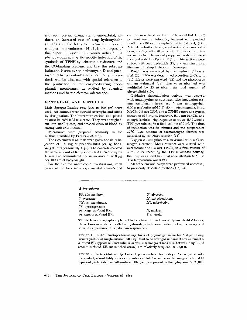
M A T E R I A L S A N D M E T H O D S
Male Spraguc-Dawley rats (200 to 300 gin) were used. All animals were starved overnight and killed by decapitation. The livers were excised and placed at once in cold 0.25 M sucrose. They were weighed, cut into small pieces, and washed clean of blood by rinsing with cold sucrose.
Microsomes were prepared according to the method described by Ernster et al. (15).
The experimental animals were given one daily in- jection of 100 mg of phenobarbital per kg body- weight intraperitoneally (i.p.). The controls received the same amount of 0.9 per cent NaCl. Actinomycin D was also administered i.p. in an amount of 8 pg per 100 gm of body-weight.
For the electron microscopic investigations, small pieces of the liver from cxperimental animals and
controls were fixed for 1.5 to 2 hours at 0-4°C in 2 per cent osmium tctroxidc, buffered with purified s-collidine (16) or a phosphate buffer (pH 7.4) (17). After dehydration in a graded series of ethanol solu- tions, starting with 70 per cent, the tissues were im- mersed in two changes of propylene oxide and were then embedded in Epon 812 (18). Thin sections were stained with lead hydroxide (19) and examined in a Siemens Elmiskop I electron microscope.
Protein was measured by the method of Lowry et al. (20). RNA was determined according to Ceriotti (21). Lipids were extracted (22) and the phosphorus content estimated (23). The value obtained was multiplied by 25 to obtain the total amount of phospholipid (15).
Oxidative demethylation activity was assayed with aminopyrine as substrate. The incubation sys- tem contained mierosomes, 5 mM aminopyrine, 0.05 M tris buffer (pH 7.5), 50 m~ nicotinamide, 5 mM MgC12, 0.5 mu TPN, and a TPNH-generating system consisting of 5 mM nL-isocitrate, 0.01 mu MnC12, and enough isocitric dehydrogenase to reduce 0.32/~moles TPN per minute, in a final volume of 2 ml. The time of incubation was 20 minutes and the temperature 37°C. The amount of formaldehyde formed was measured by the Nash reaction (24).
Oxygen consumption was measured with a Clark oxygen electrode. Measurements were started with microsomes and 0.3 mM TPNH, in a final volume of 3 mi. After recording the TPNH oxidase activity, the drug was added to a final concentration of 5 raM. The temperature was 30°C.
All other enzyme assays were performed according to previously described methods (15, 25).
Abbreviations
BC, bile capillary. C, cytosome. CM, cell membrane. CS, cytosegresome erg, rough-surfaced ER. ers, smooth-surfaced ER.
Gl, glycogen. M, mitochondrion. Mb, microbody.
N, nucleus. S, sinusoid.
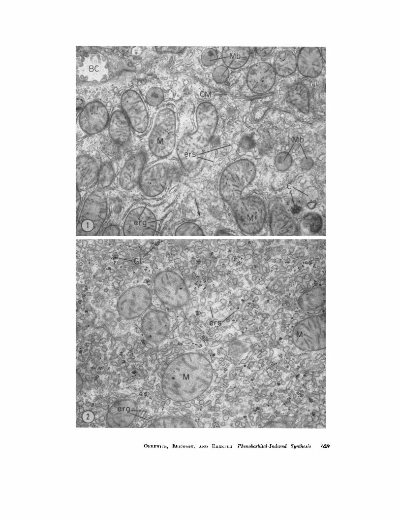
The electron micrographs in plates 1 to 6 are from thin sections of Epon-embedded tissues; the sections were stained with lead hydroxide prior to examination in the microscope and show the appearance of hepatic parenchymal cells.
FmURE 1 Control (intraperitoneal injections of physiologic saline for 5 days). Long, slender profiles of rough-surfaced ER (erg) tend to be arranged in parallel arrays. Smooth- surfaced ER appears as short tubular or vesicular images. Transitions between rough- and smooth-surfaced ER (unattached arrow) are relatively frequent. )< 15,000.
FIGURE ~ Intraperitoneal injections of phenobarbital for 3 days. As compared with the control, considerably increased numbers of tubular and vesicular images, believed to represent proliferated smooth-surfaced ER (ers), are present in the cytoplasm. × ~3,000.
628 THE JOURNAL OF CELL BIOLOGY • VOLUME ~5, 1965
ORRENIUS, ERICSSON, AND ERNSTER Phenobarbital-Induced Synthesis 629
The thyroidectomized rats were a generous gift from Dr. J . R. Tata, Mill Hill, London. Actinomycin D was made available to us by Merck, Sharp & Dohme, International. All chemicals employed were standard commercial products.
A part of this work has already been reported briefly (26).
R E S U L T S
A. Electron .~licroscopy 2
The cytoplasm of hepatic parenchymal cells from control animals given physiologic saline i.p. (Fig. l) showed an appearance similar to that in normal rat and mouse. In experimental animals given phenobarbital alone, alterations of the endoplasmic reticulum (ER) were noted in some of the animals sacrificed 2 days after the beginning of the injections, and in all of those sacrificed on the 3rd day and later. The earliest changes (ob- served in animals sacrificed on the 2nd and 3rd day) consisted in proliferation of the E R (Fig. 2). Smooth-surfaced tubular and vesicular profiles of E R were more abundant than in control ani- mals, but otherwise showed the same appearance as in the latter. Unequivocal alterations in the structure or disposition of the rough-surfaced E R were not observed. Moderate numbers of "free" R N P particles were present in the cytoplasmic ground substance. On the 4th and 5th days (Figs. 3 to 5), the cytoplasmic ground substance was filled with tightly packed smooth- and rough- surfaced profiles of the ER. Rough-surfaced E R appeared to be more abundant than in control
2 A brief account of the observations is given below. The ultrastructural alterations will be described more comprehensively elsewhere.
animals, and large areas of cytoplasm occupied by conglomerations of rough-surfaced tubular pro- files arranged in parallel arrays were often en- countered (Fig. 4). Smooth-surfaced E R usually appeared as circular or oval vesicles. Both smooth- and rough-surfaced E R often showed dilatation to a variable degree. Significant alterations in cytoplasmic organelles other than those of the E R were not observed.
A moderate proliferation of the E R occurred in hepatic parenchymal cells of experimental animals which had received both phenobarbital and actinomycin D (Fig. 6). This alteration was first observed on the 3rd day. Dilatation and extreme proliferation of the ER, as noted in animals given phenobarbital alone, was not present. Administra- tion of actinomycin D alone did not cause any appreciable alterations in liver cell structure.
B. Biochemistry
Phenobarbital treatment resulted in increased amounts of microsomal protein, RNA, and phos- pholipid (Fig. 7). The phospholipid content was already significantly higher after one phenobarbi- tal injection and exhibited a greater increase than R N A and protein.
Fig. 8 shows the response of several microsomal enzymes to phenobarbital treatment. After five injections, there was a 4- to 5-fold increase over controls in the aminopyrine-demethylating and TPNH-cytochrome c reductase activities and in the amount of CO-binding pigment, all as calcu- lated on the protein basis. The specific IDPase activity, measured in the presence of 0.1 per cent DOC, and the content of cytochrome bs, as calcu- lated per mg protein, were about the same as in the controls, while the specific activities of glucose-
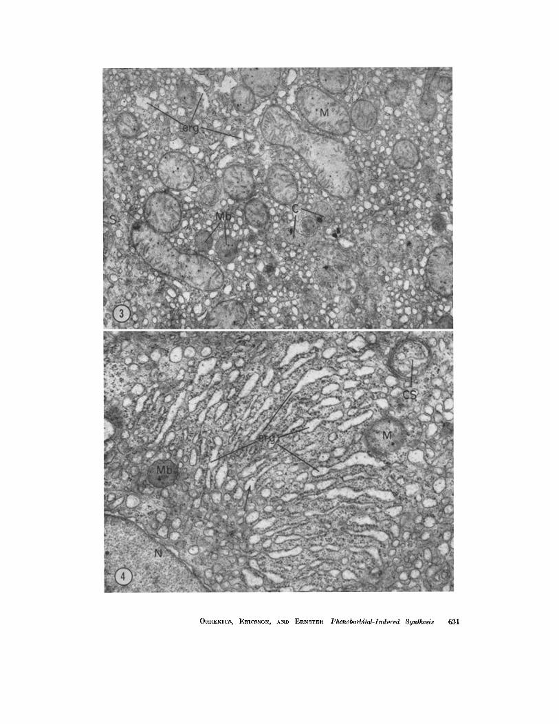
FIGURE 3 Four intraperitoneal injections of phenobarbital. The cytoplasmic ground substance contains numerous vesicles, most of which are smooth-surfaced. The rough- surfaced ER (erg) shows dilatation. X 15,000.
FIGURE 4 Four intraperitoneal injections of phenobarbital. Area of cytoplasm containing numerous cisternae of rough-surfaced ER (erg) arranged in a parallel fashion. Most of the cisternae are expanded at their ends where some become smooth-surfaced (mlattached arrow). The cytoplasm surrounding the stacked cisternae is mainly composed of smooth- surfaced vesicular profiles. In the right upper corner is an area (indicated by CS) con- taining ER which seems to have been segregated from the remainder of the cytoplasm by a system of roughly parallel membranes. Such images have been termed "cytosegresomes" and probably contain acid phosphatase (46). X 20,000.
630 TRE JOURNAL OF CELL BIOLOGY • VOLUME ~5, 1965
ORRENIUS, ERICSSON, AND ERNSTER Phenobarbital-Induced Synthesis 631

6-phosphatase, ATPase, and DPNH-cytochrome c reductase were lower in the treated animals than in the controls.
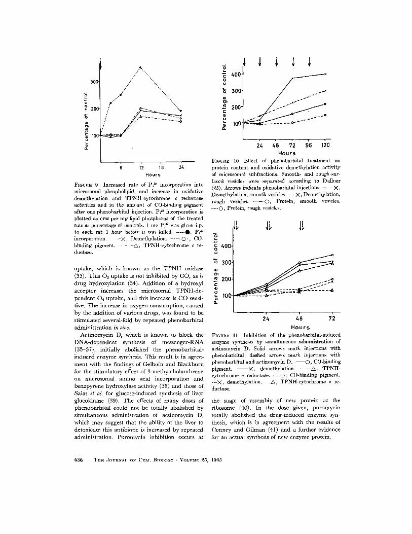
Twelve hours after one single dose of pheno- barbital, there was a significant increase in the rate of oxidative demethylation and the concen- tration of the participating enzymes (Fig. 9). The rate of P132 incorporation into microsomal phospho- lipid, however, exhibited more than a 2-fold increase already 3 hours after the administration of phenobarbital, and reached its maximum 12 hours after the injection.
In untreated rats, as well as in mice and guinea pigs, the TPNH-cytoehrome c reductase and oxidative demethylation activities and the amount of CO-binding pigment have been found to be equally distributed within microsomal subfrac- tions; i.e. rough- and smooth-surfaced vesicles (27). Fig. 10 shows that following stimulation with phenobarbital there was a greater increase in the amount of protein and oxidative demethyla- tion activity in the fraction containing smooth vesicles as compared to the one predominantly composed of rough vesicles; the latter, however, also exhibited a highly significant response to phenobarbital stimulation which is in agreement with previous findings (28).
Fig. 11 shows the effect of actinomycin D on drug-induced enzyme synthesis. This antibiotic exerted a strong inhibitory effect at the beginning of the treatment; however, following repeated injections of phenobarbital, it was no longer pos- sible to prevent an increase in enzymatic activities with simultaneous administration of actinomycin D. Puromycin completely abolished the pheno- barbital-stimulated increase in the concentration of CO-binding pigment and oxidative demethylation activity (Table I).
There was a 3- to 4-fold increase in the rate of Pi 32 incorporation into microsomal phospholipid
12 hours after a single phenobarbital injection (Fig. 12). No significant increase over the controls was found in this incorporation rate when actino- mycin D was given alone. Simultaneous adminis- tration of phenobarbital and this antibiotic caused an increased rate of Pi :~2 incorporation into micro- somal lipid phosphorus; although this stimulation of the incorporation rate was found to be sig- nificantly lower than the stimulation caused by giving phenobarbital alone. Trea tment with phenobarbital also increased the rate of Pi 32 in- corporation into mitochondrial phospholipid, to a much lesser extent, however. I t cannot be ex- cluded that the stimulation, at least in part, de- pended on contamination of the mitochondrial fraction with microsomes.
Table I I shows the stimulation of the T P N H - dependent oxygen uptake caused by addition of various drugs to microsomes isolated from pheno- barbital-treated and control animals. For all the drugs added, the increase in oxygen consumption was found to be stimulated several times by re- peated phenobarbital administration in vivo. It may be noted that the microsomal TPNH-oxidiz- ing activity, measured in the absence of substrate, was not significantly increased by phenobarbital treatment. The molar amount of formaldehyde formed from the oxidatively demethylated drugs was the same as the amount of oxygen consumed.
There are many similarities between the re- sponse of rat liver to phenobarbital stimulation and the effects of administration of thyroid hor- mone to a thyroidectomized rat (cf. review, 29); e.g., increased liver weight, increased protein synthesis, increased TPNH-cytochrome c re- ductase activity, sensitivity to actinomycin D, and the relatively long time lag before measurable changes occur. In order to study the possible role of the thyroid hormone in drug-induced enzyme synthesis, thyroidectomized rats that had received
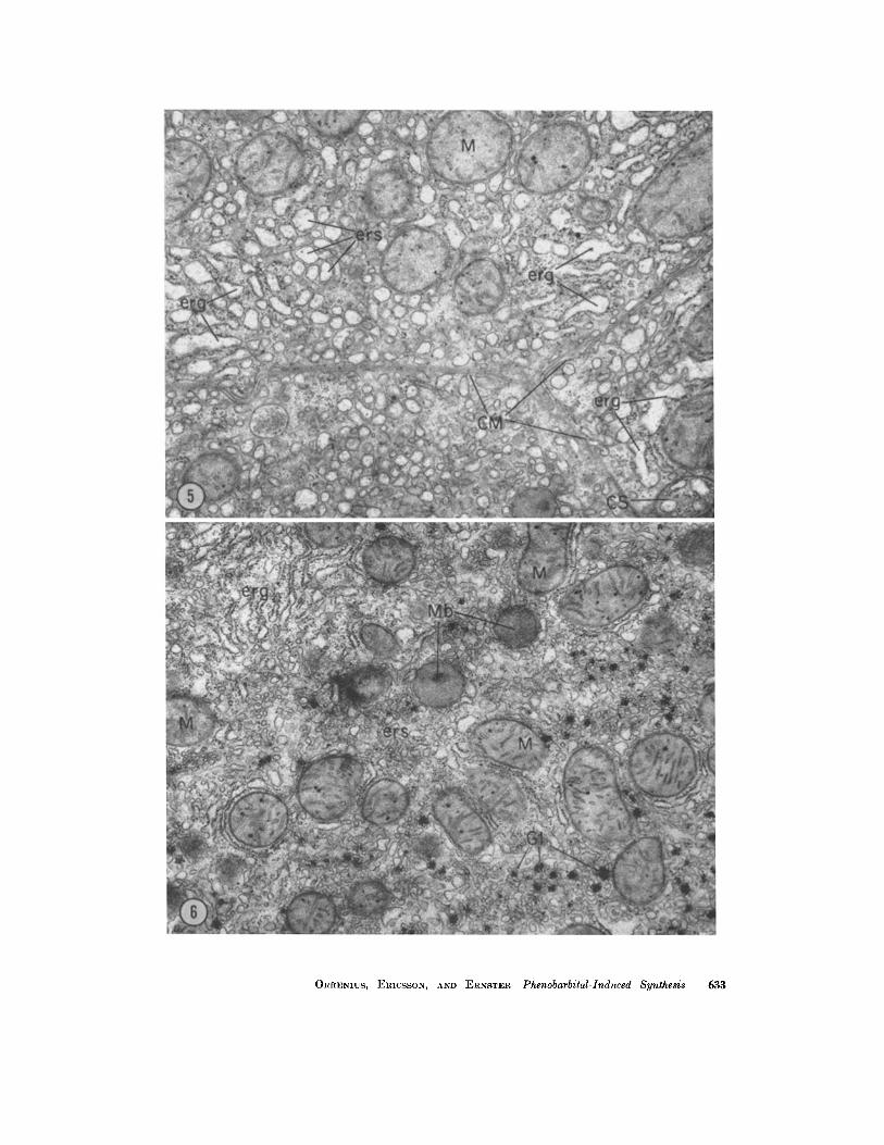
FIGURE 5 Five intraperitoneal injections of phenobarbital. The cytoplasm contains numerous dilated cisternae of rough-surfaced ER (erg), often showing irregular outlines. There are also abundant profiles of smooth-surfaced vesicular, oval, or short tubular images, probably representing dilated (possibly also fragmented), smooth-surfaced ER. In the right lower corner is a "eytosegresome" similar to that shown in Fig. 4. X 16,000.
FmVRE 6 Five intraperitoneal injections of phenobarbital and actinomyein D. Abundant smooth- (ers) and rough-surfaced (erg) ER in the cytoplasm. (Compare with Fig. 1). X 1~,500.
632 T ~ E JOURNAL OF CELL BIOLOGY • VOLUME ~5, 1965
ORRENIUS, ERICSSON, AND ERNSTER Phenobarbital-Induced Synthesis 633

200. o
g
E 100- o~
lipid
A i0 7'2 9; li0 hours
I~GURE 7 Effect of phenobarbital treatment on micro- somal protein, RNA, and phospholipid. Mean values (in mg per gm liver) of six phenobarbital-treated rats are plotted, expressed as percentage of the mean values obtained from a control gloup of six rats. Phenobarbital injections are marked with arrows. The averages 4- standard deviations for the phenobarbital-treated groups (Exp.) and the control groups (Con.) were the following:
No, of pheno- barbital
treatmenls
C o n . 1 Exp.
C o n . 2 Exp.
Coi l . 3 Exp.
C o n . 4 Exp.
C o n . 5 Exp.
Protein
mg/gm liver
'24.1 -4- 1.46 23.9 4- 1.54
23.1 4- 1.34 27.7 4- 1.28
i24.1 -4- 1.30 31.4 4- 1.52
RNA
mg/gm liver
4.82 4- 0.38 4.62 4- 0.32
4.50 4- 0.34 5.31 -4- 0.41
4.28 4- 0.29 5.56 -4- 0.40
Phospholipid
mg/gm liver
6.75 4- 1.27 10.00 4- 1.27
6.40 -4- 1.20 10.77 4- 2.00
6.22 4- 1.18 11.70 4- 2.10
0 37 [ 6 05 4- 1.18 22.8 4- 1.384.30 -4- i 33.1 4- 2 .226.84 4- 0171,12170 4- 2.72
22.0 4- 1.50,4.40 -4- 0.46 6.30 4- 1.40 35.8 4- 3.0017.08 4- 0.90i15.10 -4- 3.20
no, one, or three injections of t r i iodothyronine, were subjected to phenobarb i ta l t r ea tment (Table I I I ) . ~[he increase in the specific aminopyr ine- demethyla t ing and TPNH-cy toch rome c reductase activities and in the amount of CO-b ind ing pig- ment , as calculated per mg protein, was about the same as in the non-thyroidectomized controls. This f inding indicates tha t thyroid hormones are no t involved in phenobarbi ta l - induced enzyme synthesis.
D I S C U S S I O N
It is well documented tha t t r ea tment of rats with cer tain drugs induces an increased tolerance to the action not only of the specific drug used for p re t rea tment bu t also of a whole group of com- pounds which share the property of being hydroxy- lated by liver microsomal enzymes in the presence of T P N H and oxygen ( l l - 1 3 , 30). I t has been postulated tha t the inducing agents act by caus- ing an increased synthesis of the microsomal drug-metabol iz ing enzymes (13, 28, 31). Adminis- t ra t ion of ethionine was shown to inh ib i t the drug- induced increase in the activities of the metabol iz- ing enzymes (13, 31), and Gelboin and Sokoloff reported tha t t rea tment with phenobarbi ta l , which is one of the most potent inducing agents, st imu- lated the incorporat ion of amino acids into micro- somal proteins of cell-free liver preparat ions (32). I t was recently shown tha t phenobarb i ta l st imu- lat ion of the activities of several drug-metabol iz ing enzymes is associated with increased numbers of endoplasmic membranes (14, 28).
Earl ier results suggested that TPNH-cy to- chrome c reductase and the CO-b ind ing p igment are involved in l iver-microsomal drug-hydroxylat- ing reactions (8-10). Strong evidence for the involvement of these two enzymes in oxidative demethyla t ion was obta ined in the present study. Repea ted adminis t ra t ion of phenobarb i ta l to rats caused a ca. 1.5-fold increase in the amount of microsomal m e m b r a n e cons t i tuents - -pro te in , RNA, and phosphol ip id- -whi le the activities of aminopyr ine demethyla t ion and TPNH-cy to- chrome c reductase and the amount of CO-b ind ing pigment , all as calculated on the protein basis, exhibited a 4- to 5-fold increase over the controls. The amount of cytochrome bs, calculated per mg protein, did not change, while DPNH-cy tochrome c reductase showed a lower specific activity in the drug-t reated animals; this was also true for ATP- ase and glucose-6-phosphatase. T h e fact tha t t r ea tment with phenobarb i ta l causes a several-fold increase in the concentra t ion of only one of the microsomal hemoproteins, the CO-b ind ing pig- ment , and in the activity of only one of the micro- somal flavoproteins TPNH-cy toch rome c re- ductase, indicates tha t the induct ion is a specific substrate-caused de novo synthesis of the metabo- lizing enzymes.
However, s t imulat ion with phenobarb i ta l also leads to proliferation of the ER. I t would thus appear tha t the newly synthesized enzymes are
634 TaE JOlYRNAL OF CELL BIOLOGY • VOLUME ~5, 1965

5"
=_ o E
" I 3 - -
E i22"
1" ==
~2- "o
¢J
E
/J.o Demethytati°n CO-bind. pigm.
/ 1-~-* TPNH-cytc red. . ,v~ -~"
"'~I~..-~- ~ "'~':U'" " .... ... - - s e
...... • DPNH-cyt.c red.
..... "-*~ ............... °'"'"'~"" ~ "jL"" ''* ATPase
24 4'8 72 9'6 120 H o u r s
FIGURE 8 Effect of phenobarbital treatment on certain mierosomal enzymes. The mean values of the specific activities of the enzymes of the phenobarbital-treated group (six rats) are plotted in relation to the same values of the control group (six rats). Arrows indicate phenobarbital injections. The averages standard deviations for the phenobarbital-treated groups (Exp.) and the control groups (Con.) were the following:
No, of pheno- barbital
ueatments
1 Con. Exp.
C o n . 2 E x p .
C o n . 3 E x p .
C o n . 4 Exp.
Con. 5 Exp.
Demethyla- TPNH-cyto- ] I DPNH-ey- CO-binding [ Cytochrome A tion " chrome ¢ IDPase G-6-Pase toehrome TPase
. . . . . . . . . p*gment reduetase b5 c reductase
m#moles ^ E lamoles T P N H I #moles P i / A E Izmoles P i / ] Izmoles #moles P i / 46o ~0o/ ~a 42~ 41o/ FA/min . / - ox./rain./ I 20 rain.~ - 20 min./ I D P N H ox./ 20 min./
mg prot. i mg prot. . rag prot. mg prot. I mg prot. rag prot. I ra*n,/mg prot mg prot.
2.80 4. 0.370.019 4. 0.0070.020 -4- 0.004L7.20 -4- 0.52 D 103 -4- 00303.12 4- 0.3510.62 4- 0.1', 1.95 4. 0.24 5.56 -4- 0.900036 4. 00090042 4- 0006!718 4- 070, D089 4- 00241240 4- 026032 4- 006101 4- 013
3.05 4. 0.400.022 4. 0.007}0.019 4. 0,003 7.00 ::t: 0.401D.090 4. 0.0293.40 4. 0.41 0.71 4. 0.142.20 4- 0.26 6.70 4. 1.120.067 4. 0.010;0.045 4. 0.0076.98 4. 0.56 3.085 4. 0.0202.16 4- 0.200.25 4- 0.04 1.36 4. 0.14
9.51 4- 1 .20
3 .1 0 4- 0 . 4 5 . 2 ::k: • • 2 4- • 7. 4- • 2 . . . . 01 i ' 4- • . ::h . 2, 2 4 . 0 . 30 13 .50 4 . 1 . 4 0 0 . 0 7 5 4 . 0 . 0 1 0 0 . 0 8 7 4- 0 . 0 0 8 7 . 4 0 4- 0 4 9 0 . 0 8 2 -4- 0 . 0 2 4 2 . 0 8 4 . 0 . 2 9 0 . 2 6 4. 0 . 0 4 ' 0 . 8 4 4- 0 . 13
311 4. 0,600.022 4. 0.00610.026 4- 0,00517.19 4- 0.3010.100 4- 0.03013.12 4- 0341062 4- 009195 4- 024 15.80 4- 1 .52 0 . 1 0 0 4 . 0 .01410.113 4- 0 . 0 1 7 7 .45 ::b 0 .53 0 . 0 8 8 4- 0 . 0 2 6 1.54 4- 0 . 1 6 i 0 . 2 7 4- 0 . 0 5 i 0 . 8 2 4- 0 .11
d e p e n d e n t u p o n the p roduc t ion of new m e m b r a n e s
in o rder to function. This view is fur ther suppor ted
by the increased rate of Pi 3~ incorpora t ion into
mic rosomal phosphol ipids , wh ich was the first
sign of p h e n o b a r b i t a l s t imulat ion.
T h e fastest and greatest increase in the sub-
s t ra te- induced enzyme levels was found in the
fract ion conta in ing smooth-sur faced vesicles. This
is in ag reemen t wi th the results of R e m m e r and
Merke r (28). An increased a m o u n t of endoplasmic
m e m b r a n e s was found by electron microscopy in
hepat ic p a r e n c h y m a l cells after two to three in-
ject ions of phenobarb i t a l . T h e earliest a l terat ion
was the appea rance of smal l smooth-surfaced
vesicular and t ubu l a r profiles in the cytoplasm.
Fol lowing fur ther injections, an increase in the
a m o u n t of rough-sur faced m e m b r a n e s was also
observed. I t is possible tha t d r u g t r e a t m e n t first
gives rise to a synthesis of smoo th m e m b r a n e s ;
some of these m a y later become coated wi th R N P
particles.
Microsomes catalyze a T P N H - d e p e n d e n t 02
O R R E N I U S , ERICSSON, AND ERNSTF.R P h e n o b a r b i t a l - I n d u c e d S y n t h e s i s 635
300
-5
o o 20(;
100' o
, " "... ," "-%
,o o* ,, ,, °,
I [ " %- .
~,'" "%,
/ / - . . . . . . ~
: k,
1'2 1'8 2'4 H o u r s
FIGURE 9 Increased rate of Pi ~ incorporation into microsomal phospholipid, and increase in oxidative demethylation and TPNH-cytochrome e reductase activities and in the amount of CO-binding pigment after one phenobarbital injection. Pi ~ incorporation is plotted as ePM per mg lipid phosphorus of the treated rats as percentage of controls. 1 m c Pi ~ was given i.p. to each rat 1 hour before it was killed. -----O, P i ~ incorporation. - - X , Demethylation. - . - - © . , CO- binding pigment. - - - A , TPNH-cytochrome c re- ductase.
uptake, which is known as the T P N H oxidase (33). This O2 uptake is not inhibited by CO, as is drug hydroxylation (34). Addition of a hydroxyl acceptor increases the microsomal TPNH-de- pendent Oo uptake, and this increase is CO sensi- tive. The increase in oxygen consumption, caused by the addition of various drugs, was found to be stimulated several-fold by repeated phenobarbital administration in vivo.
Actinomycin D, which is known to block the DNA-dependent synthesis of messenger-RNA (35-37), initially abolished the phenobarbital- induced enzyme synthesis. This result is in agree- ment with the findings of Gelboin and Blackburn for the stimulatory effect of 3-methylcholanthrene on microsomal amino acid incorporation and benzpyrene hydroxylase activity (38) and those of Salas et al. for glucose-induced synthesis of liver glucokinase (39). The effects of many doses of phenobarbital could not be totally abolished by simultaneous administration of actinomycin D, which may suggest that the ability of the liver to detoxicate this antibiotic is increased by repeated administration. Puromycin inhibition occurs at
O k. "5 400' 0 u
'~ 300'
200" e "
• 100' n
s ~ ' s ~ ' ~ " x
i4 4'8 7'2 i6 H o u r s
FmURE 10 Effect of phenobarbital treatment on protein content and oxidative demethylation activity of mierosomal subfractions. Smooth- and rough-sur- faced vesicles were separated according to Dallner (~5). Arrows indicate phenobarbital injections. - - X , Demethylation, smooth vesicles. ----X, Demethylation, rough vesicles. ©, Protein, smooth vesicles. .... C), Protein, rough vesicles.
-6 t_
8 400 (J
~5 300
m 200,
1oo. O-
i
i ! !
24 48 72
H o u r s FmvnE 11 Inhibition of the phenobarbital-induced enzyme synthesis by simultaneous administration of actinomycin D. Solid arrows mark injections with phenobarbital; dashed arrows mark injections with phenobarbital and actinomycin D . - - - - O , CO-binding pigment. - - X , demethylation. - - A , TPNH- cytochrome c reductase. ----0, CO-binding pigment. ---)<, demethylation. ---~, TPNH-cytochrome c re- ductase.
the stage of assembly of new protein at the ribosome (40). In the dose given, puromycin totally abolished the drug-induced enzyme syn- thesis, which is in agreement with the results of Conney and Gilman (41) and a further evidence for an actual synthesis of new enzyme protein.
636 T H E JOURNAL OF CELL BIOLOaY - VOLUME ~5, 1965
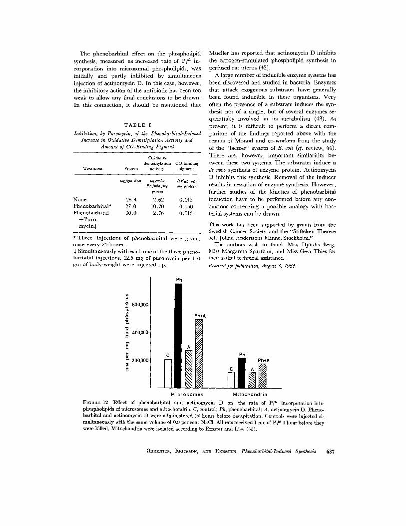
T h e phenobarb i t a l effect on the phospholipid synthesis, measured as increased rate of Pi ~2 in- corpora t ion into microsomal phospholipids, was initially and part ly inhibi ted by simultaneous injection of act inomycin D. In this case, however, the inhibi tory action of the antibiotic has been too weak to allow any final conclusions to be drawn. In this connection, it should be ment ioned tha t
T A B L E I
Inhibition, by Puromycin, of the Phenobarbital-Induced Increase in Oxidative Demethylation Activity and
Amount of CO-Binding Pigment
Oxidative demethylation CO-binding
Treatment Protein ac t i v i t y pigment
mg/gm liver ml~moles AE450_ 500 / FA/min./mg mg protein
protein
None 26.4 2.62 0.013 Phenobarbi ta l* 27.8 10,70 0.050 Phenobarbi ta l 30.0 2,76 0.013
+ P u r o - mycin :~
* Three injections of phenobarb i ta l were given, once every 24 hours.
Simultaneously with each one of the three pheno- barb i ta l injections, 12.5 mg of puromycin per 100 gm of body-weight were injected i.p.
Muel ler has reported tha t ac t inomycin D inhibits the estrogen-st imulated phospholipid synthesis in perfused ra t uterus (42).
A large n u m b e r of inducible enzyme systems has been discovered and studied in bacteria. Enzymes t ha t at tack exogenous substrates have generally been found inducible in these organisms. Very often the presence of a substrate induces the syn- thesis not of a single, but of several enzymes se- quent ial ly involved in its metabol ism (43). At present, it is difficult to perform a direct com- parison of the findings reported above wi th the results of Monod and co-workers from the study of the "lactose" system of E. coli (cf. review, 44). There are, however, impor t an t similarities be- tween these two systems. The substrates induce a de novo synthesis of enzyme protein. Act inomycin D inhibits this synthesis. Remova l of the inducer results in cessation of enzyme synthesis. However, fur ther studies of the kinetics of phenobarb i ta l induct ion have to be performed before any con- clusions concerning a possible analogy wi th bac- terial systems can be drawn.
This work has been supported by grants from the Swedish Cancer Society and the "Stiftelsen Therese och Johan Anderssons Minne, Stockholm."
The authors wish to thank Miss Hj6rdis Berg, Miss Margareta Sparthan, and Miss Gesa Thies for their skilful technical assistance.
Received for publication, August 3, 1964.
600,000. ,.c: o. u~ o .J¢ o. .lo "~ 400,O00-
E
o. 200 ,000 . z n. u
Ph
~h*A
¢/,
Ph ~ Ph*A
M i c r o s o m e s M i t o c h o n d r i a
]?IGURE 12 Effect of phenobarbital and actinomycin D on the rate of Pi m incorporation into phospholipids of microsomes and mitochondria. C, control; Ph, phenobarbital; A, actinomycin D. Pheno- barbital and actinomycin D were administered 12 hours before decapitation. Controls were injected si- multaneously with the same volume of 0.9 per cent NaC1. All rats received 1 mc of Pi ~ 1 hour before they were killed. Mitochondria were isolated according to Ernster and Liiw (45).
ORRENIUS, ERICSSON, AND ERNSTER Phenobarbital-Induced Synthesis 637

T A B L E II
Effect of Phenobarbital Treatment on the Microsomal TPNH-Dependent Oxygen Uptake, as Stimulated by Various Drugs
Exp. No.
No. of phenobarbital treatments 0 1 3
Additions Oxygen consumption
mt~moles 02 consumed/rain/rag protein
T P N H 2.60 6.00 4.85 T P N H + aminopyr ine 5.35 12.50 16.60
T P N H 3.40 4.20 4.00 T P N H + d ime thy ln i t ro samine 6.15 11.10 14.00
T P N H 3.90 3.00 7.42 T P N H + codeine 5.84 9.70 16.50
T P N H 3.72 3.00 3.12 T P N H + p h e n o b a r b i t a l 5.00 6.00 9.40
T A B L E I I I
Effect of Phenobarbital Treatment on the Oxidative Demethylation System of Thyroidectomized Rats and Thyroidectomized + Triiodothyronine (T3) - Treated Rats
No. of phenobarb,
treatm.
TPNH- Oxidative cytochrome c
demethylation redactase CO-binding Treatment Protein activity activity pigment
mg/gm liver
Non- thy ro idec tomized 25.2 Thyro idec tomized 16.6 Thyro idee tomized + I T3 inj.* 19.0 Thy ro idee tomized + 3 T3 inj.:~ 20.2
ml~moles FA/ t~moles TPNH AE4~o_b~/mg min./mg protein ox./min./mg protein protein
2.95 0.029 0.020 3.30 0.018 0.019 3.45 0.036 0.014 4.70 0.036 0.021
Non- thy ro idec tomized 27.1 9.10 0.045 0.074 Thyro idec tomized 26.4 14.90 0.046 0.083 Thy ro idec tomized + 1 T3 inj.* 25.0 17.80 0.052 0.093 Thy ro idec tomized + 3 Ta inj. :~ 23.8 11.50 0.049 0.101
* 45 /zg of t r i i odo thyron ine was in jec ted subcutaneous ly on the 3rd day before beg inn ing of p h e n o b a r b i t a l s t imula t ion . :~ 45 #g of t r i iodo thyron ine was in jec ted subcutaneous ly on the 3rd, 6th, and 8th day before beg inn ing of the p h e n o b a r b i t a l s t imulat ion.
R E F E R E N C E S
1. PALADE, G. E., and SIEKEVITZ, P., J. Biophysic. and Biochem. Cytol., 1956, 2, 171.
2. BRODm, B. B., GILLETTE, J . R., and LA Du, B. N., Ann. Rev. Biochem., 1958, 27, 427.
3. HORECK~R, B. L., J. Biol. Chem., 1950, 183, 593. 4. WILLIAMS, CI. H., and KAmN, H., J. Biol. Chem.,
1962, 237, 587.
5. PHILLIPS, A. H., and LANGDON, R. G., or. Biol. Chem., 1962, 237, 2652.
6. KLINGENBERG, M., Arch. Biochem. and BiophyKcs, 1958, 75, 376.
7. OMURA, T., and SATO, R., private communica- tion through N I H Informat ion Group I, 1964.
638 T~E J-OU~N.~L OF C~LL BIOLOaY • VOLV'~E 25, 1965

8. ORRENIUS, S., DALLNER, G., and ERNSTER, L., Abstracts, First Meeting of the Federation of European Biochemical Societies, London, 1964, 13,
9. ORRENIUS, S., DALLNER, G., and ERNSTER, L. Biochem. and Biophysic. Research Com., 1964, 14, 329.
10. ESTAEROOK, R. W., COOPER, D. Y., and ROSEN- THAL, O., Biochem. Z., 1963, 338, 741.
11. REM~ER, H., Arch. Exp. Path. Pharmakol., 1959, 235,279.
12. KATO, R., Experientia, 1960, 16, 9. 13. CONNEY, A. H., DAVISON, C., GASTEL, R., and
BURNS, B. B., J. Pharmacol. and Exp. Therap., 1960, 130, 1.
14. REMMER, H., and MERKER, H. J., Klin. Woch., 1963, 41,276.
15. ERNSTER, L., SIEKEVITZ, P., and PALADE, G. E., J. Cell. Biol., 1962, 15, 541.
16. BENNETT, H. S., and LUFT, J. H., J. Biophysic. and Biochem. Cytol., 1959, 6, 113.
17. MILLONIG, G., J. Appl. Physics, 1961, 32, 1637. 18. LUFT, J. H., J. Biophysic. and Biochem. Cytol., 1961,
9, 409. 19. KARNOVSKY, M., J. Biophysic. and Biochem. Cytol.,
1961, 11, 729. 20. LOWRY, O. H., ROSEEROUGH, N. J., FARR, A. L.,
and RANDALL, R. J., J. Biol. Chem., 1951, 193, 265.
21. CERIOTTI, G., J. Biol. Chem., 1955, 214, 59. 22. FOLCH, J., LEES, M., and SLOANE STANLEY,
G. H. S., J. Biol. Chem., 1957, 226, 497. 23. KING, E. J., Biochem. J., 1932, 26, 292. 24. NASrI, T., Biochem. J., 1953, 55, 416. 25. DALLNER, G., Acta Path. et Microbiol. Scand., 1963,
suppl. 166. 26. ORRENIUS, S., and ERNSTER, L., Biochem. and
Biophysic. Research Com., 1964, 16, 60. 27. ORRENIUS, S., DALLNER, G., and ERNSTER, L.,
data in manuscript.
28. REMMER, H., and MERKER, H. J., Science, 1963, 142, 1657.
29. TATA, J . R., in Advances in Metabolic Disorders, (R. Levine and R. Luft, editors), New York, Academic Press, Inc., 1964, 1, 153.
30. KATO, R., CHIESARA, E., and VASSANELLI, P., Biochem. Pharmacol., 1962, l l , 913.
31. CONNEY, A. H., MILLER, E. C., and MILLER, J. A., J. Biol. Chem., 1957, 228, 753.
32. GELBOIN, H. V., and SOKOLOFF, L., Science, 1961, 134, 611.
33. GILLETTE, J . R., BRODIE, B. B., and La Do, B. N., J. Pharmacol. and Exp. Therap., 1957, 119, 532.
34. NILSSON, R., ORRENIUS, S., and EaNSTER, L., Biochem. and Biophysic. Research Com., 1964, 17, 303.
35. GOLDBERO, I. M., and RABINOWITZ, M., Science, 1962, 136, 315.
36. REICH, E., GOLDEERG, I. M., and RABINOWITZ, M., Nature, 1962, 196, 745.
37. HURWITZ, J., FURTH, J. J., MALAMV, M., and ALEXANDER, M., Proc. United States Nat. Acad. Sc., 1962, 48, 1222.
38. GELBOIN, H. V., and BLACKBURN, N. R., Bio- chim. et Biophysica Acta, 1963, 72, 657.
39. SALAS, M., VINUELA, E., and SOLS, A., J. Biol. Chem., 1963, 238, 3535.
40. MORRIS, A., FAVELUKES, S., .~xRLINGHAUS, R., and SCHWEET, R., Biochem. and Biophysic. Re- search Com., 1962, 7,326.
41. CONNEY, A. H., and GILMAN, A. G., d. Biol. Chem., 1963, 238, 3682.
42. MUELLER, G. C., personal communication, 1964. 43. STANIER, R. Y., Ann. Rev. Microbiol., 1951, 5, 35. 44. JACOB, F., and MONOD, J., J. Mol. Biol., 1961, 3,
318. 45. ERNSTER, L., and L6w, H., Exp. Cell Research,
1955, suppl. 3, 133. 46. ERICSSON, J. L. E., and TRUMP, B. F., Lab.
Invest., 1964, 13, 1427.
ORRENIUS, ERICSSON, AND ERNSTER Phenobarbital-Induced Synlhesis 639


![Postnatal phenobarbital for the prevention of ...perinatal.com.br/9simposiointneorj/pdf/Aula11 - Whitelaw prevencao... · [Intervention Review] Postnatal phenobarbital for the prevention](https://img.dokumen.tips/doc/110x75/5af131777f8b9ac2468f06a7/postnatal-phenobarbital-for-the-prevention-of-whitelaw-prevencaointervention.jpg)