Embed Size (px)
Citation preview

2ND YEAR RESEARCH ELECTIVE RESIDENT’S JOURNAL Volume VII, 2002-2003
Phase III trial of L-alanosine in advanced pancreatic cancer
Matt Maurer
A. Study purpose and Rationale Background on tumor: Pancreatic cancer is the fifth leading cause of cancer mortality in the
United States. Approximately 30,000 Americans die of this disease each year with an approximate same number who are diagnosed with pancreatic cancer. Incidence with carcinoma of the pancreas increases with age and it is associated with cigarette smoking, chronic alcohol consumption, diabetes and recurrent pancreatitis. Over 90% of patients have tumors that are inoperable at the time of diagnoses and of those who undergo successful Whipple surgical procedures less than half are cured. Thus, five or less patients from a cohort of 100 patients diagnosed with this disease are ever cured. This disease has one of the poorest five year survival rates of any cancer (<4%). Progress in chemotherapy of this disease has been extremely slow. Until 1997 fluorouracil (5FU) was the standard of care and had been for over 25 years. Gemcitabine or Gemzar has been demonstrated to prolong survival over weekly bolus 5FU. Gemcitabine has an objective response rate of between 7 and 11% and those who respond to treatment improve survival by only 3 months. Gemcitabine is the current single agent of choice for pancreatic cancer. This was shown in a Phase III trial by Burris et al in which 126 patients were randomized to either Gemcitabine 1000mg/m2 weekly for 7 weeks followed by a week of rest then 3 out of 4 weeks thereafter or bolus 5-FU 600mg/m2 once weekly(36). The primary outcome was the clinical benefit response (CBR) defined as either an improvement in pain and/or performance status. The results of Gemzar vs. 5-FU were as follows(36):
• 24% vs. 5% CBR (P=.0022) • 5.65 vs. 4.41 month median survival (P=.0025) • 9 weeks vs. 4 weeks as median time to progression (P=.0002) • 18% vs. 2% one year survival
The CBR was not validated but the significance of the secondary outcomes were impressive enough to change the standard of care. Unfortunately, survival is still poor and of 56 patients with measurable disease treated with Gemxar, only three had a partial response. Many clinicians still must weigh the modest benefit with the cost of toxicity when counseling patients on whether to embark on a course of chemotherapy.
Background on new treatment: L-alanosine is an amino acid analog anti-metabolite that was extensively investigated in trials sponsored by the National Cancer Institute (NCI) during the 1970s and 1980s. L-alanosine is a potent inhibitor of de novo purine biosynthesis. L-alanosine is converted intracellularly into L-alanosinyl-5-amino-4-imidazole carboxylate acid ribonucleotide (L-alanosinyl-AICOR). L-alanosinyl-AICOR potently inhibits adenylosuccinate synthetase (ASS), which catalyses the conversion of IMP to AMP, the final common pathway for de novo AMP synthesis.
Methylthioadenosine phosphorylase (MTAP) is required in the synthesis of adenosine triphosphate (ATP) via the purine salvage pathway. Pre-clinical research conducted at the University of California, San Diego (UCSD) in the mid 1990’s demonstrated that one could increase the activity of L-alanosine by preferential treatment of tumors deficient of MTAP and therefore the purine salvage pathway. Because all normal tissues express MTAP, L-alanosine should exert selective toxicity towards MTAP-deficient malignant cells in vivo.
Studies performed at UCSD demonstrated that the MTAP gene is encoded on human chromosome 9p21, in close proximity to the p16 and p14ARF tumor suppressor genes. These genes are homozygously deleted at high frequency in certain cancers. MTAP and p16 are co-deleted in ~35% of pancreatic carcinomas, ~38% of primary NSCLC, ~66% of mesothelioma, ~75% of malignant gliomas, ~38% of osteosarcomas, ~37% soft tissue sarcomas, and ~33% of childhood T-cell acute lymphoblastic leukemias (T-ALL) (1-20).
Columbia University College of Physicians and Surgeons
153

2ND YEAR RESEARCH ELECTIVE RESIDENT’S JOURNAL Volume VII, 2002-2003
Salmedix Inc. has optimized diagnostic studies to preferentially select patients whose tumors do
not express MTAP. They developed an anti-MTAP monoclonal antibody that has been shown to be suitable for immunohistochemical (IHC) staining of paraffin-fixed tumor specimens. The IHC assay has been cross-correlated with PCR-based and RT-PCR based DNA assays to provide independent validation for MTAP-deletion.
Clinical Experience with L-alanosine National Cancer Institute (NCI) Studies Effect in Cancer Patients Without Regard to MTAP. StatusThe clinical pharmacology,
toxicity, and anti-tumor activity of L-alanosine were studied by the NCI over an 8-year period from 1978 to 1985. A total of 16 Phase I and II clinical trials were sponsored by the NCI. These studies were conducted under a variety of doses and schedules; primarily bolus intravenous (IV) doses given daily for 3 or 5 days repeated every 3 or 4 weeks. Dose-limiting toxicities included stomatitis, myelosuppression, rash, and fatigue. Responses were rare. One durable complete response (43+ months) was seen among 37 patients with renal cell carcinoma, and 2 partial responses in 39 melanoma patients (32). Retrospectively, many of the cancers treated in preclinical trials are known today to be MTAP+.
Effect in MTAP-Deficient. TumorsIn 1996, through an investigator-sponsored Investigational
New Drug application, UCSD initiated 2 studies in MTAP-negative tumors using continuous dosing schedules. Study No. 961081 examined MTAP-deficient patients with Stage IV (metastatic) non-small cell lung cancer (NSCLC) at 80 mg/m2 CIVI x 5 days q 4 weeks, in which 1 of 3 relapsing patients achieved stable disease until the 4-month evaluation (34). Study No. 960770 was transferred to Triangle Pharmaceuticals and conducted under a separate IND at a dose of 80 mg/m2 CIVI x 5 days q 4 weeks. One partial response out of 4 patients was observed in MTAP-negative recurrent glioblastoma multiforme. Stomatitis was the dose-limiting toxicity under the lower dose, continuous infusion schedules, was ameliorated by supportive care, and resolved within days after the end of infusion (28). A large multi center, multi tumor site Phase II trial is now ongoing and this protocol is contingent upon L-alanosine showing efficacy in advanced pancreatic cancer.
B. Study Design and Statistical Analysis
This is a phase III study of intravenous L-alanosine for the treatment of patients with MTAP-
deficient metastatic pancreatic tumors. Design:
• Multi-center • Prospective • Interventional – 2 arms
– L analosine alone vs. Gemcitabine alone • Randomized
– Stratification based on Center, WHO Performance Status 0, 1, or 2 (see appendix) and absence/presence of metastatic disease.
– Randomization will occur at a centralized site. • Double blind
Patients will be screened for the absence of MTAP. Fixed tumor specimens will be sent to
Impath Predictive OncologyTM Laboratory for MTAP determination. Patients meeting all eligibility requirements will receive either:
Columbia University College of Physicians and Surgeons
154

2ND YEAR RESEARCH ELECTIVE RESIDENT’S JOURNAL Volume VII, 2002-2003
L-alanosine as a continuous 5 day IV infusion (CIVI) (120 hours) at a dose of 80 mg/m2 daily, repeated every 3 weeks (21-day schedule).
Gemcitabine 1000 mg/m2 weekly 3 weeks of every 4 Therapy will continue until disease progression, drug intolerance, or other illness requiring study
drug discontinuation. The primary outcome is time to progression. Progression is defined as: Any of the following
• An increase in the product of two perpendicular diameters of any measured lesion by > 25% over the size present at study entry.
• The appearance of new areas of malignant disease including CNS lesions or ascites which must be cytologically proven.
• Deterioration in performance status. At the point of progression the study drug will be stopped and the patient will be unblinded and
counseled on alternative therapy options (see alternative therapies). The secondary outcome is overall survival. Patient sample size (n=130) was statistically determined based on:
Median time to progression with standard of care is ~8 weeks Assumed parametric data estimating standard deviation of time to progression to be ~8
weeks. Conservative given the likelihood of non parametric data. 80% power to detect 50% increase (12 weeks vs. 8 weeks) in time to progression.
An interim analysis at six months after the point of 50% accrual will assess safety and efficacy
using a p value of 0.01. The interim analysis will be done by a data safety monitoring board. At the end of the study the significance of the primary outcome will be tested with a p value of 0.04. The primary and secondary outcomes will be assessed using a Life Table Analysis with Kaplan Meier Curves. A Cox proportional hazard analysis will be employed to identify risk factors associated with the time to progression. In previous studies both performance status and metastatic disease status correlated with the time to disease progression and that is why the study subjects are stratified by these parameters.
The goal will be an accrual period of 6 months with an 18 month follow up period to complete
the study in two years. Estimating the needed number of centers to accrue 128 patients. Assuming one new pancreatic cancer referral each day to each center:
• ½ are chemotherapy naïve, • ~33% are MTAP deficient, • ½ consent rate, • A few will fail other inclusion/exclusion criteria.
Therefore there will about two accruals per month per center so over six months we will conservatively estimate a goal of 10 referrals per center. We would thus require 13 centers. If accrual is slow, centers will be added as needed.
Addressing why a head to head study is better than a placebo controlled study with all patients
receiving standard of care. • Historical precedent: Gemcitabine beat out 5-FU in a head to head study and a follow up
study showed adding 5-FU to Gemcitabine had no added benefit. If the original study was
Columbia University College of Physicians and Surgeons
155

2ND YEAR RESEARCH ELECTIVE RESIDENT’S JOURNAL Volume VII, 2002-2003
Gemcitabine + 5-FU vs. Placebo + 5-FU then it is likely that the current standard of care would be Gemcitabine + 5-FU.
• Cost vs. benefit: The benefit of standard of care is small and thus the cost of treating experimentally without it is small and single agent therapy will avoid the toxicity of combined therapy.
C. Study Procedure
Baseline Evaluation After documentation of MTAP-negative tumor status pretreatment evaluations will occur within
14 days of starting treatment and will consist of the following: • Performance status measurement. • Physical examination including vitals, height, weight, and BSA. • Medical history. • Tumor measurements using contrast-enhanced CT or MRI.
Within 28 days of study treatment initiation, radiological imaging studies will be performed. The same imaging modality will be used for tumor evaluation throughout the study.
• CBC with differential, Chem-7, LFTs • Ca19-9, uric acid, LDH, serum β-HCG (women only). • Assessment of baseline adverse events. • Documentation of concurrent medications.
MTAP Deletion Assessment MTAP expression will be assessed in advance of the patient receiving therapy. Screening for the
presence or absence of MTAP in tumor tissue may occur any time from diagnosis before study treatment. Fixed tumor specimens will be sent to Impath Predictive OncologyTM, Los Angeles, CA, for determination of MTAP status on each patient. Salmedix generated a new anti-MTAP monoclonal antibody that has been shown to be suitable for immunohistochemical (IHC) staining of paraffin-fixed tumor specimens of the same type included in this clinical trial protocol. The IHC assay has been cross-correlated with PCR-based and RT-PCR-based DNA assays providing an independent validation for MTAP-deletion. The same diagnostic laboratory (Impath Predictive OncologyTM) that developed the MTAP IHC procedure will be used for the determination of the MTAP deletion for all patients screened.
The final result of the MTAP IHC assay is qualitative, the tumor cells can be labeled as MTAP-expressing (displaying MTAP staining) or MTAP-deleted (displaying no MTAP staining). An MTAP-negative tumor will have at least 80% of the tumor cells staining at 0 and no tumor cells staining above 1+. Based on this threshold, 22 tumors out of the 80 analyzed by IHC at IMPATH were considered MTAP-negative. A total of 18 of 22 of these tumor cases displayed an H-score of 0, indicating that no staining of the neoplastic cells was observed. A total of 4 of 22 displayed a long H-score of 20 or less. In these latter 4 cases, 80% of the tumor cells contained no stain, whereas the remaining 20% stained faintly positive (+1). With the results obtained by IMPATH, we established a cut-off threshold for the definition of MTAP-deleted tumors at Long H-score of 20 or less.
Radiological Assessment Tumor assessments will be evaluated using contrast-enhanced CT scan or MRI for determination
of measurable disease. Patients must have measurable disease outside of a previously radiated area or recurrence within the radiation field to be eligible for study enrollment. If, in the judgment of the investigator, a contrast-enhanced CT scan is not obtainable, an MRI will be performed.
Treatment Plan Each patient will have a routine medical history and physical examination performed within 14
days of starting therapy and a physical examination including weight on the first day of each month.
Columbia University College of Physicians and Surgeons
156

2ND YEAR RESEARCH ELECTIVE RESIDENT’S JOURNAL Volume VII, 2002-2003
Each patient will be queried regarding baseline symptoms, and once therapy begins, any adverse events that occur during or following treatments by either a site visit or phone call on day 4 of each week (day 1 of each week will be the treatment beginning day)
Patients will receive either L-alanosine or ½ normal saline (same volume) intravenously as a continuous infusion for five days (at a dose of 80 mg/m2 per day if L-alanosine), repeated every 21 days. Gemcitabine 1000 mg/m2 or ½ normal saline (same volume) will be administered intravenously weekly for 3 weeks out of every 4. Every patient will require the placement of PICC line. Patients must sign a separate consent for PICC line placement before entry into the study. Continuous infusions will be given on an outpatient basis. Patients will be educated and trained in the use of the continous infusion equipment (which may very depending on center). Patients will be given assistance with a combination of visiting nurse services and home health aids as needed.
Body surface area will be calculated at baseline and should be recalculated prior to subsequent courses only if there is a documented change in weight (+ 10%).
Lab values will obtained weekly before the next dose of treatment. Tumor progression parameter evaluations:
• Performance status will be evaluated every two weeks with interview and physical exam. A change in PS must be confirmed at the next interval evaluation to take effect.
• CT with contrast or MRI imaging will be performed monthly. • If new ascites is found on radiologic evaluation then diagnostic paracentesis is required
for cytologic determination. Treatment will be stopped for patients demonstrating progressive disease at any time during the
study. Once treatment has stopped the patients or family will be contacted every three months for vital status and date of demise if appropriate.
Criteria For Removal From Study • Patient develops progressive disease. • Patient develops unacceptable toxicity (any WHO grade 4). • Patient is found to be ineligible. • Patient is non-compliant with study requirements. • Patient withdraws consent for further study participation.
D. Study Drugs
L-alanosine Type: Investigational Safety: The plasma clearance of L-alanosine after bolus intravenous administration to patients is
biphasic, with t1/2α = 14 min and t1/2β = 99 min. Less than 4% of the parent compound was excreted in the urine over 24h. The major route of metabolism appears to be hepatic transamination (34-35).
Expected Adverse Events with L-alanosine The most common adverse events reported during clinical trials using L-alanosine:
• Mucositis/stomatitis • Thrombocytopenia, neutropenia, leucopenia
Other reported toxicities include: malaise, headache, diarrhea, nausea, vomiting, hypotension, hypertension, rash, renal and hepatic insufficiency (at high doses), leg cramps, somnolence, confusion and disorientation.
Although 300 patients have been treated with L-alanosine since 1978, relatively few of those patients have been treated using continuous infusion schedules. Of the 18 studies in which patients have been enrolled, 4 have been conducted using continuous infusion schedules. A study of ANLL
Columbia University College of Physicians and Surgeons
157
2ND YEAR RESEARCH ELECTIVE RESIDENT’S JOURNAL Volume VII, 2002-2003
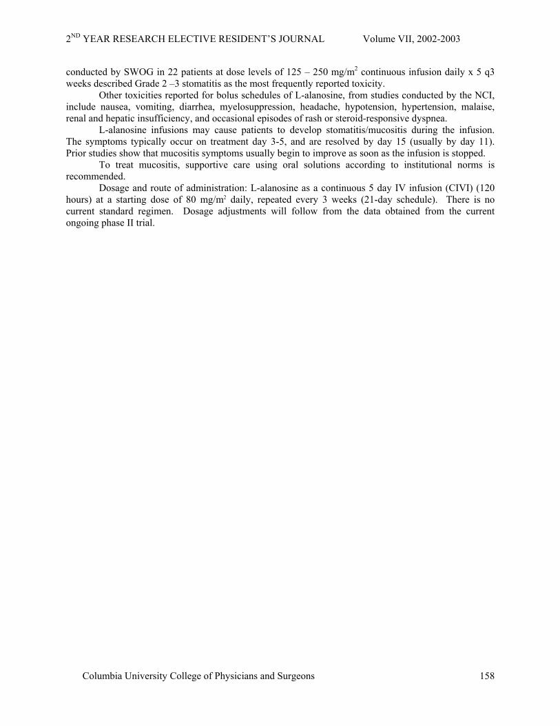
conducted by SWOG in 22 patients at dose levels of 125 – 250 mg/m2 continuous infusion daily x 5 q3 weeks described Grade 2 –3 stomatitis as the most frequently reported toxicity.
Other toxicities reported for bolus schedules of L-alanosine, from studies conducted by the NCI, include nausea, vomiting, diarrhea, myelosuppression, headache, hypotension, hypertension, malaise, renal and hepatic insufficiency, and occasional episodes of rash or steroid-responsive dyspnea.
L-alanosine infusions may cause patients to develop stomatitis/mucositis during the infusion. The symptoms typically occur on treatment day 3-5, and are resolved by day 15 (usually by day 11). Prior studies show that mucositis symptoms usually begin to improve as soon as the infusion is stopped.
To treat mucositis, supportive care using oral solutions according to institutional norms is recommended.
Dosage and route of administration: L-alanosine as a continuous 5 day IV infusion (CIVI) (120 hours) at a starting dose of 80 mg/m2 daily, repeated every 3 weeks (21-day schedule). There is no current standard regimen. Dosage adjustments will follow from the data obtained from the current ongoing phase II trial.
Columbia University College of Physicians and Surgeons
158

2ND YEAR RESEARCH ELECTIVE RESIDENT’S JOURNAL Volume VII, 2002-2003
Gemcitabine Type: FDA approved for use in advanced pancreatic cancer. Mechanism Of Action Gemcitabine exhibits cell phase specificity, primarily killing cells undergoing DNA synthesis (S-
phase) and also blocking the progression of cells through the G1/S-phase boundary. Gemcitabine is metabolized intracellularly by nucleoside kinases to the active diphosphate (dFdCDP) and triphosphate (dFdCTP) nucleosides. The cytotoxic effect of gemcitabine is attributed to a combination of two actions of the diphosphate and the triphosphate nucleosides, which leads to inhibition of DNA synthesis. First, gemcitabine diphosphate inhibits ribonucleotide reductase, which is responsible for catalyzing the reactions that generate the deoxynucleoside triphosphates for DNA synthesis. Inhibition of this enzyme by the diphosphate nucleoside causes a reduction in the concentrations of deoxynucleotides, including dCTP. Second, gemcitabine triphosphate competes with dCTP for incorporation into DNA. The reduction in the intracellular concentration of dCTP (by the action of the diphosphate) enhances the incorporation of gemcitabine triphosphate into DNA (self potentiation). After the gemcitabine nucleotide is incorporated into DNA, only one additional nucleotide is added to the growing DNA strands. After this addition, there is inhibition of further DNA synthesis. DNA polymerase epsilon is unable to remove the gemcitabine nucleotide and repair the growing DNA strands (masked chain termination).
Dosage And Administration Gemcitabine use will follow the well established guidelines described below. Single-Agent Use : should be administered by intravenous infusion at a dose of 1000 mg/m2 over
30 minutes once weekly for up to 7 weeks (or until toxicity necessitates reducing or holding a dose), followed by a week of rest from treatment. Subsequent cycles should consist of infusions once weekly for 3 consecutive weeks out of every 4 weeks.
Dose Modifications -adjustment is based upon the degree of hematologic toxicity experienced by the patient.
Patients receiving Gemzar should be monitored prior to each dose with a complete blood count (CBC), including differential and platelet count. If marrow suppression is detected, therapy should be modified or suspended according to the guidelines below.
Dosage Reduction Guidelines
Absolute granulocyte count
(x 106/L)
Platelet count
(x 106/L)
% of full dose
>1,000 and >100,000 100 500-999 or 50,000-99,000 75
<500 or <50,000 Hold Single-Agent Use: Myelosuppression is the principal dose-limiting toxicity with Gemzar therapy.
Dosage adjustments for hematologic toxicity are frequently needed and are described in the DOSAGE AND ADMINISTRATION section above.
The data in tabulated below are based on 979 patients receiving Gemzar as a single-agent administered weekly as a 30-minute infusion for treatment of a wide variety of malignancies. The Gemzar starting doses ranged from 800 to 1250 mg/m2. The frequency of all grades and severe (WHO grade 3 or 4) adverse events were generally similar in the single-agent safety database of 979 patients and the subset of patients with pancreatic cancer. Adverse reactions reported in the single-agent safety database resulted in discontinuation of Gemzar therapy in about 10% of patients. In the comparative trial in pancreatic cancer, the discontinuation rate for adverse reactions was 14.3% for the gemcitabine arm and 4.8% for the 5-FU arm.
Columbia University College of Physicians and Surgeons
159

2ND YEAR RESEARCH ELECTIVE RESIDENT’S JOURNAL Volume VII, 2002-2003
Selected WHO-Graded Adverse Events in Patients Receiving Single Agent Gemzar WHO Grades (% incidence)
All Patients a Pancreatic Cancer Patients b
Descontinuations (%)c
All Grades
Grade 3
Grade 4
All Grades
Grade 3
Grade 4
All Patients
Laboratory d Hematologic Anemia 68 7 1 73 8 2 <1Leukopenia 62 9 <1 64 8 1 <1Neutropenia 63 19 6 61 17 7 -Thrombocytopenia 24 4 1 36 7 <1 <1Hepatic <1ALT 68 8 2 72 10 1 AST 67 6 2 78 12 5 Alkaline Phosphatase 55 7 2 77 16 4 Bilirubin 13 2 <1 26 6 2Renal <1Proteinuria 45 <1 0 32 <1 0Hematuria 35 <1 0 23 0 0 BUN 16 0 0 15 0 0Creatinine 8 <1 0 6 0 0Non-laboratory e Nausea and Vomiting 69 13 1 71 10 2 <1 Pain 48 9 <1 42 6 <1 <1Fever 41 2 0 38 2 0 <1Rash 30 <1 0 28 <1 0 <1Dyspnea 23 3 <1 10 0 <1 <1Constipation 23 1 <1 31 3 <1 0Diarrhea 19 1 0 30 3 0 0Hemorrhage 17 <1 <1 4 2 <1 <1 Infection 16 1 <1 10 2 <1 <1Alopecia 15 <1 0 16 0 0 0 Stomatitis 11 <1 0 10 <1 0 <1Somnolence 11 <1 <1 11 2 <1 <1 Paresthesias 10 <1 0 10 <1 0 0Grade based on criteria from the World Health Organization (WHO) a N = 699-974; all patients with laboratory or non-laboratory data b N = 161-241; all pancreatic cancer patients with laboratory data c N = 979 d Regardless of causality eTable includes non-laboratory data with incidence for all patients >/=³ 10%. For approximately 60% of the patients, non-laboratory events were graded only if assessed to
be possibly drug-related.
Columbia University College of Physicians and Surgeons
160

2ND YEAR RESEARCH ELECTIVE RESIDENT’S JOURNAL Volume VII, 2002-2003
E. Medical Device:
None
F. Study Questionaires:
None
G. Study Subjects
Inclusion Criteria • Histologically or cytologically documented pancreatic adenocarcinoma. • Documented absence of MTAP on fixed tumor specimens • Patients ≥ 18 years of age. • WHO Performance Status 0 to 2 (see appendix) • Estimated life expectancy of at least 3 months • Absolute granulocyte count ≥ 1500/mm3 (1.5 x 10⁹/L), • Platelets ≥ 100,000/mm3 (100 x 109/L) • Creatinine ≤ 1.5 x upper limit laboratory normal value, • SGOT (AST), SGPT (ALT), and alkaline phosphatase (liver fraction) ≤ 2.5 x upper limit
laboratory normal. In the presence of liver metastasis, SGOT and SGPT must be ≤ 5 x the institutional upper limits of normal.
• Bilirubin ≤ 1.5 x upper limit laboratory normal value. • Female patients of childbearing potential must have a negative pregnancy test (serum β-human
chorionic gonadotropin, β-HCG). • Both male and female patients must employ effective contraceptive measures, including at least
one barrier method prior to the start of therapy until four weeks after the last dose of study drug. Oral contraceptives alone are not considered adequate for this study.
• Patient (or patient’s legal representative) must be capable of giving written informed consent
Exclusion Criteria
• Pregnant or lactating female patients, female patients of childbearing potential not using effective contraception, and men not using effective contraception.
• Concurrent, active malignancy besides pancreatic cancer, except completely excised, non-melanoma skin cancer, in situ cervical or bladder cancer.
• Previous chemotherapy (except adjuvent therapy with > 6 month interval between treatment and recurrence)
• Previous radiation within the last 4 weeks or absence of measurable disease outside the radiation field and lack of progression within the field.
• Brain metastasis • Serious infection, medical condition, or psychiatric condition that, in the opinion of the
investigator, might interfere with the achievement of the study objectives.
H. Recruitment of Subjects All new pancreatic cancer referrals at each center will be first screened by a coordinator verifying
that the patient’s primary physician agrees that the patient is suitable for the study and that the patient is willing to discuss the study with the research team and primary investigator.
Columbia University College of Physicians and Surgeons
161

2ND YEAR RESEARCH ELECTIVE RESIDENT’S JOURNAL Volume VII, 2002-2003
An IRB-approved consent form will be signed prior to MTAP analysis of the excised tumor tissue. Patients found to have MTAP-deficient tumors will be considered for protocol eligibility. Copies of pathology reports verifying the stage (when applicable), and histology will be required.
I. Confidentiality of Study Data
The investigator must ensure that the patient’s confidentiality will be maintained. Patients should
not be identified by name on any documents submitted to the sponsor or during verbal communications. Initials and a protocol-assigned number will identify patients.
The investigator shall maintain the written consent forms in strict confidence. The investigator will maintain a separate log of patient’s initials and a hospital or clinic accession number.
Only a coded number will identify all laboratory specimens and evaluation forms submitted to the sponsor in order to maintain confidentiality. All records will be kept in a secured area in the clinical research unit. Computer entry and networking programs will be performed using coded numbers.
The patient will be informed that all clinical information is confidential, but that the sponsor, or designee, the IRB or EC, and regulatory authorities may need to inspect the records. The sponsor may review the records for monitoring and audits.
J. Potential Conflict of Interest
The primary investigators at each gain prestige and publishing rights of data obtained from the
study. Also, the biotech company salmedix, which is producing the drug, may supply grant money to the investigators to support their laboratories and offices.
K. Location of the study
MultiCenter in appropriate ECOG sites
L. Potential Risks
The potential risks of posed to subjects include: • The investigation treatment may not be as effective as the standard treatment and the patients
condition may worsen as a result. • The investigation treatment may cause more toxicity then the standard treatment. • Placement of a PICC line places the subjects at risk of line sepsis. • Intra peritoneal bleeding, infection, or bowel perforation as a complication of diagnostic
paracentesis, if required. • All toxicities as outlined in the study drug section.
M. Potential Benefits
Patients may or may not benefit from the study but Gemcitabine has been shown in previous phase III trials to increase time to progression and slow decline of performance status. The investigational drug L-alanosine has been shown in Phase II studies to be efficacious. Thus the patient has a reasonable chance to obtain benefit and the risk to any individual patient may be offset by a societal benefit in defining the role that L-alanosine can play in treating advanced pancreatic cancer.
N. Alternative Therapies
There are several alternative therapies to advanced pancreatic cancer.
Columbia University College of Physicians and Surgeons
162

2ND YEAR RESEARCH ELECTIVE RESIDENT’S JOURNAL Volume VII, 2002-2003
CIVI 5-FU in combination with Mitomycin has been shown to have similar efficacy to single agent Gemcitabine in a Phase III trial but the drugs were not placed head to head (37).
Many other chemotherapy regimens are being tried in all phases of study and include among many others: irinotecan, xeloda, cisplatin.
Non chemotherapeutic interventions applied to treat individual lesions include chemo-embolization, cryotherapy, and radiofrequency ablation, among others.
O. Compensation to Subjects
None
P. Cost to Subjects
Patients will not incur any costs associated with actions required by the study. Patients will have to pay for any transportation costs. Hospitalization costs should be covered by the patients own insurance.
Q. Minors as Research Subjects
None
R. Radiation or Radioactive Substances
None S. References
1. Schmid M, Sen M, Rosenbach MD, Carrera CJ, Friedman H, Carson DA. A methylthioadenosine phosphorylase (MTAP) fusion transcript identifies a new gene on chromosome 9p21 that is frequently deleted in cancer. Oncogene. 2000;19:5747-5754.
2. Schmid M, Malicki D, Nobori T, Rosenbach MD, Campbell K, Carson DA, Carrera CJ. Homozygous deletions of methylthioadenosine phosphorylase (MTAP) are more frequent than p16INK4A (CDKN2) homozygous deletions in primary non-small cell lung cancers (NSCLC). Oncogene. 1998;17:2669-2675.
3. Garcia-Castellano JM, Villanueva A, Healey J, et al. Methylthioadenosine phosphorylase (MTAP) gene deletions are common in osteosarcoma. Clinical Cancer Research. 2002; 8:782-787.
4. Brat DJ, James CD, Jedlicka AE, Connolly DC, Chang E, Castellani RJ, Schmid M, Schiller M, Carson DA, Burger PC. Molecular genetic alterations in radiation-induced astrocytomas. Am J Pathol. 1999;154:1431-1438.
5. Perry A, Nobori T, Ru N, Anderl K, Borell TJ, Mohapatra G, Feuerstein BG, Jenkins RB, Carson DA. Detection of p16 gene deletions in gliomas: a comparison of fluorescence in situ hybridization (FISH) versus quantitative PCR. J Neuropathol Exp Neurol. 1997; 56: 999-1008.
6. M'Soka T J, Nishioka J, Taga A, Kato K, Kawasaki H, Yamada Y, Yu A, Komada Y, Nobori T. Detection of methylthioadenosine phosphorylase (MTAP) and p16 gene deletion in T cell acute lymphoblastic leukemia by real-time quantitative PCR assay. Leukemia. 2000;14:935-940.
7. Caldas C, Hahn SA, da Costa LT, Redston MS, Schutte M, Seymour AB, Weinstein CL, Hruban RH, Yeo CJ, Kern SE. Frequent somatic mutations and homozygous deletions of the p16 (MTS1) gene in pancreatic adenocarcinoma. Nat Genet. 1994;8:27-32.
Columbia University College of Physicians and Surgeons
163

2ND YEAR RESEARCH ELECTIVE RESIDENT’S JOURNAL Volume VII, 2002-2003
8. Heinmoller E, Dietmaier W, Zirngibl H, Heinmoller P, Scaringe W, Jauch KW, Hofstadter F, Ruschoff J. Molecular analysis of microdissected tumors and preneoplastic intraductal lesions in pancreatic carcinoma. Am J Pathol. 2000;157:83-92.
9. Illei P, Rusch V, Ladanyi M. Detection of homozygous deletion of CDKN2A and methylthioadenosine phosphorylase (MTAP) by FISH in 95 cases of pleural mesothelioma.U.S. and Canadian Academy of Pathology Meeting. 2002.
10. Gronbaek K, de Nully Brown P, Moller MB, Nedergaard T, Ralfkiaer E, Moller P, Zeuthen J, Guldberg P. Concurrent disruption of p16INK4a and the ARF-p53 pathway predicts poor prognosis in aggressive non-Hodgkin's lymphoma. Leukemia. 2000;14:1727-1735.
11. Dreyling MH, Roulston D, Bohlander SK, Vardiman J, Olopade OI. Codeletion of CDKN2 and MTAP genes in a subset of non-Hodgkin's lymphoma may be associated with histologic transformation from low-grade to diffuse large-cell lymphoma. Genes Chromosomes Cancer. 1998;22:72-78.
12. Stadler WM, Steinberg G, Yang X, Hagos F, Turner C, Olopade OI. Alterations of the 9p21 and 9q33 chromosomal bands in clinical bladder cancer specimens by fluorescence in situ hybridization. Clin Cancer Res. 2001;7:1676-1682.
13. Tsutsumi M, Tsai YC, Gonzalgo ML, Nichols PW, Jones PA. Early acquisition of homozygous deletions of p16/p19 during squamous cell carcinogenesis and genetic mosaicism in bladder cancer. Oncogene. 1998;17:3021-3027.
14. Halling KC, King W, Sokolova IA, Meyer RG, Burkhardt HM, Halling AC, Cheville JC, Sebo TJ, Ramakumar S, Stewart CS, Pankratz S, O'Kane DJ, Seelig SA, Lieber MM, Jenkins RB. A comparison of cytology and fluorescence in situ hybridization for the detection of urothelial carcinoma. J Urol. 2000;164:1768-1775.
15. Hebert J, Cayuela JM, Berkeley J, Sigaux F. Candidate tumor-suppressor genes MTS1 (p16INK4A) and MTS2 (p15INK4B) display frequent homozygous deletions in primary cell from T- but not from B-cell lineage acute lymphoblastic leukemias. Blood. 1994. 84:4038-4044.
16. Ogawa S, Hirano N, Sato N, et al. Homozygous loss of the cyclin-dependent kinase 4-inhibitor (p16) gene in human leukemias. Blood. 1994. 84:2431-2435.
17. Stranks G, Height SE, Mitchell P, et al. Deletions and rearrangement of CDKN2 in lymphoid malignancy. Blood. 1995. 85:893-901.
18. Kai M, Arakawa H, Sugimoto Y, et al. Infrequent somatic mutation of the MTS1 gene in primary bladder carcinomas. Japanese J Cancer Res. 1995. 86:249-251.
19. Quesnel B, Preudhomme C, Phillipe N, et al. p16 gene homozygous deletions in acute lymphoblastic leukemia. Blood. 1995. 85:657-663.
20. Dreyling MH, Bohlander SK, Adeyanju MO, Olopade OI: Detection of CDKN2 deletions in tumor cell lines and primary glioma by interphase fluorescence in situ hybridization. Cancer Res 55:984-988, 1995.
21. Murthy YK, Thiemann JE, Coronelli C, Sensi P: Alanosine, a new antiviral and antitumour agent isolated from a Streptomyces. Nature. 1966; 211:1198-1199.
22. Gale GR, Schmidt GB: Mode of action of alanosine. Biochem Pharmacol. 1968;17:363-368. 23. Graff JC, Plagemann PG: Alanosine toxicity in Novikoff rat hepatoma cells due to inhibition of
the conversion of inosine monophosphate to adenosine monophosphate. Cancer Res. 1968; 36:1428-1440.
24. Tyagi AK, Cooney DA: Identification of the antimetabolite of L-alanosine, L-alanosyl-5-amino-4-imidazolecarboxylic acid ribonucleotide, in tumors and assessment of its inhibition of adenylosuccinate synthetase. Cancer Res.1980; 40:4390-4397.
25. Anandaraj SJ, Jayaram HN, Cooney DA, Tyagi AK, Han N, Thomas JH, Chitnis M, Montgomery JA: Interaction of L-alanosine (NSC 153, 353) with enzymes metabolizing L-aspartic acid, L-glutamic acid and their amides. Biochem Pharmacol.1980; 29:227-245.
26. Hurlbert RB, Zimmerman CJ, Carrington DG: Inhibition of adenylosuccinate synthetase by a metabolite of alanosine. Proc Am Assoc Cancer Res.1977; 18:234.(Abstract)
Columbia University College of Physicians and Surgeons
164

2ND YEAR RESEARCH ELECTIVE RESIDENT’S JOURNAL Volume VII, 2002-2003
27. Hurlbert RB, Carrington D, Wassick K: AICOR-Alanosine: unambiguous enzymic & chromatographic preparation and its metabolic excretion product. Proc Am Assoc Cancer Res.1982;23:211.(Abstract)
28. Hori H, Tran P, Carrera CJ, Hori Y, Rosenbach MD, Carson DA, Nobori T: Methylthioadenosine phosphorylase cDNA transfection alters sensitivity to depletion of purine and methionine in A549 lung cancer cells. Cancer Res. 1996; 56:5653-5658.
29. Batova A, Diccianni MB, Omura-Minamisawa M, Yu J, Carrera CJ, Bridgeman LJ, Kung FH, Pullen J, Amylon MD, Yu AL: Use of alanosine as a methylthioadenosine phosphorylase-selective therapy for T-cell acute lymphoblastic leukemia in vitro. Cancer Res. 1999; 59:1492-1497.\
30. Jayaram HN, Tyagi AK, Anandaraj S, Montgomery JA, Kelley JA, Kelley J, Adamson RH, Cooney DA: Metabolites of alanosine, an antitumor antibiotic. Biochem Pharmacol.1979;28:3551.
31. Kelley JM, Adamson RH, Cooney DA, Jayaram H, Anandaraj S: Pharmacologic disposition of DL-alanosine in mice, rats, dogs, and monkeys. Cancer Treat Rep.1977; 61:1471-1484.
32. Powis G, Ames, M: Disposition of L-alanosine in plasma and urine by reversed-phase high-performance liquid chromatography of the Dns derivative. Journal of Chromatography. 1979; 170:195-201.
33. Final report to the Food and Drug Administration: L-alanosine (NSC 153353), IND 14,247. 1986.
34. University of California, San Diego (UCSD) IND 52,312 Annual report to FDA, October, 2000. 35. Berlin, J. D., P. Catalano, et al. (2002). "Phase III study of gemcitabine in combination with
fluorouracil versus gemcitabine alone in patients with advanced pancreatic carcinoma: Eastern Cooperative Oncology Group Trial E2297." J Clin Oncol 20(15): 3270-5.
36. Burris, H. A., 3rd, M. J. Moore, et al. (1997). "Improvements in survival and clinical benefit with gemcitabine as first-line therapy for patients with advanced pancreas cancer: a randomized trial." J Clin Oncol 15(6): 2403-13.
37. Maisey, N., I. Chau, et al. (2002). "Multicenter randomized phase III trial comparing protracted venous infusion (PVI) fluorouracil (5-FU) with PVI 5-FU plus mitomycin in inoperable pancreatic cancer." J Clin Oncol 20(14): 3130-6.
Columbia University College of Physicians and Surgeons
165
2ND YEAR RESEARCH ELECTIVE RESIDENT’S JOURNAL Volume VII, 2002-2003
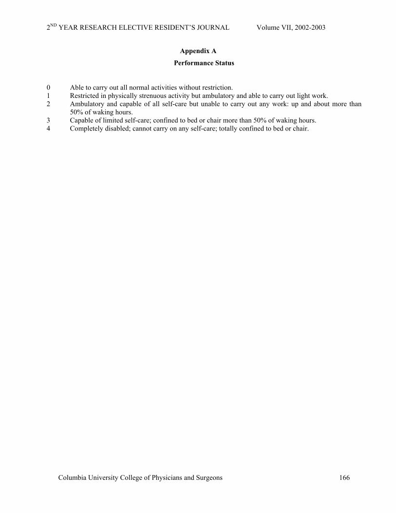
Appendix A
Performance Status
0 Able to carry out all normal activities without restriction. 1 Restricted in physically strenuous activity but ambulatory and able to carry out light work. 2 Ambulatory and capable of all self-care but unable to carry out any work: up and about more than
50% of waking hours. 3 Capable of limited self-care; confined to bed or chair more than 50% of waking hours. 4 Completely disabled; cannot carry on any self-care; totally confined to bed or chair.
Columbia University College of Physicians and Surgeons
166
2ND YEAR RESEARCH ELECTIVE RESIDENT’S JOURNAL Volume VII, 2002-2003
Columbia University College of Physicians and Surgeons
167
Appendix B
Staging
Definitions
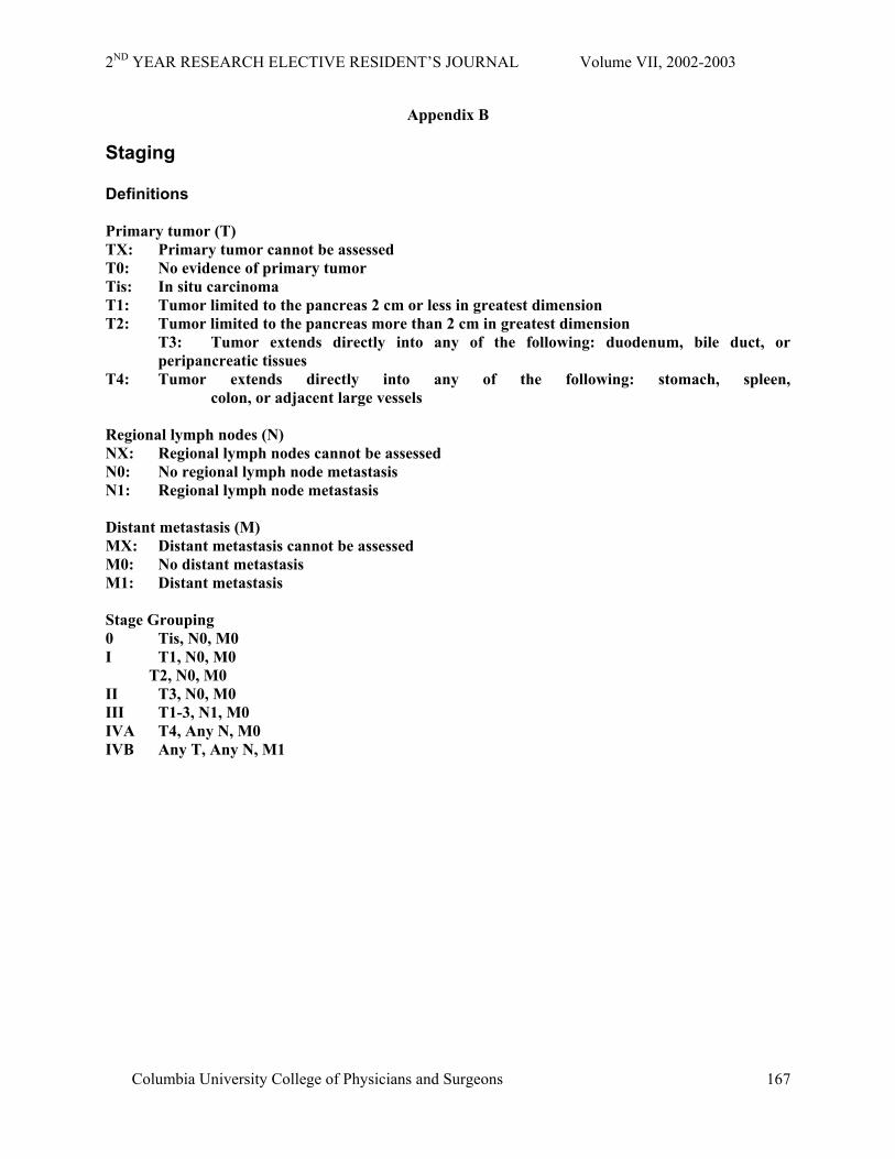
Primary tumor (T) TX: Primary tumor cannot be assessed T0: No evidence of primary tumor Tis: In situ carcinoma T1: Tumor limited to the pancreas 2 cm or less in greatest dimension T2: Tumor limited to the pancreas more than 2 cm in greatest dimension T3: Tumor extends directly into any of the following: duodenum, bile duct, or
peripancreatic tissues T4: Tumor extends directly into any of the following: stomach, spleen,
colon, or adjacent large vessels Regional lymph nodes (N) NX: Regional lymph nodes cannot be assessed N0: No regional lymph node metastasis N1: Regional lymph node metastasis Distant metastasis (M) MX: Distant metastasis cannot be assessed M0: No distant metastasis M1: Distant metastasis Stage Grouping 0 Tis, N0, M0 I T1, N0, M0 T2, N0, M0 II T3, N0, M0 III T1-3, N1, M0 IVA T4, Any N, M0 IVB Any T, Any N, M1