Embed Size (px)
Citation preview
Pertanika J. Sci. & Technol. 25 (S): 29 - 40 (2017)
SCIENCE & TECHNOLOGYJournal homepage: http://www.pertanika.upm.edu.my/
ISSN: 0128-7680 © 2017 Universiti Putra Malaysia Press.
ARTICLE INFO
Article history:Received: 25 October 2016Accepted: 17 March 2017
E-mail addresses: [email protected] (Nik Norziehana Che Isa),[email protected] (Yusairie Mohd),[email protected] (Mohammad Hafizudden Mohd Zaki),[email protected] (Sharifah Aminah Syed Mohamad) *Corresponding Author
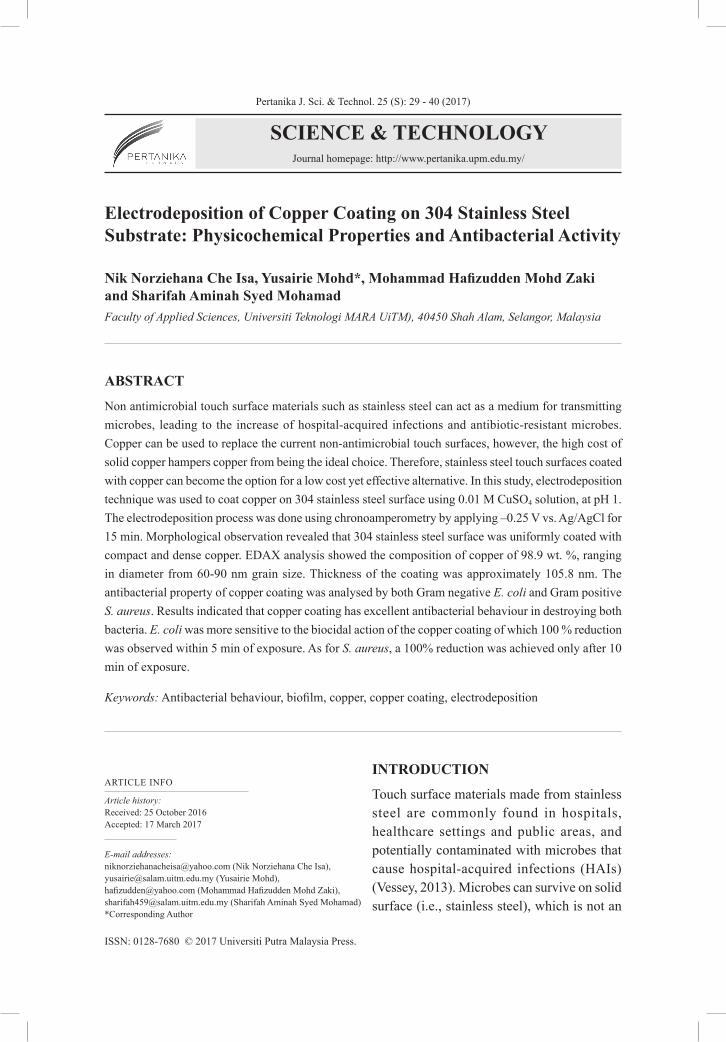
Electrodeposition of Copper Coating on 304 Stainless Steel Substrate: Physicochemical Properties and Antibacterial Activity
Nik Norziehana Che Isa, Yusairie Mohd*, Mohammad Hafizudden Mohd Zaki and Sharifah Aminah Syed Mohamad Faculty of Applied Sciences, Universiti Teknologi MARA UiTM), 40450 Shah Alam, Selangor, Malaysia
ABSTRACT
Non antimicrobial touch surface materials such as stainless steel can act as a medium for transmitting microbes, leading to the increase of hospital-acquired infections and antibiotic-resistant microbes. Copper can be used to replace the current non-antimicrobial touch surfaces, however, the high cost of solid copper hampers copper from being the ideal choice. Therefore, stainless steel touch surfaces coated with copper can become the option for a low cost yet effective alternative. In this study, electrodeposition technique was used to coat copper on 304 stainless steel surface using 0.01 M CuSO4 solution, at pH 1. The electrodeposition process was done using chronoamperometry by applying –0.25 V vs. Ag/AgCl for 15 min. Morphological observation revealed that 304 stainless steel surface was uniformly coated with compact and dense copper. EDAX analysis showed the composition of copper of 98.9 wt. %, ranging in diameter from 60-90 nm grain size. Thickness of the coating was approximately 105.8 nm. The antibacterial property of copper coating was analysed by both Gram negative E. coli and Gram positive S. aureus. Results indicated that copper coating has excellent antibacterial behaviour in destroying both bacteria. E. coli was more sensitive to the biocidal action of the copper coating of which 100 % reduction was observed within 5 min of exposure. As for S. aureus, a 100% reduction was achieved only after 10 min of exposure.
Keywords: Antibacterial behaviour, biofilm, copper, copper coating, electrodeposition
INTRODUCTION
Touch surface materials made from stainless steel are commonly found in hospitals, healthcare settings and public areas, and potentially contaminated with microbes that cause hospital-acquired infections (HAIs) (Vessey, 2013). Microbes can survive on solid surface (i.e., stainless steel), which is not an
Nik Norziehana Che Isa, Yusairie Mohd, Mohammad Hafizudden Mohd Zaki and Sharifah Aminah Syed Mohamad
30 Pertanika J. Sci. & Technol. 25 (S): 29 - 40 (2017)
antimicrobial active material and formed biofilm. The microbes can persist for extended periods of time, acting as microbes’ reservoir, multiplying their pathways of transmission (Dancer, 2004; Page, Wilson, & Parkin, 2009). Bacteria in biofilms are drastically more resistance to antibiotics and external forces that withstand host immune response (Beech et al., 2002). One of the solutions to curb the problem is by preventing bacterial adherence onto the solid surface and inevitable stop the formation of biofilms. The use of copper in surface engineering represents an attractive solution due to the broad-spectrum biocide activity of copper towards microbes. Copper can inhibit growth of microbial biofilms by interacting with the thiol groups of bacteria proteins and enzymes (Gant et al., 2007; Sierra et al., 2013; Warnes et al., 2010).
From the literature, copper and its alloy surfaces have been thoroughly investigated for their antimicrobial activity both in laboratory and in clinical environment (Airey & Verran, 2007; Casey et al., 2010; Cassandra et al., 2013; Champagne & Helfritch, 2013; Michels, Noyce, & Keevil, 2009; Michels et al., 2005; Noyce, Michels, & Keevil, 2006; Ojeil et al., 2013; Schmidt, Attaway, & Fairey, 2013). Research shows that microbes’ survival on many copper surfaces is limited to just a few hours or even minutes compared to stainless steel (Airey & Verran, 2007; Ojeil et al., 2013). The findings indicated that copper surface is a promising material to replace the contemporary use of stainless steel surfaces in the hospital and healthcare environment, since bacterial placed in contact with the dry copper surface suffered the great impact (Espirito et al., 2011). However, the use of pure solid copper is expensive (Elguindi et al., 2011; Nejad et al., 2013), and to replace the currently stainless steel touch surfaces with solid copper is very costly. Hence, one way to address the issue of material cost is by modifying the stainless steel surface with copper via coating process.
There are various techniques available for the copper-based coating process on the substrates such as vapour deposition, ion implantation, sputtering, sol-gel and electrodeposition (Alam et al., 2015; Daniel et al., 2009; Dorogov et al., 2015; Jakupi et al., 2015; Triantou et al., 2015; Zhang, An, & Chang, 2009). Recently, numerous research was found focusing on the development of processing methods for the fabrication of copper-based coating in achieving the desired structural and functional coating properties for limiting bacterial adhesion and biofilm formation (Cloutier, Mantovani, & Rosei, 2015; Palza, Delgado, & Curotto, 2015; Sharifahmadian et al., 2013; Wan et al., 2007). Despite a lot of studies revealed the available techniques to prepare good copper-based coating, only a few have discussed about the copper-based coating prepared by the electrodeposition technique for antimicrobial purpose.
Electrodeposition is a less expensive and straight-forward method with an ability to control the coating properties by effectively adjusting the experiment parameters such as applied potential or current, deposition time, source of metal ions and concentration, pH of the electrolyte solution, temperature, as well as nature of the substrate. This present work is aimed at evaluating and comparing the antimicrobial efficacy of the copper coating prepared by electrodeposition technique with stainless steel (SS304) towards E. coli (gram negative) and S. aureus (gram positive) bacteria.
Electrodeposition of Copper Coating on 304 Stainless Steel Substrate
31Pertanika J. Sci. & Technol. 25 (S): 29 - 40 (2017)
METHOD
Substrate Preparation for Coating
The substrate used in this study is 304 stainless steel coupons (20 mm x 20 mm x 1 mm). Prior to electrodeposition process, the substrate was polished with SiC paper from P800 to P4000 grit, followed by ultrasonically cleaned in acetone, subsequently rinsed with ultrapure water and dried at room temperature. An adhesive tape was used to mask off of the substrate except for 20 mm2 area on which deposition was desired.
Electrodeposition of Copper on Stainless Steel Substrate
The electrodeposition process was performed via chronoamperometry method on the polished 304 stainless steel as a working electrode, platinum rod as a counter electrode and Ag/AgCl as a reference electrode. All the three electrodes were immersed into 0.01 M Cu2+ ions (pH 1) solution. The electrodeposition process was controlled using an Autolab Potentiostat (Aut302 FRA2), and interfaced with a PC running NOVA software. The copper coating on the 304 stainless steel substrate was deposited by applying a constant potential at –0.25 V vs Ag/AgCl for 15 min.
Characterisation of Copper Coating. The surface morphology images of the uncoated and coated 304 stainless steel were examined using a Field Emission Scanning Electron Microscope (FESEM, Carl Zeiss SMT Supra 40VP) at various magnifications. An electron accelerating voltage of 5 kV was used to observe the morphology of the prepared samples. The elemental composition and mapping of the coated 304 stainless steel were determined by Energy Dispersive X-ray (EDAX). The control software (the Oxford INCA X-max 51-XMX 0021), equipped with FESEM, was used for EDAX analysis. Si(Li), cooled to cryogenic temperature with liquid nitrogen, was used as detector to convert X-ray energy into voltage signals. Thickness of the prepared coating was measured on at least three random areas by Surface profiler (P-6).
Antibacterial Test. The antibacterial activity of the prepared samples was tested using intimate contact cell suspension test but modified according to the standard method, JIZ S 2801/ ISO 22196. Two bacterial species, Gram negative (E. coli) and Gram positive (S. aureus) were selected as the test bacteria. The tested bacteria were cultured in nutrient agar overnight at 37°C, and then diluted in 10 mL of saline (0.9 % of NaCl) to become an optical density OD600 nm of 0.05, which is equivalent to 107 cells/mL. Subsequently, bacterial suspension of approximately 105 cells/mL was prepared.
Copper coated stainless steel was sterilised by immersing in 95% ethanol for a few seconds, and dried at room temperature before placing it on sterile 90 mm diameter Petri dishes. The uncoated stainless steel substrate was also tested and used as a negative control. An amount of 20 μL of the diluted bacterial suspension was added onto each substrate and covered with a sterile glass cover slip in order to maintain the same contact area of suspension on each tested substrate surface during the designated contact time, to monitor the reduction rate. All
Nik Norziehana Che Isa, Yusairie Mohd, Mohammad Hafizudden Mohd Zaki and Sharifah Aminah Syed Mohamad
32 Pertanika J. Sci. & Technol. 25 (S): 29 - 40 (2017)
the plates were incubated at room temperature at the designated contact time (i.e., 0, 5, 10, 15, 20, 25, 30 min).
After incubation, the substrate was transferred to 10 mL of sterile phosphate-buffered saline (PBS) for 10 s to dislodge the cover slip and suspend the surviving bacteria in the PBS solution. 100 μL aliquots of the bacterial suspension was evenly spread onto nutrient agar using a sterile glass spreader and incubated overnight at 37°C. The number of colony forming units (CFUs), resulting from the growth of the viable bacteria at 37°C kept overnight, was calculated using colony counter, while reduction of exposure time of bacteria was measured. The percentage of reduction was calculated according to the following formula [1]:
[1]
Where, N0 is the mean CFU/mL for the same substrate at 0 hour, and Nt is the mean CFU/mL from a test substrate after a designated contact time. Metal samples were removed immediately after inoculation at zero time to determine the initial number of viable bacteria.
RESULTS AND DISCUSSION
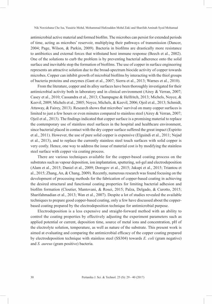
The authors have successfully deposit the copper element onto the 304 stainless steel surface area under the laboratory set condition of, V = –0.25 V vs Ag/AgCl and t = 15 min, in 0.01 M Cu2+ ions (pH 1) solution. Figure 1 shows chronoamperometric curve of the deposition of copper onto the 304 stainless steel surface, coating the 304 stainless steel surface (inset image). From the curve, the current density shows an abrupt decrease for a short time (i.e., 5 s) at the beginning of the process. This behaviour indicates that the double layer charging of non-faradaic current has occurred. Right after, a plateau current density was observed until the end of the process indicates the nucleation growth of the copper, coating onto the stainless steel surface. The inset picture in Figure 1 exhibits the entire 304 stainless steel substrate surface that was soaked in the 20 mm2 solution during electrodeposition process, was coated with smooth and uniform red-brown colour copper.
Figure 1. Chronoamperometric curve of copper coating formed on 304 stainless steel substrate at –
0.25 V for 15 min. (Inset: visual observation of the coating produced on the exposed 304 stainless
steel surface)
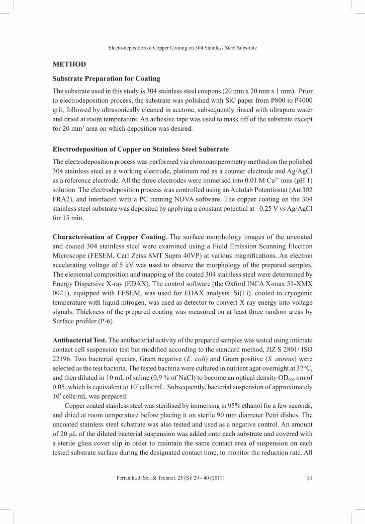
Figure 2 shows the surface morphologies of the polished 304 stainless steel (Figure
2(a)) and copper coating produced on the 304 stainless steel surface at different
magnifications (Figure 2(b) and Figure 2(c)). Polishing the stainless steel surface produced
a flat surface structure with relatively minor grooves in the polishing direction. After the
deposition process, the 304 stainless steel surface was coated with uniform, compact and
dense copper on the entire exposed surface. At a higher magnification (Figure 2(c)), the
coating with compact and homogeneous grain structure was obviously seen, ranging in 60-
90 nm diameter.
(a) (b) (c)
Figure 1. Chronoamperometric curve of copper coating formed on 304 stainless steel substrate at –0.25 V for 15 min. (Inset: visual observation of the coating produced on the exposed 304 stainless steel surface)
Electrodeposition of Copper Coating on 304 Stainless Steel Substrate
33Pertanika J. Sci. & Technol. 25 (S): 29 - 40 (2017)
Figure 2 shows the surface morphologies of the polished 304 stainless steel (Figure 2(a)) and copper coating produced on the 304 stainless steel surface at different magnifications (Figure 2(b) and Figure 2(c)). Polishing the stainless steel surface produced a flat surface structure with relatively minor grooves in the polishing direction. After the deposition process, the 304 stainless steel surface was coated with uniform, compact and dense copper on the entire exposed surface. At a higher magnification (Figure 2(c)), the coating with compact and homogeneous grain structure was obviously seen, ranging in 60-90 nm diameter.
Figure 1. Chronoamperometric curve of copper coating formed on 304 stainless steel substrate at –
0.25 V for 15 min. (Inset: visual observation of the coating produced on the exposed 304 stainless
steel surface)
Figure 2 shows the surface morphologies of the polished 304 stainless steel (Figure
2(a)) and copper coating produced on the 304 stainless steel surface at different
magnifications (Figure 2(b) and Figure 2(c)). Polishing the stainless steel surface produced
a flat surface structure with relatively minor grooves in the polishing direction. After the
deposition process, the 304 stainless steel surface was coated with uniform, compact and
dense copper on the entire exposed surface. At a higher magnification (Figure 2(c)), the
coating with compact and homogeneous grain structure was obviously seen, ranging in 60-
90 nm diameter.
(a) (b) (c) Figure 2. SEM images of: (a) 304 stainless steel; (b) copper coating prepared on 304 stainless steel substrate from 0.01 M CuSO4 solution (pH 1) at –0.25 V for 15 min at magnification: (b) 5000×; and (c) 50000×
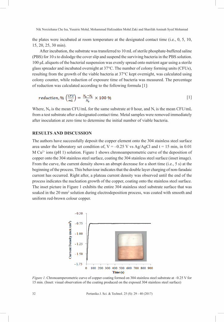
The EDAX analysis on the FESEM image of the copper coating (Figure 3) shows the composition of copper and oxygen is 98.9 wt.% and 1.10 wt.%, respectively. This indicates that the high percentage of copper deposited on the stainless steel surface comprises only of a minor distribution of oxide content. From the mapping images (Figure 4), it can be seen that a uniform distribution of the nano-grains copper (Figure 4(a)) on the entire exposed 304 stainless steel surface comprises only of a minor distribution of oxygen content (Figure 4(b)).
Figure 2. SEM images of: (a) 304 stainless steel; (b) copper coating prepared on 304
stainless steel substrate from 0.01 M CuSO4 solution (pH 1) at –0.25 V for 15 min at
magnification: (b) 5000×; and (c) 50000×
The EDAX analysis on the FESEM image of the copper coating (Figure 3) shows the
composition of copper and oxygen is 98.9 wt.% and 1.10 wt.%, respectively. This
indicates that the high percentage of copper deposited on the stainless steel surface
comprises only of a minor distribution of oxide content. From the mapping images (Figure
4), it can be seen that a uniform distribution of the nano-grains copper (Figure 4(a)) on the
entire exposed 304 stainless steel surface comprises only of a minor distribution of oxygen
content (Figure 4(b)).
Figure 3. The elemental composition of copper coating prepared on 304 stainless steel substrate
analysed by EDAX
Figure 3. The elemental composition of copper coating prepared on 304 stainless steel substrate analysed by EDAX

Nik Norziehana Che Isa, Yusairie Mohd, Mohammad Hafizudden Mohd Zaki and Sharifah Aminah Syed Mohamad
34 Pertanika J. Sci. & Technol. 25 (S): 29 - 40 (2017)
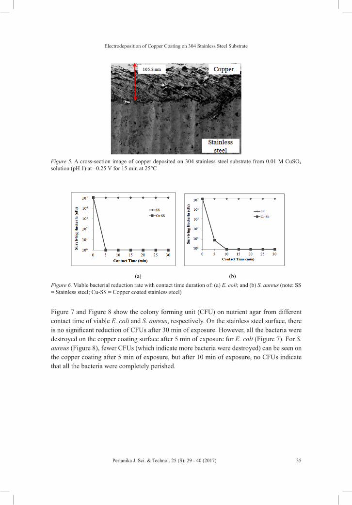
Proper adhesion of coating on a substrate is one of the very important factor for determining the mechanical behaviour and performance of the coated components (Okamoto, Wang, & Watanabe, 2004). Metallic films electrodeposited on the metal substrates are commonly thought to have a favourable adhesive strength since the electrodeposited films are usually bonded with substrates metallically, without any interruption from hydrogen evolution reaction during the electrodeposition process. The adhesion quality of the copper coating on the stainless steel was observed at the cross section image by FESEM. The cross section image (Figure 5) exhibits well intact between coating and substrate. There are no voids or gaps in between the coating and substrate. In addition, nano-sized grain structures have filled-up the vacancies, of which the same condition is not possible via conventional micro-structure coating. The thickness of the coating was about 105.8 nm.
Figure 6 indicates the reduction rate of viable bacteria within the designated contact time under ambient room temperature. E. coli was more sensitive to the inhibitory action of the copper coating (100% reduction within 5 min of exposure), whereas 100% reduction of S. aureus was achieved only after 10 min of exposure. On the other hand, no obvious reduction of viable bacteria on the 304 stainless steel surface was observed even after 30 min of exposure. These findings strongly showed that copper coating has an excellent antimicrobial property than stainless steel in which there is no sign of antibacterial activity against both the tested bacteria. It is suggested that copper accumulation within the cell, cell death and DNA damage assays that copper has lethal effects towards bacteria, as stated by Ibrahim et al. (2011). Thus, stainless steel surface exerted no lethal effect.
(a) (b)
Figure 4. The EDAX elemental mapping of: (a) copper; and (b) oxygen present on the coating
prepared on 304 stainless steel substrate from 0.01 M CuSO4 solution (pH 1) at –0.25 V for 15 min
Proper adhesion of coating on a substrate is one of the very important factor for
determining the mechanical behaviour and performance of the coated components
(Okamoto, Wang, & Watanabe, 2004). Metallic films electrodeposited on the metal
substrates are commonly thought to have a favourable adhesive strength since the
electrodeposited films are usually bonded with substrates metallically, without any
interruption from hydrogen evolution reaction during the electrodeposition process. The
adhesion quality of the copper coating on the stainless steel was observed at the cross
section image by FESEM. The cross section image (Figure 5) exhibits well intact between
coating and substrate. There are no voids or gaps in between the coating and substrate. In
addition, nano-sized grain structures have filled-up the vacancies, of which the same
condition is not possible via conventional micro-structure coating. The thickness of the
coating was about 105.8 nm.
Figure 6 indicates the reduction rate of viable bacteria within the designated contact
time under ambient room temperature. E. coli was more sensitive to the inhibitory action
of the copper coating (100% reduction within 5 min of exposure), whereas 100%
Figure 4. The EDAX elemental mapping of: (a) copper; and (b) oxygen present on the coating prepared on 304 stainless steel substrate from 0.01 M CuSO4 solution (pH 1) at –0.25 V for 15 min
Electrodeposition of Copper Coating on 304 Stainless Steel Substrate
35Pertanika J. Sci. & Technol. 25 (S): 29 - 40 (2017)
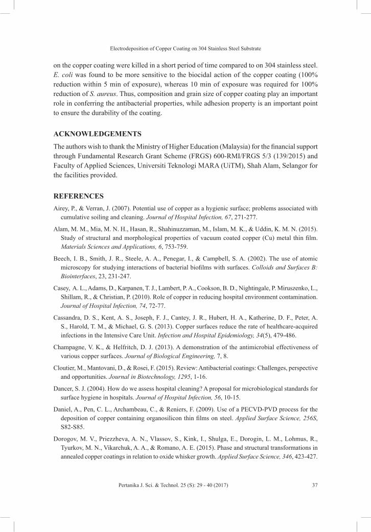
Figure 7 and Figure 8 show the colony forming unit (CFU) on nutrient agar from different contact time of viable E. coli and S. aureus, respectively. On the stainless steel surface, there is no significant reduction of CFUs after 30 min of exposure. However, all the bacteria were destroyed on the copper coating surface after 5 min of exposure for E. coli (Figure 7). For S. aureus (Figure 8), fewer CFUs (which indicate more bacteria were destroyed) can be seen on the copper coating after 5 min of exposure, but after 10 min of exposure, no CFUs indicate that all the bacteria were completely perished.
reduction of S. aureus was achieved only after 10 min of exposure. On the other hand, no
obvious reduction of viable bacteria on the 304 stainless steel surface was observed even
after 30 min of exposure. These findings strongly showed that copper coating has an
excellent antimicrobial property than stainless steel in which there is no sign of
antibacterial activity against both the tested bacteria. It is suggested that copper
accumulation within the cell, cell death and DNA damage assays that copper has lethal
effects towards bacteria, as stated by Ibrahim et al. (2011). Thus, stainless steel surface
exerted no lethal effect.
Figure 5. A cross-section image of copper deposited on 304 stainless steel substrate from 0.01 M
CuSO4 solution (pH 1) at –0.25 V for 15 min at 25oC
(a) (b)
Figure 5. A cross-section image of copper deposited on 304 stainless steel substrate from 0.01 M CuSO4
solution (pH 1) at –0.25 V for 15 min at 25°C
reduction of S. aureus was achieved only after 10 min of exposure. On the other hand, no
obvious reduction of viable bacteria on the 304 stainless steel surface was observed even
after 30 min of exposure. These findings strongly showed that copper coating has an
excellent antimicrobial property than stainless steel in which there is no sign of
antibacterial activity against both the tested bacteria. It is suggested that copper
accumulation within the cell, cell death and DNA damage assays that copper has lethal
effects towards bacteria, as stated by Ibrahim et al. (2011). Thus, stainless steel surface
exerted no lethal effect.
Figure 5. A cross-section image of copper deposited on 304 stainless steel substrate from 0.01 M
CuSO4 solution (pH 1) at –0.25 V for 15 min at 25oC
(a) (b) Figure 6. Viable bacterial reduction rate with contact time duration of: (a) E. coli; and (b) S. aureus (note: SS = Stainless steel; Cu-SS = Copper coated stainless steel)
Nik Norziehana Che Isa, Yusairie Mohd, Mohammad Hafizudden Mohd Zaki and Sharifah Aminah Syed Mohamad
36 Pertanika J. Sci. & Technol. 25 (S): 29 - 40 (2017)
Figure 6. Viable bacterial reduction rate with contact time duration of: (a) E. coli; and (b) S.
aureus (note: SS = Stainless steel; Cu-SS = Copper coated stainless steel)
Figure 7 and Figure 8 show the colony forming unit (CFU) on nutrient agar from
different contact time of viable E. coli and S. aureus, respectively. On the stainless steel
surface, there is no significant reduction of CFUs after 30 min of exposure. However, all
the bacteria were destroyed on the copper coating surface after 5 min of exposure for E.
coli (Figure 7). For S. aureus (Figure 8), fewer CFUs (which indicate more bacteria were
destroyed) can be seen on the copper coating after 5 min of exposure, but after 10 min of
exposure, no CFUs indicate that all the bacteria were completely perished.
(a) (b) (c)
Figure 7. Colony forming unit of viable E. coli after being in contact with: (a) stainless steel for 0
min; (b) stainless steel for 30 min; and (c) copper coating for 5 min
(a) (b)
Figure 7. Colony forming unit of viable E. coli after being in contact with: (a) stainless steel for 0 min; (b) stainless steel for 30 min; and (c) copper coating for 5 min
Figure 6. Viable bacterial reduction rate with contact time duration of: (a) E. coli; and (b) S.
aureus (note: SS = Stainless steel; Cu-SS = Copper coated stainless steel)
Figure 7 and Figure 8 show the colony forming unit (CFU) on nutrient agar from
different contact time of viable E. coli and S. aureus, respectively. On the stainless steel
surface, there is no significant reduction of CFUs after 30 min of exposure. However, all
the bacteria were destroyed on the copper coating surface after 5 min of exposure for E.
coli (Figure 7). For S. aureus (Figure 8), fewer CFUs (which indicate more bacteria were
destroyed) can be seen on the copper coating after 5 min of exposure, but after 10 min of
exposure, no CFUs indicate that all the bacteria were completely perished.
(a) (b) (c)
Figure 7. Colony forming unit of viable E. coli after being in contact with: (a) stainless steel for 0
min; (b) stainless steel for 30 min; and (c) copper coating for 5 min
(a) (b)
Figure 8. Colony forming unit of viable S. aureus after being in contact with: (a) stainless steel for 0 min; (b) stainless steel for 30 min; (c) copper coating for 5 min; and (d) copper coating for 10 min
(c) (d)
Figure 8. Colony forming unit of viable S. aureus after being in contact with: (a) stainless steel for
0 min; (b) stainless steel for 30 min; (c) copper coating for 5 min; and (d) copper coating for 10
min
CONCLUSION
Copper coating was successfully deposited on the 304 stainless steel substrate through the
electrodeposition technique for antibacterial applications. The copper coating showed
good surface coverage with uniform distribution of copper nano-grains on the 304
stainless steel surface. In addition, the copper coating showed a very good and intact
adhesion property on the 304 stainless steel based on the observation of cross section
image by FESEM. Viable bacteria on the copper coating were killed in a short period of
time compared to on 304 stainless steel. E. coli was found to be more sensitive to the
biocidal action of the copper coating (100% reduction within 5 min of exposure), whereas
10 min of exposure was required for 100% reduction of S. aureus. Thus, composition and
grain size of copper coating play an important role in conferring the antibacterial
properties, while adhesion property is an important point to ensure the durability of the
coating.
CONCLUSION
Copper coating was successfully deposited on the 304 stainless steel substrate through the electrodeposition technique for antibacterial applications. The copper coating showed good surface coverage with uniform distribution of copper nano-grains on the 304 stainless steel surface. In addition, the copper coating showed a very good and intact adhesion property on the 304 stainless steel based on the observation of cross section image by FESEM. Viable bacteria
Electrodeposition of Copper Coating on 304 Stainless Steel Substrate
37Pertanika J. Sci. & Technol. 25 (S): 29 - 40 (2017)
on the copper coating were killed in a short period of time compared to on 304 stainless steel. E. coli was found to be more sensitive to the biocidal action of the copper coating (100% reduction within 5 min of exposure), whereas 10 min of exposure was required for 100% reduction of S. aureus. Thus, composition and grain size of copper coating play an important role in conferring the antibacterial properties, while adhesion property is an important point to ensure the durability of the coating.
ACKNOWLEDGEMENTS
The authors wish to thank the Ministry of Higher Education (Malaysia) for the financial support through Fundamental Research Grant Scheme (FRGS) 600-RMI/FRGS 5/3 (139/2015) and Faculty of Applied Sciences, Universiti Teknologi MARA (UiTM), Shah Alam, Selangor for the facilities provided.
REFERENCESAirey, P., & Verran, J. (2007). Potential use of copper as a hygienic surface; problems associated with
cumulative soiling and cleaning. Journal of Hospital Infection, 67, 271-277.
Alam, M. M., Mia, M. N. H., Hasan, R., Shahinuzzaman, M., Islam, M. K., & Uddin, K. M. N. (2015). Study of structural and morphological properties of vacuum coated copper (Cu) metal thin film. Materials Sciences and Applications, 6, 753-759.
Beech, I. B., Smith, J. R., Steele, A. A., Penegar, I., & Campbell, S. A. (2002). The use of atomic microscopy for studying interactions of bacterial biofilms with surfaces. Colloids and Surfaces B: Biointerfaces, 23, 231-247.
Casey, A. L., Adams, D., Karpanen, T. J., Lambert, P. A., Cookson, B. D., Nightingale, P. Miruszenko, L., Shillam, R., & Christian, P. (2010). Role of copper in reducing hospital environment contamination. Journal of Hospital Infection, 74, 72-77.
Cassandra, D. S., Kent, A. S., Joseph, F. J., Cantey, J. R., Hubert, H. A., Katherine, D. F., Peter, A. S., Harold, T. M., & Michael, G. S. (2013). Copper surfaces reduce the rate of healthcare-acquired infections in the Intensive Care Unit. Infection and Hospital Epidemiology, 34(5), 479-486.
Champagne, V. K., & Helfritch, D. J. (2013). A demonstration of the antimicrobial effectiveness of various copper surfaces. Journal of Biological Engineering, 7, 8.
Cloutier, M., Mantovani, D., & Rosei, F. (2015). Review: Antibacterial coatings: Challenges, perspective and opportunities. Journal in Biotechnology, 1295, 1-16.
Dancer, S. J. (2004). How do we assess hospital cleaning? A proposal for microbiological standards for surface hygiene in hospitals. Journal of Hospital Infection, 56, 10-15.
Daniel, A., Pen, C. L., Archambeau, C., & Reniers, F. (2009). Use of a PECVD-PVD process for the deposition of copper containing organosilicon thin films on steel. Applied Surface Science, 256S, S82-S85.
Dorogov, M. V., Priezzheva, A. N., Vlassov, S., Kink, I., Shulga, E., Dorogin, L. M., Lohmus, R., Tyurkov, M. N., Vikarchuk, A. A., & Romano, A. E. (2015). Phase and structural transformations in annealed copper coatings in relation to oxide whisker growth. Applied Surface Science, 346, 423-427.

Nik Norziehana Che Isa, Yusairie Mohd, Mohammad Hafizudden Mohd Zaki and Sharifah Aminah Syed Mohamad
38 Pertanika J. Sci. & Technol. 25 (S): 29 - 40 (2017)
Elguindi, J., Hao, X., Lin, Y., Alwathnani, H. A., Wei, G., & Rensing, C. (2011). Advantages and challenges of increased antimicrobial copper use and copper mining. Applied Microbiology and Biotechnology, 91, 237-249.
Espirito, S. C., Lam, E. W., Elowsky, C. G., Quaranta, D., Domaille, D. W., Chang, C. J., & Grass, G. (2011). Bacteria killing by dry metallic copper surfaces. Applied Environmental Microbiology, 77, 794-800.
Gant, V. A., Wren, M. W. D., Rollins, M. S. M., Jeanes, A., Hickok, S. S., & Hall, T. J. (2007). Three novel highly charged copper-based biocides: safety and efficacy against healthcare-associated organisms. Journal of Antimicrobial Chemotherapy, 60, 294-299.
Ibrahim, M., Wang, F., Lou, M., Xie, G., Li, B., Bo, Z., Zhang, G., Liu, H., & Wareth, A. (2011). Copper as an antibacterial agent for human pathogenic multidrug resistant Burkholderia cepacia complex bacteria. Journal of Bioscience and Bioengineering, 112(6), 570-576.
Jakupi, P., Keech, P. G., Barker, I., Ramamurthy, S., Jacklin, R. L., Shoesmith, D. W., & Moser, D. E. (2015). Characterization of commercially cold sprayed copper coatings and determination of the effects of impacting copper powder velocities. Journal of Nuclear Materials, 466, 1-11.
Michels, H. T., Wilks, S. A., Noyce, J. O., & Keevil, C. W. (2005). Copper alloys for human infectious disease control. Materials Science and Technology Conference, 25-28 September 2005, Pittsburgh, PA Copper for the 21st Century Symposium.
Michels, H. T., Noyce, J. O., & Keevil, C. W. (2009). Effects of temperature and humidity on the efficacy of methicillin-resistant Staphylococcus aureus challenged antimicrobial materials containing silver and copper. Letters in Applied Microbiology, 49, 191 – 195.
Nejad, M., Pershin, L., Mostaghimi, J., & Ringuette, M. (2013). Evaluation of bioactivity of copper alloy coatings. 21st International Symposium on Plasma Chemistry (ISPC 21) 4-9 August 2013, Cairns Convention Centre, Queensland, Australia.
Noyce, J. O., Michels, H., & Keevil, C. W. (2006). Use of copper cast alloys to control Escherichia coli 0157 cross contamination during food processing. Applied Environmental Microbiology, 72(6), 4239-4244.
Ojeil, M., Jermann, C., Holah, J., Denyer, S. P., & Maillard, J. Y. (2013). Evaluation of new in vitro efficacy test for antimicrobial surface activity reflecting UK hospital conditions. Journal of Hospital Infection, 85, 274-281.
Okamoto, N., Wang, F., & Watanabe, T. (2004). Adhesion of electrodeposited copper, nickel, and silver films on copper, nickel and silver subsrates. Materials Transactions, 45(12), 3330-3333.
Page, K., Wilson, M., & Parkin, P. (2009). Antimicrobial surfaces and their potential in reducing the role of the inanimate environment in the incidence of hospital-acquired infections. Journal of Materials Chemistry, 19, 3819-3819.
Palza, H., Delgado, K., & Curotto, N. (2015). Synthesis of copper nanostructures on silica-based particles for antimicrobial organic coatings. Applied Surface Science, 357, 86-90.
Schmidt, M. G., Attaway, H. H., & Fairey, S. E. (2013). Copper continuously limits the concentration of bacteria resident on bed rails within the intensive care unit. Infection Control Hospital Epidemiology, 34(5), 530-533.
Electrodeposition of Copper Coating on 304 Stainless Steel Substrate
39Pertanika J. Sci. & Technol. 25 (S): 29 - 40 (2017)
Sharifahmadian, O., Salimijazi, H. R., Fathi, M. H., Mostaghimi, J., & Pershin, L. (2013). Study of the antibacterial behavior of wire arc sprayed copper coatings. Journal of Thermal Spray Technology, 22, 371-379.
Sierra, M., Sanhueza, A., Alcantara, R., & Sanchez, G. (2013). Antimicrobial evaluation of copper sulphate (II) on strains of Enterococcus faecalis. In vitro study. Journal of Oral Research, 2(3), 114-118.
Triantou, K. I., Pantelis, D. I., Guipont, V., & Jeandin, M. (2015). Microstructure and tribological behaviour of copper and composite copper + alumina cold sprayed coatings for various alumina contents. Wear, 336 – 337, 96-107.
Vessey, A. (2013). Copper – a weapon in the war on pathogens. Health Estate Journal, 65-69.
Wan, Y. Z., Raman, S., He, F., & Huang, Y. (2007). Surface modification of medical metals by ion implantation of silver and copper. Vacuum, 81, 1114-1118.
Warnes, S. L., Green, S. M., Michels, H. T., & Keevil, C. W. (2010). Biocidal efficacy of copper alloys against pathogenic enterococci involves degradation of genomic and plasmid DNAs. Applied Environmental Microbiology, 76, 5390-5401.
Zhang, J., An, M., & Chang, L. (2009). Study of the electrochemical deposition of Sn-Ag-Cu alloy by cyclic voltammetry and chronoamperometry. Electrochimica Acta, 54, 2883-2889.