Embed Size (px)
Citation preview
Research Article
Persistence of Bronchial Dysplasia Is Associatedwith Development of Invasive Squamous CellCarcinomaDaniel T. Merrick1, Dexiang Gao2, York E. Miller3,4, Robert L. Keith3,4, Anna E. Baron5,William Feser5, Timothy C. Kennedy4, Patrick J. Blatchford2, Sarah Braudrick5,Fred R. Hirsch1, Lynn Heasley6, Paul A. Bunn, Jr.7, and Wilbur A. Franklin1
Abstract
Bronchial dysplasia (BD), a presumed precursor of pulmo-nary squamous cell carcinoma (SCC), rarely progresses toinvasive cancer. A high-risk cohort at the University of Color-ado provided an opportunity to directly sample airway epithe-lium at mapped sites on successive bronchoscopies. We havehypothesized that persistent dysplastic lesions showing a sim-ilar or higher level of dysplasia on follow-up biopsy, areassociated with increased risk for the development of SCC.Endoscopic biopsies from 188 high-risk subjects were histo-logically classified according to the current WHO classificationfor BD using a numeric histology score ranging from 1 to 8representing normal bronchial mucosa through invasive lungcancer. Differences in follow-up histology scores were com-pared between sites classified by clinical, histologic, and immu-nohistochemical variables. Subjects with a higher frequency of
sites that persist or progress to high-grade dysplasia (�37.5%persist/progress, N ¼ 35 versus <37.5% persist/progress, N ¼114) show a significant association with development of inci-dent invasive SCC (adjusted HR, 7.84; 95% confidence interval,1.56–39.39), and those with incident lung SCC have adjustedmean follow-up histology scores 1.55 U higher than in subjectswithout lung cancer. Current smoking, elevated Ki67 growthfraction, histologic features of angiogenic squamous dysplasia(ASD) and higher histology score in baseline biopsies aresignificantly associated with increased follow-up histologyscores. These results show that persistent BD is associated withthe development of invasive SCC. Furthermore, increasedexpression of Ki67, the presence of angiogenic change anddegree of baseline atypia are associated with persistence of BD.Cancer Prev Res; 9(1); 96–104. �2015 AACR.
IntroductionBronchial dysplasia (BD; ref. 1) is a presumed precursor lesion
of squamous cell carcinoma (SCC) of the lung, and elimination ofthese lesions has been proposed as a way to prevent invasive SCCof the lung (2). A scoring system for BD that stratifies these lesionsby severity of squamous atypia has provided a framework forusing histology scores to assess the effect of chemopreventionagents on bronchial mucosa (3–6). Understanding the behavior
of BD could provide means to assess risk for the development oflung cancer and elucidate mechanisms underlying progression.
Molecular alterations that parallel those seen in invasive cancerfor site-specific loss of heterozygosity (LOH), andmRNA,miRNA,and immunohistochemical marker expression levels that becomemore prominent in higher grades of BD have provided data tosupport a relationship between BD and invasive SCC of the lung(7–10). In addition, chromosomal aneusomy, gene copy-numbergains, increased PI3K pathway activation, and alterations intelomere length have been shown to be more common in BDfrom patients with known lung cancer as compared with thosewithout (11–14). A relationship between increased expression ofmultiple tumor-related markers or specific LOH in BD and sub-sequent development of carcinoma-in-situ (CIS) or SCC hasalso been shown (15, 16). In a few selected cases, an increase inSOX2 amplification has been described as sites of BD progressedin atypia and ultimately developed invasive SCC (17). Progres-sion of atypia or development of cancer has also been relatedto baseline histologic atypia and other clinical features, buthas frequently produced contradictory findings. Although somestudies have correlated baseline histology with smokingstatus, more frequent progression of atypia or development ofcancer (18–23), others have failed to detect significant relation-ships with these parameters (15, 18, 24). Important issuescompromising interpretation of these data include use of CISas a malignant outcome, small numbers of cases per individualreport, frequent pre-selection of cases with some cohorts beingcomposed entirely of patients with previous lung or head and
1Department of Pathology, University of Colorado Anschutz MedicalCampus, Aurora, Colorado. 2Department of Pediatrics, University ofColorado Anschutz Medical Campus, Aurora, Colorado. 3Division ofPulmonary Medicine, Department of Medicine, Denver VeteransAffairs Medical Center, Denver, Colorado. 4Division of PulmonaryMedicine, Department of Medicine, University of Colorado AnschutzMedical Campus, Aurora, Colorado. 5Department of Biostatistics andInformatics, Colorado School of Public Health, Aurora, Colorado.6Department ofCraniofacial Biology,UniversityofColoradoAnschutzMedical Campus, Aurora, Colorado. 7Division of Medical Oncology,Department of Medicine, University of Colorado Anschutz MedicalCampus, Aurora, Colorado.
Note: Supplementary data for this article are available at Cancer PreventionResearch Online (http://cancerprevres.aacrjournals.org/).
Corresponding Author: Daniel T. Merrick, University of Colorado AnschutzMedical Campus, Mail Stop 8104, 12801 East 17th Avenue, Room 5114, Aurora,CO 80045. Phone: 303-724-4321; Fax: 303-724-3096; E-mail:[email protected]
doi: 10.1158/1940-6207.CAPR-15-0305
�2015 American Association for Cancer Research.
CancerPreventionResearch
Cancer Prev Res; 9(1) January 201696
Research. on June 15, 2018. © 2016 American Association for Cancercancerpreventionresearch.aacrjournals.org Downloaded from
Published OnlineFirst November 5, 2015; DOI: 10.1158/1940-6207.CAPR-15-0305

neck cancer, confounding of outcome by use of therapeuticintervention, and variable, often short periods of time tofollow-up assessment of lesions. The limitations associatedwith these factors were also noted in a recent meta-analysis,in which findings suggested that higher degrees of atypia in BDare associated with more frequent progression or persistencethan are lesions of lower grade (25). These findings were notrelated to development of lung cancer.
Prediction of outcome in BD is of paramount importance to theestablishment of reliable, informative screening programs, andeffective prevention measures. Surveillance techniques such asautofluorescence bronchoscopy have improved sensitivity fordetection of BD (26–28), thus providing an opportunity toaccurately follow these lesions over time. We have examined therelationship between differences in follow-up histology scoresand a variety of parameters and found that more atypical out-comes are associatedwith subsequent incidence of SCC, increasedbaseline histologic atypia, smoking status, features of angiogenicsquamous dysplasia (ASD), and Ki67 expression levels. We haveidentified a pattern of persistent BD that defines a subset ofsubjects with aggressive airway disease and increased risk fordevelopment of invasive SCC that may benefit from close fol-low-up and potential preventive measures.
Materials and MethodsPatients at high risk for the development of lung cancer were
recruited to bronchoscopy protocols established through theColorado SPORE in Lung Cancer program. High-risk subjectsincluded those with tobacco smoking histories of greater than 20pack years and thosewith apersonal history of lung cancer or priorcancer of the upper aerodigestive tract. In subjects without ahistory of cancer, sputum screening was performed and bron-choscopy was offered to individuals with moderate or worsecytologic atypia. Informed consent for enrollment in the proto-cols and collection of associated clinical data were approved bythe Colorado Multiple Institutional Review Board (CoMIRB).Histologic, clinical, and other data from subjects for which therewas any bronchial site that was sampled on at least two occasions
were assembled for this study. Specimens that were collectedless than 3 months after the original baseline biopsy at aspecific site were excluded. During the timeframe of specimencollection (1993–2010), two Colorado Lung SPORE sponsoredchemoprevention trials were conducted in which all-trans reti-noic acid or the prostacyclin analog, Iloprost, were used aspotential chemopreventive agents (3, 4). Iloprost, but not all-trans retinoic acid, was shown to be associated with a signif-icant reduction in histology score on follow-up biopsy amongformer smokers as compared with former smokers on theplacebo arm of the study. Therefore, all biopsies from treatmentarm subjects collected after the Iloprost trial enrollment bron-choscopy were excluded.
Biopsy site classificationBiopsies were assigned a numeric histology score (see Fig. 1)
ranging from 1 to 8 for normal (score¼ 1), basal cell hyperplasia(score ¼ 2), squamous metaplasia without atypia (score ¼ 3),mild dysplasia (score¼ 4),moderate dysplasia (score¼ 5), severedysplasia (score ¼ 6), CIS (score ¼ 7), and invasive carcinoma(score ¼ 8) with each group being defined by the histologicfeatures described in the WHO classification (1). Baseline histo-logic score was defined as the diagnosis for the first biopsy at agiven site. Follow-up histologic scores were classified into threegroups: those with biopsies collected between 3 months to 2years, 2 to 4 years and>4years after baseline biopsies. Ifmore thanone biopsy had been collected during the timeframe of one ofthese groups, thebiopsywith thehighest diagnosiswasused.Mostanalyses used grouping of histology scores into nondysplastic(scores 1–2), low-grade dysplasia (LGD, scores 3–4), and high-grade dysplasia (HGD, scores 5–7) histologic groups. In analysescomparing groups defined by a pre-assigned persistent/progres-sive or regressive classification, persistent/progressive dysplasia(referred to as "persistent" BD throughout manuscript) wasdefined as any baseline LGD that showed LGD or higher histo-logic score on follow-up and anybaselineHGD that showedHGDor higher histologic score on follow-up unless otherwise noted.Biopsies of histology score 3 or greater were also characterized asASDs if they showed projections of vascular structures into
A
E F G H
B C DHS=1
HS=5 HS=6 HS=7 HS=8
HS=2 HS=3 HS=4
Figure 1.Photomicrographs demonstrating histologic features and histology scores (HS) of bronchial epithelium from sites of nondysplasia (A, normal; B, reserve cellhyperplasia), low-grade dysplasia (C, squamous metaplasia without atypia; D, mild dysplasia), high-grade dysplasia (E, moderate dysplasia; F, severedysplasia; G, CIS) and invasive SCC (H). Note, vascular papillary projections of ASD in E and F; Magnification, A–G, �400; H, �100.
Persistence of Bronchial Dysplasia and Lung Cancer Risk
www.aacrjournals.org Cancer Prev Res; 9(1) January 2016 97
Research. on June 15, 2018. © 2016 American Association for Cancercancerpreventionresearch.aacrjournals.org Downloaded from
Published OnlineFirst November 5, 2015; DOI: 10.1158/1940-6207.CAPR-15-0305

overlying epithelium as described in previous publications (29)and shown in Fig. 1E and F.
Cancer status was based on subject-level tissue diagnoses withincident carcinoma defined as those cases in which a diagnosisof invasive carcinoma was made 6 months or longer after thedate of the baseline bronchoscopy. All other subjects withknown lung cancer diagnoses were considered prevalent (orprior) cancer cases. Twenty three of the 41 cases with associatedlung carcinomas demonstrated incident carcinomas (in onesubject, two synchronous, incident carcinomas occurred incontralateral lung lobes). Fourteen of these were SCC, 4 ade-nocarcinoma, and 6 not otherwise specified (NOS). Tumordiagnoses and sites were established by bronchoscopic biopsyin 8 cases (all SCC), cytologic sampling in 3 (2 SCC and 1adenocarcinoma) or resection specimens in 7 (4 SCCs, 2 ade-nocarcinomas, and 1 NOS), whereas in 6 cases the method ofdiagnosis/site were unknown (1 adenocarcinoma and 5 NOS).Among the incident cases, 6 represented second or higherprimaries (4 SCC, 1 adenocarcinoma, and 1 NOS). Among the18 cases associated only with prevalent lung carcinomas, 12were SCC, 5 adenocarcinomas, and 1 NOS. Throughout thearticle, analyses of cancer-associated cases indicate whether thisgroup includes prevalent and incident cancers or incident orprevalent cancers alone. Each baseline biopsy was associatedwith clinical data that were indexed at the time of the baselinebiopsy, including subject age, gender, smoking status (current,former, and never), and pack-year smoking history. A subjectwas considered a former smoker if they had quit at least 12months before the baseline biopsy was collected.
ImmunohistochemistryAll immunohistochemical (IHC) stains had been performed
previously in studies assessing the relationship between bronchialhistology and marker expression levels (3, 4, 9). None of themarkers, except for a subset of Ki67, had been previously corre-lated with follow-up histologic scores of biopsy sites. The Ki67scores represent an expanded set of data, and this analysis of therelationship between expression of this parameter and lesionoutcome has not been previously published. IHC analyses usedmarker levels from thefirst biopsy at a site stainedwith themarker.Follow-up histology scores were based on biopsies collectedsubsequent to the biopsy on which the IHC was performed. IHCscoring has been previously described (3, 4, 9). Briefly, EGFR andHER2 immunostains were classified as normal if the staining wasconfined to the basal layer of the bronchial epithelium andoverexpressed if the staining was seen to extend into the upperhalf of the epithelium. Ki67, MCM2, and p53 were scored as thepercentage of positive epithelial cells with a goal of counting 400cells per biopsy from the area that established the diagnosis forthat site.
Statistical analysisDescriptive statistics were used to summarize demographic and
clinical characteristics of the data. Mean, SD, median, and rangewere used for continuous variables and frequency and percentagewere used for categorical variables. The follow-up time wasgrouped into three periods: 3 months-2 years, 2–4 years, andgreater than 4 years. The dysplasia status was categorized intopersistent/progressive or regressive as described in the earliersection. The c2 test was used to assess the association betweengrouped baseline histologic score distribution (nondysplastic,
LGD, and HGD) and demographic or clinical characteristics.Multivariable linear mixed effects models were used to evaluatethe association between the follow-up histology score, primarilyat the window of 3 months to 2 years, and predictor variablesincluding dysplasia status, cancer status, tobacco use, smokingstatus, and biomarker expression, while adjusting for baselinehistology score and gender, etc. Correlation among biopsies atdifferent sites from the same patient was accounted for by includ-ing a random patient effect. Kaplan–Meier survival curves wereobtained for developing SCC based on categorized persistentdysplasia. Cox proportional hazards regressionmodels were usedto estimate HRs for developing cancer for persistent dysplasiawhile adjusting for worst baseline diagnosis score and smokingstatus only due to a limited number of available events. Persistentdysplasia was modeled as a continuous variable and also as acategorical variable based on quartile cutoff values of its distri-bution initially, and then combining categories if similar survivalfunctions were observed based on the log[�log(survival curves)].The more appropriate functional form between the continuousand categorical was then selected on the basis of the smaller AIC(Akaike information criterion). A bootstrap sampling approachwas used to obtain robust HR estimates and their associated 95%confidence limits. Overall predictive accuracy of the Cox modelwas assessed using ROC curves following the approach of Heag-erty and Zheng (Heagerty and Zheng 2005 survival model pre-dictive accuracy and ROC curves). SAS 9.2 (SAS Inc. Cary) and R(3.12) were used for analysis. A P value of �0.05 was consideredstatistically significant throughout the article.
ResultsStudy subject and biopsy site characteristics
Three thousand and forty-two biopsies representing 1,170biopsy sites from 188 subjects were included in the analysis (seeSupplementary Table S1). Four hundred and two sites weresampled on three or more occasions. Subjects were more fre-quentlymale than female (72.3%male), and ages ranged from39to 83 (mean andmedian 61) years. There were fewer current (79)than former (104) smokers and five subjects were never smokers.One hundred and forty-eight biopsy sites were from 41 subjectswith diagnoses of lung cancer, and 75 of these sites were from 23subjectswith incident carcinomas. Twohundred and twobiopsieswith histologic features of ASD were associated with follow-upbiopsies. Table 1 describes the relationship between baselinehistology score and a variety of clinical characteristics. Strong,significant correlation between higher baseline histology scoreand carcinoma status, cancer subtype, tobacco use, tobacco packyears, gender and age were noted. As described below, several ofthese variables were also associated with differences in follow-uphistology scores although age and gender were never or onlyinfrequently associated with differences in follow-up histologyscores, respectively. This lead to the inclusion of all of theseparameters except age in multivariable analyses of differences infollow-up histology score presented below. Male gender wasadjusted for in those analyses when sample size or number ofevents was adequate to allow for this.
Baseline histology is directly correlated with persistence onfollow-up biopsy
Each site was evaluated for follow-up histology score at three,2-year interval follow-up time periods. A direct comparison of the
Merrick et al.
Cancer Prev Res; 9(1) January 2016 Cancer Prevention Research98
Research. on June 15, 2018. © 2016 American Association for Cancercancerpreventionresearch.aacrjournals.org Downloaded from
Published OnlineFirst November 5, 2015; DOI: 10.1158/1940-6207.CAPR-15-0305

frequency of follow-up histology scores at each unique baselinehistologic diagnosis indicates two distinct groups of lesions at 3months to 2 years of follow-up: Those that show dysplasia(histology score� 3) and those that are nondysplastic (histologyscore < 3) on repeat biopsy (Fig. 2A). These follow-up data showthat the proportion of follow-up biopsies with dysplastic mor-phology increases with increasing baseline histology score. Toinvestigate this relationship, sites with LGD or HGD were com-pared with those with nondysplastic histology at baseline andfound to show significantly higher frequencies of persistence(follow-up histology score of 3 or greater) in the 3 months to2 years follow-up period with crude risk ratios (RR) of 2.68 [95%confidence interval (CI), 1.66–4.34] and 5.22 (95% CI, 3.39–8.04), respectively (Fig. 2B). Persistence was more frequent in theHGD versus the LGD baseline groups in this time interval (crudeRR 1.95, 95% CI, 1.21–3.14). Significantly higher frequencies ofpersistence for baseline LGD and HGD compared with nondys-plastic sites were also seen at 2 to 4 years of follow-up (LGD: crudeRR, 2.19; 95%CI, 1.15–4.18;HGD: crude RR, 2.13; 95%CI, 1.14–3.95). Although a similar trend was seen in the analysis of siteswith follow-up biopsies collected greater than 4 years post-base-line biopsy (Fig. 2B), these differences in frequency were notstatistically significant possibly due to much smaller number ofsites followed for this length of time (see Supplementary TableS1). Multivariable linear mixed effects model analyses showedthat adjusted follow-up histology scores in the 3months to 2 yearpost-baseline groupwereonaverage 0.68 (95%CI) units higher inHGD compared with LGD baseline sites and 0.78 (95% CI) unitshigher in LGD compared with nondysplastic baseline sites fol-lowing adjustment for tobacco status (current/former/never),pack-year tobacco exposure and cancer status (SupplementaryTable S2). Similar, statistically significant, but progressively smal-ler differences were seen in the 2 to 4 year and >4 year follow-upgroups with the exception that the mean follow-up histology
scores were nearly identical in the baseline LGD andHGD groupsfor the 2 to 4 year follow-up period (Supplementary Table S2).
Relationship between development of lung cancer andpersistence of BD
Per subject classifications of airway disease were undertaken todetermine whether persistence of BD could act as an indicator ofrisk for lung cancer in patients undergoing bronchoscopic eval-uation. Two subgroups of cases with baseline BD (LGDor higher)were assessed. These subgroups were composed of cases with oneormore BDs at baseline or cases withmultiple (2 ormore) BDs atbaseline. For each of these subgroups two subject level definitionsof persistent BDwere used. In one set, persistent BDwasdefined aspresence of LGD orHGD in follow-up biopsies of sites with BD atbaseline, and in the second, presence of HGD only on follow-upbiopsy of dysplastic baseline sites was considered to representpersistent BD. c2 analyses revealed a statistically significant cor-relation with SCC for the persistent group defined by multipledysplastic sites persisting as or progressing toHGDbut not for thethree other alternatively defined persistent BD groups (Fig. 3A).
Table 1. Histologic and clinical characteristics of baseline biopsies
Non-dys (%) LGD (%) HGD (%) P
Lung cancer statusa
Cancer negative 598 (59.0) 179 (17.7) 237 (23.4) <0.001Prevalent lung cancer 34 (46.6) 8 (11.0) 31 (42.5)Incident lung cancer 33 (42.9) 14 (18.2) 30 (40.0)
Lung cancer subtypeb
Cancer negative 598 (58.7) 179 (17.8) 237 (23.5) <0.001SCC positive 34 (37.6) 19 (16.8) 53 (45.5)Adenocarcinoma positive 16 (85.0) 1 (5.0) 2 (10.0)
Tobacco statusCurrent smoker 231 (42.8) 138 (25.6) 171 (31.7) <0.001Former smoker 423 (68.9) 66 (10.8) 125 (20.4)Never smoker 13 (81.3) 0 (0.0) 3 (18.8)
Tobacco—pack yearsNever smoker 13 (81.3) 0 (0.0) 3 (18.8) <0.0010–40 pack years 273 (64.8) 73 (17.3) 75 (17.8)40–72 pack years 238 (48.7) 107 (21.9) 144 (29.4)>72 pack years 143 (58.6) 24 (9.8) 77 (31.6)
GenderMale 478 (54.9) 162 (18.6) 231 (26.5) 0.0369Female 189 (63.2) 42 (14.1) 68 (22.7)
Age<55 y.o. 189 (59.1) 66 (20.6) 65 (20.3) 0.007955–65 y.o. 259 (52.6) 88 (17.9) 145 (29.5)>65 y.o. 219 (61.2) 50 (14.0) 89 (24.8)
NOTE: Correlation between baseline histology and clinical variables.aCancer status unknown for 6 sites.bCancer subtype unknown for 31 sites.
9
8
7
6
5
4
3
2
1
0
80
70
60
50
40
30
20
10
0
0 1 2 3 4 5 6 7 8
0.25–2 years 2–4 years >4 years
Baseline histology score
BL = ND
BL = LGD
BL = HGD
Fol
low
-up
hist
olog
y sc
ore
Freq
uenc
y of
per
sist
ence
A
B
Figure 2.A, the plot of the frequency of specific follow-up histology scores (3months—2 years post-baseline biopsy) associated with a given baseline histologyscore. The area of each sphere is proportional to the percentage of biopsieswith the baseline histology score shown on the x-axis that correspond to thefollow-up histology score represented on the y-axis. B, the frequency ofpersistence (follow-up histology score greater than or equal to 3) innondysplastic (ND), low-grade (LGD), and high-grade (HGD) dysplasiabaseline (BL) groups at three follow-up time intervals. � , P < 0.05 afteradjustment for age, tobacco status (current/former/never), pack-yearexposure, and cancer status.
Persistence of Bronchial Dysplasia and Lung Cancer Risk
www.aacrjournals.org Cancer Prev Res; 9(1) January 2016 99
Research. on June 15, 2018. © 2016 American Association for Cancercancerpreventionresearch.aacrjournals.org Downloaded from
Published OnlineFirst November 5, 2015; DOI: 10.1158/1940-6207.CAPR-15-0305

The same relationship was foundwhen considering persistence orprogression to dysplasia for sites of any baseline histology (Sup-plementary Fig. S1). Cox proportional hazards regression analysiswas used to assess the association between the time todiagnosis ofincident SCC and the percentage of sites with HGD on follow-upbiopsy. The model fit criteria indicated that the data fit either acontinuous or dichotomized model equally well. When consid-ered as a continuous variable with correction for tobacco statusand baseline diagnosis, the percentage of persistent sites showedanHRof 1.34 (bootstrap 95%CI, 1.03–1.97; P¼ 0.017; incidenceof SCC, n ¼ 9) for every 10% increase in the percentage ofpersistent sites. Dichotomization was based on a cutoff(�37.5% of dysplastic sites showing persistence vs. less) value
selected using AIC as described in the statistical methods sectionthat corresponded to the 75th percentile of the percentage ofpersistent dysplastic sites. An HR of 7.84 (95% CI, 1.56–39.39;P ¼ 0.003) was obtained for the group in which 37.5% or moresites showed persistence as or progression toHGDcomparedwiththe group with less frequent persistence (Fig. 3B). However, thisrelationship was not significant and showed a much wider con-fidence interval using a bootstrap sampling approach (HR, 6.63;95% CI, 0.44 – >107), indicating that larger numbers of caseevents are needed to define a reliable cutoff for risk and a moreprecise estimate of theHR. Themeanbaseline histologic score, butnot highest baseline histology score, also showed a correlationwith SCC in univariable analyses (Supplementary Fig. S2). Whenmean baseline histology score was treated as a continuous var-iable, a 2.4-fold (95% CI, 1.2–4.6) increase in the hazard fordeveloping SCC with each unit increase in histology score wasseen.However, a statistically significant cutoff value incorporatingtime to diagnosis for risk of invasive SCC based onmean baselinehistology score could not be identified.
Comparison of follow-up histology scores in SCC,adenocarcinoma, and non–cancer-associated cases
A determination of the degree of difference in follow-uphistology score for sites from SCC-associated cases as comparedwith those from adenocarcinoma-associated cases or cases inwhich there hadbeennodocumented lung cancerwas undertakento characterize the features of persistence that are associated withincreased risk for progression to invasive SCC. When consideringfollow-up histology scores from all prevalent and incident cancercases together, multivariable linear mixed effects model analysesshowed a significantmean increase of 0.82U(95%CI, 0.32–1.32)in follow-uphistology scores fromSCC-associated sites comparedwith the non–cancer-associated sites (Table 2). Comparisons ofsites from adenocarcinoma-associated cases with non–cancer- orSCC-associated sites did not show significant differences in fol-low-up histology scores. Compared with non–cancer-associatedcases, sites from cases with prior SCC but not adenocarcinoma-associated cases again showed significantly increased follow-uphistology score with a mean difference of 0.73 (95% CI, 0.17–1.29; Table 2). The highest increase in adjusted mean follow-uphistology score was seen in the group of biopsies from cases withincident SCC. Incident SCC-associated sites showed adjustedmean follow-up histology scores that were 1.55 (95% CI,0.80–2.30) higher than those from non-cancer sites, and thedifferences were statistically significant regardless of whether sitesfrom cases in which the incident SCC represented a secondprimarywere includedor excluded (Table 2). In addition, incidentand prevalent SCC-associated sites showed significantly higherfrequencies of progression to or persistence as HGD in follow-upbiopsies as compared with non–cancer-associated sites with thehighest frequency seen in sites from patients with incident SCC(Supplementary Fig. S3).
Relationship between tobacco history and follow-up histologyTobacco history showed a significant relationship with follow-
up histology scores. In multivariable analyses, bronchial sitesfrom current smokers showed a mean 0.37 (95% CI, 0.08–0.69) unit increase in follow-up histology score when comparedwith former smokers (Table 3). In addition, an interaction wasnotedbetween smoking status and lung cancer status. The effect oftobacco use was also evaluated by comparisons of subjects
25
20
15
10
5
0
PBD
RBD
Multiple persistent BD are associated with SCC
Kaplan–Meier plot: percentage of PBD and SCC probability
Mult FU=HGD Mult FU=L/HGD≥1 FU=HGD ≥1 FU=L/HGD
Freq
uenc
y of
SC
C (
%)
P = 0.003
0.0
0.2
0.4
0.6
0.8
1.0
0 50 100 150
Time to SCC (months)
Pro
babi
lity
of o
vera
ll S
CC
-fre
e su
rviv
al
A
B
< 37.5% Persistent BD≥ 37.5% Persistent BD
Figure 3.A, univariable analysis comparing frequency of SCC using four differentdefinitions of persistent BD (PBD; RBD ¼ regressive BD) for subjects withdysplasia at baseline (BLHS > 3). The presence of two ormore dysplastic sitesthat persist or progress to HGD (Mult FU¼ HGD, n¼ 96) shows a statisticallysignificant correlation with presence or development of SCC (�P ¼ 0.02).When PBD is defined as one or more dysplastic sites with HGD on follow-up(>1 FU ¼ HGD, n ¼ 120; P ¼ 0.14), two or more with low- or high-gradedysplasia on FU (Mult FU ¼ L/HGD, n ¼ 96; P ¼ 0.07) or one or more withL/HGD on FU (>1 FU ¼ HGD, n ¼ 120; P ¼ 0.63), PBD is not correlated withSCC. B, the Kaplan–Meier plot demonstrating significantly higher probabilityof developing SCC in patients (n ¼ 35) with 37.5% or more of all sitesshowing persistence as or progression to HGD compared with those forpatients (n ¼ 114) with less than 37.5% of their dysplastic sites showingpersistence as or progression to HGD (P ¼ 0.003 by log-rank test).
Merrick et al.
Cancer Prev Res; 9(1) January 2016 Cancer Prevention Research100
Research. on June 15, 2018. © 2016 American Association for Cancercancerpreventionresearch.aacrjournals.org Downloaded from
Published OnlineFirst November 5, 2015; DOI: 10.1158/1940-6207.CAPR-15-0305
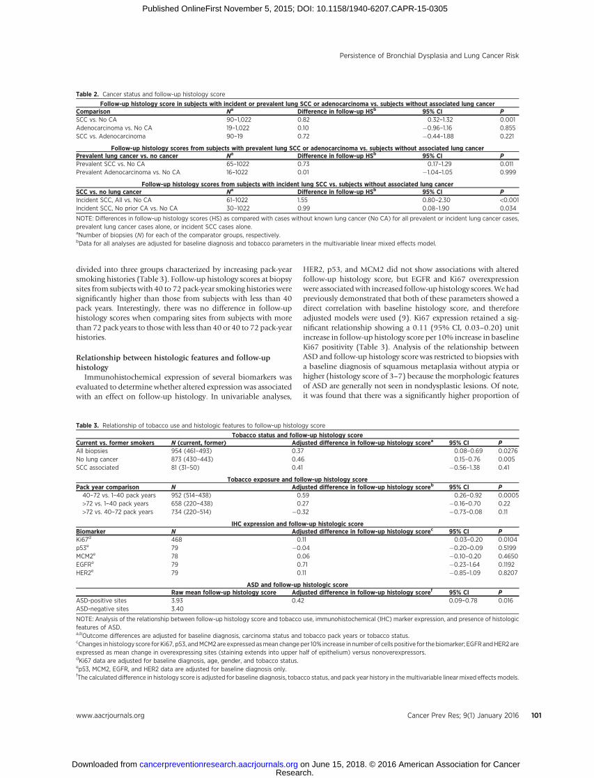
divided into three groups characterized by increasing pack-yearsmoking histories (Table 3). Follow-up histology scores at biopsysites from subjects with 40 to 72 pack-year smoking histories weresignificantly higher than those from subjects with less than 40pack years. Interestingly, there was no difference in follow-uphistology scores when comparing sites from subjects with morethan 72 pack years to those with less than 40 or 40 to 72 pack-yearhistories.
Relationship between histologic features and follow-uphistology
Immunohistochemical expression of several biomarkers wasevaluated to determine whether altered expression was associatedwith an effect on follow-up histology. In univariable analyses,
HER2, p53, and MCM2 did not show associations with alteredfollow-up histology score, but EGFR and Ki67 overexpressionwere associatedwith increased follow-uphistology scores.Wehadpreviously demonstrated that both of these parameters showed adirect correlation with baseline histology score, and thereforeadjusted models were used (9). Ki67 expression retained a sig-nificant relationship showing a 0.11 (95% CI, 0.03–0.20) unitincrease in follow-up histology score per 10% increase in baselineKi67 positivity (Table 3). Analysis of the relationship betweenASD and follow-up histology score was restricted to biopsies witha baseline diagnosis of squamous metaplasia without atypia orhigher (histology score of 3–7) because the morphologic featuresof ASD are generally not seen in nondysplastic lesions. Of note,it was found that there was a significantly higher proportion of
Table 2. Cancer status and follow-up histology score
Follow-up histology score in subjects with incident or prevalent lung SCC or adenocarcinoma vs. subjects without associated lung cancerComparison Na Difference in follow-up HSb 95% CI PSCC vs. No CA 90–1,022 0.82 0.32–1.32 0.001Adenocarcinoma vs. No CA 19–1,022 0.10 �0.96–1.16 0.855SCC vs. Adenocarcinoma 90–19 0.72 �0.44–1.88 0.221
Follow-up histology scores from subjects with prevalent lung SCC or adenocarcinoma vs. subjects without associated lung cancerPrevalent lung cancer vs. no cancer Na Difference in follow-up HSb 95% CI PPrevalent SCC vs. No CA 65–1022 0.73 0.17–1.29 0.011Prevalent Adenocarcinoma vs. No CA 16–1022 0.01 �1.04–1.05 0.999
Follow-up histology scores from subjects with incident lung SCC vs. subjects without associated lung cancerSCC vs. no lung cancer Na Difference in follow-up HSb 95% CI PIncident SCC, All vs. No CA 61–1022 1.55 0.80–2.30 <0.001Incident SCC, No prior CA vs. No CA 30–1022 0.99 0.08–1.90 0.034
NOTE: Differences in follow-up histology scores (HS) as compared with cases without known lung cancer (No CA) for all prevalent or incident lung cancer cases,prevalent lung cancer cases alone, or incident SCC cases alone.aNumber of biopsies (N) for each of the comparator groups, respectively.bData for all analyses are adjusted for baseline diagnosis and tobacco parameters in the multivariable linear mixed effects model.
Table 3. Relationship of tobacco use and histologic features to follow-up histology score
Tobacco status and follow-up histology scoreCurrent vs. former smokers N (current, former) Adjusted difference in follow-up histology scorea 95% CI PAll biopsies 954 (461–493) 0.37 0.08–0.69 0.0276No lung cancer 873 (430–443) 0.46 0.15–0.76 0.005SCC associated 81 (31–50) 0.41 �0.56–1.38 0.41
Tobacco exposure and follow-up histology scorePack year comparison N Adjusted difference in follow-up histology scoreb 95% CI P40–72 vs. 1–40 pack years 952 (514–438) 0.59 0.26–0.92 0.0005>72 vs. 1–40 pack years 658 (220–438) 0.27 �0.16–0.70 0.22>72 vs. 40–72 pack years 734 (220–514) �0.32 �0.73–0.08 0.11
IHC expression and follow-up histologic scoreBiomarker N Adjusted difference in follow-up histology scorec 95% CI PKi67d 468 0.11 0.03–0.20 0.0104p53e 79 �0.04 �0.20–0.09 0.5199MCM2e 78 0.06 �0.10–0.20 0.4650EGFRe 79 0.71 �0.23–1.64 0.1192HER2e 79 0.11 �0.85–1.09 0.8207
ASD and follow-up histologic scoreRaw mean follow-up histology score Adjusted difference in follow-up histology scoref 95% CI P
ASD-positive sites 3.93 0.42 0.09–0.78 0.016ASD-negative sites 3.40
NOTE: Analysis of the relationship between follow-up histology score and tobacco use, immunohistochemical (IHC) marker expression, and presence of histologicfeatures of ASD.a,bOutcome differences are adjusted for baseline diagnosis, carcinoma status and tobacco pack years or tobacco status.cChanges in histology score for Ki67, p53, andMCM2are expressed asmean change per 10% increase in number of cells positive for the biomarker; EGFRandHER2 areexpressed as mean change in overexpressing sites (staining extends into upper half of epithelium) versus nonoverexpressors.dKi67 data are adjusted for baseline diagnosis, age, gender, and tobacco status.ep53, MCM2, EGFR, and HER2 data are adjusted for baseline diagnosis only.fThe calculated difference in histology score is adjusted for baseline diagnosis, tobacco status, and pack year history in themultivariable linear mixed effects models.
Persistence of Bronchial Dysplasia and Lung Cancer Risk
www.aacrjournals.org Cancer Prev Res; 9(1) January 2016 101
Research. on June 15, 2018. © 2016 American Association for Cancercancerpreventionresearch.aacrjournals.org Downloaded from
Published OnlineFirst November 5, 2015; DOI: 10.1158/1940-6207.CAPR-15-0305

high-grade dysplasia at baseline in the ASD versus dysplasiawithout ASD groups (71.9% vs. 53.0%, respectively, P <0.001). Nonetheless, after adjustment for a number of clinicalparameters, including baseline diagnosis, the presence of histo-logic features of ASD was also associated with a significantincrease in follow-up histology score as compared with siteswithout features of ASD (Table 3).
DiscussionThe potential to use lesion-specific changes over time as an
indication of risk for the development of invasive SCC of the lungwas explored in this study. The findings demonstrated that anincreased frequency of sites that show persistence as or progres-sion toHGDwas associatedwith a significant 7.84 (95%CI, 1.56–39.39) fold increase in risk for development of invasive SCC, andindicated that subjects withmultiple dysplastic sites that persist orprogress to HGD represent a subset of patients with aggressiveairway disease. The demonstration that subjects with multiplesites of persistent disease show the strongest association withdevelopment of SCC emphasizes the importance of performing athorough evaluation of the airway, and adds support to the role offield carcinogenesis in the development of invasive lung cancer.Increased risk for invasive SCC has previously been associatedwith multiple sites of abnormal appearing mucosa by autofluor-escence bronchoscopy (AFB; ref. 30). In addition, the data dem-onstrate that higher histology scores in follow-up biopsies implyimportant differences in potential for progression. For instance,cases with dysplastic sites that persisted or progressed to HGDshowed a significant increase in risk for development of SCCwhereas those in which persistence as low- or high-grade BD didnot, despite the fact that the latter definition of persistenceallowed for inclusion of several more cases in the overall evalu-ation. This finding is similar to those of Alaa and colleagues (31),who demonstrated that the development of new severely dys-plastic lesions, regardless of the baseline histology, was morecommon in subjects that developed invasive cancer or CIS. Ourdata showed similar findings, demonstrating a significant rela-tionship between development of SCC and presence of multiplepersistent HGDs when sites of any baseline histology score wereincluded (see Supplementary Fig. S2). However, as discussedbelow, we and others have observed that CIS often regresses(20). Therefore, establishment of a relationship between persis-tence of BD and risk for invasive SCC is an important extension ofthese previous findings.
The demonstration of a relationship between HGD in follow-up biopsies and risk for invasive SCCmay also have implicationsfor the management of patients at risk for aggressive airwaydisease. The tumors that develop in association with persistentBD are more centrally located and are less likely to be associatedwith identifiable radiographic abnormalities in the early course ofdisease. Thus, screening for BD and identification of patients athigh risk for progression to invasive SCC will likely require adifferent modality from high-resolution CT to be effective. Ourfindings suggest that multiply sampling the airways may beimportant and that the employment of a bronchoscopic tech-nique that increases the sensitivity for detection of BD, such asAFB, may be advisable (27, 30). Furthermore, in patients withfeatures of aggressive airway disease, close follow-up would likelybe indicated and consideration of potential benefit from preven-tive therapy might be suggested. With respect to invasive cancer,
our study showed that subjects with persistent BD developedinvasive cancer both at sites that were associated with and remotefrom those with baseline dysplastic change. Eight subjects devel-oped incident SCC at sites that were previously biopsied and fiveat sites that had not been previously biopsied (including onepatient that developed synchronous, incident SCCs in two dif-ferent contralateral lung lobes). Six of the previously biopsiedsites showed dysplasia in baseline biopsies, of which two haddemonstrated persistence before development of SCC (SCC wasdiagnosed in the second biopsy for the other four). Thus, twoSCCs developed at nondysplastic sites and six others developed atsites that had not been previously sampled, suggesting that theydid not appear abnormal on AFB. This may indicate that chemo-preventive rather than local therapy will be necessary to signifi-cantly reduce the incidence of SCC in this setting. Finally, thefindings also support the use of reduced bronchial histologyscores as an informative endpoint in trials evaluating efficacy ofpotential preventive agents. Although we have shown that thefrequency of persistent BD is associated with subsequent devel-opment of SCC, a potential drawback associatedwith our analysisis the inclusion of some incident SCC cases in which this tumorrepresents a second lung primary. Information regarding thera-pies that patientswithprior lung cancermayhave receivedwas notavailable. It is possible that such treatments could influence thecourse of BD in the group of patients with prior carcinoma.However, our finding that primary incident SCC is also associatedwith increased follow-up histology scores further supports arelationship between persistence or progression of BD and riskfor the development of invasive SCC.
The association of higher grades of dysplasia at baseline withincreased histologic scores on follow-up corroborates findingsfrom the prospective study of Bota and colleagues (21) and themeta-analysis of follow-up data from four different chemopre-vention trials performed in the British Columbia Lung HealthStudy that included more than 700 subjects (2). Although dif-ferent classifications of outcomewere used in the latter study, theirfinding of a 4- to 5-fold higher rate of progression in sites withbaseline diagnoses of moderate or severe dysplasia as comparedwith those with lower diagnoses is consistent with the findings inour analysis. Strengths of our study thatmaymorefirmly establishsome of these relationships include fewer numbers of sites com-ing fromsubjectswithprior lungor head andneck cancers (8.8%),inclusion of sites with lengthy follow-up (48.2% with >2 years),and confinement of our study group to subjects from the non-treatment protocols or the placebo arm of prevention trials withpositive findings. Previously, CIS has been reported to progress toinvasive frequently with the majority progressing to cancer insome reports (19). In our cohort, 9 subjects had 23 sites thatshowed CIS and had follow-up biopsies. Although histologicallynormal at the baseline biopsy, one of these sites developed CISand progressed to invasive SCC 7 months later. In addition, 2other subjects developed incident SCCs, but not at their sites ofCIS. Although 16 of 22 (72.7%) CIS sites persisted as HGD,including all of those in cases with associated SCC, six sites in2 subjects regressed to nondysplastic histology, including five thatwere followed over a course of 35months and were re-biopsied 1to 3 times. Taking the biopsy with the highest diagnosis beforedevelopment of SCC, one site with CIS at baseline (1/22, 4.54%),five sites with baseline moderate or severe dysplasia (5/282,1.77%), one with baseline LGD (1/204, 0.49%) and four withnondysplastic baseline diagnoses progressed to SCC (4/667,
Merrick et al.
Cancer Prev Res; 9(1) January 2016 Cancer Prevention Research102
Research. on June 15, 2018. © 2016 American Association for Cancercancerpreventionresearch.aacrjournals.org Downloaded from
Published OnlineFirst November 5, 2015; DOI: 10.1158/1940-6207.CAPR-15-0305

0.60%). Although the overall number of CIS lesions is small inthis cohort, the findings support the aggressive nature that otherpublications have found to be associated with these lesions, butalso suggests that the rate of progression is not high anddocumentregression of CIS.
Our data show that tobacco use has an impact on the course ofBD with current tobacco users having higher follow-up histologyscores than former tobacco users. These findings compliment thefindings of Cl�ement-Duchenea and colleagues (32) in whichduration of smoking historywas found to correlate with increasedincidence of BD. This information could be useful clinically forphysicians counseling their patients to quit tobacco use as ameasure to prevent the development of lung cancer.
ASDs were also shown to be associated with an increased levelof atypia on follow-up biopsy. Angiogenesis is well established asa prognostic factor in invasive carcinoma, and we have previouslyshown that expression of VEGF increases with higher grades of BD(33). Furthermore, our recent analysis of vandetanib, the VEGFR2inhibitor with multitarget inhibitory capacity, showed preventiveactivity of this agent in a mouse model of lung carcinogenesis(34). These findings also correlate with previous work that hasdemonstrated more frequent ASD in subjects with lung cancerthan in those without (35). Angiogenic changes could support anincreased level of epithelial cell proliferation that may be impor-tant in promoting BD persistence and progression. Given thatpoor vascular integrity has been associated with VEGF dominantneoangiogenesis (36), it is also possible that ASD lesions areassociated with an altered microenvironment that promotesprogression. In addition, our IHC analyses suggest that higherlevels of expression of Ki67 could also serve as biomarkers ofincreased risk in BD.
The results of this study suggest that an important subset ofaggressive airway disease is represented by cases that show thepresence of multiple dysplastic lesions that persist or progress toHGD, and demonstrate that in patients with this presentationthere is increased risk for invasive SCC. Further characterization of
these persistent lesions should allow for the development ofmoreprecise predictive markers. Furthermore, obtaining an under-standing of the biologic characteristics that drive these BD witha high risk for progression to invasive lung cancer will helpidentify effective targets for prevention.
Disclosure of Potential Conflicts of InterestNo potential conflicts of interest were disclosed.
Authors' ContributionsConception and design: D.T. Merrick, Y.E. Miller, R.L. Keith, T.C. Kennedy,P.J. Blatchford, F.R. Hirsch, P.A. Bunn Jr, W.A. FranklinDevelopment of methodology: D.T. Merrick, Y.E. Miller, W. Feser,T.C. Kennedy, F.R. Hirsch, W.A. FranklinAcquisition of data (provided animals, acquired and managed patients,provided facilities, etc.): D.T. Merrick, Y.E. Miller, R.L. Keith, T.C. Kennedy,F.R. Hirsch, P.A. Bunn Jr, W.A. FranklinAnalysis and interpretation of data (e.g., statistical analysis, biostatistics,computational analysis): D.T. Merrick, D. Gao, A.E. Baron, P.J. Blatchford,S. Braudrick, F.R. Hirsch, W.A. FranklinWriting, review, and/or revision of the manuscript: D.T. Merrick, D. Gao,Y.E.Miller, R.L. Keith, A.E. Baron,W. Feser, T.C. Kennedy, F.R.Hirsch, L.Heasley,P.A. Bunn Jr, W.A. FranklinAdministrative, technical, or material support (i.e., reporting or organizingdata, constructing databases): D.T. Merrick, P.J. Blatchford, S. Braudrick,W.A. FranklinStudy supervision: D.T. Merrick, F.R. Hirsch, P.A. Bunn Jr, W.A. Franklin
Grant SupportThis study was supported by Lung Specialized Programs of Research Excel-
lence P50 CA058187 and Cancer Center Support grant P30 CA046934 (allauthors received P30 CA046934)
The costs of publication of this articlewere defrayed inpart by the payment ofpage charges. This article must therefore be hereby marked advertisement inaccordance with 18 U.S.C. Section 1734 solely to indicate this fact.
Received August 5, 2015; revisedOctober 9, 2015; acceptedOctober 26, 2015;published OnlineFirst November 5, 2015.
References1. Travis WD, Brambilla E, Burke AP, Marx A, Nicholson AG. WHO classi-
fication of tumours of the lung, pleara, thymus and heart, 4th Edition.Lyon: IARC; 2015. p. 59–63.
2. Ishizumi T,McWilliams A,Macaulay C,Gazdar A, LamS.Natural history ofbronchial preinvasive lesions. Cancer Metastasis Rev 2010;29:5–14.
3. Keith RL, Blatchford PJ, Kittelson J, Minna JD, Kelly K, Massion PP, et al.Oral iloprost improves endobronchial dysplasia in former smokers.Cancer Prev Res 2011;4:793–80.
4. Kelly K, Kittelson J, Franklin WA, Kennedy TC, Klein CE, Keith RL, et al. Arandomized phase II chemoprevention trial of 13-CIS retinoic acid with orwithout alpha tocopherol or observation in subjects at high risk for lungcancer. Cancer Prev Res 2009;5:440–9.
5. Lam S, leRiche JC, McWilliams A,MacAulay C, Dyachkova Y, Szabo E, et al.A randomized phase IIb trial of pulmicort turbuhaler (budesonide) inpersons with dysplasia of the bronchial epithelium. Clin Cancer Res2004;10:6502–11.
6. Lam S, McWilliams A, Leriche J, MacAulay C, Wattenburg L, Szabo E. Aphase I study of myo-inositol for lung cancer chemoprevention. CancerEpidemiol Biomarkers Prev 2006;15:1526–31.
7. Wistuba II, Behrens C, Milchgrub S, Bryant D, Hung J, Minna JD, et al.Sequential molecular abnormalities are involved in the multistagedevelopment of squamous cell lung carcinoma. Oncogene 1999;18:643–50.
8. Wistuba II, Behrens C, Virmani Ak, Mele G, Milchgrub S, Girard L, et al.High resolution chromosome 3p allelotyping of human lung cancer and
bronchial epithelium reveals multiple, discontinuous sites of 3pallele lossand three regions of frequent breakpoints. Cancer Res 2000;60:1949–60.
9. Merrick DT, Kittelson J, Winterhalder R, Kotantoulas G, Ingeberg S, KeithRL, et al. Analysis of c-ErbB1/epidermal growth factor receptor and c-ErbB2/HER-2 expression in bronchial dysplasia: evaluation of potentialtargets for chemoprevention of lung cancer. Clin Cancer Res 2006;12:2281–8.
10. Mascaux C, Laes JF, Anthoine G, Haller A, Ninane V, Burny A, et al.Evolution of microRNA expression during human bronchial squamouscarcinogenesis. Eur Respir J 2009;33:352–9.
11. Jonsson S, Varella-Garcia M, Miller Y, Wolf HJ, Byers T, Braudrick S, et al.Chromosomal aneusomy in bronchial high-grade lesions is associatedwith invasive lung cancer. Am J Respir Crit Care Med 2008;177:342–7.
12. Massion P, Zou Y,UnerH, Kiatsimkul P,WolfHJ, Baron AE, et al. Recurrentgenomic gains in preinvasive lesions as a biomarker of risk for lung cancer.PLoS ONE 2009;4:e5611.
13. Gustafson AM, Soldi R, Anderlind C, Scholand MB, Qian J, Zhang X, et al.Airway PI3K pathway activation is an early and reversible event in lungcancer development. Sci Transl Med 2010;2:1–11.
14. Lantuejol S, Raynaud C, Salameire D, Gazzeri S, Moro-Sibilot D, Soria J-C,et al. Telomere maintenance and DNA damage response during lungcarcinogenesis. Clin Cancer Res 2010;16:2979–88.
15. Jeanmart Lantuejoul S, Fievet F, MoroD, SturmN, Brambilla C, et al. Valueof immunohistochemical markers in preinvasive bronchial lesions in riskassessment of lung cancer. Clin Cancer Res 2003;9:2195–203.
Persistence of Bronchial Dysplasia and Lung Cancer Risk
www.aacrjournals.org Cancer Prev Res; 9(1) January 2016 103
Research. on June 15, 2018. © 2016 American Association for Cancercancerpreventionresearch.aacrjournals.org Downloaded from
Published OnlineFirst November 5, 2015; DOI: 10.1158/1940-6207.CAPR-15-0305

16. Salaun M, Sesboue R, Moreno-Swire S, Metayer J, Bota S, Bourguignon J,et al. Molecular predictive factors for progression of high-grade preinvasivebronchial lesions. Am J Respir Crit Care Med 2008;177:880–6.
17. McCaughan F, Pole JCM, Bankier AT, Konfortov BA, Carroll B, Falzon M,et al. Progressive 3q amplification consistently targets SOX2 in preinvasivesquamous lung cancer. Am J Respir Crit Care Med 2010;182:83–91.
18. Breuer RH, Pasic A, Smit EF, van Vliet E, VonkNoordegraaf A, Risse EJ, et al.The natural course of preneoplastic lesions in bronchial epithelium.Clin Cancer Res 2005;11:537–43.
19. Venmans B, van Boxem A, Smit E, Postmus P, Sutedja T. Outcome ofbronchial carcinoma in situ. Chest 2000;117:1572–6.
20. Moro-SibilotD, Fievet F, JeanmartM, Lantuejoul S, Arbib F, LaverribreMH,et al. Clinical prognostic indicators of high-grade pre-invasive bronchiallesions. Eur Respirol J 2004;24:24–9.
21. Bota S, Auliac JB, Paris C, Metayer J, Sesboue R, Nouvet G, et al. Follow-upof bronchial precancerous lesions and carcinoma in situ using fluorescenceendoscopy. Am J Respir Crit Care Med 2001;164:1688–93.
22. SalaunM, Bota S, Thiberville L. Long-term followupof severe dysplasia andcarcinoma in situ of the bronchus. J Thorac Oncol 2009;4:1187–8.
23. Hoshino H, Shibuya K, Chiyo M, Iyoda A, Yoshida S, Sekine Y, et al.Biological features of bronchial squamous dysplasia followed up byautofluorescence bronchoscopy. Lung Cancer 2004;46:187–96.
24. Pasic A, van Vliet E, Breuer RH, Risse EJ, Snijders PJ, Postmus PE, et al.Smoking behavior does not influence the natural course of pre-invasivelesions in bronchial mucosa. Lung Cancer 2004;45:153–4.
25. Banerjee AK. Preinvasive lesions of the bronchus. J Thorac Oncol 2009;4:545–51.
26. Lam S, Kennedy T, Unger M, Miller YE, Gelmont D, Rusch V, et al.Localization of bronchial intraepithelial neoplastic lesions by fluorescencebronchoscopy. Chest 1998;113:696–702.
27. Hirsch FR, Prindiville SA, Miller YE, Franklin WA, Dempsey EC, Murphy JR,et al. Fluorescence versus white-light bronchoscopy for detection of preneo-plastic lesions: a randomized study. J Natl Cancer Inst 2001;93:1385–91.
28. Edell E, Lam S, Pass H,Miller YE, Sutedja T, Kennedy T, et al. Detection andlocalization of intraepithelial neoplasia and invasive carcinoma usingfluorescence-reflectance bronchoscopy: an international, multicenter clin-ical trial. J Thorac Oncol 2009;4:49–54.
29. Keith RL, Miller YE, Gemmill RM, Drabkin HA, Dempsey EC, Kennedy TC,et al. Angiogenic squamous dysplasia in bronchi of individuals at high riskfor lung cancer. Clin Cancer Res 2000;6:1616–25.
30. Pasic A, Vonk-Noordegraaf A, Risse EK, Postmus PE, Sutedja TG. Multiplesuspicious lesions detected by autofluorescence bronchoscopy predictmalignant development in the bronchial mucosa in high risk patients.Lung Cancer 2003;41:295–301.
31. Alaa MRM, Shibuya K, Fujiwara T, Wada H, Hoshino H, Yoshida S, et al.Risk of lung cancer in patients with preinvasive bronchial lesions followedby autofluorescence bronchoscopy and chest computed tomography. LungCancer 2011;72:303–8.
32. Cl�ement-Duchenea C, Alla F, Gauchotte G, Marie B, Carnin C, Menard O,et al. Is there a relationship between the presence of lung mucosa pre-invasive lesions and lung cancer incidence? Influence of tobacco consump-tion. Lung Cancer 2014;84:134–8.
33. Merrick DT, Haney J, Petrunich S, Sugita M, Miller YE, Keith RL, et al.Overexpression of vascular endothelial growth factor and its receptors inbronchial dysplasia demonstrated by quantitative RT-PCR analysis.Lung Cancer 2005;48:31–45.
34. Karoor V, Le M, Merrick D, Dempsey EC, Miller YE. Cancer Vascularendothelial growth factor receptor 2-targeted chemoprevention of murinelung tumors. Prev Res 2010;3:1141–7.
35. Karimi S, Mohammadi F, Khodadad K, Sadr M, Seyfollahi L, Masjedi MR.Relationship between angiogenic squamous dysplasia and bronchogeniccarcinoma in patients undergoing white light bronchoscopy. Can Respir J2012;19:201–8.
36. Greenberg JI, Cheresh DA. VEGF as an inhibitor of tumor vessel matura-tion: implications for cancer therapy. Expert Opin Biol Ther 2009;9:1347–56.
Cancer Prev Res; 9(1) January 2016 Cancer Prevention Research104
Merrick et al.
Research. on June 15, 2018. © 2016 American Association for Cancercancerpreventionresearch.aacrjournals.org Downloaded from
Published OnlineFirst November 5, 2015; DOI: 10.1158/1940-6207.CAPR-15-0305

2016;9:96-104. Published OnlineFirst November 5, 2015.Cancer Prev Res Daniel T. Merrick, Dexiang Gao, York E. Miller, et al. of Invasive Squamous Cell CarcinomaPersistence of Bronchial Dysplasia Is Associated with Development
Updated version
10.1158/1940-6207.CAPR-15-0305doi:
Access the most recent version of this article at:
Material
Supplementary
1
http://cancerpreventionresearch.aacrjournals.org/content/suppl/2016/01/09/1940-6207.CAPR-15-0305.DCAccess the most recent supplemental material at:
Cited articles
http://cancerpreventionresearch.aacrjournals.org/content/9/1/96.full#ref-list-1
This article cites 35 articles, 10 of which you can access for free at:
Citing articles
http://cancerpreventionresearch.aacrjournals.org/content/9/1/96.full#related-urls
This article has been cited by 2 HighWire-hosted articles. Access the articles at:
E-mail alerts related to this article or journal.Sign up to receive free email-alerts
Subscriptions
Reprints and
To order reprints of this article or to subscribe to the journal, contact the AACR Publications Department at
Permissions
Rightslink site. Click on "Request Permissions" which will take you to the Copyright Clearance Center's (CCC)
.http://cancerpreventionresearch.aacrjournals.org/content/9/1/96To request permission to re-use all or part of this article, use this link
Research. on June 15, 2018. © 2016 American Association for Cancercancerpreventionresearch.aacrjournals.org Downloaded from
Published OnlineFirst November 5, 2015; DOI: 10.1158/1940-6207.CAPR-15-0305