Embed Size (px)
Citation preview

PERIODONTAL DISEASE, BONE LOSS, AND ANTI-ANDROGEN THERAPY
by
Pouran Famili
D.M.D., University of Pittsburgh School of Dental Medicine, 1985
M.P.H., University of Pittsburgh Graduate School of Public Health, 2000
Submitted to the Graduate Faculty of
the Department of Epidemiology
of the Graduate School of Public Health in partial fulfillment
of the requirements for the degree of
Doctor of Philosophy
University of Pittsburgh
2005

UNIVERSITY OF PITTSBURGH
Graduate School of Public Health
This dissertation was presented by
Pouran Famili
It was defended on 6 September 2005
and approved by
Dissertation Advisor
Jane A. Cauley, Dr.P.H. Professor
Department of Epidemiology Graduate School of Public Health
University of Pittsburgh
Susan L. Greenspan, M.D. Professor of Medicine
Director, Osteoporosis Prevention and Treatment Center Division of Endocrinology and Metabolism
School of Medicine University of Pittsburgh
Jon B. Suzuki, D.D.S., Ph.D.
Professor of Periodontics Associate Dean for Graduate Education, Research, and International Affairs
School of Dentistry Temple University
Joseph M. Zmuda, PhD.
Assistant Professor of Epidemiology and Human Genetics Department of Epidemiology
Graduate School of Public Health University of Pittsburgh
Joseph P. Costantino, Dr. P.H.
Professor, Department of Biostatistics Director, NSABP Biostatistical Center
Graduate School of Public Health University of Pittsburgh
ii

Copyright by Pouran Famili 2005
iii

Jane A. Cauley Dr.P.H.
PERIODONTAL DISEASE, BONE LOSS, AND ANTI-ANDROGEN THERAPY
Pouran Famili, Ph.D.
University of Pittsburgh, 2005
Periodontitis is a multifactorial disease with microbial dental plaque as the etiological agent. The
manifestation and progress of periodontitis is influenced by a wide variety of determinants and
factors, including subject characteristics, social and behavioral factors, systemic factors, genetic
factors, the microbial composition of dental plaque, and others. The pathogenesis of periodontal
disease results in resorption of alveolar bone and loss of the attachment apparatus to the teeth.
There is a biological potential that periodontal destruction may be influenced by systemic bone
loss. Since alveolar bone loss is a prominent feature of periodontal disease, disturbances in bone
mineral density (BMD), especially in the jaws, are suspected of being an aggravating factor in
periodontal disease. In previously published research, the severity of osteoporosis may be related
to tooth loss in post-menopausal women. Considering the relationship among bone mineral
density, osteoporosis, and periodontitis in men, it is known that men completing androgen
ablation therapy for control of prostate cancer are at higher risk for osteoporosis. As androgen
deprivation therapy is the recommended treatment for men with metastatic or locally-advanced
nonmetastatic prostate carcinoma, and as prostate carcinoma is the most common visceral
malignancy and the second leading cause of death from cancer in men, the relationship between
androgen deprivation therapy and loss of bone mineral density is a matter of public health
importance.
This dissertation assesses the association between bone mineral density, the presence of
periodontal disease, and the possible subsequent onset of clinical osteoporosis, as seen among a
population of older women followed longitudinally; a set of men with prostate carcinoma
undergoing androgen ablation therapy; and those men in the same set not receiving androgen
ablation for prostate cancer. We believe our research, using the model of periodontal bone
density and oral bone loss, shows additional clear empirical evidence pointing to a cause-and-
effect relationship between androgen deprivation therapy and loss of bone mineral density.
iv

TABLE OF CONTENTS 1.0 Chapter One Introductory Remarks The relationships among periodontal disease, bone loss, and anti-androgen therapy, by a consideration of the epidemiology and etiology of periodontal disease and osteoporosis: A review of the literature..................................................................................................................1
1.1 The etiology and epidemiology of periodontal disease……………………………….2 1.1.1 The epidemiology of periodontal disease: Prevalence and incidence………...2 1.1.2 The epidemiology of periodontal disease: Periodontal attachment loss……....4 1.1.3 The epidemiology of periodontal disease: Probing pocket depth……………..5 1.1.4 The epidemiology of periodontal disease: Gingival recession………………..6 1.1.5 The etiology of periodontal disease: Summary and risk factors………………6 1.1.6 The risk factors for periodontal disease: Smoking……………………………7 1.1.7 The risk factors for periodontal disease: Drinking alcohol…………………....8 1.1.8 The risk factors for periodontal disease: Diabetes …………………………....8
1.1.8.1 Insulin-Dependent Diabetes Mellitus (Type I)………………………..8 1.1.8.2 Non-Insulin-Dependent Diabetes Mellitus (Type II diabetes)...............9 1.1.9 The risk factors for periodontal disease: Obesity……………………………10 1.1.10 The risk factors for periodontal disease: Atherosclerosis……………............11 1.1.11 The risk factors for periodontal disease: Osteoporosis………….…………...12 1.1.12 The risk factors for periodontal disease: Hormone replacement therapy……13 1.2 The epidemiology and etiology of osteoporosis, and the relationship between systemic bone mineral density and oral bone mineral density……......……………..14 1.2.1 Epidemiology of osteoporosis: Prevalence and incidence…………………...14 1.2.2 Etiology of osteoporosis……………………………......................................15 1.2.3 Etiology of osteoporosis: Detection…………………………………….........16 1.2.4 Risk factors for osteoporosis…………………………………………………17 1.2.5 Risk factors for osteoporosis: Vitamin D deficiency………………………...18 1.2.6 The relationship between systemic bone mineral density and oral bone mineral density…….........................................................................................18 1.2.7. The relationship of periodontal disease and osteoporosis…………………....20 1.3 The relationships among osteoporosis, bone loss, and periodontal disease in men generally and in men with prostate cancer under androgen deprivation therapy..........................................................................................................................23 1.3.1 Osteoporosis in men……………………………….........................................23 1.3.2 Bone loss in men with prostate cancer……………………………………….24 1.3.3 Osteoporosis in men with prostate cancer under androgen suppression therapy………………………………………………………………..............25 1.4 The relationship of periodontal disease in men with prostate cancer under androgen deprivation therapy. A brief summary preface to the subsequent chapters………….27 1.5 The literature cited in the introductory remarks…………..........................................28 v

2.0 Chapter Two Longitudinal study of periodontal disease and edentulism with rates of bone loss in older women (Published in the Journal of Periodontology 76:11-15, January 2005)......................38
2.1 Abstract of the research……………...………………………………..………....39 2.2 Background and rationale for the research…..……………..................................40 2.3 The design/setting/patients and the materials and methods of the research, as
described in the chapter……………………………………………………….....41 2.4 The results of the research, as described for this chapter…..................................42 2.5 A discussion of the research, as described for this chapter……………………...42 2.6 The literature cited in the chapter summary of the research.................................48
3.0 Chapter Three The effect of androgen deprivation therapy on periodontal disease in men, with a consideration of the relationships between periodontal disease and bone loss on men with prostate cancer, both with and without receiving androgen deprivation therapy................49
3.1 Abstract of the research………………………………………………………….50 3.2 Background and rationale for the research…………............................................51 3.3 The design/setting/patients and the materials and methods of the research, as
described in the chapter……...……………………………………………….….51 3.4 The results of the research, as described in the chapter… ………………………53 3.5 A discussion of the research, as described in the chapter….….............................54 3.6 The literature cited in the chapter summary of the research.…………………….60
4.0 Chapter Four Summary and Conclusions The relationships between periodontal disease and bone loss on men with prostate cancer under androgen deprivation therapy. A brief summary analysis of the research conducted, and a discussion of the related issues, limitations, and conclusions of the research and the dissertation………………………………………………………………………………………62 4.1 The limitations of the research...............................................................................65 4.2 Conclusions, and future recommendations for research........................................65 Bibliography………………………………………….................................................................66 vi

LIST OF TABLES Table 1.1 Prevalence rate ratios of periodontal variables, by gender and race-ethnicity........................................................................................................................................5 Table 1.2 Clinical measurement methods to assess bone mineral density...........................17 Table 1.3 Relationship between systemic bone mineral density (BMD) and oral bone mineral density (BMD).............................................................................................................20 Table 1.4 Relationship of periodontal destruction and bone mineral density (BMD)........21 Table 1.5 Percent change in lumbar spine and hip bone mineral density (BMD) in men with prostate carcinoma receiving androgen-deprivation therapy.............................26 Table 2.1 Characteristics of dentate and edentulous postmenopausal women (n=398). Values are mean (SD), number (%), or median (range)........................................44 Table 2.2 Age-adjusted BMD and annualized percent change by edentulous status.........45 Table 2.3 Characteristics of dentate women with and without periodontal disease (n=202).......................................................................................................................................46 Table 2.4 Age-adjusted BMD and annualized percent change in BMD of dentate subjects by periodontal status (n=202)....................................................................................47 Table 3.1 Characteristics of men with prostate cancer with and without anti-androgen therapy (n=68)..........................................................................................................56 Table 3.2 Characteristics of men with prostate cancer with and without receiving androgen deprivation therapy and with periodontal disease variables (n=68)...................57 Table 3.3 OR of periodontal disease by androgen therapy adjusted by different conditions...................................................................................................................................58 Table 3.4 Bone mineral density in men with and without periodontal disease, adjusted for receiving androgen deprivation therapy...........................................................59 vii

1.0 CHAPTER ONE: INTRODUCTORY REMARKS
THE RELATIONSHIPS AMONG PERIODONTAL DISEASE, BONE LOSS, AND ANTI-ANDROGEN THERAPY, BY A CONSIDERATION OF THE EPIDEMIOLOGY AND ETIOLOGY OF PERIODONTAL
DISEASE AND OSTEOPOROSIS: A REVIEW OF THE LITERATURE
Periodontal disease is a local chronic inflammatory disease of the tooth-supporting tissues, characterized
by inflammation of those tissues. The disease has a prevalence of thirty-five (35) percent in the adult
population of the United States (Consensus Report Annals of Periodontology 1996; Consensus Report
Journal of the American Dental Association 1998), and is a major cause of tooth loss and edentulism
among adults. Periodontal destruction---pathologic inflammation of the gingival and hard tissues---often
results in bone loss in the jaw, mobility of teeth, and ultimately tooth loss. The health and social impacts
of periodontal disease are enormous and adversely influence nutrition, speech, oral function, and facial
esthetics.
The pathogenesis of periodontal disease results in resorption of alveolar bone and loss of the
attachment apparatus to the tooth. There is the biological potential that periodontal destruction may be
influenced by systemic bone loss (Consensus Report Annals of Periodontology 1996). Since alveolar
bone loss is a prominent feature of periodontal disease, disturbances in bone mineral density (BMD),
especially in the jaws, are suspected of being an aggravating factor in the case of periodontal disease
(Wactawski-Wende et al. 1996).
Our incomplete understanding of the role of various risk factors obscures the specific causes and
underlying mechanisms of the periodontal disease process (Albandar 2002, Albandar 2002). Established
risk factors for periodontal disease include dental plaque, calculus, smoking, systemic disease, stress, and
genetic traits. It is clearly documented that the incidence of periodontal disease increases with advancing
age. Significant age-demographic shifts exist in the United States populations and, as people live longer
and retain more teeth, the number of people developing periodontal disease is expected to increase in the
next few decades. Changes in our knowledge of the etiology of periodontal disease, and better recognition
of the potential importance of the susceptibility factors as they affect the initiation and progression of
periodontal disease, have led to an intense study of specific risk factors for periodontal disease.
Several convincing epidemiologic studies, such as the surveys conducted by the National Center
for Health Statistics (Ronderos 2004) and those of the National Institute of Dental and Craniofacial
1

Research (NIDCR), with additional studies from abroad, have challenged the popular perception that
humans have a universal susceptibility to periodontal disease (Irfan 2001).
The relationship of osteoporosis in men and periodontal disease due to androgen ablution is not
well established. Studies to evaluate the relationship between osteoporosis due to androgen ablution and
periodontal disease are essential for enhancing our understanding of systemic bone pathogenesis. The
objective of this research is to assess, and thus better understand, the association between bone mineral
density, the presence of periodontal disease, and the possible subsequent onset of clinical osteoporosis, as
seen among a set of men with prostate carcinoma undergoing androgen ablation therapy, and those in the
same set not receiving androgen ablation for control of prostate cancer.
1.1 THE ETIOLOGY AND EPIDEMIOLOGY OF PERIODONTAL DISEASE Periodontal disease is a pathological condition, a chronic inflammatory disease, affecting the supporting
structures of teeth, characterized by inflammation of the teeth-supporting tissues at the gums. Periodontal
disease is identified by a bacterial challenge that can destroy the host tissues, the response leading to
periodontal attachment loss, bone loss, and ultimately possible tooth loss (Nunn 2003; Consensus Report
Annals of Periodontology 1996; Journal of the American Dental Association 1998).
Thus, periodontitis is clearly a multi-factorial disease with microbial dental plaque as the initiator
(Nunn 2003; Consensus Report Annals of Periodontology 1996; Journal of the American Dental
Association 1998). However, the manifestation and progression of periodontitis is influenced by a wide
variety of determinants and risk factors, including subject characteristics, social and behavioral factors,
systemic factors, genetic factors, the microbial composition of dental plaque, and others (Nunn 2003;
Annals of Periodontology 1996; Journal of the American Dental Association 1998). If periodontitis is not
treated, the disease will slowly progress and painlessly destroy the bone which supports the teeth.
Untreated, the disease will eventually cause tooth loss.
1.1.1 The epidemiology of periodontal disease: Prevalence and incidence
Although many epidemiological studies have been conducted concerning periodontal disease using the
parameters of clinical attachment level, attachment loss, probing pocket depth, and gingival recession, the
evidence pertaining to prevalence, incidence, and risk factors in an older adult population of either sex is
limited (Albandar 2002).
2

The NHANES III (1994) study showed that among dentate adults in the United States (six teeth
or more present) age 30 and older, 3.1 percent had advanced periodontitis (defined as pocket depth upon
probing greater than 6mm); 9.5 percent had moderate periodontitis (defined as pocket depth upon probing
4-6 mm), 21.8 percent had mild periodontitis (defined as pocket depth upon probing 3-5 mm) and 65.5
percent had no periodontitis (defined as pocket depth less than 3 mm). The prevalence of periodontitis (all
severities) increases steadily with increasing age (Albandar 2002, Albandar 2002). Using the measure of
tooth loss to definitively diagnose periodontitis in his six-year study, the youngest age group (18-33)
presented at baseline with the fewest mean number of teeth lost (1) and concluded with the least (0).
Subjects over age 57 entered with the most teeth lost (3.6), and concluded with the most teeth lost (0.4).
Periodontal disease and aging are commonly thought to be correlated with each other. The
relationship is intuitively thought to be more related to the breakdown of the periodontal tissues over
time, rather than an actual function of the accumulation of chronological years causing a deficiency that
increases the individual’s susceptibility to periodontal disease (Nunn 2003; Genco 1996).
Epidemiological studies have shown more periodontal disease, both in terms of attachment loss as well as
bone loss, among older age groups than younger age groups (Grossi et al. 1994; Grossi et al. 1995;
Streckfus et al. 1999). Other research has shown that certain physiological changes in the periodontium
occur with age (Johnson et al. 1989 and van der Velden 1984) although these physiological changes
alone are not responsible for the breakdown in the gingival tissues (the gingival recession, the attachment
loss, the eventual tooth loss).
Pocket probing depth is the measure from the free gingival margin (FGM) to the base of the
sulcus/pocket with the use of a calibrated probe (HuFriedy Michigan 0 probe, diameter of probe is 0.5
mm). Pocket depth is probed on six sites per tooth (distobuccal, midbuccal, mesiobuccal, distolingual,
midlingual, and mesiolingual). Clinical periodontal attachment level and the loss of periodontal
attachment are defined as the distance in millimeters (mm) from the cementoenamel junction (CEJ) to the
base of the pocket. Probing depth is the distance from the free gingival margin (FGM) to the base of the
sulcus/pocket. Other essential periodontal measures are the presence or absence of gingival recession or
gingival bleeding, and the measures of supragingival plaque and calculus. Gingival bleeding will be
measured as (0: no bleeding; 1: bleeding); supragingival plaque will be measured as (0: no plaque; 1:
plaque). Calculus will be measured as (0: no calculus; 1: supragingival calculus).
Among adult Americans, males have a greater prevalence of periodontitis than females. Albandar
(2000) reports that different racial and ethnic groups within a given population show marked differences
in periodontal disease indicators, and the general diagnosis of periodontitis. An important finding of the
NHANES I study was the trend that African-Americans in the U.S. population have a much higher
occurrence of periodontitis than Caucasians (1.28 to 0.76 respectively, using the general indicator of
3
Periodontal Index) and a higher prevalence of periodontitis than Mexican-Americans. Non-Hispanic
whites had significantly less periodontitis than either of the other two racial groups (Albandar 2002). The
same ratios were confirmed in Albandar’s 2002 study of periodontitis among 14,000 U.S. adolescents
(13-17 years old): early onset forms of periodontitis occurred in 10 percent of the adolescent African-
Americans, five percent of Hispanics, and 1.3 percent of whites. This gives a prevalence ratio of 7.7 for
African-American adolescents, and 3.9 percent for Hispanic adolescents, compared to the white
adolescents.
Nunn (2003) points out that race as a risk factor for periodontitis is a complex issue. She notes
that Prevotella intermedia presents a risk factor for blacks, but not for whites. When socioeconomic risk
factors for periodontitis were not considered, blacks were three times more likely than whites to exhibit
advanced periodontal destruction. When subjects were in the same socioeconomic status, differences in
gum disease often disappeared.
Albandar (2002) takes the conclusions of the NHANES III data regarding gender, race and
ethnicity, and the common variables indicating periodontal disease, and presents this data regarding
prevalence rate ratios (duplicated here as Table 1, next page). In the U.S., adult blacks show the highest
prevalence of periodontitis and the most loss of periodontal tissue, followed by Mexican-Americans.
Whites show the least disease and tissue loss. As shown in the table, blacks have a much higher
probability of having attachment loss and increased probing depth, and Mexican-Americans have a
moderately increased probability, compared to whites. For attachment loss ≥3mm, the ratio was 1.26 for
blacks, and 1.10 for Mexican-Americans, whereas for attachment loss of ≥5mm, the ratio was 1.73 and
1.28, respectively. Hence, in the U.S. adult population, the probabilities of having attachment loss of
≥3mm and ≥5mm, respectively, were 26 percent and 73 percent higher for blacks and 10% and 28%
higher for Mexican-Americans compared to whites. A similar pattern was observed for the other
periodontal disease indicators, and is also summarized in Table 1.
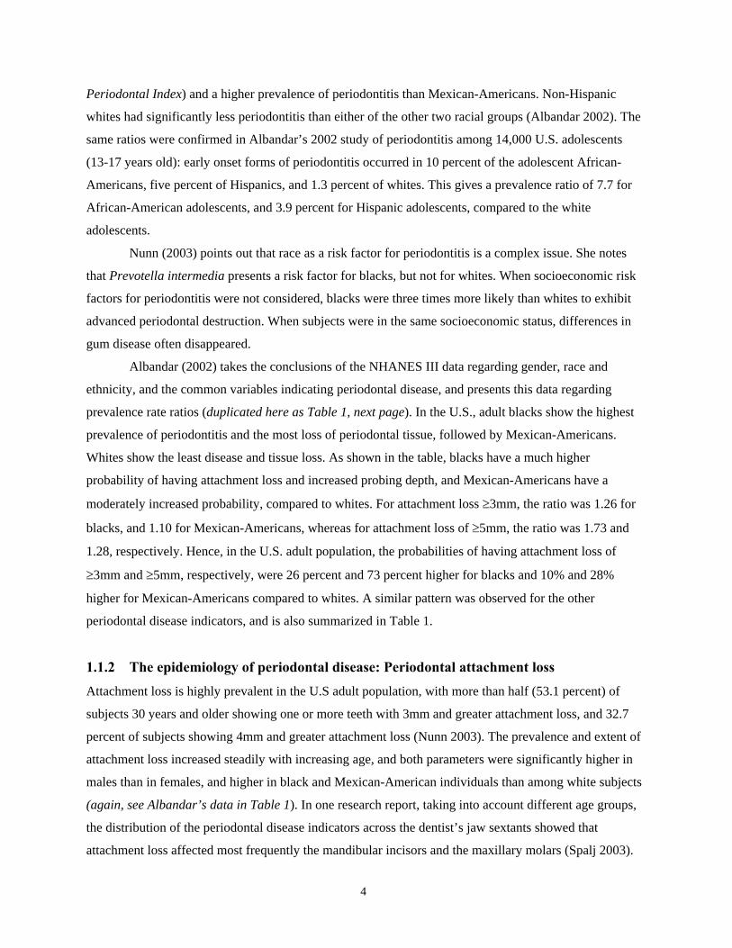
1.1.2 The epidemiology of periodontal disease: Periodontal attachment loss Attachment loss is highly prevalent in the U.S adult population, with more than half (53.1 percent) of
subjects 30 years and older showing one or more teeth with 3mm and greater attachment loss, and 32.7
percent of subjects showing 4mm and greater attachment loss (Nunn 2003). The prevalence and extent of
attachment loss increased steadily with increasing age, and both parameters were significantly higher in
males than in females, and higher in black and Mexican-American individuals than among white subjects
(again, see Albandar’s data in Table 1). In one research report, taking into account different age groups,
the distribution of the periodontal disease indicators across the dentist’s jaw sextants showed that
attachment loss affected most frequently the mandibular incisors and the maxillary molars (Spalj 2003).
4

Table 1.1 Prevalence rate ratios of periodontal variables, by gender and race-ethnicity [Data show males v.females (blacks v.whites, and Mexican-Americans v.whites) 30 years and older examinedin the third National Health and Nutrition Examination Survey (NHANES III) U.S. 1988-1994]
Gender Race-Ethnicity Variable Males/females Blacks/whites Mexican-
Americans/whites Prevalence (percent of persons)
Attachment loss ≥3mm 1.23 1.26 1.10 Attachment loss ≥4mm 1.44 1.46 1.22 Attachment loss ≥5mm 1.55 1.73 1.28 Probing depth ≥3mm 1.14 1.24 1.22 Probing depth ≥4mm 1.46 1.97 1.61 Probing depth ≥5mm 1.73 2.71 1.79
Gingival bleeding 1.17 1.15 1.31 Gingival recession ≥1mm 1.12 1.03 0.94 Gingival recession ≥3mm 1.54 1.29 1.09
Dental calculus 1.02 1.05 1.06 Extent (percent of teeth per person)
Attachment loss ≥3mm 1.48 1.54 1.17 Attachment loss ≥4mm 1.65 1.75 1.24 Attachment loss ≥5mm 1.93 2.06 1.33 Probing depth ≥3mm 1.39 1.79 1.48 Probing depth ≥4mm 1.63 2.47 1.65 Probing depth ≥5mm 2.00 3.31 1.85
Gingival bleeding 1.24 1.31 1.53 Gingival recession ≥1mm 1.30 1.13 0.95 Gingival recession ≥3mm 1.67 1.49 1.13
Dental calculus 1.21 1.49 1.35 [Data compiled by Albandar et al. in 2000]
1.1.3 The epidemiology of periodontal disease: Probing pocket depth Sixty-four percent of U.S. adults in this study had one or more teeth with a probing depth of 3 mm or
greater (mild periodontitis). Approximately 22.3 percent had a probing depth of 4mm or greater
(moderate periodontitis). The prevalence of probing depth at 4mm and greater increased steadily with
age. The prevalence and extent of probing depth at 4mm and more was significantly greater among males
than in females and among blacks and Mexican–American than whites. Blacks had the highest probing
depth measurement among the three race-ethnicity groups (Consensus Report Annals of Periodontology
1996; Journal of the American Dental Association 1998).
5

Again, Albandar’s data best summarizes all of these periodontal disease indicators---attachment
loss, probing depth, gingival bleeding, gingival recession, and dental calculus---related to gender and race
and ethnicity (Table 1.1).
1.1.4 The epidemiology of periodontal disease: Gingival recession In the same study, 22.5 percent of the subjects had gingival recession at 3mm and greater. The prevalence
and extent of gingival recession also increased steadily with increasing age: 10 percent of subjects 30-39
years old and 60 percent of subjects 80-90 years old had one or more teeth with 3mm gingival recession.
Males had a significantly greater prevalence and extent of gingival recession than females. The
prevalence and extent of gingival recession were comparable in the three race ethnic groups (Consensus
Report Annals of Periodontology 1996).
1.1.5 The etiology of periodontal disease: Summary and risk factors Clinicians have long believed they had a profile of the patient with periodontal disease, but reviewing the
literature regarding the etiology of periodontitis shows little hard evidence to identify the assumed
specific predisposing factors (Rees 2003). The profile, according to Rees, includes a patient who is a
smoker, is emotionally stressed, possibly malnourished, prone to disease, has a family history of
periodontitis, possesses local factors predisposing to the accumulation of plaque (clinically recognizable
in short, again, as a smoker with poor oral hygiene) and basically not motivated and not compliant with
the oral hygiene maintenance protocols (brushing, flossing, recalls every three, six---at most---twelve,
months) and not good at oral hygiene when brushing or flossing is undertaken.
While the profile is clinically valid, the risk factors are more profound than the profile indicates,
and detailed, thorough, examination of the underlying risk factors---the systemic risk factors---is
warranted and necessary. All persons are not equally susceptible to periodontal disease and do not
respond equally well to periodontal therapy. In a classic longitudinal study of a population with no access
to dental care and poor oral hygiene (Albandar 1990), enormous variability among individuals in the rates
of periodontal disease progression was observed. In this mostly homogenous population, some persons
developed disease at very rapid rates, while others had little or no disease progression (Albandar 1990).
Again, the established risk factors for periodontal disease are dental plaque, calculus (a hard mineral
deposit produced by the action of bacteria and body calcium, requiring removal with instruments by a
professional), smoking, systemic disease, age, genetic traits, stress, and others. While each of these
factors alone may have a detrimental effect on etiology, prognosis, and treatment outcomes, it should be
kept in mind that the presence of any of these factors alone or in combination may influence yet a
different outcome (Journal of the American Dental Association 1998).
6

While it is true that some strong current epidemiologic research has challenged the notion that
humans face a universal susceptibility to periodontal disease, it is also true that between five and twenty
percent of the population have severe forms of periodontal disease. The etiology of chronic adult
periodontitis is bacterial in nature, involving pathogens such as Actinobacillus actinomycetmcomitans,
Porphyromonas gingivalis, and Bacteroides forsythus. Current concepts in the etiology of periodontal
disease emphasize systemic factors which may place patients at risk to develop periodontitis. Conversely,
periodontitis may be a risk factor in the development of a variety of systemic conditions. A clearer
understanding of the relationship between oral health and systemic disease will aid healthcare providers in
the management of a variety of conditions and earlier detection and preventive strategies may be
developed to address potential systemic problems. A brief closer look at the most current information
regarding the association between periodontal disease and the chief systemic risk factors of smoking,
diabetes mellitus, obesity, atherosclerosis, hormone replacement therapy, and osteoporosis is worthwhile
at this point. We will conclude the risk factor discussion of osteoporosis by transiting into an in-depth
look at osteoporosis in men, and then beginning the discussion of our research interest, the relationships
between periodontal disease, bone loss and anti-androgen therapy.
1.1.6 The risk factors for periodontal disease: Smoking Periodontal disease long has been associated with smoking. Although smokers do have greater levels of
plaque and calculus than nonsmokers, these factors alone cannot account for the increased incidence of
periodontal disease. S.G. Grossi and colleagues found a direct linear relationship between the level of
smoking (pack years) and destructive periodontitis even after adjusting for confounding variables such as
age, oral hygiene, gender, and socioeconomic status (Grossi et al. 1995). Smoking was shown to be a
strong risk indicator for periodontal disease, with an odds ratio of 2.0 to 5.0 when evaluating connective
tissue attachment loss and 1.5 to 7.0 when measuring bone loss. Zambon and Grossi (1996) further
directly concluded that cigarette smoking increased the risk for periodontitis, by increasing the risk for
subgingival infection with periodontal pathogens. In other research, Grossi evaluated the effects of
smoking and not smoking on healing after mechanical forms of periodontal therapy, such as scaling and
root planing (1997) and has also investigated the independent responses of smokers and diabetics to
periodontal therapy (1996). Smoking is a major risk factor for destructive periodontal disease and should
be addressed when evaluating each patient, regardless of the individual’s gender, age, or other risk
factors. This deleterious relationship between smoking and periodontal disease is seen in smokers
regardless of their overall levels of plaque accumulation. However, the specific microbial flora in smokers
may shift to a more pathogenic profile (Van Winkelhoff 2001). There are conflicting reports regarding the
effects of smoke on the qualitative and quantitative composition of periodontal pathogens. Recent reports
7

have shown that the rate of recovery of such harmful pathogens from relatively shallow pockets is greater
in smokers (Egger 2001). These finding suggest that tobacco smoking itself may promote the
development of local environments that favor the growth of such pathogenic species. Tobacco products
may also exert a destructive effect on the periodontium by impairing the normal defense of the host
response, or by stimulating the destructive effects of the host response (Person 2001).
1.1.7 The risk factors for periodontal disease: Drinking alcohol In her comprehensive discussion of the etiology of periodontitis (2003), on discussing the risk factors,
Nunn (citing Tezal 2001) summarizes the risk factor of alcohol consumption, concluding that excessive
alcohol consumption has been associated with an increased risk of clinical attachment loss as well as an
increased risk of gingival bleeding. However, no association between alcohol consumption and alveolar
bone loss has been found (Nunn 2003, Tezal 2001).
1.1.8 The risk factors for periodontal disease: Diabetes The severity of diabetes mellitus (Type I) or non insulin-dependent diabetes mellitus (Type II) is related
to the incidence and severity of periodontal disease (Cianciola 1982; Shlossman 1990). Synthesis of
collagen appears to be affected by glucose levels. Gingival fibroblasts from diabetic patients synthesize
less collagen compared to non-diabetic subjects (Willenshausen-Zonnchen 1991). In addition to finding
decreased collagen production in association with diabetes, investigators have found increased crevicular
fluid in diabetic patients. The increased crevicular fluid collagenase activity appears to be primarily of
neutrophil origin. Interestingly, the increased crevicular fluid collagenase levels found in patients with
diabetes can be inhibited in vitro by tetracycline (Sorsa 1991). Collagen in a hyperglycemic environment
undergoes non-enzymatic glycosylation and cross-linking between the collagen molecules. This collagen
cross-linking significantly contributes to reduced solubility and decreases turnover rates. Consistent with
this, diabetic gingival collagen shows decreased solubility. Significantly, a return to near-normal
solubility of collagen can be achieved by insulin treatment (Sorsa 1991). Also defects in PMN function;
induction of insulin resistance (or increased insulin resistance in the diabetic subject); and vascular
changes can all contribute to increased susceptibility to infection. Importantly, control of serum glucose
levels appears to partly reverse these factors and should therefore be closely monitored with infections.
1.1.8.1 Insulin–Dependent Diabetes Mellitus (Type I) In initial studies to investigate a relation between periodontal disease and insulin-dependent diabetes
mellitus (Type I), the periodontal status of 263 patients was compared to 59 non-diabetics (Cianciola
8

1982). No periodontal disease was found among subjects under the age of 12, while 13.6 percent of
individuals 13- to 18-years-old had periodontal disease. Individuals from 19- to 32-years-old had a
prevalence of 39 percent. Investigators noted the duration of diabetes was greater in groups with severe
periodontal disease. Thus, it appears that individuals with insulin-dependent diabetes mellitus (Type I
diabetes) have an increased risk for developing periodontal disease with age, and the severity of
periodontal disease increases with the increased duration of the diabetes.
1.1.8.2 Non-Insulin-Dependent Diabetes Mellitus (Type II diabetes) Research conducted among Native American communities of (North American) Pima Indians, a
population with an extremely high prevalence of non-insulin-dependent diabetes (Type II diabetes), has
correlated the relationship of periodontal disease and diabetes. The initial study of periodontal disease in
this community [(Nelson 1990) a cross- sectional analysis diagnosing periodontal disease by periodontal
attachment loss (tooth loss) and percentage of interproximal crestal alveolar bone loss determined by
panoramic radiograph], determined that the rate of periodontal disease in subjects with diabetes was 2.6
times that seen in subjects without diabetes. Regardless of age or gender, subjects with diabetes had a
higher prevalence of periodontal disease, indicating that diabetes is a risk factor for periodontal disease.
Further studies among the population showed individuals with non-insulin-dependent diabetes
mellitus (Type II diabetes) to be 2.8 times more likely to have periodontal disease defined by clinical
attachment loss, and 3.4 times more likely to have periodontal disease defined by radiographic bone loss.
The increased risk of developing periodontal disease could not be explained on the basis of age, sex, or
hygiene (Emrich Scholossman Genco 1991). A review of the literature on the relationship between
periodontitis and diabetes by Soskolne a decade later (1998) reiterated the likelihood of the relationship,
and considered the influence of periodontal disease on the control of the diabetic state, confirming the
clinical observation that effective control of periodontal infection in patients with diabetes reduces the
level of advanced glycosylation end products in the serum. Soskolne concluded that periodontal infection
control must be considered an integral part of diabetic control.
Diabetes mellitus Type II and periodontitis are both common diseases in the United States.
Many studies have established the clinical observation that periodontal disease may be more severe in
patients with diabetes. In fact, periodontal disease has been referred to as the sixth most significant
complication of diabetes Type II. Uncontrolled diabetics may present with multiple or frequent
periodontal abscesses, generalized gingival swelling and bleeding, and advanced bone loss. It is possible
that the increased severity of periodontitis in this population may be related to increased susceptibility,
impaired host response, and excessive collagenolytic activity. Periodontal healing may be compromised
in poorly controlled diabetics; however, patients who are well-controlled seem to respond as well as non-
9

diabetics. Diabetic control may be negatively influenced by the presence of chronic infections, such as
periodontitis, and treatment of a diabetic patient's periodontal condition may result in more favorable
diabetic status. It is important to recognize periodontal disease as an indicator of a potential underlying
systemic condition, such as diabetes mellitus, and consider periodontal health as an important component
in the treatment of diabetic patients (Cianciola 1982; Shlossman and Genco 1990; Pucher 2004).
1.1.9 The risk factors for periodontal disease: Obesity The growing prevalence of increased body weight and obesity in the United States has raised significant
public health concerns (Mokdad et al., writing in the Journal of the American Medical Association 1999;
also the American Journal of Public Health (2004) editorializing obesity as ‘the public health challenge
of our time’). Obesity is implicated as a risk factor for several chronic health conditions, is associated
with increased mortality, and, moreover, exists in complexes of multiple, clustered, behavioral risk
factors. Identified by one group of authors (Fine et al. 2004) as among the four most common risk factors
contributing to chronic disease [cigarette smoking, risky drinking of alcoholic beverages, physical
inactivity, and overweight] and applied to the 2001 National Health Interview Survey, showed that
seventeen percent of 29,000 subjects had three or more risk factors for chronic disease. The number of
obese people is increasing rapidly in both western and eastern countries [James and others (2001) the
report of the International Obesity Task Force (London), analyzing cross-cultural standards of acceptable
body mass index, reported in Obesity Research]. The health implications of obesity when viewed in
conjunction with raised cholesterol and hypertension is cited as the major cause of mortality and disease
in Europe (same authors 2004). Obesity is further seen as an issue for developing countries (Caballero
2001) and a review of the literature indicates analyses of the nature of obesity in South Africa, Pakistan,
Brazil, China, Mexico, Australia, Gibraltar, Cyprus, Spain, and Greece; among Singaporean Chinese
children, French and Italian children, black Jamaican children, postmenopausal Japanese women; and
among twenty years of British children.
A 1998 report in The New England Journal of Medicine finds a relation between obesity and
periodontitis in a healthy Japanese population (Saito Shimazaki Sakamoto). Investigating that relationship
is further explored by al-Zahrani and colleagues of Case Western Reserve University (2003), who
examined the link between body weight and periodontal disease using data from the third National Health
and Nutrition Examination Survey (NHANES III).
A ten-year-old Swedish study concludes that counseling on a balanced diet and smoking
cessation by dental professionals might possibly generally improve risk factors for cardiovascular disease
(Johansson et al.1994). Adipocytes in the adipose tissues of obese people produce quantities of
biologically active molecules such as leptin, an important molecule regulating energy expenditure and
10

body weight. Adipocyte-derived active molecules---adipocytokines---are candidate molecules accounting
for the close association between obesity and other multiple risk factor syndromes.
The proinflammatory cytokine tumor necrosis factor-alpha (TNF-alpha) is produced by
adipocytes, and its blood concentration is elevated in obese patients and declines with weight loss. Studies
have demonstrated that TNF-alpha suppresses insulin action via its specific receptor; hence, TNF-alpha
exacerbates insulin resistance. TNF-alpha produced by adipose tissues of obese patients acts as a risk
factor for periodontal inflammation, and TNF-alpha produced due to periodontal inflammation may be an
additional important factor influencing insulin sensitivity in both obese and Type II diabetes patients. This
interaction is a possible mechanism accounting for the two-way relationship between Type II diabetes and
periodontal disease (Nishimura and Murayama 2001; Nishimura et al. 2003).
1.1.10 The risk factors for periodontal disease: Atherosclerosis
Atherosclerosis is a progressive disease involving large-to-medium-size muscular and large elastic
arteries. The advanced lesion or atheroma consists of elevated focal intimal plaques with a necrotic
central core containing lysed cells, cholesterol ether crystals, lipid-laden foam cells, and surface plasma
proteins including fibrin and fibrinogen. The presence of an atheroma provides a surface for enhanced
platelet aggregation and thrombus formation. According to data compiled by the American Heart
Association, nearly one of every two Americans dies of cardiovascular disease that is attributed to
complications of atherosclerosis manifested as coronary thrombosis and myocardial infarction (Kuller
1988; Consigny 1995).
Periodontitis patients and coronary heart disease patients have a number of common
characteristics, including variables such as age (positive correlation), education (negative association),
gender (males higher), finances (negative association), tobacco use (positive correlation), alcohol use
(positive correlation), hypertension (positive association), stress (positive association), and social
isolation (positive association). Many studies have suggested a relationship between periodontitis and
heart disease (Genco 2002). It is believed that, once established, periodontitis provides a biological
burden of endotoxin (lipopolysaccharide) and inflammatory cytokines (especially IL-lß, PGE2, and TNF-
a) which initiate and exacerbate atherogenesis and thromboembolic events.
Odds ratios comparing systemic or periodontal bone loss and coronary heart disease, adjusted
for age and other established cardiac risk factors, are reported to be 1.4 for all coronary heart diseases; 1.6
for fatal coronary heart disease; and 2.0 for stroke. Major research endeavours [the Oral Infections and
Vascular Disease Epidemiology Study (Desvarieux and others 2003) and the Atherosclerosis Risk in
Communities Study (Beck and others from the University of North Carolina 2001; Slade and others
2003)] are currently underway that will ultimately improve our understanding of the relationship between
11

periodontal disease and cardiovascular disease. The relationship of periodontal disease to cardiovascular
disease was also the subject of a symposium by the American Dental Association in 2001 (Ciancio 2002).
1.1.11 The risk factors for periodontal disease: Osteoporosis Osteopenia is a reduction in bone mass due to an imbalance between bone resorption and formation,
favoring resorption, and resulting in demineralization of bone and osteoporosis. Osteoporosis is a disease
characterized by a low bone mass and fragility and a consequent increase in the risk for fracture
(Wactawski-Wende et al. 1996). Periodontal disease is characterized by resorption of alveolar bone and
loss of connective tissue attachment. The bacterial etiology of periodontal disease is well established;
however, loss of oral bone may be important in the creation of a susceptible host. The strongest
associations between osteopenia and periodontal disease are found when evaluating hard-tissue
measurements, such as bone mineral density, and measures of loss of oral bone, such as alveolar crest
height, residual ridge resorption, and tooth loss (Jeffcoat 1998). Women with osteoporosis demonstrate
greater attachment loss and reduced bone density in the mandible. Measurements of mandibular bone
density appear to correlate well with systemic bone density.
Weyant and others in 1999, in a cross-sectional study, assessed the association between systemic
bone mineral density and the clinical measurement of attachment loss in a group of 292 elderly dentate
women with a mean age of 75.5 years. The results showed that, after controlling for age, smoking, and the
number of remaining teeth, no statistically significant associations were disclosed between five indicators
of periodontal disease and systemic bone mineral density.
Tezal and Wactawski-Wende in 2000 examined seventy postmenopausal Caucasian women with
a mean age of 62 years, and found a significant association between interproximal alveolar bone loss and
systemic bone mineral density. However, their data also showed a weak and statistically insignificant
association between clinical attachment loss and systemic bone mineral density.
As we have already established, osteoporosis and periodontitis affect a large portion of the
population. It has been hypothesized that osteoporotic persons are at increased risk for developing
periodontitis. The role of osteoporosis in the etiology of periodontal disease is not fully understood. In
some patients, there is a correlation between a decrease of mandibular bone mass and tooth loss (Mattson
2002). While some reports suggest the lack of a significant association between periodontitis and
systemic bone mass (Chestnut 2001), most observational studies support the possible role of low skeletal
bone mineral density as a risk indicator for periodontitis (Jeffcoat 1998 and 2000; Reddy 2002; Tezal
2000; Wactawski-Wende and others 1996; Elders 1992). Ronderos (2000), analyzing the NHANES III
data, found that in women with high dental calculus scores, the mean clinical attachment loss per person
12

was higher in women with low compared to normal femoral bone mineral density. Meanwhile, no
difference was found when the same comparison was made in women with low or moderate calculus
scores. The clear interaction between calculus and bone mineral density suggests that, in the presence of
the local irritant (the calculus), osteoporosis and, to a lesser extent, osteopenia, increase the risk for
periodontitis present among osteoporotic and osteopenic persons, and that this risk may be prevented or
attenuated by frequent calculus removal. Further (speaking to the risk factor next under discussion),
Ronderos found that postmenopausal women who reported having used estrogen replacement therapy had
significantly less attachment loss than those who had never used estrogen.
As we pointed out earlier, since alveolar bone loss is a prominent feature of periodontal disease,
disturbances in bone metabolism density (BMD), especially in the jaws, are suspected of being an
aggravating factor in the case of periodontal disease (Wactawski-Wende et al. 1996).
1.1.12 The risk factors for periodontal disease: Hormone replacement therapy Since estrogen depletion is an important risk factor for the development of osteoporosis, it is critical to
consider the role of estrogen as a possible underlying cause for the association between periodontitis and
low skeletal bone mineral density (Pacifici 1996). In postmenopausal women, estrogen supplementation
prevents loss of skeletal and alveolar bone mineral density and may be protective against gingival
inflammation (Reinhardt et al. 1999) and attachment loss. The effect of steroid hormones as metabolic
mediators on the expression of cytokines may be a plausible explanation for the protective effect of
estrogen supplementation against bone loss, infectious disease, and periodontitis (Pacifici 1996; Reinhardt
et al. 1999). It is possible that hormone replacement therapy, calcium, Vitamin D supplementation, and
other medications used to prevent low bone density have positive effects on the outcome of periodontal
therapy (Orwoll et al. 1990; Simon 2002; Streckfus et al. 1997).
The sum of these studies suggests that women with osteoporosis and poor oral hygiene are at a
higher risk for attachment loss than women without osteoporosis, or osteoporotic women with good oral
hygiene. Further, the data suggest that this general risk may be attenuated by the use of estrogen
replacement therapy. However, given the reported risks of estrogen replacement therapy to other body
systems, and the fact that the magnitude of the increase in risk is not established, without further
longitudinal research it is not possible to justify intervention with hormone replacement therapy for the
purpose of modulating tooth attachment loss (Albandar 2002).
13

1.2 THE EPIDEMIOLOGY AND ETIOLOGY OF OSTEOPOROSIS, AND THE RELATIONSHIP BETWEEN SYSTEMIC BONE MINERAL DENSITY AND ORAL
BONE MINERAL DENSITY 1.2.1 Epidemiology of osteoporosis: Prevalence and incidence Osteoporosis, or porous bone, is characterized by reduced bone mass and structural deterioration of bone
tissue, leading to abnormal bone architecture, fragility of bone, and increased risk of fragility fracture,
particularly of the hip, spine, and wrist. Osteoporosis is a major public health threat for 44 million
Americans, and approximately 1.5 million will suffer fragility fractures each year, including 300,000 hip
fractures, approximately 700,000 vertebral fractures, 250,000 wrist fractures, and more than 300,000
fractures at other sites. Ten million Americans already have osteoporosis, and 34 million more have low
bone mass, placing them at risk for osteoporosis. Half of all women and one-quarter of men over 50 will
have an osteoporosis-related bone fracture in their lifetimes.
The incidence of osteoporosis is estimated at twenty (20) percent of postmenopausal white
women (Cummings and Melton 2002). An alternative approach to taking bone mass measurements is to
use morphological deformities as the variable to define osteoporosis. When using defined vertebral
deformities as the delimiting variable, the prevalence of osteoporosis fell to an average 12 percent in both
men and women. Among women, rates ranged from five percent at age 50-54 years and rose to 18 percent
at age 75-79 years (Cummings and Melton 2002). Among men the reported incidence of osteoporosis was
10 percent at age 50-54 years, rising to 18 percent at age 75-79 years. Furthermore, the already-high
prevalence of this silent disease is on the rise. Projections indicate a three-fold increase in osteoporosis
hip fracture in men within ten years (Cummings and Melton 2002). The likelihood of suffering a hip
fracture accelerates in the ten years following the menopause in women and takes off similarly after age
70 in men.
Osteoporosis in men also has profound clinical implications (Seeman 1983, 1995, 1997).
Osteoporosis is a significant health problem and contributor to disability and premature mortality among
older men. Osteoporosis occurring in middle-aged and elderly men is commonly associated with alcohol
abuse, smoking, immobilization, and medications. The incidence of hip fracture increases dramatically
with age in both men and women, but the impact of hip fracture on mortality may actually be greater in
men (Kenny Taxel 2000; Kenny Joseph Taxel 2003; Seeman) such that the number of fractures in men is
nearing that observed several decades ago in women. Men suffer one-third of all hip fractures due to
osteoporosis. More than two million American men suffer from osteoporosis, and millions more are at
risk. Each year, 80,000 men suffer a hip fracture (among 300,000 reported annually) and one-third of
14

these men die within a year. Estimated national direct expenditures (hospitals and nursing homes) for
osteoporosis and related fractures are $14 billion each year; men comprise one-third of all osteoporosis
cases, and account for one-third of the national health care expense deriving from osteoporosis care.
1.2.2 Etiology of osteoporosis Osteoporosis is characterized by low bone mass and micro-architectural deterioration with a consequent
increase in bone fragility and susceptibility to fracture. According to the United Nations’ World Health
Organization (Kanis 2002), osteoporosis is operationally defined when bone mineral density (BMD) is
2.5 standard deviations (SD) or more below that which is the standard in the young normal individual.
Osteopenia is defined as bone density levels between 1 SD and 2.5 SD below normal BMD (same; also
Ahmed 1997)
Bone is a living, growing tissue. It is made mostly of collagen, a protein that provides a soft
framework, and calcium phosphate, a mineral that adds strength and hardens the framework. This
combination of collagen and calcium makes bones strong, yet flexible to withstand stress. More than 99
percent of the body's calcium is contained in the bones and teeth. The remaining one percent is found in
the blood. Throughout one’s lifetime, old bone is removed (resorption) and new bone is added to the
skeleton (formation). During childhood and teenage years, new bone is added faster than old bone is
removed. As a result, bones become larger, heavier, and denser. Bone formation continues at a pace faster
than resorption until peak bone mass (maximum bone density and strength) is reached around age 30.
After age 30, bone resorption slowly begins to exceed bone formation.
Osteoporosis develops when bone resorption and bone formation become uncoupled---that is,
when rates of bone resorption exceed rates of bone formation. The bone mass attained during childhood is
perhaps the most important determinant of life-long skeletal health. Achieving optimum bone mass early
in life reduces the impact of bone loss related to aging. Genetic factors exert a strong influence upon peak
bone mass, but controllable environmental and lifestyle factors also play a role. These include good
nutrition, particularly adequate calcium and vitamin intake. Only ten percent of girls and 25 percent of
boys between ages nine and seventeen obtain an adequate amount of calcium in their diet through the
consumption of dairy products and vegetables. There is strong evidence that physical activity early in life
contributes to higher peak bone mass. Osteoporosis is more likely to develop if one did not reach optimal
bone mass during the bone building years.
15

1.2.3 Etiology of osteoporosis: Detection Osteoporosis is called the silent disease because bone loss occurs without symptoms. In fact, clinicians
recognize no symptoms, except possibly back pain from a vertebral fracture. The bones become so weak
that a sudden strain, bump, or fall causes a hip to fracture or a vertebra to collapse; a simple fall or lifting
a heavy object may cause a vertebral fracture. It is collapsed vertebra that may initially be felt or seen in
the form of severe back pain, loss of height, or spinal deformities such as kyphosis [literally,
humpbacked], defined as abnormal backward curvature of the spine.
There have been dramatic advances in methods to diagnose osteoporosis and assess the risk of
future fractures. Technology is now available to determine bone mass (or density) safely, conveniently,
and at a relatively low cost in appendicular and axial skeletal sites with an accuracy exceeding 95 percent
and a precision error less than one percent. Single photon absorptiometry (SPA) and single x-ray
absorptiometry (SXA) are applicable to peripheral appendicular bones; dual x-ray absorptiometry (DXA)
has largely replaced dual photon absorptiometry (DPA) as the optimum method to estimate axial,
proximal appendicular, and total body mass. Quantitative computed tomography (QCT) has been adapted
to bone densitometry, but limited accessibility and high cost have precluded the widespread routine use of
quantitative computed tomography. Less expensive methods such as ultrasound, x-ray photodensitometry,
and others remain to be validated in careful clinical studies. These clinical measurement methods used to
assess bone mineral density are summarized in Table 1.2, on the next page.
Substantial evidence from prospective studies indicates that bone mass measurement is the most
accurate available predictor of fracture risk (Black 2000). Measuring any site is probably equally
predictive of the risk of all osteoporotic fractures, while measurement of the hip appears to be a better
predictor of the risk of hip fracture (Delmas 2002).
Bone mineral density (BMD) tests measure bone density in the spine, wrist, or hip (the most
common sites of fractures due to osteoporosis), while others measure bone in the heel or hand. These tests
are painless, noninvasive, and safe. Bone density tests can detect low bone density before a fracture
occurs; characterize the bone loss after a fracture has occurred; predict the individual’s likelihood of
fracturing in the future; and determine the individual’s rate of bone loss, or monitor the effects of
treatment, if the test is conducted comparatively with baseline data (Jeffcoat 1998; Jeffcoat Lewis Reddy
Wang 2000).
16

Table 1.2. Clinical measurement methods to assess bone mineral density Method Sites Units Single photon absorptiometry Peripheral due to Bone mineral content need for water bath g/cm g/cm2
Dual energy X-ray absorption Central or peripheral Bone mineral density areal density
g/cm2
Bone mineral content in grams
refers to scanned area Bone mineral apparent density
g per approximation of volume Quantitative computed tomography Central or peripheral Apparent bone density
mg/cm2
1.2.4 Risk factors for osteoporosis Risk factors for osteoporosis can be divided into non-modifiable and modifiable risk factors. The non-
modifiable risk factors are gender (2:1 female to male); age (bones becomes less dense and weaker with
age); early menopause; small or thin body frame; ethnicity (Caucasian and Asian women are at highest
risk; African-American and Latino women have a lower but significant risk); and family history (people
whose parents have a history of fractures also seem to have reduced bone mass and may be at risk for
fractures) (Cummings Nevitt Browner 1995; Cummings and Melton 2003).
Risk factors that individuals can change are a lifetime diet lacking in calcium and Vitamin D; a
lack of exercise, an inactive lifestyle, or extended bedrest; cigarette smoking and the excessive use of
alcohol; anorexia; and levels of sex hormones off the norm [an abnormal absence of menstrual periods
(amenorrhea), low estrogen level (menopause), and low testosterone level in men]. Low bone mass,
certain medications (glucocorticoids or some anticonvulsants), the propensity to fall, and the presence of
systemic disease such as hypothyroidism are modifiable to some extent (Cummings Nevitt Browner 1995;
Cummings and Melton 2003).
Large cross sectional studies have found a strong inverse relation between bone mass and age.
Results of early prospective studies of forearm bone loss in women suggested that the rate of bone loss
decreases with increasing age. More recently, several longitudinal studies indicated that bone loss
accelerates about 0.4-1.3 percent per year in the peripheral skeleton of men and women up to age 80
(Greenspan Dresner-Pollak Parker London Ferguson 1997; Heyse 1993).
17

Osteoporosis research has historically been focused on the disease in postmenopausal women.
Although osteoporosis is less common in older men, the disease nonetheless represents a major health
concern for men (above). The reason for the rising incidence of osteoporosis fractures in men is unclear,
but the extension of the actual human life span, improvements in the quality of a long-lived-life, and
better management of other chronic disease most likely play a role. At the present time, men over 50
years of age have a 19-25 percent lifelong risk of any osteoporotic fracture (Seeman Melton O’Fallon
Riggs 1983; other Seeman 1995, 1997, 2001). Testosterone is thought to be important in the development
of peak bone mass but its role in age-related bone loss is not established.
1.2.5 Risk factors for osteoporosis: Vitamin D deficiency
Vitamin D intake is essential for maintenance of the skeleton throughout the lifespan, and often it is
compromised in the elderly. Vitamin D is also derived from the conversion of precursors in the skin that
are stimulated by sunlight. Decreased mobility and avoidance of the sun contribute to Vitamin D
deficiency in the elderly. The skeletal effects of Vitamin D are two-fold: the vitamin ensures
mineralization of the organic matrix of bone and the vitamin mediates mobilization of stored calcium and
phosphate from bone to blood to achieve mineral homeostasis. Inadequate levels of Vitamin D produce a
secondary increase in the release of parathyroid hormone that stimulates bone resorption and can result in
osteoporosis (Simon LeBoff Wright Glowacki 2002).
1.2.6 The relationship between systemic bone mineral density [BMD] and oral bone
mineral density [BMD]
Studies discussing the relationship between systemic bone mineral density (BMD) and oral bone mineral
density (BMD) are summarized in Table 1.3. Most studies reported to date concerning this relationship
are cross-sectional, using different populations and different methods to assess bone mineral density.
Kribbs and colleagues (1990, 1989, 1983, and Table 1.2) were the first to study the relationship
between systemic bone mineral density and oral bone mineral density in both normal and osteoporotic
women. Assessments of total body calcium were taken by neutron activation analysis, and found to be
associated with mandibular density as measured by quantitative analysis of intra-oral radiographs. In a
comparison of 85 individuals in two groups, the osteoporotic group had 20 percent less mandibular bone
mass and density and a thinner cortex at the gonion than the normal group. The osteoporotic group also
had a greater percentage of subjects who were edentulous.
Von Wowern and colleagues (1994) found less mandibular bone mineral content as measured by
dual photon absorptiometry than in fourteen normal women. Streckfus and colleagues (1997) used
quantitative measurements of vertical bitewing and hand radiographs in patients with active periodontitis.
18

The radiographs showed postmenopausal women on estrogen therapy had more alveolar bone loss, more
missing teeth, and reduced alveolar and second metacarpal bone density than pre-menopausal women.
Shrout and colleagues (2000) used morphologic measurements from digitized images of bitewing
radiographs to correlate with lumbar and femoral bone mineral density in 45 post menopausal women
who had no or only mild periodontal disease (no probing depths >5 mm). The complexity of the pattern of
the trabecular bone weakly correlated with lumbar spine and femoral bone mineral density.
Most studies relate systemic bone mineral density with mandibular mineral density. In one study
of both maxilla and the mandible (Southard 1992), forty-one dentate Caucasian women ages 20-78 were
evaluated using quantitative intraoral radiography and systemic bone densities determined by dual energy
x-ray absorptiometry (DXA). The density of maxillary alveolar process bone was significantly related to
the density of the mandibular alveolar process, lumbar spine, hip, and radius in healthy women. The
results also indicated that maxillary alveolar process bone density declined with age.
In a preliminary report of the oral ancillary study of the Women’s Health Initiative, 158 patients
with a mean age of 62.2 years were evaluated (Jeffcoat 1998). Hip bone mineral density was measured by
dual energy x-ray absorptiometry (DXA) and mandibular bone density by quantitative digital intraoral
radiography. A significant correlation was found between mandibular basal bone and hipbone mineral
density.
In summary, the collective data reported in most of these cross-sectional studies appears to
indicate a relationship between systemic bone mineral density and oral bone mineral density, but the
sample size on all of these studies was very small and only one investigator (Jeffcoat 1998) adjusted for
age. Generally, most researchers used differing populations and did not adjust for the other co-variants
such as smoking. The exception to this is Kribbs, studying both normal and osteoporotic women (1990),
who showed 20 percent less mandibular density as measured by quantitative analysis of intra-oral
radiographs. Additional data from ongoing longitudinal studies will further elaborate this relationship.
All of the research discussed above and others is summarized in Table 1.3, next.
19

Table 1.3 Relationship between systemic bone mineral density (BMD) and oral bone mineral density (BMD) Authors Population Major Result Type of study
Jeffcoat et al. 158 postmenopausal Significant correlation between Cross-sectional women, ages 62.2±7.6 years hip BMD and mandibular-basal BMD Shrout et al. 45 postmenopausal women Complexity of the trabecular Cross-sectional with no or mild periodontitis. pattern weakly correlated with lumbar Mean age 57.4 ± 5.8 spine and femoral BMD Southard et al. 41 dentate Caucasian women, Significant correlation between the Cross-sectional ages 20 to 78 years density of maxillary and mandibular alveolar process, lumbar spine, hip, and radius in healthy women. Streckfus et al. 28 healthy women, ages 23-78 Strong correlation between alveolar Cross-sectional bone and the second metacarpal densities. Both reduced in post- menopausal women. Jacobs et al. 69 women receiving HRT, Correlation between spinal density Cross-sectional aged 32-64 at entry and mandibular bone mass at the second examination (average follow-up 5.1 years) von Wowern 12 women with osteoporotic Osteoporotic subjects had less bone Cross-sectional et al. fractures mineral content Kribbs 85 osteoporotic women and Osteoporotic group had less Cross-sectional 27 normal women, ages 50-85 mandibular bone mass and density Kribbs 85 osteoporotic women Total body calcium, bone mass at Cross-sectional radius, and bone density at spine correlated with mandibular mass Kribbs et al. 50 normal women ages 20-90 Mandibular bone mass correlated Cross-sectional with bone mass at spine and wrist Kribbs et al. 30 postmenopausal women Total bone calcium associated Cross-sectional with mandibular bone density Famili et al. 398 dentate women Statistically significant relation Cross-sectional (average age 75.5 years) between tooth loss and systemic BMD 1.2.7 The relationship of periodontal disease and osteoporosis The relationship of tooth loss to bone mineral density has been studied. Several reports find a correlation
between tooth losses and diminished systemic bone mineral density. Several mostly cross-sectional
reports have used a variety of parameters to evaluate periodontal disease severity in subjects with
decreased bone mineral density. These reports are summarized in Table 1.4, which examines the
relationship of periodontal destruction and bone mineral density (BMD). The previous Table 1.3 examined
the relationship between systemic bone mineral density (BMD) and oral bone mineral density (BMD).
The next table, Table 1.4, examines the relationship between periodontal destruction and bone mineral
density.
20

Table 1.4 Relationship of periodontal destruction and bone mineral density (BMD)
Authors Population Major Result Type of study
Lundstron et al. 15 women with osteoporosis, No statistically significant differences Cross-sectional 21 women with normal BMD in gingival bleeding, probing pocket
depths, gingival recession, and marginal bone level
Taxel et al. 70 postmenopausal Caucasian Mean ABL significantly correlated Cross-sectional women aged 51-78 with BMD Weyant et al. 292 dentate women No statistically significant relation Cross-sectional (average age 75.5 years) between periodontal disease and systemic BMD Payne Female periodontal maintenance Greater ABL, crestal and subcrestal 2-year longitudinal patients within five years of density bone loss in the osteoporotic clinical study menopause; 21 with normal BMD and estrogen-deficient women and 17 osteoporotic women Reinhardt et al. Women within five years of Non-smoking osteopenic/ 2-year prospective menopause, 59 with adult osteoporotic periodontitis patients longitudinal study periodontitis and 16 non-periodontitis. with estrogen deficiency had more Stratified by serum estradiol levels bleeding on probing and clinical attachment levels Hildebolt et al. 135 postmenopausal women Attachment loss was correlated with Cross-sectional aged 41-70 years, no moderate or tooth loss but not with BMD severe periodontitis Streckfus et al. 28 healthy women, ages 23-78 More ABL, more missing teeth, in Cross-sectional postmenopausal women on estrogen therapy than in premenopausal women von Wowern et al. 12 women with osteoporotic Osteoporotic subjects had more loss Cross-sectional . fractures of attachment than normal subjects Elders et al. 216 females between 46 and 55 No significant correlation was Cross-sectional observed between probing depth, bleeding on probing, missing teeth, alveolar bone height, and bone mass In the research by Elders and colleagues (1992), lumbar bone mineral density and metacarpal
cortical thickness were compared to alveolar bone height measured on bitewing radiographs, and to
clinical parameters of periodontitis. No significant relationship was observed between the bone mass
measurements and alveolar bone height, or the periodontal parameters. The mean age in this group was
relatively young, between 46 and 55 years of age, and the sample size was very small, which could have
contributed to the lack of correlation. Similar findings were reported in a study of tooth loss and
attachment loss when related to vertebral and proximal femoral bone mineral density. One-hundred-
thirty-five (135) women with at least ten teeth and no evidence of moderate or severe periodontal disease
were examined. Attachment loss correlated with tooth loss but not with vertebral or proximal femur bone
density (Looker1997). In an age cohort of 70-year-old women, fifteen subjects with osteoporosis were
compared to twenty-one subjects with normal bone mineral density. No statistically significant
21

differences were found in gingival recession or marginal bone level between the women with osteoporosis
and the women with normal bone mineral density (Jeffcoat 2001).
In contrast to these reports, other authors have noted a significant relationship between systemic
osteopenia and periodontal bone loss. Von Wowern and colleagues (1994) found greater amounts of loss
of attachment in osteoporotic women, in a small population with a mean age of 68. Osteoporosis was
assessed using the bone mineral content of the mandible and forearm determined by dual photon
scanning.
In a two-year longitudinal clinical study, alveolar bone height and bone mass density changes
were studied in seventeen osteoporotic/osteopenic women, compared with twenty-one women with
normal lumbar spine bone mineral density. Osteoporotic women exhibited a higher frequency of alveolar
bone height loss and crestal and subcrestal density loss relative to women with normal bone mineral
density (Payne 1999).
Fifty-nine moderate/advanced adult periodontitis patients and sixteen non-periodontitis subjects,
all within five years after menopause at baseline, were stratified based on serum estradiol levels.
Attachment loss was assessed over a two-year period and correlated to bone mineral density and serum
estradiol levels. Serum estradiol levels did not influence the percentage of sites losing attachment for
either the periodontitis or the non-periodontitis groups. The estradiol-deficient group had a trend toward a
higher frequency of sites with attachment loss>2 mm. Larger prospective longitudinal studies are needed
to further evaluate osteoporosis as a risk factor for periodontal disease (Reinhardt et al. 1999).
Research conducted by the author and colleagues (Famili et al. 2005) concluded that there was
little evidence of an association between periodontal disease and longitudinal changes in bone mineral
density, but there was significant association between tooth loss and change in bone mineral density.
Cross-sectional studies have limitations. No information relevant to the diseases studied is
available prior to the date of the exam. Although both osteopenia and periodontal disease are chronic
diseases and can be assumed to have been present prior to the observations, most of the studies adjust
only for age and do not consider other risk factors. Another common deficiency among these studies is
that the sample size is usually too small to make definite associations between periodontal disease and
osteoporosis as measured by using bone mineral density.
22
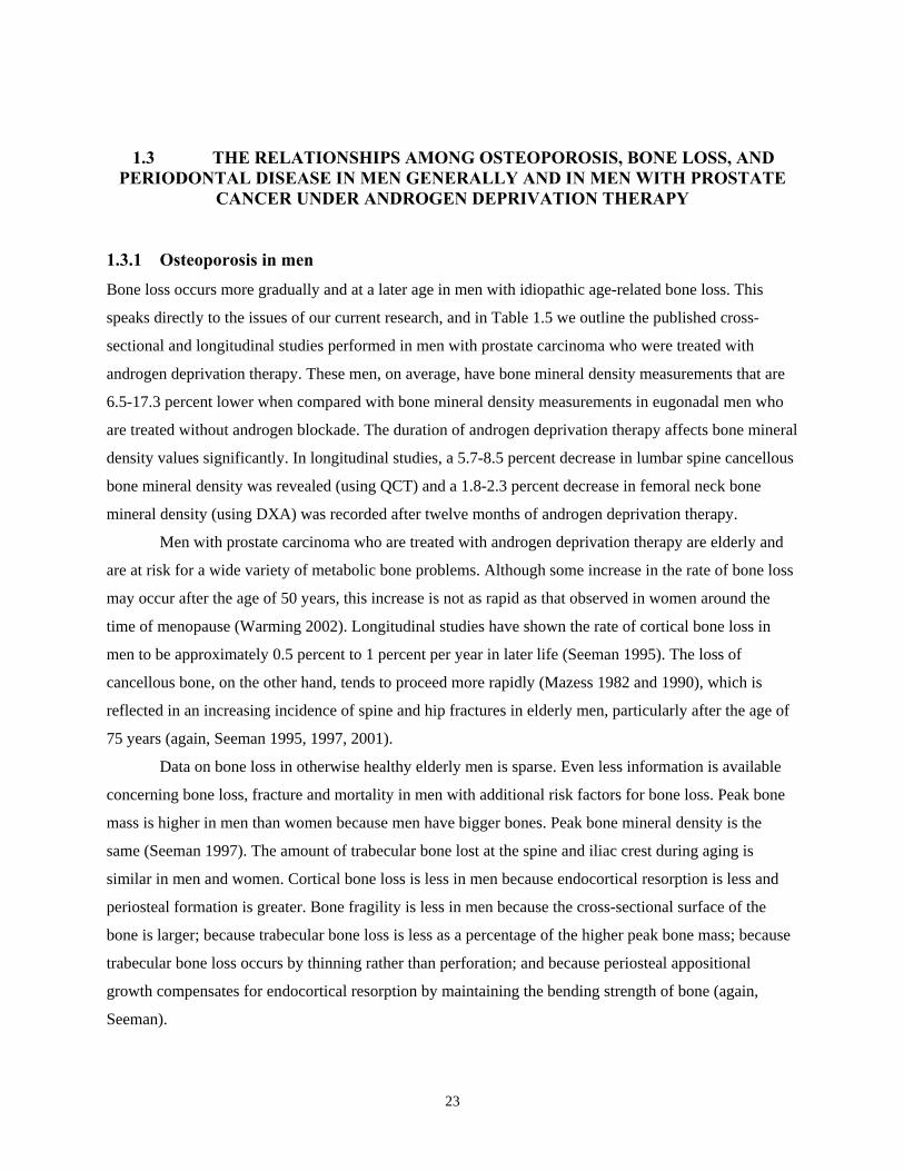
1.3 THE RELATIONSHIPS AMONG OSTEOPOROSIS, BONE LOSS, AND PERIODONTAL DISEASE IN MEN GENERALLY AND IN MEN WITH PROSTATE
CANCER UNDER ANDROGEN DEPRIVATION THERAPY 1.3.1 Osteoporosis in men Bone loss occurs more gradually and at a later age in men with idiopathic age-related bone loss. This
speaks directly to the issues of our current research, and in Table 1.5 we outline the published cross-
sectional and longitudinal studies performed in men with prostate carcinoma who were treated with
androgen deprivation therapy. These men, on average, have bone mineral density measurements that are
6.5-17.3 percent lower when compared with bone mineral density measurements in eugonadal men who
are treated without androgen blockade. The duration of androgen deprivation therapy affects bone mineral
density values significantly. In longitudinal studies, a 5.7-8.5 percent decrease in lumbar spine cancellous
bone mineral density was revealed (using QCT) and a 1.8-2.3 percent decrease in femoral neck bone
mineral density (using DXA) was recorded after twelve months of androgen deprivation therapy.
Men with prostate carcinoma who are treated with androgen deprivation therapy are elderly and
are at risk for a wide variety of metabolic bone problems. Although some increase in the rate of bone loss
may occur after the age of 50 years, this increase is not as rapid as that observed in women around the
time of menopause (Warming 2002). Longitudinal studies have shown the rate of cortical bone loss in
men to be approximately 0.5 percent to 1 percent per year in later life (Seeman 1995). The loss of
cancellous bone, on the other hand, tends to proceed more rapidly (Mazess 1982 and 1990), which is
reflected in an increasing incidence of spine and hip fractures in elderly men, particularly after the age of
75 years (again, Seeman 1995, 1997, 2001).
Data on bone loss in otherwise healthy elderly men is sparse. Even less information is available
concerning bone loss, fracture and mortality in men with additional risk factors for bone loss. Peak bone
mass is higher in men than women because men have bigger bones. Peak bone mineral density is the
same (Seeman 1997). The amount of trabecular bone lost at the spine and iliac crest during aging is
similar in men and women. Cortical bone loss is less in men because endocortical resorption is less and
periosteal formation is greater. Bone fragility is less in men because the cross-sectional surface of the
bone is larger; because trabecular bone loss is less as a percentage of the higher peak bone mass; because
trabecular bone loss occurs by thinning rather than perforation; and because periosteal appositional
growth compensates for endocortical resorption by maintaining the bending strength of bone (again,
Seeman).
23

[Seeman (1999) even is capable of presenting data regarding racial differences in the fragility of
bone, in addition to gender differences in fragility. Blacks have shorter vertebrae than whites, but
vertebral width is similar. Trabecular thickness is greater in blacks than whites. Men have wider long
bones than women. Blacks have wider long bones than whites. He summarizes the issue by saying that
racial differences in vertebral strength are likely to be bone mineral density-, not size-, dependent. Greater
periosteal expansion during growth in males than females, and in blacks than whites, establishes the
gender and racial differences in peak bone size.]
The higher mortality rate (one-third greater) associated with hip fracture in men (Seeman 1995)
compared with women may in fact be due to the frequent presence of underlying medical conditions or
risk factors, such as a slow decrease in serum-free testosterone, alcoholism, tobacco use, hypogonadism,
and disorders of calcium and Vitamin D absorption or metabolism. In one case control study,
approximately a third of the men studied had a significant underlying condition that could affect calcium
or bone metabolism (Seeman 1995, 2001; Deng 2004). Seeman (1997) sums up the risk factors for
fracture in men by citing the age-related decline in testosterone, adrenal androgens, growth hormone, and
insulin-like growth factor 1. Men with vertebral fractures often have hypogonadism or other illnesses with
what Seeman calls suspiciously few clinical features---alcoholism, myeloma, malabsorption, primary
hyperparathyroidism, haemochromatosis, and Cushing’s disease.
[In 2004, conceding that the single most important consideration in the treatment of osteoporosis
is the individual’s absolute risk of fracture, Seeman considered what was out there that worked in the
treatment of osteoporosis. Reported to reduce vertebral fractures are the bisphosphonates, raloxifene,
parathyroid hormone, and strontium ranelate. The bisphophanates and hormone replacement therapy have
been used to reduce fractures in one population of women, but hormone replacement is clearly not
recommended (the discussion above). Calcium plus Vitamin D and the mechanical device of hip
protectors reduced fractures in one study of the institutionalized elderly. Seeman saw little evidence to
support treatment with calcitonin, fluoride, anabolic steroids, or active Vitamin D metabolites, and could
only recommend lifestyle changes to maintain lifetime bone strength, not statistically reduce fracture
risk.]
1.3.2 Bone loss in men with prostate cancer There has been a gradual increase in the incidence of prostate cancer around the world since the 1960s.
Prostate cancer occurs in older men and, as previously mentioned, aging is associated with bone loss in
the range of 0.5 percent to 1 percent per year (Higano 2003, Mazess 1982). Decreased testosterone levels
and hypovitaminosis D also occur in this population (Simon 2002, Higano 2003). These factors may help
explain why osteopenia may be present in 10 percent to 25 percent of men with prostate cancer even
24

before androgen deprivation therapy is begun. While androgen deprivation therapy is an effective
measure to control advanced prostate cancer, the development of osteoporosis in patients treated with
androgen deprivation therapy has not aroused general attention until recent years (Wei et al. 1999;
Diamond 1998; Morote et al. 2003; Deng 2004). Ross and Small indicated very recently (2002) that
osteoporosis was an important and debilitating side effect of androgen deprivation therapy, but the precise
estimates of its incidence and importance have not yet been made fully clear.
1.3.3 Osteoporosis in men with prostate carcinoma under androgen suppression therapy *The literature regarding this subject is summarized as Table 1.5, below.
Prostate carcinoma is the most common visceral malignancy and the second leading cause of death from
cancer in men (Greenlee 2001). Androgen deprivation therapy is the recommended treatment for men
with metastatic or locally advanced, non-metastatic prostate carcinoma (Stoch et al. 2001). Although it
has been demonstrated that this form of therapy significantly reduces tumor growth and improves survival
beyond three years after completion of the androgen deprivation therapy, there is growing concern
regarding the negative effects of androgen deprivation therapy on the skeleton. Accelerated bone loss
(Higano 2003), osteoporosis, and a potential for increased fracture rates have been reported in men with
prostate carcinoma who are receiving androgen deprivation therapy. Because many patients who present
with prostate carcinoma are elderly and may have pre-existing osteoporosis, sub-clinical Vitamin D
deficiency, or any of a multitude of medical problems, the risk of skeletal deterioration is increased
(Simon 2002). At present the prevalence of osteoporosis in men under androgen suppression is not well
known, and neither is the impact of the time spent under this treatment.
25

Table 1.5 Percent change in lumbar spine and hip bone mineral density (BMD) in men with prostate carcinoma receiving androgen-deprivation therapy Percent change in BMD per year Study
Number of Patients
Treatment LS DXA LS QCT Hip DXA
Mallefert1999 12 LHRH agonist -4.6 _ -3.9 Daniell 2000 16 Orchiectomy/LHRH agonist _ _ -2.4 Higano1999 18 CAB -4.5 - -2.5 Diamond 1998 12 CAB _ -6.6 -6.5 Smith 2001 21 CAB -3.3 -8.5 -1.8 Diamond 2001 21 CAB _ -5.7 -2.3 Smith 2003 51 LHRH agonist/ CAB -2.2 _ -2.8 Mittan 2002 15 CAB -2.8 _ -3.3
Berruti 2002 42 LHRH -2.3 _ -0.5
Footnote: Table 1.5 outlines the published cross-sectional and longitudinal studies performed in men with prostate carcinoma who were treated with androgen deprivation therapy (ADT). On average, these men have BMD measurement that are 6.5-17.3% lower compared with BMD measurement in eugonadal men who are treated with androgen blockade. The duration of ADT affects BMD values significantly. In longitudinal studies, a 5.7-8.5% decrease in lumbar spine cancellous bone mineral density was shown (using QCT) and 1.8-2.3% decrease in femoral neck BMD.
26

1.4 THE RELATIONSHIP OF PERIODONTAL DISEASE IN MEN WITH PROSTATE CANCER UNDER ANDROGEN DEPRIVATION THERAPY. A
BRIEF SUMMARY PREFACE TO THE SUBSEQUENT CHAPTERS The relation of periodontal disease and osteoporosis due to androgen ablution is not well
established. Studies to evaluate the relationship between osteoporosis due to androgen deprivation
therapy and periodontal disease are essential for enhancing our understanding of systemic bone
pathogenesis. There is no literature on the association between periodontal disease and prostate
cancer under androgen deprivation therapy.
To address whether the two diseases are linked, we will analyze two data sets. In the first
analysis, we will test the hypothesis that older women with periodontal disease will experience
faster rates of bone loss (Chapter Two).
The second analysis will involve men undergoing androgen deprivation therapy for
prostate cancer. These men have been enrolled in Bone loss in men with prostate cancer treated
with androgen deprivation therapy (Susan L. Greenspan, MD, principal investigator), which is
testing the hypotheses (1) that androgen deprivation therapy in men with prostate cancer is
associated with a rapid increase in bone resorption and bone loss, as well as a change in body
composition; and (2) that androgen deprivation in men with advanced prostate cancer therapy is
associated with progressive loss of bone and lean body mass compared to normal controls; and
(3) that bone resorption, bone loss, and general bone health will be further altered by additional
systemic or local therapy for prostate cancer. As part of the current research, we will add a
periodontal assessment and will test the additional hypothesis (4) that men with prostate cancer
undergoing androgen deprivation therapy with have a higher prevalence of periodontal disease
(Chapter Three).
Finally, we will examine the correlation between measures of bone mineral density and
bone turnover to the existence of periodontal disease in this population of older men (again,
Chapter Three).
27

1.5 THE LITERATURE CITED IN THE FIRST CHAPTER
1. Ahmed AI. Blake GM. Rymer JM. Fogelman I. Screening for osteopenia and osteoporosis: do the accepted normal ranges lead to over-diagnosis? Osteoporosis International 7(5):432-8, 1997. 2. Akesson K. Principles of bone and joint disease control programs–osteoporosis. Journal of Rheumatology Supplement 67:21-5, August 2003. 3. Albandar JM. A 6-year study on the pattern of periodontal disease progression. Journal of Clinical Periodontology 17(7 Pt 1):467-471, August 1990. 4. Albandar JM. Ram TE. Global epidemiology of periodontal diseases: An overview. Periodontology 2000 29:7-10, 2002. 5. Albandar JM. Tinoco EM. Global epidemiology of periodontal diseases in children and young persons. Periodontology 2000 29:153-176, 2002. 6. Al-Zahrani MS. Bissada NF. Borawskir EA. Obesity and periodontal disease in young, middle-aged and older adults. Journal of Periodontology 74(5):610-615.2003. 7. Anonymous. Consensus Report. Periodontal diseases: Epidemiology and diagnosis. Consensus Development Conference. Annals of Periodontology 1(1):216-222, November 1996. 8. Anonymous. Periodontal disease: Epidemiology and diagnosis. Consensus Development Conference. Journal of the American Dental Association 129(Supplement):9S-14S, Sept. 1978. 9. Bassett MT. Perl S. Obesity: The Public Health Challenge of Our Time. American Journal of Public Health 94(9):1477, September 2004. 10. Bilezikian JP. Osteoporosis in men. Journal of Clinical Endocrinology and Metabolism 84(10):3431-3434, 1999. 11. Black DM. Cooper C. Epidemiology of fractures and assessment of fracture risk. Clinics in Laboratory Medicine 20(3):439-53, September 2000.
28

12. Caballero B. Introduction. Symposium: Obesity in developing countries: Biological and ecological factors. Journal of Nutrition 131(3):866S-870S, 2001.
13. Cauley JA. Murphy PA, et al. Effects of fluoridated drinking water on bone mass and fractures:
The study of osteoporotic fractures. Journal of Bone and Mineral Research 10(7):1076-1085, 1995.
14. Cauley JA. Robbins J. Chen Z. Cummings SR. Jackson RD. LaCroix AZ. Leboff M. Lewis CE.
McGiwan J. Neuner J. Pettinger M. Stefanick ML. Wactawski-Wende J. Witts NB. Women’s Health Initiative Investigators. Effect of estrogen plus progestin on risk fracture and bone mineral density: The Women’s Health Initiative Randomized Trial. Journal of the American Medical Association 290(13):1729-1738.2003.
15. Chestnut CH 3rd. The relationship between skeletal and oral bone mineral density: an overview.
Annals of Periodontology 6(1):193-196, December 2001.
16. Ciancio SG. Taking oral health to heart: an overview. Journal of the American Dental Association 133Suppl:4S-6S, June 2002.
17. Cianciola LJ. Park BH. Bruck E. Mosovich L. Genco RJ. Prevalence of periodontal disease in
insulin-dependent diabetes mellitus (juvenile diabetes). Journal of the American Dental Association 104(5):653-660, May 1982.
18. Consigny PM. Pathogenesis of atherosclerosis. AJR:American Journal of Roentgenology
164(3):553-558, 1995.
19. Cummings SR. Black DM. Rubin SM. Lifetime risk of hip, Colles’ or vertebral fracture and coronary heart disease among white postmenopausal women. Archives of Internal Medicine 149(11):2445-2448, 1989.
20. Cummings SR. Nevitt MC. Browner WS, et al. Risk factors for hip fracture in white women: The
Study of Osteoporotic Fractures Research Group. New England Journal of Medicine 332(12):767-773, 1995.
21. Cummings SR. Melton LJ. Epidemiology and outcomes of osteoporotic fractures. Lancet
359(9319):1761-1767, May 18, 2002.
22. DeLaet CE. Pols HA. Fractures in the elderly: Epidemiology and demography. Best Practice and Research Clinical Endocrinology and Metabolism 14(2):171-179, 2000.
23. Delmas PD. Bone mass measurement: how, where, when and why? International Journal of
Fertility and Menopausal Studies 38 Suppl 2:70-6, 1993.
24. Deng JH. Yang LP. Wang LS. Zhou DF. Effect of androgen deprivation therapy on bone mineral density in prostate cancer patients. Asian Journal of Andrology 6(1):75-7, March 2004.
25. Desvarieux M. Demmer RT. Rundek T. Boden-Albala B. Jacobs DR Jr. Papapanou PN. Sacco
RL. Oral Infections and Vascular Disease Epidemiology Study (INVEST). Relationship between periodontal disease, tooth loss, and carotid artery plaque: the Oral Infections and Vascular Disease Epidemiology Study (INVEST). Stroke 34(9):2120-5, September 2003.
29
therapy in representative elderly women: A randomized clinical trial. Journal of Bone and Mineral Research 13(9):1431-1438, 1998.
41. Grossi SG. Genco RJ. Machtei EE. Ho A. Koch G. Dunford R. Zambon JJ. Hausmann E.
Assessment of risk for periodontal disease. I. Risk indicators for alveolar bone loss. Journal of Periodontology 66(1):260-267, 1994.
42. Grossi SG. Genco RJ. Machtei EE. Ho A. Koch G. Dunford R. Zambon JJ. Hausmann E.
Assessment of risk for periodontal disease. II. Risk indicators for alveolar bone loss. Journal of Periodontology 66(1):23-29, 1995.
43. Grossi SG. Skrepcinski FB. DeCaro T. Zambon JJ. Cummins D. Genco RJ. Response to
periodontal therapy in diabetics and smokers. Journal of Periodontology 67(10 Suppl.):1094-1102, October 1996.
44. Grossi SG. Zambon JJ. Machtei EE. Schifferle R. Andreana S. Genco RJ. Cummins D. Harrap G.
Effects of smoking and smoking cessation on healing after mechanical periodontal therapy. Journal of the American Dental Association 128(5):599-607, May 1997.
45. Heyse SP. Epidemiology of hip fractures in the elderly: A cross-national analysis of mortality
rates for femoral neck fractures. Osteoporosis International 3Supplement1:16-19, 1993.
46. Higano CS. Management of bone loss in men with prostate cancer. Journal of Urology 170(6 Pt 2):S259-63; Discussion S64, 2003 December.
47. Hildebolt CF. Pilgram TK. Dotson M. Cohen SC. Hauser JF. Kardaris E. Civitelli R.
Relationships between clinical attachment level and spine and hip bone mineral density: data from healthy postmenopausal women [Erratum appears in Journal of Periodontology 75(5):780]. Journal of Periodontology 73(3):298-301, 2002 Mar.
48. Irfan UM, Dawson DV, Bissada NF. Epidemiology of periodontal disease: Review and clinical
perspectives. Journal of the International Academy of Periodontology 3(1):14-21, 2001 January.
49. Jacobs R. Ghyselen J. Koninckx P. van Steenberghe D. Long-term bone mass evaluation of mandible and lumbar spine in a group of women receiving hormone replacement therapy. European Journal of Oral Sciences 104(1):10-16, February 1996.
50. Jacobsen SJ. Goldberg J. Miles TP et al. Regional variation in the incidence of hip fracture: US
white women aged 65 years and older. Journal of the American Medical Association 264(4):500-502, 1990.
51. Jacqmin-Gadda H. Fourrier A. Commenges D. Dartigues JF. Risk fractures in the elderly.
Epidemiology 9(4):417-423, 1998.
52. Jaglal SB, Kreiger N, Darlington G. Past and recent physical activity and risk of hip fracture. American Journal of Epidemiology 138(2):107-118, 1993.
53. James PT. Leach R. Kalamara E. Shayeghi M. International Obesity Task Force, London, United
Kingdom. The worldwide obesity epidemic. Obesity Research 9Supplement4:228S-233S, November 2001.
30

54. James PT. Rigby N. Leach R. International Obesity Task Force, London, United Kingdom. The obesity epidemic, metabolic syndrome and future prevention strategies. European Journal of Cardiovascular Prevention and Rehabilitation 11(1):3-8, February 2004.
55. Jeffcoat MK. Osteoporosis: A possible modifying factor in oral bone loss. Annals of
Periodontology 3(1):312-321, July 1998.
56. Jeffcoat MK. Lewis CE. Reddy MS. Wang CY. Redford M. Post-menopausal bone loss and its relationship to oral bone loss. [Erratum appears in Periodontology 2000 26:169, 2001]. Periodontology 2000 23:94-102, June 2000.
57. Johansson I. Tidehag P. Lundberg V. Hallmans G. Dental status, diet and cardiovascular risk
factors in middle-aged people in northern Sweden. Community Dentistry and Oral Epidemiology 22(6):431-436, December 1994.
58. Johnson RB. Gilbert JA. Cooper.RC. Dai X. Newton BI. Tracy RR. West WF. DeMoss TL.
Meyers PJ. Streckfus CF. Alveolar bone loss one year following ovariectomy in sheep. Journal of Periodontology 68(9):864-871, 1997.
59. Kanis JA. Assessment of fracture risk and its application to screening for postmenopausal
osteoporosis: Synopsis of a WHO report. WHO Study Group. Osteoporosis International 4(6):368-81, November 1994.
60. Kanis JA. Diagnosis of osteoporosis and assessment of fracture risk. [See comment, Lancet
360(9343):1429, November 2002.] Lancet 359(9321):1929-1936, June 2002.
61. Kannus P. Niemi S. Parkkari J. Palvanen M. Vuori I. Jarvinen M. Hip fractures in Finland between 1970 and 1997 and predictions for the future. Lancet 353(9155):802-805, March 1999.
62. Kenny AM. Taxel P. Osteoporosis in older men. Clinical Cornerstone 2(6):45-51, 2000.
63. Kenny AM. Joseph C. Taxel P. Prestwood KM. Osteoporosis in older men and women.
Connecticut Medicine 67(8):481-486, September 2003.
64. Kinane DF. Periodontitis modified by systemic factors. Annals of Periodontology 4(1):54-64, December 1999.
65. Kinane DF. Periodontal diseases' contributions to cardiovascular disease: an overview of
potential mechanisms. Annals of Periodontology 3(1):142-50, July 1998.
66. Kribbs PJ. Comparison of mandibular bone in normal and osteoporotic women. Journal of Prosthetic Dentistry 63(2):218-22, February 1990.
67. Kribbs PJ. Smith DE. Chesnut CH 3rd. Oral finding in osteoporosis. Part II: Relationship between
residual ridge and alveolar bone resorption and generalized skeletal osteopenia. Journal of Prosthetic Dentistry 50(5):719-724, 1983.
68. Kribbs PJ. Chesnut CH 3rd. Ott SM. Kilcoyne RF. Relationships between mandibular and
skeletal bone in an osteoporotic population. Journal of Prosthetic Dentistry 62(6):703-7, December 1989.
31
69. Kribbs PJ. Chesnut CH 3rd. Ott SM. Kilcoyne RF. Relationships between mandibular and skeletal bone in a population of normal women. Journal of Prosthetic Dentistry 63(1):86-9, January 1990.
70. Kribbs PJ. Comparison of mandibular bone in normal and osteoporotic women. Journal of
Prosthetic Dentistry 63(2):218-22, February 1990.
71. Kuller LH. Orchard TJ. The epidemiology of atherosclerosis in 1987: unraveling a common-source epidemic. Clinical Chemistry 34(8B):B40-8, 1988.
72. Lalla E. Lamster IB, Stern DM. Schmidt AM. Receptor for advanced glycation end products,
inflammation, and accelerated periodontal disease in diabetes; mechanisms and insights into therapeutic modalities. Annals of Periodontology 6(1):113-118, 2001.
73. Levy SM. Warren JJ. Chowdhury J. DeBus B. Watkins CA. Cowen HJ. Kirchner HL. Hand JS.
The prevalence of periodontal disease measures in elderly adults, aged 79 and older. Special Care in Dentistry 23(2):50-57, 2003.
74. Lips P. Epidemiology and predictors of fractures associated with osteoporosis. American Journal
of Medicine 103(2A):3S-8S; discussion 8S-11S, August 1997.
75. Locker D. Slade GD. Murray H. Epidemiology of periodontal disease among older adults: Periodontology 2000 16:16-33, 1998.
76. Loe H. Anerud A. Boysen H. Morrison E. Natural history of periodontal disease in man. Rapid,
moderate and no loss of attachment in Sri Lankan laborers 14 to 46 years of age. Journal of Clinical Periodontology 13(5):431-445, 1986.
77. Looker AC. Orwoll ES. Johnston CC Jr. Lindsay RL. Wahner HW. Dunn WL. Calvo MS. Harris
TB. Heyse SP. Prevalence of low femoral bone density in older U.S. adults from NHANES III. Journal of Bone and Mineral Research 12(11):1761-8, November 1997.
78. Luckey MM. Wallenstein S. Lapinski R. Meier DE. A prospective study of bone loss in African-
American and white women--A clinical research center study. Journal of Clinical Endocrinology and Metabolism 81(8):2948-56, August 1996.
79. Lundstrom A. Jendle J. Stenstrom B. Toss G. Ravald N. Periodontal conditions in 70-year-old
women with osteoporosis. Swedish Dental Journal 25(3):89-96, 2001.
80. Mann T. Oviatt SL. Wilson D. Nelson D. Orwoll ES. Vertebral deformity in men. Journal of Bone and Mineral Research 7(11):1259-1265, 1992.
81. Mattson JS. Cerutis DR. Diabetes mellitus: a review of the literature and dental implications.
Compendium of Continuing Education in Dentistry 22(9):757-60, 762, 764 passim; quiz 773, September 2001.
82. Mattson JS. Cerutis DR. Parrish LC. Osteoporosis: a review and its dental implications.
Compendium of Continuing Education in Dentistry 23(11):1001-4, 1006, 1008 passim; quiz 1014, November 2002.
32

83. Mazess RB. On aging bone loss. Clinical Orthopedics and Related Research (165):239-252, May 1982.
84. Mazess RB. Barden HS. Drinka PJ. Bauwens SF. Orwoll ES. Bell NH. Influence of age and body
weight on spine and femur bone mineral density in U.S. white men. Journal of Bone and Mineral Research 5(6):645-652, June 1990.
85. Melton LJ III, Riggs BL. Epidemiology of age related fractures. In: The Osteoporotic Syndrome.
Edited L. V. Avioli. New York: Grune and Stratton, pp. 45-72, 1998.
86. Mokdad AH. Serdula MK. Dietz WH. Bowman BA. Marks JS. Koplan JP. The spread of the obesity epidemic in the United States, 1991-1998. Journal of the American Medical Association 282(16):1519-1522, October 1999.
87. Morote J. Martinez E. Trilla E. Esquena S. Abascal JM. Encabo G. Reventos J. Osteoporosis
during continuous androgen deprivation: Influence of the modality and length of treatment. European Urology 44(6):661-5, 2003.
88. Nares S. The genetic relationship to periodontal disease. Periodontology 2000 32:36-49, 2003.
89. Nelson RG. Shlossman M. Budding LM. Pettitt DJ. Saad MF. Genco RJ. Knowler WC.
Periodontal disease and NIDDM in Pima Indians. Diabetes Care 13(8):836-40, August 1990.
90. Nguyen TV. Center JR. Sambrook PN. Eisaman JA. Risk factors for proximal humerus, forearm, and wrist fractures in elderly men and women: the Dubbo Osteoporotic Epidemiology Study. American Journal of Epidemiology 153(6):587-595, 2001.
91. Nindl BC. Harman EA. Marx JO. Gotshalk LA. Frykman PN. Lammi E. Palmer C. Kraemer WJ.
Regional body composition changes in women after six months of periodized physical training. Journal of Applied Physiology 88(6):2251-9, June 2000.
92. Nishimura F. Murayama Y. Periodontal inflammation and insulin resistance---lessons from
obesity. Journal of Dental Research 80(8):1690-1694, August 2001.
93. Nishimura F. Iwamoto Y. Mineshiba J. Shimizu A. Soga Y. Murayama Y. Periodontal disease and diabetes mellitus: the role of tumor necrosis facor-alpha in a two-way relationship. Journal of Periodontology 74(1):97-102, January 2003.
94. Nunn ME: Understanding the etiology of periodontitis: An overview of periodontal risk factors.
Periodontology 2000 32:11-23, 2003.
95. Orwoll ES. Oviatt SK. McClung MR. Deftos LJ. Sexton G. The rate of bone mineral loss in normal men and the effects of calcium and cholecalciferol supplementation. Annals of Internal Medicine 112(1):29-34, January 1990.
96. Pacifici R. Estrogen, cytokines, and pathogenesis of postmenopausal osteoporosis. Journal of
Bone and Mineral Research 8(11):1043-1051, August 1996.
97. Patel A. Coates PS. Nelson JB. Trump DL. Resnick NM. Greenspan SL. Does bone mineral density and knowledge influence health-related behaviors of elderly men at risk for osteoporosis? Journal of Clinical Densitometry 6(4):323-30, 2003 Winter.
33

98. Paulander J. Axelsson P. Lindhe J. Association between level of education and oral health status
in 35-, 50-, 65-, and 75-year olds. Journal of Clinical Periodontology 30(8):697-704, August 2003.
99. Payne JB. Reinhardt RA. Nummikoski PV. Patil KD. Longitudinal alveolar bone loss in
postmenopausal osteoporotic/osteopenic women. Osteoporosis International 10(1):34-40, 1999.
100. Payne JB. Reinhardt RA. Nummikoski PV. Dunning DG. Patil KD. The association of cigarette smoking with alveolar bone loss in postmenopausal females. Journal of Clinical Periodontology 27(9):658-64, September 2000.
101. Persson L. Bergstrom J. Ito H. Gustafsson A. Tobacco smoking and neutrophil activity in patients
with periodontal disease. Journal of Periodontology 72(1):90-95, January 2001. 102. Pucher J. Stewart J. Periodontal disease and diabetes mellitus. Current Diabetes Reports 4(1):46-
50, February 2004. 103. Reddy MS. Oral osteoporosis: is there an association between periodontitis and osteoporosis?
Compendium of Continuing Education in Dentistry 23(10 Suppl):21-8, 2002. 104. Rees TD. A profile of the patient with periodontal disease? Periodontology 2000 32(1):9-13, June
2003. 105. Reinhardt RA. Payne JB. Maze CA. Patil KD. Gallagher SJ. Mattson JS. Influence of estrogen
and osteopenia/osteoporosis on clinical periodontitis in postmenopausal women. Journal of Periodontology 70(8):823-828, August 1999.
106. Rodrigo F. Neiva JS. Khalaf F. Al-Shammari. Wang H-L. Effect of specific nutrients on
periodontal disease onset. Progression and Treatment 30(7):579-589, 2003. 107. Ronderos M. Jacobs DR. Himes JH. Pihlstrom BL. Associations of periodontal disease with
femoral bone mineral density and estrogen replacement therapy: cross-sectional evaluation of US adults from NHANES III. Journal of Clinical Periodontology 27(10):778-786, 2000.
108. Ronderos M. Ryder MI. Risk assessment in clinical practice. Periodontology 2000 34:120-135,
2004. 109. Ross RW. Small EJ. Osteoporosis in men treated with androgen deprivation therapy for prostate
cancer. Journal of Urology 167(5):1952-6, 2002 May. 110. Saito T. Shimazaki Y. Sakamoto M. Obesity and periodontitis. New England Journal of
Medicine 339(7):482-483, 1998. 111. Sasaki T. Ramammurthy NS. Golub LM. Insulin-deficient diabetes impairs osteoblast and
periodontal ligament fibroblast metabolism but does not affect ameloblasts and odontoblasts: Response to tetracycline(s) administration. Journal de Biologie Buccale 18(3):215-226, September 1990.
112. Seeman E. Melton LJ III. O’Fallon WM. Riggs BL. Risk factors for spinal osteoporosis in men.
American Journal of Medicine 75(6):977-983, December 1983.
34

113. Seeman E. The dilemma of osteoporosis in men. American Journal of Medicine 98(2A):76S-88S,
February 1995. 114. Seeman E. Osteoporosis in men. Baillieres Clinical Rheumatology 11(3):613-29, 1997. 115. Seeman E. The structural basis of bone fragility in men. Bone 25(1):143-147, July 1999. 116. Seeman E. Unresolved issues in osteoporosis in men. Reviews in Endocrine and Metabolic
Disorders 2(1):45-64, January 2001. 117. Seeman E. Eisman JA. Treatment of osteoporosis: why, whom, when and how to treat. The single
most important consideration is the individual’s absolute risk of fracture. Medical Journal of Australia 180(6):298-303, March 2004.
118. Shlossman M. Knowler WC. Pettitt DJ. Genco RJ. Type II diabetes mellitus and periodontal
disease. Journal of the American Dental Association 121(4):532-536, Oct. 1990. 119. Shrout MK. Hildebolt CF. Potter BJ. Brunsden TK. Pilgram TK. Dotson M. Yokoyama-Crothers
N. Hauser J. Cohen S. Kardaris E. Civitelli R. Hanes P. Comparison of morphological measurements extracted from digitized dental radiographs with lumbar and femoral bone mineral density measurements in postmenopausal women. Journal of Periodontology 71(3):335-340, March 2000.
120. Simon J. Leboff M. Wright J. Glowacki J. Fractures in the elderly and Vitamin D. Journal of
Nutrition, Health and Aging 6(6):406-412 , 2002. 121. Slade GD. Ghezzi EM. Heiss G. Beck JD. Riche E. Offenbacher S. Relationship between
periodontal disease and C-reactive protein among adults in the Atherosclerosis Risk in Communities study. Archives of Internal Medicine 163(10):1172-9, 2003 May 26.
122. Slots J. Update of general health risk of periodontal disease. International Dental Journal
53(Supplement)3:200-207, 2003. 123. Sorsa T. Ingman T, Suomalainen K, et al. Cellular source and tetracycline inhibition of gingival
crevicular fluid collagenase of patients with labile diabetes mellitus. Journal of Clinical Periodontology 19(2):146-149, 1992.
124. Soskolne WA. Epidemiological and clinical aspects of periodontal diseases in diabetics. Annals of
Periodontology 3(1):3-12, July 1998. 125. Southard KA. Southard TE. Comparison of digitized radiographic alveolar features between 20-
and 70-year-old women. A preliminary study. Oral Surgery, Oral Medicine, Oral Pathology 74(1):111-7, July 1992.
126. Spalj S, Plancak D. The distribution of periodontal disease and loss of attachment in jaw sextants
in different age groups--cross sectional study. Collegium Antropologicum 27(Suppl) 1:183-190, 2003.
35
127. Stoch SA. Parker RA. Chen L. Bubley G. Ko Y-J. Vincelette A. Greenspan SL. Bone loss in men with prostate cancer treated with gonadotropin-releasing hormone agonists. Journal of Clinical Endocrinology and Metabolism 86(6):2787-2791, 2001.
128. Streckfus CF. Johnson RB. Nick T. Tsao A. Tucci M. Comparison of alveolar bone loss, alveolar
bone density and second metacarpal bone density, salivary and gingival crevicular fluid interleukin-6 concentrations in healthy premenopausal and postmenopausal women on estrogen therapy. Journals of Gerontology Series A-Biological Sciences and Medical Sciences 52(6):M343-51, November 1997.
129. Streckfus CF, Parsell DE, Streckfus JE, Pennington W, Johnson RB. Relationship between oral
alveolar bone loss and aging among African-American and Caucasian individuals. Gerontology 45(2):110-114, 1999.
130. Teng YT. The role of acquired immunity and periodontal disease progression. Critical Reviews in
Oral Biology and Medicine 14(4):237-52, 2003. 131. Tezal M. Wactawski-Wende J. Grossi SG. Ho A. Dunford R. The relationship between bone
mineral density and periodontitis in postmenopausal women. Journal of Periodontology 71(9):1492-1498, 2000.
132. Tezal M. Grossi SG, Ho AW, Genco RJ. The effect of alcohol consumption on periodontal
disease. Journal of Periodontology 72(2):183-189, 2001. 133. Tobin JD. Fox KM. Cejku ML. Bone density changes in normal men: A 4-19 year longitudinal
study. Journal of Bone and Mineral Research Supplement 8(3):142, 1993. 134. [Tobin JD]. Hochberg MC. Lethbridge-Cejku ML. Bone mineral density and osteoarthritis: data
from the Baltimore Longitudinal Study of Aging. Osteoarthritis and Cartilage 12 Suppl A:S45-8, 2004.
135. van Winkellhoff AJ. Bosch-Tijhof CJ. Winkel EG. van der Reijden WA. Smoking affects the
subgingival microflora in periodontitis. Journal of Periodontology 72(5):666-671, May 2001. 136. von Wowern N. Klausen B. Kollerup G. Osteoporosis: A risk factor in periodontal disease.
Journal of Periodontology 65(12):1134-1138, 1994. 137. Wactawski-Wende J. Grossi SG. Trevisan M. Genco RJ, et al. The role of osteopenia in oral bone
loss and periodontal disease. Journal of Periodontology 67(10 Supplement):1076-1084, 1996. 138. Warming L. Hassager C. Christiansen C. Changes in bone mineral density with age in men and
women: a longitudinal study. Osteoporosis International 13(2):105-12, 2002. 139. Wei JT. Gross M. Jaffee CA. Gravlin K. Lahaie M. Faerber GJ. Cooney KA. Androgen
deprivation therapy for prostate cancer results in significant loss of bone density. Urology 54(4):607-11, 1999.
140. Werning JW. Downey NM. Brinker RA. Khuder SA. Davis WJ. Rubin AM. Elsamaloty HM. The
impact of osteoporosis on patients with maxillofacial trauma. Archives of Otolaryngology–Head and Neck Surgery 130(3):353-356, March 2004.
36

141. Weyant RJ. Pearlstein ME. Churak AP. Forrest K. Famili P. Cauley JA. The association between osteopenia and periodontal attachment loss in older women. Journal of Periodontology 70(9):982-991, 1999.
142. Willershausen-Zonnchen B. Lemmen C. Hamm G. Influence of high glucose concentrations on
glycosaminoglycan and collagen synthesis in cultured human gingival fibroblasts. Journal of Clinical Periodontology 18(3):190-195, 1991.
143. Zambon JJ. Grossi SG. Machtei EE. Ho AW. Dunford R. Genco RJ. Cigarette smoking increases
the risk for subgingival infection with periodontal pathogens. Journal of Periodontology 67(10 Suppl):1050-4, October 1996.
37
2.0 CHAPTER TWO
LONGITUDINAL STUDY OF PERIODONTAL DISEASE AND EDENTULISM WITH RATES OF BONE LOSS IN OLDER WOMEN
By Pouran Famili, Jane A. Cauley, Jon B. Suzuki, Robert J. Weyant
Published in the Journal of Periodontology 76:11-15, January 2005.
Pouran Famili, D.M.D, M.D.S., M.P.H., Ph.D. Professor and Chairman, Department of Periodontics School of Dental Medicine, University of Pittsburgh
Jane A. Cauley, Ph.D. Associate Professor
Department of Epidemiology Graduate School of Public Health
University of Pittsburgh
Jon B. Suzuki, D.D.S., Ph.D., M.B.A. Professor, Department of Periodontics
Associate Dean for Graduate Education, Research, and International Affairs Temple University School of Dentistry, Philadelphia
Robert J. Weyant, M.S., D.M.D., Dr.PH
Professor and Chair Department of Dental Public Health
University of Pittsburgh School of Dental Medicine 38

2.1 Abstract of the research Purpose: Previous cross-sectional studies have suggested a link between periodontal disease and
osteoporosis. The purpose of the present study is to evaluate the association between changes in bone
mineral density (BMD) and clinical signs of periodontal tissue destruction and tooth loss over a two-year
period.
Methods: A total of 398 women (mean age 75.5 years) were randomly selected for an ancillary study of
periodontal disease. We evaluated osteoporosis in association with the presence or absence of teeth, and
further evaluated osteoporosis in association with periodontal disease. All subjects were participants at the
Pittsburgh Clinical Center for the Study of Osteoporotic Fractures (SOF), a prospective cohort study of
women 65 years+ designed to determine risk factors for fractures. Oral health examinations, including
periodontal probing and attachment loss, were performed at the fourth clinical visit, on average six years
after baseline. BMD of the total hip and its sub-regions were measured using dual energy X-ray
absorptiometry at the time of dental examination, and two years later. Results are expressed as annual
percentage change.
Data analysis: Generalized linear models were used to assess the association between BMD and the
presence or absence of teeth and periodontal health status. Periodontal variables among dentate women
included average loss of periodontal attachment (LOA); the number of sites with at least four millimeters
(4mm) of attachment loss; and the presence or absence of calculus.
Results: A total of 145 (36.4 percent) women were edentulous and 163 (80.7 percent) had periodontal
disease. Dentate women reported higher education (p<0.001), and a higher calcium intake (p=0.002).
Absolute BMD and percentage change in BMD were similar in dentate and edentulous women. We found
no difference in BMD, or in absolute or percentage change in BMD between women with or without
periodontal disease.
Conclusion: Little evidence exists for an association between edentulousness, periodontal disease, and
longitudinal changes in BMD.
39

2.2 Background and rationale for the research Osteoporosis is a systemic skeletal disease characterized by low bone mass and micro-architectural
deterioration of bone tissue, with a consequent increase in bone fragility and susceptibility to fracture.
Osteoporosis is a major public health problem that most often affects postmenopausal women.
In the United States osteoporosis affects more than 25 million people, and predisposes patients to
more than 1.3 million fractures annually (Annals of Periodontology Consensus Report 1996). Since
alveolar bone loss is a clinical feature of periodontal disease, disturbances in bone metabolism and
decreases in the bone mineral density (BMD) of the skeleton, especially in the jaws, may be a factor in
periodontal disease (Wactawski-Wende et al. 1996). Osteoporosis and periodontal disease lead to
significant morbidity, mortality, and expense.
The clinical importance of generalized bone loss as a contributor to alveolar bone loss and
subsequent tooth loss is unclear. To date, the evidence for an association between tooth loss and bone
mineral density (BMD) in the extra-cranial skeleton has been derived from cross-sectional studies, and
results have been inconclusive (Tezal 2000). Several large studies conducted in early postmenopausal
women failed to find significant associations between tooth status and BMD. Elders (1992) found no
relationship between the number of missing teeth and either spine BMD or metacarpal thickness. Krall et
al. (1996) conducted a longitudinal study on associations between tooth loss and bone loss in whole
bodies, in the femoral neck, and in the spine, including 189 healthy white dentate postmenopausal women
who participated in three intervention trials conducted within a seven-year period. Forty-five women (24
percent) reported having lost one or more teeth. The rates of BMD changes at all three sites (whole body,
femoral neck, spine) were independent predictors of tooth loss in the multivariate models, supporting a
role for systemic bone loss in the development of tooth loss among postmenopausal women.
Bando et al. (1998) suggested that sufficient masticatory function with periodontally healthy
dentition may inhibit or delay the progress of osteoporotic changes in skeletal bone, or that edentulous
women may be more susceptible to osteoporosis. The role of osteoporosis or osteopenia in the etiology of
periodontal disease is not fully understood. Observational studies support the possible role of low skeletal
BMD or osteoporosis as risk indicators for reduced alveolar crestal height (Wactawski-Wende et al.
1996). Ronderos et al. (2000) studied the possible association of periodontal disease with femoral bone
mineral density (BMD) in a large sample of U.S. adults (n=11,655). Their finding indicates that, in the
presence of high calculus scores, females with osteoporosis are at an increased risk for attachment loss.
In our earlier report (Weyant et al. 1999), we found no association between BMD and periodontal
disease. However, no longitudinal studies have been reported to date. One intent of the present study was
to extend the earlier report (Weyant et al. 1999) and evaluate the link between changes in bone mineral
40

density and clinical signs of periodontal tissue destruction. In addition, we examined the association of
between edentulism and changes in BMD.
2.3 The design/setting/patients and the materials and methods of the research A total of 398 women (mean age 75.5 years) were randomly selected for an ancillary study of periodontal
disease, presence or absence of teeth, and osteoporosis. All subjects participated in the Pittsburgh Clinical
Center for the Study of Osteoporotic Fractures (SOF), a prospective study of a cohort of elderly women
>65 years of age at baseline, to determine risk factors for fractures.
From September 1986 to October 1988, a total of 2,401 women who were at least 65 years of age
or older were recruited for the SOF from the Monongahela Valley near Pittsburgh, Pennsylvania. Age-
eligible women were identified on voter registration lists for zip codes within a 25-mile radius of
Monessen, Pennsylvania. To be eligible to participate in this ancillary study, women had to be willing to
continue participation in the SOF study, willing to have a dental examination, and willing to voluntarily
sign a University of Pittsburgh Institutional Review Board (IRB)-approved consent form. Exclusion
criteria for this study included presence of medical conditions that would preclude conducting an
adequate oral examination. All women presenting for SOF examinations when the dentist examiner was
present and who met the above eligibility criteria were eligible for enrollment into the dental study.
BMD measurement Bone mineral density (g/cm) of the total hip and its sub regions (the femoral neck, the inter-trochanteric
region, the trochanter) was measured using dual X-ray absorptiometry with Hologic QDR-1000 scanners
(Hologic Inc., Waltham, MA). Bone mineral density of the calcaneus was measured with Osteo Analyzers
(Siemens-Osteon, Wahiawa, HI) using single- photon absorptiometry at the baseline examination. Details
of these measurement methods and densiotometry quality control procedures have been published
elsewhere (Ensrud 1995).
Periodontal assessment The assessment for periodontal destruction was made at three sites on the buccal aspect (mesiobuccal,
midbuccal, distobuccal) of all remaining natural teeth (Weyant 1999). Only fully erupted teeth were
measured. Loss of attachment was defined both clinically and quantitatively as the distance in millimeters
(mm) from the cementoenamel junction (CEJ) to the base of the pocket. Probing depth was the distance
from the free gingival margin (FGM) to the base of the sulcus/pocket. A woman was diagnosed with
periodontal disease if she had more than three millimeters (>3mm) attachment loss (Weyant et al. 1999).
Statistical analysis All data were screened for accuracy and completeness. SAS 8.2e™ software was used to analyze the data.
Descriptive statistics were calculated for all variables including means, standard deviations, ranges and
41

percentages. Linear regressions were used to compare the rate of bone loss in edentulous vs. dentate
women. Among dentate women, women with attachment loss >4 mm vs. women with <4 mm of
attachment loss on more than twelve teeth were also compared. We initially adjusted for age. Multivariate
adjusted models included variables known to influence bone density and fracture outcomes (age, weight,
education, dietary calcium intake, alcohol use, walking for exercise).
2.4 The results of the research, as described for this chapter Dentate vs. edentulous status of 398 women in the study was examined. Most (253) or 64 percent, were
dentate, and 145 (36 percent) were edentulous. Dentate women were more likely to report a higher
education, greater calcium intake, drinking alcohol and a dental examination in the last two-years
compared with edentulous women. There were no differences in age, body weight, smoking, hormone
use, and physical activity between the two groups. Similar proportions of dentate and edentulous women
reported a history of fracture or osteoporosis.
For the most part there was no difference in age-adjusted BMD, absolute rates of bone loss, or
percentage rate of bone loss between dentate and edentulous women for tooth loss at other sites. Further
adjustments for age, weight, education, dietary calcium intake, drinking alcohol, and walking for exercise
had little effect on these results. The rate of bone loss at the trochanter was somewhat greater among
edentulous women (0.86 percent per year) compared to dentate women (-0.52 percent per year).
Periodontal disease
The majority (80 percent) of dentate women had some evidence of periodontal disease (Table 1.4). Women
with periodontal disease were slightly older than women without periodontal disease (p=0.07), but all
other characteristics were remarkably similar in the two groups. We found no difference in BMD, or in
absolute or percentage change in BMD between women with and without periodontal disease (Table 1.4).
We also correlated several other measures of periodontal disease with BMD, including average
periodontal attachment loss, number of sites with > 6 mm attachment loss, site with > 4 mm attachment
loss, average number of sites with bleeding, calculus and probing depth. In general there was no
association between periodontal disease and rate of bone loss.
2.5 A discussion of the research, as described for this chapter
The overall prevalence of edentulism in this population of older women was higher (36 percent)
compared with the 23 percent edentulism rate reported for the U.S. population of women ages 70 to 74
years as published in the NHANES III study. The higher prevalence observed in our study could have
reflected a continued increase in edentulism with advancing age. The women in our study ranged from 73 to 84
years in age. We found little evidence to support an association between osteoporosis and edentulism.
42

BMD and rate of bone loss were similar in these two groups even after adjustments for established
correlating factor of edentulism.
Tooth loss has been observed to be related to bone density in the oral cavity in a limited number
of studies. When interpreting results in various anatomic regions, it is necessary to remember that tooth
loss is highly influenced not only by periodontal disease, but also by the local practices of the dental
community, as well as other factors related to oral health.
The majority (80 percent) of dentate women had some evidence of periodontal disease (p=0.07).
We found no statistically significant associations between periodontal disease variables (average
periodontal attachment loss, average number of sites with bleeding, and average number of sites with
calculus) and BMD at trochanter, inter-trochanter and femoral neck either cross-sectionally or
longitudinally. This was true even after controlling variables for age, weight, and other important
correlating factors. A possible explanation of our lack of an association between periodontal disease and
BMD may be because our population was chronologically older, and were missing among them a large
number of teeth. We have no information on the individual reasons for missing teeth. This edentulousness
could be due to periodontal disease that occurred much earlier in the individual’s life.
Historical events such as tooth loss can alter the interpretation of the current status of their
periodontal disease. Loss of several periodontally involved teeth can result in an improvement of
periodontal summary scores such as average loss of attachment. Compared to the other NHANES III
studies (Winn 1999; Slade 1999), average attachment loss for the U.S. population was reported to be 2.1
mm, while our data for our population showed an average attachment loss\ of 2.49 mm. The major
strengths of this current study are its large sample size and well-controlled measurement of variables,
which could confound relationship with BMD. The sample is drawn randomly from a large population of
community-dwelling women.
Limitations Interpretations of the present study may be limited, since periodontal measures of bone such as
subtraction radiography or Shei bone score measurement were not available. In addition, blinded dental
examiners were only able to record a single assessment of periodontal measurements.
Conclusion There was little evidence of an association between edentulousness, clinical and periodontal disease, and
longitudinal changes in BMD in this population of older women. Future studies may be needed to
longitudinally correlate periodontal disease and BMD over the same period.
43

Table 2.1. Characteristics of postmenopausal women with teeth and without teeth (N=398) Values are mean (SD), number (%), or median (range)
$ = Analysis was performed log transformed variable Values are mean (SD), number (%).
Dentate (n= 253)
Edentulus (n= 145)
p-value
Age (yrs) 75.9 (4.12) 78.8 (4.40) 0.741 Height (cm) 158.0 (5.45) 157.3 (6.01) 0.239 Weight (kg) 68.5 (12.69) 67.85 (11.82) 0.587 BMI (kg/m2) 27.4 (4.80) 27.4 (4.55) 0.935 Age at menopause (yrs) 47.2 (6.5) 46.8 (5.6) 0.523 Smoking (n, %) Past Current
75 (29.64) 11 (4.35)
47 (32.64) 7 (4.86)
0.503
Education (n, %) > 12 years > 16 years
145 (57.31) 91 (35.97)
103 (71.03) 12 (8.28)
<0.001
Dietary Calcium Intake (mg/day)$ 500.82 (1.81) 411.13 (1.93) 0.002 Calcium supplement use, n (%) 103 (40.71) 49 (33.79) 0.172 Alcohol Drinking, n (%) 97 (38.49) 43 (29.66) 0.076 Walking for exercise, n (%) 111 (43.9) 46 (31.7) 0.017 Diabetes, n (%) 17 (6.72) 8 (5.52) 0.634 Osteoporosis, n (%) 25 (10.00) 12 (8.39) 0.599 History of Fracture since 50 years, n (%)
114 (45.1) 67 (46.2) 0.825
Maternal history of fracture, n (%) 53 (25.85) 23 (21.30) 0.371 Thiozide use (n, %) 14 (17.00) 22 (15.17) 0.636 Estrogen use (n, %) Past Current
46 (18.47) 18 (7.23)
24 (16.78) 8 (5.59)
0.423
Glucocorticoid use (n, %) 20 (7.91) 13 (8.97) 0.712 Dental examination (n, %) Less than 6 months One year ago Two years ago > 2 years ago
177 (69.86) 44 (17.39) 12 (4.74) 20 (7.91)
12 (8.28) 18 (12.41) 23 (15.86) 92 (63.45)
<0.001
44

Table 2.2 Age- adjusted BMD and annualized per cent change in BMD by edentulous status Dentate
(n=253) Edentulous (n=145)
p-value
BMD (g/cm2)
Total Hip
Femoral Neck
Intertrochanter
Trochanter
0.74 (0.01)
0.63 (0.01)
0.87 (0.01)
0.57 (0.01)
0.73 (0.01)
0.63 (0.01)
0.86 (0.01)
0.56 (0.01)
0.528
0.729
0.661
0.401
Absolute rate of change in BMD (mg/cm2,yr)
Total Hip
Femoral Neck
Intertrochanter
Trochanter
-4.31 (0.55)
-3.18 (0.57)
-4.92 (0.71)
-2.96 (0.54)
-5.07 (0.73)
-3.03 (0.75)
-5.88 (0.94)
-4.60 (0.71)
0.408
0.871
0.415
0.068
Per cent rate of change in BMD (%/yr)
Total Hip
Femoral Neck
Intertrochanter
Trochanter
-0.60 (0.08)
-0.50 (0.09)
-0.59 (0.08)
-0.52 (0.10)
-0.72 (0.10)
-0.49 (0.12)
-0.72 (0.11)
-0.86 (0.13)
0.343
0.958
0.325
0.045
45

Table 2.3 Characteristics of women with and without periodontal disease (n=202)
Periodontal Disease ( > 4 mm) Yes
(n= 163) No (n= 39)
p-value
Age (yrs) 76.39 (4.25) 75.08 (3.67) 0.078 Height (cm) 157.65 (5.11) 158.43 (5.88) 0.407 Weight (kg) 68.01 (12.40) 69.33 (9.83) 0.539 BMI (kg/m2) 27.37 (4.87) 27.71 (4.19) 0.690 Age at menopause (yrs) 47.83(6.03) 46.02 (7.99) 0.118 Current Smoking (n, %) 9 (5.52) 0 (0.0) 0.211 Education (n, %) > 12 years > 16 years
97 (59.5) 54 (33.1)
21 (53.9) 16 (41.0)
0.330
Dietary Calcium Intake (mg/day) 484.91(1.81) 498.66(1.78) 0.796
Alcohol Drinking, n (%) 61 (37.65) 14 (35.90) 0.839 Walking for exercise, n (%) 71 (43.6) 15 (38.5) 0.563 Diabetes (n, %) 9 (5.52) 1 (2.56) 0.691 Osteoporosis (n, %) 15 (9.38) 5 (12.82) 0.521 History of Fracture since 50 (n, %) 70 (42.9) 19 (48.7) 0.514 Maternal history of fracture 32 (24.81) 14 (41.18) 0.059 NSAID use (n, %) 38 (23.31) 9 (23.08) 0.975 Current HRT use (n, %) 13 (7.98) 0 (0.0) 0.077 Glucocorticoid use (n, %) 13 (7.98) 3 (7.69) 1.000 Dental examination (n, %) Less than 6 months
One year ago Two year ago > 2 yrs ago
119 (73.0) 30 (18.4) 6 (3.7) 8 (4.9)
31 (79.5) 4 (10.3) 1 (2.6) 8 (4.9)
0.564
46

Table 2.4 Age adjusted BMD and annualized percent change in BMD by periodontal status (N=202) (comments, n=51 not availed) Periodontal Disease
Yes (n=163) No (n=39)
P-value
BMD (g/cm2) Total Hip Femoral Neck Intertrochanter Trochanter
0.74 (0.01) 0.63 (0.01) 0.87 (0.01) 0.56 (0.01)
0.74 (0.02) 0.63 (0.02) 0.87 (0.02) 0.57 (0.02)
0.991 0.839 0.928 0.931
Absolute rate of change in BMD (g/cm2,yr) Total Hip Femoral Neck Intertrochanter Trochanter
-4.17 (0.70) -2.76 (0.70) -4.48 (0.91) -2.76 (0.68)
-3.94 (1.44) -3.08 (1.43) -4.50 (1.87) -3.45 (1.40)
0.888 0.844 0.994 0.657
Percentage rate of change in BMD (%/yr) Total Hip Femoral Neck Intertrochanter Trochanter
-0.54 (0.10) -0.42 (0.11) -0.53 (0.11) -0.47 (0.12)
-0.59 (0.20) -0.50 (0.23) -0.65 (0.25) -0.58 (0.22)
0.906 0.756 0.837 0.504
47

2.6 The literature cited in the chapter summary of the research
1. Anonymous. Consensus report. Periodontal diseases: epidemiology and diagnosis. Annals of Periodontology 1(1):216-22, 1996 November.
2. Bando K. Nitta H. Matsubara M. Ishikawa I. Bone mineral density in periodontally healthy and edentulous
postmenopausal women. Annals of Periodontology 3(1):322-6, 1998 July. 3. Elders PJ. Habets LL. Netelenbos JC. van der Linden LW. van der Stelt PF. The relation between periodontitis
and systemic bone mass in women between 46 and 55 years of age. Journal of Clinical Periodontology 19(7):492-6, 1992 August.
4. Ensrud KE. Palermo L. Black DM. Cauley J. Jergas M. Orwoll ES. Nevitt MC, Fox KM. Cummings SR. Hip
and calcaneal bone loss increase with advancing age: longitudinal results from the study of osteoporotic fractures. Journal of Bone & Mineral Research 10(11):1778-1787, 1995 November.
5. Krall EA. Garcia RI. Dawson-Hughes B. Increased risk of tooth loss is related to bone loss at the whole body,
hip, and spine. Calcified Tissue International 59:433-437, 1996 December. 6. Ronderos M. Jacobs DR. Himes JH. Pihlstrom BL. Associations of periodontal disease with femoral bone
mineral density and estrogen replacement therapy: cross-sectional evaluation of US adults from NHANES III. Journal of Clinical Periodontology 27(10):778-786, 2000 October.
7. Slade GD. Beck JD. Plausibility of periodontal disease estimates from NHANES III. Journal of Public Health
Dentistry. 59(2):67-72, 1999 Spring. 8. Tezal M, Wactawski-Wende J, Grossi SG, Ho AL, Dunford R. The relationship between bone mineral density
and periodontitis in postmenopausal women. Journal of Periodontology 71:1492-1498, 2000. 9. Wactawski-Wende J. Grossi SG. Trevisan M. Genco RJ. Tezal M. Dunford RG. Ho AW. Hausmann E.
Hreshchyshyn MM. The role of osteopenia in oral bone loss and periodontal disease. Journal of Periodontology 67(10 Suppl):1076-84, 1996 October.
10. Weyant RJ. Pealstein ME. Churak AP. Forrest K. Famili P. Cauley JA. The association between osteopenia and
periodontal attachment loss in older women. Journal of Periodontology 70(9):982-991, 1999 September. 11. Winn DM. Johnson CL. Kingman A. Periodontal disease estimates in NHANES III: clinical measurement and
complex sample design issues. Journal of Public Health Dentistry 59(2):73-8, 1999 Spring.
48

3.0 CHAPTER THREE
THE EFFECT OF ANDROGEN DEPRIVATION THERAPY ON PERIODONTAL DISEASE IN MEN, WITH A CONSIDERATION OF THE RELATIONSHIPS BETWEEN PERIODONTAL DISEASE AND BONE LOSS ON MEN WITH
PROSTATE CANCER
By Pouran Famili, Jane A. Cauley, J.P. Costantino, J.M. Zmuda, Jon B. Suzuki, Susan L. Greenspan
Pouran Famili, D.M.D, M.D.S., M.P.H., Ph.D.
Professor and Chairman, Department of Periodontics School of Dental Medicine, University of Pittsburgh
Jane A. Cauley, Ph.D. Associate Professor
Department of Epidemiology Graduate School of Public Health
University of Pittsburgh
Joseph P. Costantino, Dr. P.H. Professor, Department of Biostatistics Director, NSABP Biostatistical Center
Graduate School of Public Health University of Pittsburgh
Joseph M. Zmuda, PhD.
Assistant Professor of Epidemiology and Human Genetics Department of Epidemiology
Graduate School of Public Health University of Pittsburgh
Jon B. Suzuki, D.D.S., Ph.D., M.B.A. Professor, Department of Periodontics
Associate Dean for Graduate Education, Research, and International Affairs Temple University School of Dentistry, Philadelphia
Susan L. Greenspan, M.D.
Professor of Medicine and Director, Osteoporosis Prevention and Treatment Center Division of Endocrinology and Metabolism
School of Medicine, University of Pittsburgh
49
3.1 Abstract of the research
Purpose: To test the hypothesis that men undergoing androgen deprivation (ADT) as treatment for
prostate cancer are at greater risk of periodontitis and tooth loss.
Materials and Methods: Eighty-one (81) men with prostate cancer (mean age 68.5 years) were
recruited among 325 enrolled in an osteoporosis study. Of these, 68 men were eligible to participate in
this study. The prevalence of periodontal disease among men with prostate cancer (n=41) undergoing
androgen deprivation (mean years treated=1.5) was compared to men with prostate cancer not undergoing
androgen deprivation (n=27, the controls). The prevalence of periodontal disease was examined in
relationship to bone mineral density (BMD) among men with prostate cancer both with and without ADT.
Outcome Measures: A periodontist blinded to ADT status recorded probing depth; periodontal
attachment loss; bleeding; Plaque Index; gingival recession; missing teeth; and calculus. Logistic
regression models tested the association between ADT and periodontal disease. Linear regression
models assessed the association between periodontal disease and bone mineral density among both groups
with prostate cancer (treated/untreated). We adjusted for variables known to influence periodontal disease
including age, race, smoking, history of periodontal disease.
Results: The prevalence of periodontal disease was 80.5% (n=33) among men on ADT compared
with 3.7% (n=1) among men not on ADT, odds ratio (95%confidence interval) =3.33(1.07-10.04). Men
on ADT had significantly greater probing depth (p<0.001) and a higher plaque index (p<0.09). Eighty-
one men (76.9 %) undergoing ADT and the controls completed bone density exams. There was no
relationship between bone mineral density and periodontal disease.
Conclusions: Men with prostate cancer undergoing ADT were more likely to have periodontal disease
than men not on ADT. If confirmed in larger studies, this observation could have important public health
implications given the increasing use of androgen deprivation therapy to treat prostate cancer.
50

3.2 Background and rationale for the research Carcinoma of the prostate is the most frequently diagnosed malignancy and the second leading cause of
cancer death among men in the western world. The peripheral blockage of androgen action remains a
critical therapeutic option in the treatment of prostate cancer. However, androgen deprivation therapy
(ADT), the most effective systemic therapy for prostate cancer, is a major risk factor for male
osteoporosis (Ross and Small 2002; Wei 1999; Diamond 1998; Morote 2003; Deng 2004). Androgen
deprivation therapy causes increased osteoclastic activity (Higano 1983), bone resorption (Seeman 1997),
and bone loss (Seeman 2004). Rates of bone loss ranged from 2 percent to 8 percent in the lumbar spine
and from 1.8 percent to 6.5 percent in the femoral neck during the initial twelve months of continuous
androgen deprivation therapy (Wei 1999; Higano 2003). An increased risk of fracture in men with
prostate carcinoma who were treated with androgen deprivation therapy was also observed (Seeman
1983).
Periodontal disease results in resorption of alveolar bone and loss of the soft tissue attachment to
the tooth. There is biological plausibility that periodontal destruction may be adversely influenced by
systemic bone loss (Rees 2003; Albandar 1990 and 2002; Slots 2003; Nunn 2003; Genco 1996). None of
this previous research, however, has examined the link between androgen deprivation therapy with
periodontal disease. The incidence of periodontal disease increases with advancing age, and current
demographic shifts in the age distribution will lead to increases in the number of people with periodontal
disease.
The purpose of the present study was to compare the prevalence of periodontal disease in men
with prostate cancer receiving androgen deprivation therapy, with a control group of men with prostate
cancer who were not receiving androgen deprivation therapy. In addition we examined the relationship
between periodontal disease and measures of bone mineral density in these men.
3.3 The design/setting/patients and the materials and methods of the research A total of 81 men (mean age 68.5 years) agreed to participate in this periodontal disease study. All men
were recruited from a larger group of 325 ambulatory men participating in a study of bone loss in men
with non-metastatic prostate cancer being treated with androgen deprivation therapy (ADT) or men not
receiving androgen deprivation therapy.
To be eligible for enrollment in the current ancillary research, the men had to be willing to submit
to a comprehensive periodontal dental examination, and be willing to sign a University of Pittsburgh
51
Institutional Review Board (IRB)-approved consent form to participate in the current research. Exclusion
criteria for this study were (1) the presence of medical conditions that preclude conducting an adequate
oral examination, such as a history of heart disease or joint replacement or any medical disease that
requires pre-medication with an antibiotic before a periodontal exam; (2) mental or legal incapacitation so
that informed consent cannot be obtained; and (3) history or evidence of metabolic bone disease including
hypercalcemia, hyperparathyroidism, Paget's disease of bone, osteomalacia, or osteogenesis imperfecta;
and (4) hyperthyroidism or hypothyroidism recognized within six months of study enrollment. The total
number of men excluded was 13 ( 11 edentulisum and two due to medical problem), leaving 68 men
participating.
Periodontal assessment A periodontist (PF), blinded to treatment status, completed comprehensive periodontal examinations
[probing depth, clinical attachment loss, bleeding upon probing, Plaque Index, gingival recession, missing
teeth, and calculus] on each subject. Probing depth was measured from the gingival margin with the use
of a calibrated probe (HuFriedy Michigan 0 probe, diameter of probe is 0.5 mm, HuFriedy Chicago IL).
Probing depth was measured on six sites per tooth (distobuccal, midbuccal, mesiobuccal, distolingual,
midlingual, and mesiolingual). The loss of clinical attachment is defined as the distance, in millimeters
(mm), from the cementoenamel junction (CEJ) to the base of the periodontal pocket. A subject was
considered to have clinical presence of periodontal disease if he had more than 3mm or more of
attachment loss (Weyant et al. 1999), as the definitive diagnostic marker. Probing depth is the distance
from the free gingival margin (FGM) to the base of the sulcus/pocket. Gingival bleeding was coded as (0:
no bleeding; 1: bleeding); supragingival plaque was coded (0: no plaque; 1: plaque), and calculus was
measured as (0: no calculus; 1: supragingival calculus). All missing teeth were recorded on a standard
dental form. History of periodontal disease, dental hygiene, frequency of tooth brushing, frequency of
flossing, and visits to the dentist were also recorded (Weyant et al. 1999).
BMD measurement Bone mineral density of the spine and hip was measured by using Fan-beam dual energy X-ray
absorptiometry (DXA), using a QDR4500 A (Hologic, Inc., Bedford MA). Standardized procedures for
patient positioning and scan analysis executed. A spine phantom was scanned daily and indicated no
shifts or drifts in scanner performance.
Statistical analysis Prior to statistical analysis, all data were screened for accuracy. Log Exact™ software was used to
analyze the data. Descriptive statistics were calculated for all variables including mean, standard
deviations, ranges, and percentages. Logistic regression models were used to compare the prevalence of
periodontal disease vs. the absence of periodontal disease, and presented as odds ratios [OR] including
52

95% confidence intervals. Linear regressions were used to examine BMD and its relation to periodontal
disease.
All models were initially adjusted for age. Multivariable adjusted models included variables
known to influence periodontal disease, specifically age, smoking, flossing, brushing, and visits made to
the dentist and history of periodontal disease (Albandar 1990 and 2000; Slots 2003; Nunn 2003; Genco
1996; Eggert 2001; Grossi; van Winkellhoff 2001)
3.4 The results of the research, as described for this chapter Sixty-eight men were examined, including 27 men with prostate cancer not undergoing androgen
deprivation therapy and 41 men with prostate cancer receiving androgen deprivation therapy for varying
lengths of time. Eleven men (13.6 per cent) were edentulous and were counted among those thirteen
individuals excluded from further analysis. The prevalence of edentulism did not differ between men
undergoing androgen deprivation therapy and those men not undergoing androgen deprivation therapy.
Completing the comprehensive periodontal exam was not possible on two subjects, due to their
compromised medical condition, leaving 68 men for the final analysis.
The mean age of the men undergoing androgen deprivation was 70.5 years, and 68.5 years among
men not undergoing androgen deprivation therapy (p=0.09). A higher proportion of men undergoing
androgen deprivation therapy were 65 years of age or older (Table 3.1). There were no statistical
differences in age, race, education, or smoking between the two groups. Three men among those
undergoing androgen deprivation therapy (7.32 percent) had a history of periodontal disease extending
two or more years in treatment, compared with eight percent of men not on androgen deprivation therapy.
Although there were no differences in dental hygiene habits [brushing, flossing, visits to the dentist]
between the men undergoing androgen deprivation therapy and those who were not, overall the men had
excellent hygiene habits, with fifty percent brushing twice a day, and about sixty percent daily flossing.
The bulk of the set (46 percent) visited the dentist regularly, at the recommended yearly intervals. Few
men had any missing teeth or dental caries (p=1). Nevertheless, eighty-two percent (34 men) of the men
undergoing androgen deprivation therapy had moderate plaque (p=0.09), and sixty-eight percent (28 men)
had bleeding upon probing (p=0.001).
Periodontal disease
The majority of subjects receiving androgen deprivation therapy had evidence of periodontal disease.
These and other indices of periodontal disease are summarized in Table 3.2. Among the 68 subjects, thirty-
three (33) men (80.95 percent) had periodontal disease compared to one man (3.75 percent) not receiving
androgen deprivation therapy who had periodontal disease. Men receiving androgen deprivation therapy
53

had greater pocket depth (>3mm) and were more prone to attachment loss, recession, and tooth mobility.
Tooth mobility was present in 14.4 percent of men undergoing androgen deprivation therapy, and not
present in men who were not on androgen deprivation therapy (p=0.074).
In unadjusted models, the odds of developing periodontal disease were three-fold greater among
men on androgen deprivation therapy compared with men not receiving androgen deprivation therapy
(Table 3.3). Adjustments for age attenuated this association but it remained borderline significant. Further
adjustments for smoking, race, and history of periodontal disease had little effect.
We examined the relationship between periodontal disease and bone mineral density in these
subjects. As shown in Table 3.4, bone mineral density was similar in both men with and without
periodontal disease after adjusting for ADT.
3.5 A discussion of the research, as described for this chapter We found that the prevalence of periodontal disease was about three times greater in the group
undergoing androgen deprivation therapy compared with the group not receiving androgen deprivation
therapy, even after we adjusted for age, race, smoking and history of periodontal treatment. To our
knowledge this is the first report of a higher prevalence of periodontal disease in men on ADT. If
confirmed in other larger studies, this observation could have major public health consequences given the
increasing number of men with prostate cancer and the increasing use of ADT to treat these men. Many
patients are now being diagnosed with early or progressive disease on the basis of a rising prostate-
specific antigen level alone, and hormonal therapy is being employed earlier in the course of the disease.
As a consequence, many patients are being treated with hormonal therapy at the asymptomatic stage and,
by virtue of the natural history of the disease, survive for years after diagnosis (Rashid and Chaudhary
2004). Clinicians treating men with ADT need to be aware of the increased need for dental visits to
identify periodontal disease at the earliest stages, before tooth loss occurs.
Although the majority of individuals in this group were highly educated, visited the dentist
regularly, and had very good plaque control, they had periodontal disease. Investigators have previously
demonstrated that androgen deprivation therapy causes severe bone loss and osteoporosis, and this may
represent the underlying factor which contributes to a relationship between periodontal disease and ADT.
Androgen deprivation therapy may enhance rapid bone loss around the teeth, and initiate or accelerate
periodontal disease, despite good plaque control. This condition appears to parallel an aggressive type of
periodontal disease in which bone loss will happen in a short period of time despite the presence of
minimal plaque, and good plaque control. In the presence of androgen ablation therapy other as-yet-
54
unknown elements may influence the impact of local factors like plaque that can cause or aggravate
periodontal disease.
The collective data reported in most of the published cross-sectional studies investigating the
relationship between periodontal disease and bone loss or bone turnover appears to indicate a relationship
between systemic bone mineral density and oral bone mineral density, but the sample size on all these
studies was very small and only one investigator (Jeffcoat 1998) adjusted for age. Generally, most
researchers used differing populations and did not adjust for essential covariants such as smoking. The
exception to this is Kribbs, studying both normal and osteoporotic women (1990), who showed twenty
percent less mandibular density as measured by quantitative analysis of intra-oral radiographs. Our
previous study found a correlation between tooth loss and diminished systemic bone mineral density
(Famili.et.al 2005). The research is strongest among reports of oral bone loss in osteoporotic women.
Interpretations of the present study may be limited since periodontal measures of bone density or
loss, such as subtraction radiography or Shei bone score measurements, were not available to the
investigators. Additionally the sample size was small. A larger sample coupled with longitudinal analysis
is needed to confirm these findings. However, the current research is significant in that the impact of
androgen deprivation therapy on periodontal disease has not been reported before.
We conclude there is an association between periodontal disease and androgen deprivation
therapy. This may be due to the osteoporotic bone loss among the group of men receiving androgen
deprivation therapy although there was no significant correlation between periodontal disease and bone
mineral density in our study. Additional studies utilizing a larger sample size and longitudinal analyses
are necessary to definitively correlate periodontal disease and androgen deprivation therapy. If these
associations can be confirmed by research conducted with larger sample sizes, this observation may have
important public health implications. Funding for this research was made available, in part, through the Osteoporosis Prevention and Treatment Center at the University of Pittsburgh, which receives support through the National Institutes of Health (K24 DK 062895-01) and the General Clinical Research Center of the University of Pittsburgh Medical Center (M01-RR0056).
55

Table 3.1 Characteristics of men with prostate cancer with and without undergoing anti-androgen therapy (n=68)
No ADT (n=27) ADT (n=41) p value for exact test
Age (mean+/-st) <=65 5(22.22) 8(19.51) 65-75 20(70.37) 21(51.22)
75+ 2(7.41) 12(29.27)
.098
Race White 24(88.89) 38(92.68) Black 3(11.11) 3(7.320
.675
Education level High School 9(33.33) 14(34.15)
College 8(29.63) 15(36.59) Beyond college 10(37.04) 12(29.27)
.781
Current smoker Yes 1(3.70) 3(7.32) 0.64
Former smoker (N, %) 2(.20) 8(.80) 0.51 Alcohol drinking (N, %) 27(39.71) 41(60.29) 0.41 Heart disease
No 26(96.30) 40(97.56) Yes 1(3.70) 1(2.44)
1.000
Kidney disease 27(39.71) 41(60.29) Brush teeth
Once a day 5(18.52) 13(31.71) Twice a day 16(59.26) 20(48.78)
.540
More than twice a day 6(22.22) 8(19.51)
Floss No 14(51.85) 24(58.54)
Yes 13(48.15) 17(41.46) .625 Visit DMD/DDS
Every six months 12(44.44) 12(29.27) Every year 9(33.33) 19(46.34)
For emergencies 2(22.22) 10(24.39) .461 Missing teeth
No 2(7.41) 1(2.44) Yes 25(92.59) 40(97.56)
.558
Caries No 26(96.30) 38(92.68)
Yes 1(3.70) 3(7.32) 1.000
Wears dentures No 27(100.00) 40(97.56)
Yes 0(0) 1(2.44) 1.000
Plaque Mild 20(74.07) 7(17.07)
Moderate 7(25.93) 34(82.93) <.09
Bleeding No 20(74.07) 13(31.71)
Yes 7(25.93) 28(68.29) .001
History of periodontal disease One year 27(100.00) 38(92.68)
Two ore more years 0(0) 3(7.32) .271
56

Table 3.2. Periodontal disease characteristics of men with prostate cancer with and without androgen deprivation therapy
Not receiving androgen deprivation therapy
Receiving androgen deprivation therapy
P value for exact test
Periodontal disease No 26(96.30) 8(19.51) Yes 27(3.70) 41(80.49)
.006
Pocket 2-3 26(96.30) 0(0) 3-4 1(3.70) 41(100.00)
<.001
Attachment loss No 25(92.59) 0(0)
Yes 2(7.41) 41(100.00) <.001
Recession present No 20(92.59) 4(9.76)
Yes 7(7.41) 37(90.24) <.001
Mobility present No 27(100.00) 35(85.37)
Yes 0.00 6(14.63) .074

Table 3.3 Odds Ratio [OR] (95 percent confidence intervals) of periodontal disease by androgen deprivation therapy
95% CI Model order
Adjustment by OR SE P Lower Upper
p value
1 No adjustment 3.333 1.927 0.037 1.073 10.352 0.037
2 Age 2.977 1.817 0.074 0.900 9.849 0.0743 Age, race 3.149 1.956 0.065 0.932 10.640 0.0654 Age, smoker 3.122 1.921 0.064 0.935 10.427 0.0645 Age, race,
smoking, history of periodontal treatment 3.201 1.991 0.061 0.946 10.835 0.061
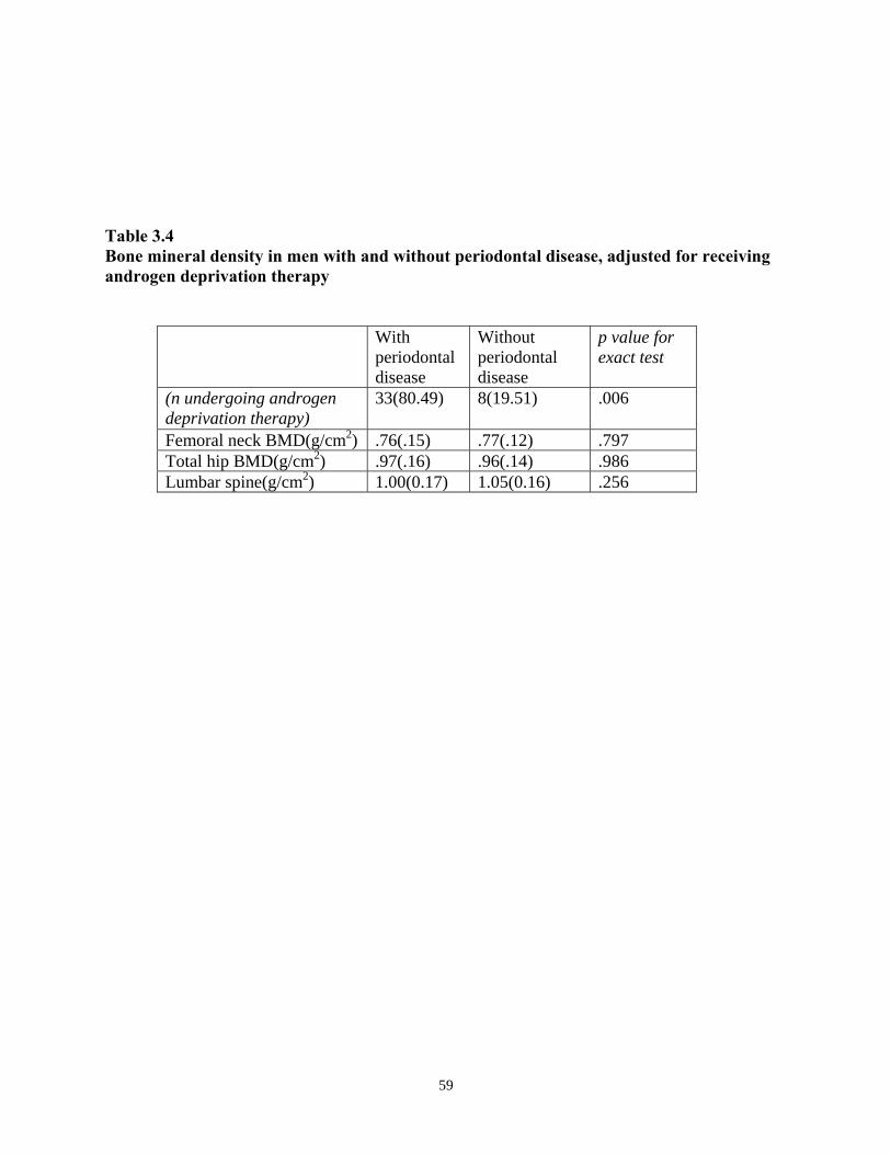
Table 3.4Bone mineral density in men with and without periodontal disease, adjusted for receiving androgen deprivation therapy
With periodontal disease
Without periodontal disease
p value for exact test
(n undergoing androgen deprivation therapy)
33(80.49) 8(19.51) .006
Femoral neck BMD(g/cm2) .76(.15) .77(.12) .797 Total hip BMD(g/cm2) .97(.16) .96(.14) .986 Lumbar spine(g/cm2) 1.00(0.17) 1.05(0.16) .256
59
3.6 The literature cited in the chapter summary of the research
1. Albandar JM. A 6-year study on the pattern of periodontal disease progression. Journal of Clinical
Periodontology 17(7 Pt 1):467-471, 1990. 2. Albandar JM. Ram TE. Global epidemiology of periodontal diseases: An overview. Periodontology 2000 29:7-
10, 2002. 3. Anonymous. Consensus Report. Periodontal diseases: Epidemiology and diagnosis. Consensus Development
Conference. Annals of Periodontology 1(1):216-222, 1996. 4. Anonymous. Periodontal disease: Epidemiology and diagnosis. Consensus Development Conference. Journal of
the American Dental Association 129(Supplement):9S-14S, Sept. 1978. 5. Cummings SR. Melton LJ. Epidemiology and outcomes of osteoporotic fractures. Lancet 359(9319):1761-1767,
2002. 6. Deng JH. Yang LP. Wang LS. Zhou DF. Effect of androgen deprivation therapy on bone mineral density in
prostate cancer patients. Asian Journal of Andrology 6(1):75-7, 2004. 7. Diamond T. Campbell J. Bryant C. Lynch W. The effect of combined androgen blockade on bone turnover and
bone mineral densities in men treated for prostate carcinoma: Longitudinal evaluation and response to intermittent cyclic etidronate therapy. Cancer 83(8):1561-1566, October 1998.
8. Eggert FM. McLeod MH. Flowerdew G. Effects of smoking and treatment status on periodontal bacteria:
Evidence that smoking influences control of periodontal bacteria at the mucosal surface of the gingival crevice. Journal of Periodontology 72(9):1210-1220, September 2001.
9. Genco RJ. Current view of risk factors for periodontal disease. Journal of Periodontology 67(10):1041-1049,
1996. 10. Grossi SG. Genco RJ. Machtei EE. Ho A. Koch G. Dunford R. Zambon JJ. Hausmann E. Assessment of risk for
periodontal disease. I. Risk indicators for alveolar bone loss. Journal of Periodontology 66(1):260-267, 1994. 11. Grossi SG. Genco RJ. Machtei EE. Ho A. Koch G. Dunford R. Zambon JJ. Hausmann E. Assessment of risk for
periodontal disease. II. Risk indicators for alveolar bone loss. Journal of Periodontology 66(1):23-29, 1995. 12. Grossi SG. Skrepcinski FB. DeCaro T. Zambon JJ. Cummins D. Genco RJ. Response to periodontal therapy in
diabetics and smokers. Journal of Periodontology 67(10 Suppl.):1094-1102, 1996. 13. Grossi SG. Zambon JJ. Machtei EE. Schifferle R. Andreana S. Genco RJ. Cummins D. Harrap G. Effects of
smoking and smoking cessation on healing after mechanical periodontal therapy. Journal of the American Dental Association 128(5):599-607, May 1997.
14. Higano CS. Management of bone loss in men with prostate cancer. Journal of Urology 170(6 Pt 2):S259-63;
Discussion S64, 2003. 15. Morote J. Martinez E. Trilla E. Esquena S. Abascal JM. Encabo G. Reventos J. Osteoporosis during continuous
androgen deprivation: Influence of the modality and length of treatment. European Urology 44(6):661-5, 2003. 16. Nunn ME: Understanding the etiology of periodontitis: An overview of periodontal risk factors. Periodontology
2000 32:11-23, 2003.
60

17. Rees TD. A profile of the patient with periodontal disease? Periodontology 2000 32(1):9-13, 2003. 18. Ross RW. Small EJ. Osteoporosis in men treated with androgen deprivation therapy for prostate cancer. Journal
of Urology 167(5):1952-6, 2002. 19. Seeman E. Melton LJ III. O’Fallon WM. Riggs BL. Risk factors for spinal osteoporosis in men. American
Journal of Medicine 75(6):977-983, 1983. 20. Seeman E. The dilemma of osteoporosis in men. American Journal of Medicine 98(2A):76S-88S, 1995. 21. Seeman E. Osteoporosis in men. Baillieres Clinical Rheumatology 11(3):613-29, 1997. 22. Seeman E. The structural basis of bone fragility in men. Bone 25(1):143-147, July 1999. 23. Seeman E. Unresolved issues in osteoporosis in men. Reviews in Endocrine and Metabolic Disorders 2(1):45-
64, 2001. 24. Seeman E. Eisman JA. Treatment of osteoporosis: why, whom, when and how to treat. The single most
important consideration is the individual’s absolute risk of fracture. Medical Journal of Australia 180(6):298-303, 2004.
25. Slots J. Update of general health risk of periodontal disease. International Dental Journal
53(Supplement)3:200-207, 2003. 26. van Winkellhoff AJ. Bosch-Tijhof CJ. Winkel EG. van der Reijden WA. Smoking affects the subgingival
microflora in periodontitis. Journal of Periodontology 72(5):666-671, 2001. 27. Wei JT. Gross M. Jaffee CA. Gravlin K. Lahaie M. Faerber GJ. Cooney KA. Androgen deprivation therapy for
prostate cancer results in significant loss of bone density. Urology 54(4):607-11, 1999. 28. Weyant RJ. Pearlstein ME. Churak AP. Forrest K. Famili P. Cauley JA. The association between osteopenia
and periodontal attachment loss in older women. Journal of Periodontology 70(9):982-991, 1999.
61

4.0 SUMMARY AND CONCLUSIONS
THE RELATIONSHIPS BETWEEN PERIODONTAL DISEASE AND BONE LOSS ON MEN WITH PROSTATE CANCER UNDER ANDROGEN DEPRIVATION THERAPY. A BRIEF SUMMARY ANALYSIS
OF THE RESEARCH CONDUCTED, AND A DISCUSSION OF THE RELATED ISSUES, LIMITATIONS, AND CONCLUSIONS OF THE RESEARCH AND THE DISSERTATION
Periodontitis is a multi-factorial disease with microbial dental plaque as the initiator (Genco 1996; Nunn
2003). The manifestation and progression of periodontitis is influenced by a wide variety of determinants
and risk factors, including subject characteristics, social and behavioral factors, systemic factors, genetic
factors, the microbial composition of dental plaque, and others (Genco 1996; Nunn 2003; Slots 2003). If
periodontitis is not treated, the disease will slowly progress and painlessly destroy the bone which
supports the teeth. Untreated, the disease will eventually cause tooth loss.
Osteoporosis, or porous bone, is characterized by reduced bone mass and structural deterioration
of bone tissue, leading to abnormal bone architecture, fragility of bone, and increased risk of fragility
fracture, particularly of the hip, spine, and wrist. Osteoporosis is a major American public health threat---
as the first-ever Surgeon General’s Report on bone health and osteoporosis made clear with its release on
October 14, 2004---and approximately 1.5 million Americans will suffer fragility fractures each year,
including 300,000 hip fractures, approximately 700,000 vertebral fractures, 250,000 wrist fractures, and
more than 300,000 fractures at other sites. Ten million Americans already have osteoporosis, and 34
million more have low bone mass, placing them at risk for osteoporosis (Surgeon General’s Report 2004).
Half of all women and one-quarter of men over 50 will have an osteoporosis-related bone fracture in their
lifetimes.
The costs, the Surgeon General points out, are tremendous: A fracture can be the beginning of the
deterioration of physical and mental health. Even among individuals who can remain independent, a
fracture diminishes the quality of life. The Surgeon General estimates that approximately 20 percent of
elderly individuals who suffer a hip fracture (the colloquial indicating fracture for osteoporosis) will die
within a year of the fracture (2004).
In fact, as osteoporosis researchers Steven R. Cummings and Joseph Melton summed up in The
Lancet in 2002, osteoporosis has clinical and public health importance only because of these fractures,
causing disability and contributing to health care costs.
Osteoporosis in men has profound clinical implications (Seeman 2001 and 2004; Slots 2003).
Osteoporosis is a significant health problem and contributor to disability and premature mortality among
older men. Osteoporosis occurring in middle-aged and elderly men is commonly associated with alcohol
abuse, smoking, immobilization, and medications. Estimated national direct expenditures (hospitals and
62

nursing homes) for osteoporosis and related fractures are $18 billion each year (U.S. Surgeon General
2004); men comprise one-third of all osteoporosis cases, and account for one-third of the national health
care expense deriving from osteoporosis care.
The issue of the morbidity of osteoporotic hip fractures is addressed directly by research
published just this year by the well-respected Swedish investigator John A. Kanis, who has made a career
of evaluating the economics of osteoporosis (Johnell and Kanis 2005). Furthermore, Kanis and the
equally well-respected Joseph Melton editorialize in the Journal of Bone and Mineral Research (June
2005) about the meaning of the reported incidence of hip fracture trends on osteoporosis treatment,
concluding that clinicians and the population may be paying better attention to controlling the causes of
hip fracture risk (falls and fragility) and that specific programs of selective screening and aggressive
treatment to prevent bone loss may really be able to reduce population fracture rates over the short term
(Melton Kanis Johnell 2005).
The Surgeon General in 2004 called on health professionals to look for “red flags” clinically and
in research that may indicate that an individual or a population is at risk for osteoporosis-related fracture.
The relationship between periodontal disease and bone loss is obvious clinically to dentists; examining
this relationship further led to the investigation of the relationship between periodontal disease and bone
loss and men with prostate cancer who are undergoing androgen deprivation therapy.
The overall prevalence of edentulism in the population of older women that we researched
(Chapter Two) was higher (36 percent), compared with the 23 percent edentulism rate reported for the
U.S. population for women 70 to 74 years as published in the NHANES III study. The higher prevalence
observed in our study could have reflected a continued increase in edentulism with advancing age. The
women in our study ranged from 73 to 84 years in age. We found little evidence to support an association
between osteoporosis and edentulism. Bone mineral density (BMD) and rate of bone loss were similar in
these two groups even after adjustments for established correlating factors of edentulism.
Tooth loss has been observed to be related to bone density in the oral cavity in a limited number
of studies. When interpreting results in various regions, it is necessary to remember that tooth loss is
highly influenced not only by periodontal disease, but also by local practices of the dental community as
well as other factors related to oral health. The majority (80 percent) of dentate women had some
evidence of periodontal disease (p=0.07). We found no statistically significant associations between
periodontal disease variables (average periodontal attachment loss, average number of sites with bleeding,
and average number of sites with calculus) and bone mineral density at trochanter, inter-trochanter and
femoral neck either cross-sectionally or longitudinally. This was true even after controlling for age,
weight, and other important correlating factors. A possible explanation of our lack of an association
between periodontal disease and bone mineral density may be because our population was older, and were
63

missing a large number of teeth. We have no information on the reasons for missing teeth. This finding
could be due to periodontal disease that occurred much earlier in a given subject’s life. Historical events
such as tooth loss can alter the interpretation of the current status of any given subject’s periodontal
disease. Loss of several periodontally-involved teeth can also result in an improvement of periodontal
summary scores such as average loss of attachment.
Compared to the other NHANES III studies, where average attachment loss for the U.S.
population was reported to be 2.1mm, our data showed an average attachment loss of 2.49mm. The major
strengths of this study (again, Chapter Two) are its large sample size and well-controlled measurement of
variables, which could confound relationships with bone mineral density. The sample is drawn randomly
from a large population of community-dwelling women.
In our current research, studying the effects and relationships of periodontal disease and androgen
deprivation therapy in men (Chapter Three), the overall prevalence of periodontal disease was very high
in the group under androgen deprivation therapy even after we adjusted for age, race, smoking and history
of periodontal treatment. Our review of the literature indicated that other comparisons between
periodontal disease and androgen deprivation therapy have not yet been reported, so we cannot compare
our findings to any others.
The majority of men under androgen deprivation therapy nevertheless had some evidence of
periodontal disease (p<0.01). We found statistically significant associations between periodontal disease
variables (these being average periodontal attachment loss, pocket depth, recession, average number of
sites with bleeding, and plaque) and androgen deprivation therapy, that we have summarized as Table 3.2 in
that chapter. These associations held true even after controlling for past history of periodontal treatment,
age, race, and smoking
Although the majority of individuals in this group were highly educated, visited the dentist
regularly, and had very good plaque control, they had periodontal disease because they were undergoing
androgen deprivation therapy. As the original study of bone loss in men with prostate cancer (by
Greenspan in 2004) demonstrated, undergoing androgen deprivation therapy causes severe bone loss and
osteoporosis, and may also precipitate rapid bone loss around the teeth and the presence of periodontal
disease, even with good plaque control. This situation creates a scenario similar to an aggressive type of
periodontal disease in which bone loss will happen in a short period of time despite the presence of
minimal plaque. In the presence of androgen ablation therapy, there may be other factors at work besides
the influence of local factors like plaque that can cause or aggravate periodontal disease.
64

4.1 The limitations of the research The obvious empirical strength of our first study (Chapter Two) was its large sample size, drawn
randomly from a large population of community-dwelling women, and its well–controlled measurement
of variables.
We believe that the current research avenue (studying the effects and relationships of periodontal
disease and androgen deprivation therapy in men (Chapter Three), is significant in that these observations
have never been reported before. Only a limited amount of research relates osteoporosis in men to the
presence of androgen deprivation therapy, and the authors believe that ours is the first empirical data
relating the presence of androgen deprivation therapy to rapid bone loss around the teeth.
Interpretations of the first study were limited because measurement of bone loss around the teeth
was not available, and we had only a single record of periodontal assessment. Interpretations of the
present study may be also limited, since periodontal measures of bone density or loss, such as subtraction
radiography or Shei bone score measurement, were not available to the investigators, and the sample size
addressed here is admittedly small. A larger research sample and longitudinal analysis is necessary to
confirm this finding.
4.2 Conclusions, and future recommendations for research We found small evidence of an association between periodontal disease and longitudinal changes in bone
mineral density in this older women population reported in Chapter Two; in the subsequent research
reported here in men (Chapter Three), we conclude that evidence of a strong association between
androgen deprivation therapy and periodontal disease exists in the corresponding male population.
Additional studies utilizing a larger sample size and longitudinal analysis may be necessary to correlate
periodontal disease and changes in bone mineral density due to androgen deprivation.
Considering the relationship among bone mineral density, osteoporosis, and periodontitis in men,
it is known that men completing androgen ablation therapy for control of prostate cancer are at higher risk
for osteoporosis. As androgen deprivation therapy is the recommended treatment for men with metastatic
or locally-advanced non-metatstatic prostate carcinoma, and as prostate carcinoma is the most common
visceral malignancy and the second leading cause of death from cancer in men, the relationship between
androgen deprivation therapy and loss of bone mineral density is a matter of public health importance.
65
BIBLIOGRAPHY
1. Ahmed AI. Blake GM. Rymer JM. Fogelman I. Screening for osteopenia and osteoporosis: do the accepted normal ranges lead to over-diagnosis? Osteoporosis International 7(5):432-438, 1997.
2. Akesson K. Principles of bone and joint disease control programs–osteoporosis. Journal of
Rheumatology Supplement 67:21-25, August 2003.
3. Albandar JM. Ram TE. Global epidemiology of periodontal diseases: An overview. Periodontology 2000 29:7-10, 2002.
4. Albandar JM. Tinoco EM. Global epidemiology of periodontal diseases in children and young
persons. Periodontology 2000 29:153-176, 2002.
5. Albandar JM. A 6-year study on the pattern of periodontal disease progression. Journal of Clinical Periodontology 17(7 Pt 1):467-471, August 1990.
6. Al-Zahrani MS. Bissada NF. Borawskir EA. Obesity and periodontal disease in young, middle-
aged and older adults. Journal of Periodontology 74(5):610-615, 2003.
7. Anonymous. Consensus Report. Periodontal diseases: Epidemiology and diagnosis. Consensus Development Conference. Annals of Periodontology 1(1):216-222, November 1996.
8. Anonymous. Periodontal disease: Epidemiology and diagnosis. Consensus Development
Conference. Journal of the American Dental Association 129(Supplement):9S-14S, Sept. 1978.
9. Bando K. Nitta H. Matsubara M. Ishikawa I. Bone mineral density in periodontally healthy and edentulous postmenopausal women. Annals of Periodontology 3(1):322-326, 1998 July.
10. Bassett MT. Perl S. Obesity: The Public Health Challenge of Our Time. American Journal of
Public Health 94(9):1477, September 2004.
11. Bilezikian JP. Osteoporosis in men. Journal of Clinical Endocrinology and Metabolism 84(10):3431-3434, 1999.
12. Black DM. Cooper C. Epidemiology of fractures and assessment of fracture risk. Clinics in
Laboratory Medicine 20(3):439-453, September 2000.
13. Caballero B. Introduction. Symposium: Obesity in developing countries: Biological and ecological factors. Journal of Nutrition 131(3):866S-870S, 2001.
14. Cauley JA. Murphy PA, et al. Effects of fluoridated drinking water on bone mass and fractures:
The study of osteoporotic fractures. Journal of Bone and Mineral Research 10(7):1076-1085, 1995.
66

15. Cauley JA. Robbins J. Chen Z. Cummings SR. Jackson RD. LaCroix AZ. Leboff M. Lewis CE.
McGiwan J. Neuner J. Pettinger M. Stefanick ML. Wactawski-Wende J. Witts NB. Women’s Health Initiative Investigators. Effect of estrogen plus progestin on risk fracture and bone mineral density: The Women’s Health Initiative Randomized Trial. Journal of the American Medical Association 290(13):1729-1738, 2003.
16. Chestnut CH 3rd. The relationship between skeletal and oral bone mineral density: an overview.
Annals of Periodontology 6(1):193-196, December 2001.
17. Ciancio SG. Taking oral health to heart: an overview. Journal of the American Dental Association 133Suppl:4S-6S, June 2002.
18. Cianciola LJ. Park BH. Bruck E. Mosovich L. Genco RJ. Prevalence of periodontal disease in
insulin-dependent diabetes mellitus (juvenile diabetes). Journal of the American Dental Association 104(5):653-660, May 1982.
19. Consigny PM. Pathogenesis of atherosclerosis. AJR:American Journal of Roentgenology
164(3):553-558, 1995.
20. Cummings SR. Black DM. Rubin SM. Lifetime risk of hip, Colles’ or vertebral fracture and coronary heart disease among white postmenopausal women. Archives of Internal Medicine 149(11):2445-2448, 1989.
21. Cummings SR. Nevitt MC. Browner WS, et al. Risk factors for hip fracture in white women: The
Study of Osteoporotic Fractures Research Group. New England Journal of Medicine 332(12):767-773, 1995.
22. Cummings SR. Melton LJ. Epidemiology and outcomes of osteoporotic fractures. Lancet
359(9319):1761-1767, 2002 May 18.
23. DeLaet CE. Pols HA. Fractures in the elderly: Epidemiology and demography. Best Practice and Research Clinical Endocrinology and Metabolism 14(2):171-179, 2000.
24. Delmas PD. Bone mass measurement: how, where, when and why? International Journal of
Fertility and Menopausal Studies 38 Suppl 2:70-76, 1993.
25. Deng JH. Yang LP. Wang LS. Zhou DF. Effect of androgen deprivation therapy on bone mineral density in prostate cancer patients. Asian Journal of Andrology 6(1):75-77, March 2004.
26. Desvarieux M. Demmer RT. Rundek T. Boden-Albala B. Jacobs DR Jr. Papapanou PN. Sacco
RL. Oral Infections and Vascular Disease Epidemiology Study (INVEST). Relationship between periodontal disease, tooth loss, and carotid artery plaque: the Oral Infections and Vascular Disease Epidemiology Study (INVEST). Stroke 34(9):2120-2125, September 2003.
27. Diamond T. Campbell J. Bryant C. Lynch W. The effect of combined androgen blockade on bone
turnover and bone mineral densities in men treated for prostate carcinoma: Longitudinal evaluation and response to intermittent cyclic etidronate therapy. Cancer 83(8):1561-1566, October 1998.
67

28. Eggert FM. McLeod MH. Flowerdew G. Effects of smoking and treatment status on periodontal bacteria: Evidence that smoking influences control of periodontal bacteria at the mucosal surface of the gingival crevice. Journal of Periodontology 72(9):1210-1220, September 2001.
29. Elders PJ. Habets LL. Netelenbos JC. van der Linden LW. van der Stelt PF. The relation between
periodontitis and systemic bone mass in women between 46 and 55 years of age. Journal of Clinical Periodontology 19(7):492-496, August 1992.
30. Emrich LJ. Shlossman M. Genco RJ. Periodontal disease in non-insulin-dependent diabetes
mellitus. [See comment]. Journal of Periodontology 62(2):123-131, Feb. 1991
31. Ensrud KE. Palermo L. Black DM. Cauley J. Jergas M. Orwoll ES. Nevitt MC, Fox KM. Cummings SR. Hip and calcaneal bone loss increase with advancing age: longitudinal results from the study of osteoporotic fractures. Journal of Bone & Mineral Research 10(11):1778-1787, 1995 November.
32. Ezzo PJ. Cutler CW. Microorganisms as risk indicators for periodontal disease. Periodontology
2000 32:24-35, 2003.
33. Ferrari S. Rizzoli R. Bonjour JP. Genetic aspects of osteoporosis. Current Opinion in Rheumatology 11(4):294-300, July 1999.
34. Fine LJ. Philogene GS. Gramling R. Coups EJ. Sinha S. Prevalence of multiple chronic disease
risk factors. 2001 National Health Interview Survey. American Journal of Preventive Medicine 27(2 Supplement):18-24, August 2004.
35. Fowler JE. Bigler SA. White PC. Duncan WL. Hormone therapy for locally advanced prostate
cancer. Journal of Urology 168(2):546-549, August 2002.
36. Genco RJ. Current view of risk factors for periodontal disease. Journal of Periodontology 67(10):1041-1049, 1996.
37. Genco R. Offenbacher S. Beck J. Periodontal disease and cardiovascular disease: epidemiology
and possible mechanisms. Journal of the American Dental Association 133 Suppl:14S-22S, June 2002.
38. Geurs NC. Lewis CE. Jeffcoat MK. Osteoporosis and periodontal disease progression.
Periodontology 2000 32:105-110, 2003.
39. Greenlee RT. Hill-Harmon MB. Murray T. Thun M. Cancer statistics 2001. Ca: A Cancer Journal for Clinicians 51(1):15-36, January-February 2001.
40. Greenspan SL. Oppenheim DS. Klibanski A. Importance of gonadal steroids to bone mass in men
with hyperprolactinemic hypogonadism. Annals of Internal Medicine 110(7): 526-531, 1989.
41. Greenspan SL. Dresner-Pollak R. Parker RA. London D. Ferguson L. Diurnal variation of bone mineral turnover in elderly men and women. Calcified Tissue International 60(5):419-423, 1997.
42. Greenspan SL. Parker RA. Ferguson L. Rosen HN. Maitland-Ramsey L. Karpf DB. Early
changes in biochemical markers of bone turnover predict the long-term response to alendronate 68

therapy in representative elderly women: A randomized clinical trial. Journal of Bone and Mineral Research 13(9):1431-1438, 1998.
43. Grossi SG. Genco RJ. Machtei EE. Ho A. Koch G. Dunford R. Zambon JJ. Hausmann E.
Assessment of risk for periodontal disease. I. Risk indicators for alveolar bone loss. Journal of Periodontology 66(1):260-267, 1994.
44. Grossi SG. Genco RJ. Machtei EE. Ho A. Koch G. Dunford R. Zambon JJ. Hausmann E.
Assessment of risk for periodontal disease. II. Risk indicators for alveolar bone loss. Journal of Periodontology 66(1):23-29, 1995.
45. Grossi SG. Skrepcinski FB. DeCaro T. Zambon JJ. Cummins D. Genco RJ. Response to
periodontal therapy in diabetics and smokers. Journal of Periodontology 67(10 Suppl.):1094-1102, October 1996.
46. Grossi SG. Zambon JJ. Machtei EE. Schifferle R. Andreana S. Genco RJ. Cummins D. Harrap G.
Effects of smoking and smoking cessation on healing after mechanical periodontal therapy. Journal of the American Dental Association 128(5):599-607, May 1997.
47. Heyse SP. Epidemiology of hip fractures in the elderly: A cross-national analysis of mortality
rates for femoral neck fractures. Osteoporosis International 3Supplement1:16-19, 1993.
48. Higano CS. Management of bone loss in men with prostate cancer. Journal of Urology 170(6 Pt 2):S259-63; Discussion S64, 2003 December.
49. Hildebolt CF. Pilgram TK. Dotson M. Cohen SC. Hauser JF. Kardaris E. Civitelli R.
Relationships between clinical attachment level and spine and hip bone mineral density: data from healthy postmenopausal women [Erratum appears in Journal of Periodontology 75(5):780]. Journal of Periodontology 73(3):298-301, 2002 March.
50. Irfan UM, Dawson DV, Bissada NF. Epidemiology of periodontal disease: Review and clinical
perspectives. Journal of the International Academy of Periodontology 3(1):14-21, 2001 January.
51. Jacobs R. Ghyselen J. Koninckx P. van Steenberghe D. Long-term bone mass evaluation of mandible and lumbar spine in a group of women receiving hormone replacement therapy. European Journal of Oral Sciences 104(1):10-16, February 1996.
52. Jacobsen SJ. Goldberg J. Miles TP et al. Regional variation in the incidence of hip fracture: US
white women aged 65 years and older. Journal of the American Medical Association 264(4):500-502, 1990.
53. Jacqmin-Gadda H. Fourrier A. Commenges D. Dartigues JF. Risk fractures in the elderly.
Epidemiology 9(4):417-423, 1998.
54. Jaglal SB, Kreiger N, Darlington G. Past and recent physical activity and risk of hip fracture. American Journal of Epidemiology 138(2):107-118, 1993.
55. Jaglal SB. Weller I. Mamdani M. Hawker G. Kreder H. Jaakkimainen L. Adachi JD. Population
trends in BMD testing, treatment, and hip and wrist fracture rates: are the hip fracture projections wrong? Journal of Bone and Mineral Research 20(6):898-905, June 2005.
69

56. James PT. Leach R. Kalamara E. Shayeghi M. International Obesity Task Force, London, United Kingdom. The worldwide obesity epidemic. Obesity Research 9Supplement4:228S-233S, November 2001.
57. James PT. Rigby N. Leach R. International Obesity Task Force, London, United Kingdom. The
obesity epidemic, metabolic syndrome and future prevention strategies. European Journal of Cardiovascular Prevention and Rehabilitation 11(1):3-8, February 2004.
58. Jeffcoat MK. Osteoporosis: A possible modifying factor in oral bone loss. Annals of
Periodontology 3(1):312-321, July 1998.
59. Jeffcoat MK. Lewis CE. Reddy MS. Wang CY. Redford M. Post-menopausal bone loss and its relationship to oral bone loss. [Erratum appears in Periodontology 2000 26:169, 2001]. Periodontology 2000 23:94-102, June 2000.
60. Johansson I. Tidehag P. Lundberg V. Hallmans G. Dental status, diet and cardiovascular risk
factors in middle-aged people in northern Sweden. Community Dentistry and Oral Epidemiology 22(6):431-436, December 1994.
61. Johnell O. Kanis J. Epidemiology of osteoporotic fractures. Osteoporosis International 16 Supp1
@:S3-S7, March 2005.
62. Johnson RB. Gilbert JA. Cooper.RC. Dai X. Newton BI. Tracy RR. West WF. DeMoss TL. Meyers PJ. Streckfus CF. Alveolar bone loss one year following ovariectomy in sheep. Journal of Periodontology 68(9):864-871, 1997.
63. Kanis JA. Assessment of fracture risk and its application to screening for postmenopausal
osteoporosis: Synopsis of a WHO report. WHO Study Group. Osteoporosis International 4(6):368-381, November 1994.
64. Kanis JA. Diagnosis of osteoporosis and assessment of fracture risk. [See comment, Lancet
360(9343):1429, November 2002.] Lancet 359(9321):1929-1936, June 2002.
65. Kannus P. Niemi S. Parkkari J. Palvanen M. Vuori I. Jarvinen M. Hip fractures in Finland between 1970 and 1997 and predictions for the future. Lancet 353(9155):802-805, March 1999.
66. Kenny AM. Taxel P. Osteoporosis in older men. Clinical Cornerstone 2(6):45-51, 2000.
67. Kenny AM. Joseph C. Taxel P. Prestwood KM. Osteoporosis in older men and women.
Connecticut Medicine 67(8):481-486, September 2003.
68. Kinane DF. Periodontal diseases' contributions to cardiovascular disease: an overview of potential mechanisms. Annals of Periodontology 3(1):142-150, July 1998.
69. Kinane DF. Periodontitis modified by systemic factors. Annals of Periodontology 4(1):54-64,
December 1999.
70. Krall EA. Garcia RI. Dawson-Hughes B. Increased risk of tooth loss is related to bone loss at the whole body, hip, and spine. Calcified Tissue International 59:433-437, 1996 Dec.

71. Kribbs PJ. Smith DE. Chesnut CH 3rd. Oral finding in osteoporosis. Part II: Relationship between residual ridge and alveolar bone resorption and generalized skeletal osteopenia. Journal of Prosthetic Dentistry 50(5):719-724, 1983.
72. Kribbs PJ. Chesnut CH 3rd. Ott SM. Kilcoyne RF. Relationships between mandibular and
skeletal bone in an osteoporotic population. Journal of Prosthetic Dentistry 62(6):703-707, December 1989.
73. Kribbs PJ. Chesnut CH 3rd. Ott SM. Kilcoyne RF. Relationships between mandibular and
skeletal bone in a population of normal women. Journal of Prosthetic Dentistry 63(1):86-89, January 1990.
74. Kribbs PJ. Comparison of mandibular bone in normal and osteoporotic women. Journal of
Prosthetic Dentistry 63(2):218-222, February 1990.
75. Kuller LH. Orchard TJ. The epidemiology of atherosclerosis in 1987: unraveling a common-source epidemic. Clinical Chemistry 34(8B):B40-B48, 1988.
76. Lalla E. Lamster IB, Stern DM. Schmidt AM. Receptor for advanced glycation end products,
inflammation, and accelerated periodontal disease in diabetes; mechanisms and insights into therapeutic modalities. Annals of Periodontology 6(1):113-118, 2001.
77. Levy SM. Warren JJ. Chowdhury J. DeBus B. Watkins CA. Cowen HJ. Kirchner HL. Hand JS.
The prevalence of periodontal disease measures in elderly adults, aged 79 and older. Special Care in Dentistry 23(2):50-57, 2003.
78. Lips P. Epidemiology and predictors of fractures associated with osteoporosis. American Journal
of Medicine 103(2A):3S-8S; discussion 8S-11S, August 1997.
79. Locker D. Slade GD. Murray H. Epidemiology of periodontal disease among older adults: Periodontology 2000 16:16-33, 1998.
80. Loe H. Anerud A. Boysen H. Morrison E. Natural history of periodontal disease in man. Rapid,
moderate and no loss of attachment in Sri Lankan laborers 14 to 46 years of age. Journal of Clinical Periodontology 13(5):431-445, 1986.
81. Looker AC. Orwoll ES. Johnston CC Jr. Lindsay RL. Wahner HW. Dunn WL. Calvo MS. Harris
TB. Heyse SP. Prevalence of low femoral bone density in older U.S. adults from NHANES III. Journal of Bone and Mineral Research 12(11):1761-1768, November 1997.
82. Luckey MM. Wallenstein S. Lapinski R. Meier DE. A prospective study of bone loss in African-
American and white women--A clinical research center study. Journal of Clinical Endocrinology and Metabolism 81(8):2948-2956, August 1996.
83. Lundstrom A. Jendle J. Stenstrom B. Toss G. Ravald N. Periodontal conditions in 70-year-old
women with osteoporosis. Swedish Dental Journal 25(3):89-96, 2001.
84. Mann T. Oviatt SL. Wilson D. Nelson D. Orwoll ES. Vertebral deformity in men. Journal of Bone and Mineral Research 7(11):1259-1265, 1992.
71
85. Mattson JS. Cerutis DR. Diabetes mellitus: a review of the literature and dental implications. Compendium of Continuing Education in Dentistry 22(9):757-760, 762, 764 passim; quiz 773, September 2001.
86. Mattson JS. Cerutis DR. Parrish LC. Osteoporosis: a review and its dental implications.
Compendium of Continuing Education in Dentistry 23(11):1001-1004, 1006, 1008 passim; quiz 1014, November 2002.
87. Mazess RB. On aging bone loss. Clinical Orthopedics and Related Research (165):239-252, May
1982.
88. Mazess RB. Barden HS. Drinka PJ. Bauwens SF. Orwoll ES. Bell NH. Influence of age and body weight on spine and femur bone mineral density in U.S. white men. Journal of Bone and Mineral Research 5(6):645-652, June 1990.
89. Melton LJ III. Riggs BL. Epidemiology of age related fractures. In: The Osteoporotic Syndrome.
Edited L. V. Avioli. New York: Grune and Stratton, pp. 45-72, 1998.
90. Melton LJ III. Kanis JA. Johnell O. Potential impact of osteoporosis treatment on hip fracture trends. Journal of Bone and Mineral Research 20(6):895-899, June 2005.
91. Mokdad AH. Serdula MK. Dietz WH. Bowman BA. Marks JS. Koplan JP. The spread of the
obesity epidemic in the United States, 1991-1998. Journal of the American Medical Association 282(16):1519-1522, October 1999.
92. Morote J. Martinez E. Trilla E. Esquena S. Abascal JM. Encabo G. Reventos J. Osteoporosis
during continuous androgen deprivation: Influence of the modality and length of treatment. European Urology 44(6):661-665, 2003.
93. Nares S. The genetic relationship to periodontal disease. Periodontology 2000 32:36-49, 2003.
94. Nelson RG. Shlossman M. Budding LM. Pettitt DJ. Saad MF. Genco RJ. Knowler WC.
Periodontal disease and NIDDM in Pima Indians. Diabetes Care 13(8):836-840, August 1990.
95. Nguyen TV. Center JR. Sambrook PN. Eisaman JA. Risk factors for proximal humerus, forearm, and wrist fractures in elderly men and women: the Dubbo Osteoporotic Epidemiology Study. American Journal of Epidemiology 153(6):587-595, 2001.
96. Nindl BC. Harman EA. Marx JO. Gotshalk LA. Frykman PN. Lammi E. Palmer C. Kraemer WJ.
Regional body composition changes in women after six months of periodized physical training. Journal of Applied Physiology 88(6):2251-2259, June 2000.
97. Nishimura F. Murayama Y. Periodontal inflammation and insulin resistance---lessons from
obesity. Journal of Dental Research 80(8):1690-1694, August 2001.
98. Nishimura F. Iwamoto Y. Mineshiba J. Shimizu A. Soga Y. Murayama Y. Periodontal disease and diabetes mellitus: the role of tumor necrosis facor-alpha in a two-way relationship. Journal of Periodontology 74(1):97-102, January 2003.
99. Nunn ME: Understanding the etiology of periodontitis: An overview of periodontal risk factors.
Periodontology 2000 32:11-23, 2003.
72

100. Payne JB. Reinhardt RA. Nummikoski PV. Dunning DG. Patil KD. The association of cigarette smoking with alveolar bone loss in postmenopausal females. Journal of Clinical Periodontology 27(9):658-64, September 2000. 101. Persson L. Bergstrom J. Ito H. Gustafsson A. Tobacco smoking and neutrophil activity in patients with periodontal disease. Journal of Periodontology 72(1):90-95, January 2001. 102. Pucher J. Stewart J. Periodontal disease and diabetes mellitus. Current Diabetes Reports 4(1):46-50, February 2004. 103. Reddy MS. Oral osteoporosis: is there an association between periodontitis and osteoporosis? Compendium of Continuing Education in Dentistry 23(10 Suppl):21-8, 2002. 104. Rees TD. A profile of the patient with periodontal disease? Periodontology 2000 32(1):9-13, June 2003. 105. Reinhardt RA. Payne JB. Maze CA. Patil KD. Gallagher SJ. Mattson JS. Influence of estrogen and osteopenia/osteoporosis on clinical periodontitis in postmenopausal women. Journal of Periodontology 70(8):823-828, August 1999. 106. Rodrigo F. Neiva JS. Khalaf F. Al-Shammari. Wang H-L. Effect of specific nutrients on periodontal disease onset. Progression and Treatment 30(7):579-589, 2003. 107. Ronderos M. Jacobs DR. Himes JH. Pihlstrom BL. Associations of periodontal disease with femoral bone mineral density and estrogen replacement therapy: cross-sectional evaluation of US adults from NHANES III. Journal of Clinical Periodontology 27(10):778-786, 2000. 108. Ronderos M. Ryder MI. Risk assessment in clinical practice. Periodontology 2000 34:120-135, 2004. 109. Ross RW. Small EJ. Osteoporosis in men treated with androgen deprivation therapy for prostate cancer. Journal of Urology 167(5):1952-6, 2002 May. 110. Saito T. Shimazaki Y. Sakamoto M. Obesity and periodontitis. New England Journal of Medicine 339(7):482-483, 1998. 111. Sasaki T. Ramammurthy NS. Golub LM. Insulin-deficient diabetes impairs osteoblast and periodontal ligament fibroblast metabolism but does not affect ameloblasts and odontoblasts: Response to tetracycline(s) administration. Journal de Biologie Buccale 18(3):215-226, September 1990. 112. Seeman E. Melton LJ III. O’Fallon WM. Riggs BL. Risk factors for spinal osteoporosis in men. American Journal of Medicine 75(6):977-983, December 1983. 113. Seeman E. The dilemma of osteoporosis in men. American Journal of Medicine 98(2A):76S-88S, February 1995. 114. Seeman E. Osteoporosis in men. Baillieres Clinical Rheumatology 11(3):613-29, 1997.
73

115. Saito T. Shimazaki Y. Sakamoto M. Obesity and periodontitis. New England Journal of
Medicine 339(7):482-483, 1998. 116. Sasaki T. Ramammurthy NS. Golub LM. Insulin-deficient diabetes impairs osteoblast and
periodontal ligament fibroblast metabolism but does not affect ameloblasts and odontoblasts: Response to tetracycline(s) administration. Journal de Biologie Buccale 18(3):215-226, September 1990.
117. Seeman E. Melton LJ III. O’Fallon WM. Riggs BL. Risk factors for spinal osteoporosis in men.
American Journal of Medicine 75(6):977-983, December 1983. 118. Seeman E. The dilemma of osteoporosis in men. American Journal of Medicine 98(2A):76S-88S,
February 1995. 119. Seeman E. Osteoporosis in men. Baillieres Clinical Rheumatology 11(3):613-629, 1997. 120. Seeman E. The structural basis of bone fragility in men. Bone 25(1):143-147, July 1999. 121. Seeman E. Unresolved issues in osteoporosis in men. Reviews in Endocrine and Metabolic
Disorders 2(1):45-64, January 2001. 122. Seeman E. Eisman JA. Treatment of osteoporosis: why, whom, when and how to treat. The single
most important consideration is the individual’s absolute risk of fracture. Medical Journal of Australia 180(6):298-303, March 2004.
123. Shlossman M. Knowler WC. Pettitt DJ. Genco RJ. Type II diabetes mellitus and periodontal
disease. Journal of the American Dental Association 121(4):532-536, Oct. 1990. 124. Shrout MK. Hildebolt CF. Potter BJ. Brunsden TK. Pilgram TK. Dotson M. Yokoyama-Crothers
N. Hauser J. Cohen S. Kardaris E. Civitelli R. Hanes P. Comparison of morphological measurements extracted from digitized dental radiographs with lumbar and femoral bone mineral density measurements in postmenopausal women. Journal of Periodontology 71(3):335-340, March 2000.
125. Simon J. Leboff M. Wright J. Glowacki J. Fractures in the elderly and Vitamin D. Journal of
Nutrition, Health and Aging 6(6):406-412, 2002. 126. Slade GD. Beck JD. Plausibility of periodontal disease estimates from NHANES III. Journal of
Public Health Dentistry. 59(2):67-72, 1999 Spring. 127. Slade GD. Ghezzi EM. Heiss G. Beck JD. Riche E. Offenbacher S. Relationship between
periodontal disease and C-reactive protein among adults in the Atherosclerosis Risk in Communities study. Archives of Internal Medicine 163(10):1172-9, 2003 May 26.
128. Slots J. Update of general health risk of periodontal disease. International Dental Journal
53(Supplement)3:200-207, 2003. 129. Sorsa T. Ingman T, Suomalainen K, et al. Cellular source and tetracycline inhibition of gingival
crevicular fluid collagenase of patients with labile diabetes mellitus. Journal of Clinical Periodontology 19(2):146-149, 1992.
74
130. Soskolne WA. Epidemiological and clinical aspects of periodontal diseases in diabetics. Annals of
Periodontology 3(1):3-12, July 1998. 131. Southard KA. Southard TE. Comparison of digitized radiographic alveolar features between 20-
and 70-year-old women. A preliminary study. Oral Surgery, Oral Medicine, Oral Pathology 74(1):111-117, July 1992.
132. Spalj S, Plancak D. The distribution of periodontal disease and loss of attachment in jaw sextants
in different age groups--cross sectional study. Collegium Antropologicum 27(Suppl) 1:183-190, 2003.
133. Stoch SA. Parker RA. Chen L. Bubley G. Ko Y-J. Vincelette A. Greenspan SL. Bone loss in men
with prostate cancer treated with gonadotropin-releasing hormone agonists. Journal of Clinical Endocrinology and Metabolism 86(6):2787-2791, 2001.
134. Streckfus CF. Johnson RB. Nick T. Tsao A. Tucci M. Comparison of alveolar bone loss, alveolar
bone density and second metacarpal bone density, salivary and gingival crevicular fluid interleukin-6 concentrations in healthy premenopausal and postmenopausal women on estrogen therapy. Journals of Gerontology Series A-Biological Sciences and Medical Sciences 52(6):M343-51, November 1997.
135. Streckfus CF, Parsell DE, Streckfus JE, Pennington W, Johnson RB. Relationship between oral
alveolar bone loss and aging among African-American and Caucasian individuals. Gerontology 45(2):110-114, 1999.
136. Teng YT. The role of acquired immunity and periodontal disease progression. Critical Reviews in
Oral Biology and Medicine 14(4):237-252, 2003. 137. Tezal M. Wactawski-Wende J. Grossi SG. Ho A. Dunford R. The relationship between bone
mineral density and periodontitis in postmenopausal women. Journal of Periodontology 71(9):1492-1498, 2000.
138. Tezal M. Grossi SG, Ho AW, Genco RJ. The effect of alcohol consumption on periodontal
disease. Journal of Periodontology 72(2):183-189, 2001. 139. Tobin JD. Fox KM. Cejku ML. Bone density changes in normal men: A 4-19 year longitudinal
study. Journal of Bone and Mineral Research Supplement 8(3):142, 1993. 140. [Tobin JD]. Hochberg MC. Lethbridge-Cejku ML. Bone mineral density and osteoarthritis: data
from the Baltimore Longitudinal Study of Aging. Osteoarthritis and Cartilage 12 Suppl A:S45-8, 2004.
141. U.S. Department of Health and Human Services. Bone Health and Osteoporosis: A Report of the
Surgeon General. Rockville, MD: U.S. Department of Health and Human Services, Office of the Surgeon General, 2004.
142. van Winkellhoff AJ. Bosch-Tijhof CJ. Winkel EG. van der Reijden WA. Smoking affects the
subgingival microflora in periodontitis. Journal of Periodontology 72(5):666-671, May 2001.
75

143. von Wowern N. Klausen B. Kollerup G. Osteoporosis: A risk factor in periodontal disease. Journal of Periodontology 65(12):1134-1138, 1994.
144. Wactawski-Wende J. Grossi SG. Trevisan M. Genco RJ, et al. The role of osteopenia in oral bone
loss and periodontal disease. Journal of Periodontology 67(10 Supplement):1076-1084, 1996. 145. Warming L. Hassager C. Christiansen C. Changes in bone mineral density with age in men and
women: a longitudinal study. Osteoporosis International 13(2):105-112, 2002. 146. Wei JT. Gross M. Jaffee CA. Gravlin K. Lahaie M. Faerber GJ. Cooney KA. Androgen
deprivation therapy for prostate cancer results in significant loss of bone density. Urology 54(4):607-611, 1999.
147. Werning JW. Downey NM. Brinker RA. Khuder SA. Davis WJ. Rubin AM. Elsamaloty HM. The
impact of osteoporosis on patients with maxillofacial trauma. Archives of Otolaryngology–Head and Neck Surgery 130(3):353-356, March 2004.
148. Weyant RJ. Pearlstein ME. Churak AP. Forrest K. Famili P. Cauley JA. The association between
osteopenia and periodontal attachment loss in older women. Journal of Periodontology 70(9):982-991, 1999.
149. Willershausen-Zonnchen B. Lemmen C. Hamm G. Influence of high glucose concentrations on
glycosaminoglycan and collagen synthesis in cultured human gingival fibroblasts. Journal of Clinical Periodontology 18(3):190-195, 1991.
150. Winn DM. Johnson CL. Kingman A. Periodontal disease estimates in NHANES III: clinical
measurement and complex sample design issues. Journal of Public Health Dentistry 59(2):73-78, 1999 Spring.
151. Zambon JJ. Grossi SG. Machtei EE. Ho AW. Dunford R. Genco RJ. Cigarette smoking increases
the risk for subgingival infection with periodontal pathogens. Journal of Periodontology 67(10 Suppl):1050-1054, October 1996.
76


![Cronicon · of periodontal ligament and alveolar bone [1] that lead to damage of the periodontal tissues, formation of intrabony defects (ID) and subsequently tooth loss [1,2]. Treatment](https://img.dokumen.tips/doc/110x75/5fa63e7602b4a8288f613a70/cronicon-of-periodontal-ligament-and-alveolar-bone-1-that-lead-to-damage-of-the.jpg)


![Periodontal Disease: General Aspects from Biofilm to the ... · gingival inflammation and loss of alveolar bone, which can be detected by x-ray (Figure 2) [15] [16]. Acute periodontal](https://img.dokumen.tips/doc/110x75/5e725d4a1a91891c5f67e737/periodontal-disease-general-aspects-from-biofilm-to-the-gingival-inflammation.jpg)


![Article Can better management of periodontal disease delay ...clok.uclan.ac.uk/17325/10/17325.pdf · progression, such as pocket depth, alveolar bone loss and tooth loss [37]. Currently,](https://img.dokumen.tips/doc/110x75/5ece409bd9590e4d3f3275a3/article-can-better-management-of-periodontal-disease-delay-clokuclanacuk173251017325pdf.jpg)