Embed Size (px)
Citation preview
Received 02/25/2013 Review began 02/26/2013 Published 06/29/2013
© Copyright 2013Vegas. This is an open access articledistributed under the terms of theCreative Commons AttributionLicense CC-BY 3.0., which permitsunrestricted use, distribution, andreproduction in any medium,provided the original author andsource are credited.
Computed Tomography Angiography,Perforator Flaps, the Surgeon, and OsirixManuel R. Vegas
1. Hospital Quiron Madrid Spain
Corresponding author: Manuel R. Vegas, [email protected] Disclosures can be found in Additional Information at the end of the article
AbstractThrough their experience with 144 patients, the authors evaluate the use of the free open-sourceDICOM viewer OsiriX for Mac in the preoperative planning of perforator flaps with threeobjectives: 1) increase the present knowledge related with the preoperative planning ofperforator flaps with computed tomography angiography (CTA), 2) evaluate the application in theimage post-processing of perforator flaps, and 3) evaluate the performance of the image post-processing when performed by a surgeon. The experience demonstrated that the use of OsiriXallowed an adequate evaluation of different structures and parameters of great preoperativeinterest in perforator flap surgery: 1) source artery, 2) diameter of artery and vein/s at thehypothetical site of microsurgical anastomoses, 3) course and branching pattern of the flappedicle, 4) perforator course in the subcutaneous fat (theoretical flap axis), 5) measurement ofthe skin and fat where the perforator pierced the deep fascia (theoretical flap thickness), 6)measurement of the distance between the point of entrance of the perforator in the subcutaneousfat to the source artery (theoretical maximal pedicle length), and 7) measurement of theperforator diameter where it pierced the deep fascia. For the home user, OsiriX is an efficientalternative, comparable to the more professional applications only available in Radiology servicesand dedicated professional PACS workstations.
Categories: Plastic Surgery, General SurgeryKeywords: ct angiography, cta, osirix, computed tomography angiography, perforator flap
IntroductionWhether or not CTA is justified in the preoperative planning of perforator flaps is still underdebate but there is a growing evidence of the associated benefits [1] and, despite the lack ofrandomized multicentric studies, a substantial and growing amount of bibliography supports theuse and security of CTA [1-2]. CTA has demonstrated to improve the surgical results, aid in theselection of the best perforators, minimize surgical trauma and donor site sequelae, reduceeconomic costs and minimize the surgeon’s surgical stress [3-7]. Its potential role in theevaluation of recipient vessels has also proven to be high [8]. Very popular in breastreconstruction with abdominal flaps, its use has rapidly expanded to other body regions becausethe technique is readily available, extremely fast and shows a low interobserver variability [9-11].However, computed tomography has drawn experts’ attention because of its relatively highradiation dose per study [12-14]. In this scenario, different measures are imperative: 1) a correctindication of CT studies, 2) the improvement of technology and 3) the optimization of theacquisition parameters to minimize the radiation dose following the ALARA (As Low AsReasonably Available) principle [4, 15-16]. At present, only magnetic resonance angiography canrival CTA [17-18], although some reports have found that the technique is still far from it becauseof a lesser resolution, higher cost and limited availability [19-21]. OsiriX is a free open-sourceimaging software that transforms an Apple Macintosh into a PACS DICOM workstation to process
1
Open Access Original Article DOI: 10.7759/cureus.125
How to cite this articleVegas M R. (2013-06-29 08:09:32 UTC) Computed Tomography Angiography, Perforator Flaps, theSurgeon, and Osirix. Cureus 5(6): e125. DOI 10.7759/cureus.125

and visualize medical images [22]. Free in its 32-bit version, the paid 64-bit version gives anextended usage of the processor’s RAM memory. The recently released MD version has obtainedthe Class II Medical Advice certification by the FDA for clinical use. IPhone and IPad HD versionsopen up a new portable dimension to radiological studies.
Materials And MethodsA retrospective review was made of the authors’ experience in the preoperative evaluation ofperforators with CTA with three objectives: 1) widen the present knowledge of the use of CTA inpreoperative planning of perforator flaps, 2) evaluate the OsiriX application in the DICOM imagepost-processing of perforator flaps and 3) analyze the performance of the post-processing whendone by a surgeon (non-specialist in Radiology). After the appropriate Institutional EthicsCommittee approval from the Hospital Fremap Majadahonda and the corresponding writtenpatient informed consent, the research included the studies of 144 consecutive patients whoundertook a CTA as one of the routine studies in the preoperative planning of a perforator-basedfree or pedicled flap reconstruction (Table 1).
Flaps Number of Studies
Free flap
Anterolateral thigh (ALT) 79
Deep inferior epigastric (DIEP) 32
Superficial inferior epigastric (SIEA) 4
Superior gluteal (SGAP) 2
Superolateral thigh (SLT) 5
Thoracodorsal (TDAP) 12
Pedicled propeller flapAnterior tibial perforator (ATP) 3
Posterior tibial perforator (PTP) 7
Total 144
TABLE 1: Preoperative planning of perforator flaps with computed tomography angiography. Patient study
The series included 56 females and 88 males with ages raging between 23 and 67 years of age(mean 42.6). All the studies were performed with a 64-slice CT (Lightspeed VCT, GE Healthcare,US) between March 2008 and February 2011. The acquisition parameters [4-16], adjusted for anoptimal relationship between radiation dose and image quality following the ALARA principle[23], are shown in Table 2.
Scanner Lightspeed VCT, GE Healthcare, US
Scan direction Craneo-caudad or caudo-craneal depending on the area of study
Range
Collimation 0.625 mm
Voltage 120 kV
Tube current 180-200 mAs
Rotation time 0,37 s
Contrast material
2013 Vegas et al. Cureus 5(6): e125. DOI 10.7759/cureus.125 2 of 11

Contrast Ultravist 300, Schering AG, Berlin, Germany
Injection system GE-Nemoto Dual-Shot Injector,GE Healthcare, Waukesha, Wisconsin, US
Volume 100 cc
Speed 4 cc/s
Saline flush
Volume 50 cc
Speed 4 cc/s
Reconstruction thickness 0,625 mm
TABLE 2: Acquisition parameters and contrast media
The images were processed and analysed by the first author with a 24-inch screen iMac and the64-bit version of OsiriX. The different reconstruction 2D and 3D modes were used as shown inTable 3.
Study Parameter Mode
First overall anatomical evaluation Orthogonal 2D MPR
Analysis of small caliber vessels (below 3-4 mm) Curved 3D MPR
Distance between points of interest Orthogonal 2D MPR
3D MPR
Measurement of flap thickness Orthogonal 2D MPR
3D MPR
Measurement of maximal theoretical length of flap pedicle Curved 3D MPR
Perforator course in subcutaneous fat Orthogonal 2D MPR
3D anatomical evaluation and photographic presentation 3D VR
TABLE 3: Reconstruction modes
Different parameters of interest in perforator flap surgery were evaluated with OsiriX: 1) sourceartery, 2) diameter of pedicle’s artery and vein at the hypothetical suture site, 3) course and side-branching of the flap pedicle, 4) measurement of the distance between the point where theperforator pierced the deep fascia and the hypothetical suture site (theoretical maximal pediclelength), 5) measurement of the perforator diameter at the point where the perforator pierced thedeep fascia, 6) measurement of the skin thickness at the perforation point (theoretical flapthickness), and 7) perforator course in the subcutaneous fat (theoretical flap axis and guidingparameter to flap thinning) (Table 4).
Studied Parameters and Structures
1. Source artery
2. Diameter of pedicle artery and vein at the hypothetical suture site
3. Course and side branching of flap pedicle
2013 Vegas et al. Cureus 5(6): e125. DOI 10.7759/cureus.125 3 of 11

4. Measurement of the distance between the suture site and the point where the perforator pierces the deep fascia
5. Measurement of the perforator diameter at the point of deep fascia perforation
6. Measurement of skin thickness at the point of deep fascia perforation (theoretical flap thickness)
7. Perforator course and disposition in the subcutaneous fat (theoretical flap axis)
TABLE 4: Studied parameters and structures
In each study, different points of interest were marked to evaluate and measure the regions ofstudy: 1) reference points with a surface translation (umbilicus, anterior superior iliac spine,greater trochanter, malleoli), 2) point of piercing of the deep fascia by the perforator and 3) site ofpedicle emergence from the source artery. The study included the following measurements: 1)distance between previously defined points of interest and 2) external diameter of small-calibrevessels (smaller than 3-4 mm) at the hypothetical suture site and at the piercing point of the deepfascia.
ResultsOverall, CTA demonstrated an excellent competence in the visualization of the vascular andmusculoskeletal anatomy. As expected, the orthogonal 2D MPR and 3D MPR were the mostvaluable and easy to learn modes of visualization (Figure 1).
FIGURE 1: 2D MPR (coronal) where three perforators (arrows) and the different branches ofthe lateral circumflex femoral artery can be seen
2D MPR (coronal) where three perforators (arrows) and the different branches of the lateralcircumflex femoral artery can be seen (1. Ascending branch. 2. Transverse branch. 3. Oblique
2013 Vegas et al. Cureus 5(6): e125. DOI 10.7759/cureus.125 4 of 11

branch (inconstant). 4. Descending branch).
They allowed a comprehensive evaluation of the muscle/vessel anatomy and easy measurementof distances among the marked points of interest (Figure 2).
FIGURE 2: 2D MPR (coronal)
2D MPR (coronal). Perforator of the descending branch of the lateral circumflex femoral artery.
The curved 3D MPR demonstrated to be the best mode for a global vessel evaluation because itcan “stretch and straighten” vessels. Of the two possible views (straightened and stretched), thelatter preserves the isometry and allowed an approximate measurement of the vessel lengthdespite its tortuous course (Figure 3).
2013 Vegas et al. Cureus 5(6): e125. DOI 10.7759/cureus.125 5 of 11

FIGURE 3: The curved 3D MPR mode permits the evaluation of the pedicle
The curved 3D MPR mode permits the evaluation of the pedicle. Also, it allows a reliablemeasurement of the pedicle diameter at the suture site and an approximate measurement of theperforator where it pierces the deep fascia.
The difficulty of an exact measurement of the external diameter of small vessels (mainly below 2mm) has been previously reflected in the literature, and none of the OsiriX tools and extensions(plugins) demonstrated a good performance. However, any of the 2D/3D modes permitted anapproximate measurement of the external diameter of the vessel/perforator. Another limitationfound, due to the tomography technique, is the poor visibility of the small calibre vessels runningclose the bony structures. The 3D VR mode exhibited excellent capabilities to evaluate and showthe 3D anatomy. The manipulation of the colour look-up table (CLUT) values permitted theassignation of a colour and opacity to each of the intensity table values to make the fat invisibleand see the perforators in the subcutaneous fat. The manipulation of the CLUT values on anindividual basis permitted a suitable 3D view of the studied perforators (Figure 4).
FIGURE 4: 3D VR showing a septocutaneous perforator of the lateral circumflex femoral artery
2013 Vegas et al. Cureus 5(6): e125. DOI 10.7759/cureus.125 6 of 11
3D VR showing a septocutaneous perforator of the lateral circumflex femoral artery. Themanipulation of the CLUT values provides illustrative images.
DiscussionDespite the growing body of literature regarding the benefits of CTA in perforator flap surgery, nopublication specifically addresses which specific data can be obtained from the technique. Basedon the study, the authors have found that CTA can provide valuable information regardingperforator flap surgery:
1. Detection of possible vessel malformations or obstructions that might even contraindicate thesurgical flap (typical examples would be the distal obstruction of the superficial femoral arterywhen planning an anterolateral thigh flap or the absence of the peroneal artery in fibulatransfers).
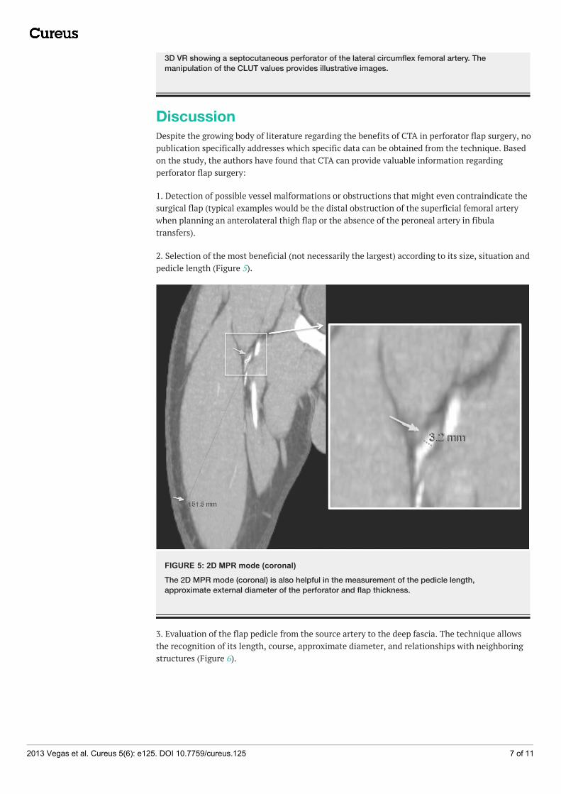
2. Selection of the most beneficial (not necessarily the largest) according to its size, situation andpedicle length (Figure 5).
FIGURE 5: 2D MPR mode (coronal)
The 2D MPR mode (coronal) is also helpful in the measurement of the pedicle length,approximate external diameter of the perforator and flap thickness.
3. Evaluation of the flap pedicle from the source artery to the deep fascia. The technique allowsthe recognition of its length, course, approximate diameter, and relationships with neighboringstructures (Figure 6).
2013 Vegas et al. Cureus 5(6): e125. DOI 10.7759/cureus.125 7 of 11

FIGURE 6: 2D MPR (coronal) (MIP 50)
2D MPR (coronal) (MIP 50). The maximum intensity projection allows the evaluation of perforatorsand pedicle in DIEP flap.
4. Evaluation of side branches arising from the flap pedicle, of great aid when planning acompound or free style free flap.
5. Evaluation of the number and size of pedicle veins at the suture site (Figure 7).
FIGURE 7: 2D MPR
2D MPR. CTA also permits the evaluation of the pedicle veins.
6. Optimized flap design. The perforator course in the subcutaneous fat defines the flap axis. Theevaluation of neighbouring perforators allows their capture, thus enabling large, reliable flapswith minimal risk of marginal skin or fat necrosis (Figure 8).
2013 Vegas et al. Cureus 5(6): e125. DOI 10.7759/cureus.125 8 of 11
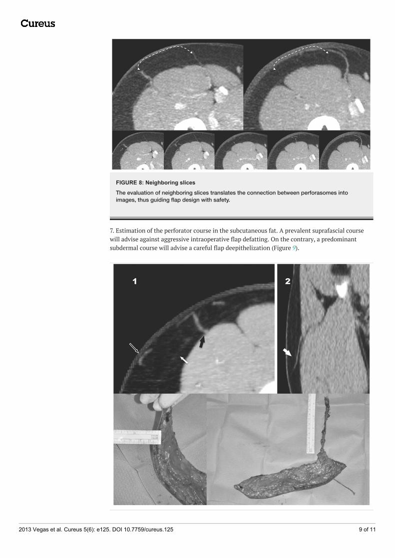
FIGURE 8: Neighboring slices
The evaluation of neighboring slices translates the connection between perforasomes intoimages, thus guiding flap design with safety.
7. Estimation of the perforator course in the subcutaneous fat. A prevalent suprafascial coursewill advise against aggressive intraoperative flap defatting. On the contrary, a predominantsubdermal course will advise a careful flap deepithelization (Figure 9).
2013 Vegas et al. Cureus 5(6): e125. DOI 10.7759/cureus.125 9 of 11

FIGURE 9: Perforator course in the subcutaneous fat
The perforator course in the subcutaneous fat provides the key for an immediate, safedeepithelization and defatting of the flap. 1. With predominantly subdermal perforators,uncontrolled deepithelization should be regarded with caution in large flaps. However, defatting isreasonably safe (Black arrow. Perforator. Empty arrow. Predominant subdermal course ofperforator. White arrow. Suprafascial course of perforator). 2. With predominantly suprafascialperforators, defatting, but not deepithelization, is hazardous.
8. Evaluation of the quality and distribution of recipient vessels and identification of possibledegenerative or traumatic vessel lesions.
Radiologists are, of course, the specialists in image post-processing but only the surgeon is ableto translate the image findings into surgical decisions. Consequently, more and more surgeonsare showing interest in image post-processing but, in most hospitals, the availability ofprofessional workstations is limited and their use is mostly restricted to radiologists under a busyclinical activity. Built-in DICOM viewers included on the CD/DVD are reduced versions of theoriginal applications with limited processing capabilities that do not allow a competent imagepost-processing. OsiriX fills the gap with efficiency and, at present, it is probably the bestalternative for the home user. Two factors have a direct influence in image post-processing ofDICOM files: software and informant. There is data enough to sustain the excellent performanceof OsiriX in the management of DICOM files. OsiriX, except for the FDA/CE-approved MDversion, is not certified as a commercial medical device for primary diagnostic imaging and itsuse in clinical practice might have ethical and legal implications. Consequently, although thebenefit of using the application to improve the preoperative knowledge of the particular patient’sanatomy is unquestionable, an adequate learning period is fundamental because a deficient useof the tool can cause erroneous information that might be potentially harmful for the patients.
ConclusionsCTA has demonstrated to be of great aid in the preoperative planning of perforator flaps. Until anon-invasive non-radiating imaging technology can replace it, great efforts must be done tominimize the risk of radiation following the ALARA (As Low As Reasonably Acceptable) principle.From a surgeon’s perspective, getting involved in image post-processing is beneficial to optimizethe translation of image findings into surgical decisions. Until the professional imagingapplications included in manufactured workstations are available to the individual user, OsiriXfills the gap with extreme efficiency.
Additional InformationDisclosuresHuman subjects: The Institutional Ethics Committee from The Hospital Fremap Majadahondaissued approval N/A. Animal subjects: This study did not involve animal subjects or tissue.
AcknowledgementsTo Dr. Rafael Acosta Rojas, good friend and better plastic surgeon, for introducing me to thestimulating world of DICOM image post-processing.
References1. Rozen WM, Whitaker IS, Stella DL, et al.: The radiation exposure of Computed Tomographic
Angiography (CTA) in DIEP flap planning: low dose but high impact. J Plast Reconstr Aesthet Surg.2009, 62:e654-655.
2. Pratt GF, Rozen WM, Chubb D, et al. : Preoperative imaging for perforator flaps in reconstructive
2013 Vegas et al. Cureus 5(6): e125. DOI 10.7759/cureus.125 10 of 11

surgery: a systematic review of the evidence for current techniques. Ann Plast Surg. 2012, 69:3-9.3. Smit JM, Dimopoulou A, Liss AG, et al.: Preoperative CT angiography reduces surgery time in
perforator flap reconstruction. J Plast Reconstr Aesthet Surg. 2009, 62:1112-1117.4. Phillips TJ, Stella DL, Rozen WM, et al.: Abdominal Wall CT Angiography: A Detailed Account of a
Newly Established Preoperative Imaging Technique. Radiology. 2008, 249:32-44.5. Masia J, Larranaga J, Clavero JA, et al.: The value of the multidetector row computed tomography
for the preoperative planning of deep inferior epigastric artery perforator flap: our experience in 162cases. Ann Plast Surg. 2008, 60:29-36.
6. Rozen WM, Paddle AM, Chubb D, et al.: Guiding local perforator flaps with preoperative imaging:revealing perforator anatomy to improve flap design. Plast Reconstr Surg. 2012, 130:130-134.
7. Uppal RS, Casaer B, Van Landuyt K, et al. : The efficacy of preoperative mapping of perforators inreducing operative times and complications in perforator flap breast reconstruction. J Plast ReconstrAesthet Surg . 2009, 62:859-864.
8. Tejerina Botella C, Márquez Cañada JM, García Andrés E, et al.: Estudio preoperatorio de vasosreceptores en reconstrucción mamaria con colgajo DIEP. Cir Plást Iberolatinoam. 2011, 37:233-238.
9. Adams AS, Wright MJ, Johnston S, et al.: The Use of Multislice CT Angiography Preoperative Studyfor Supraclavicular Artery Island Flap Harvesting. Ann Plast Surg. 2012, 69:312-5.
10. Karanas YL, Antony A, Rubin G, et al. : Preoperative CT angiography for free fibula transfer .Microsurgery. 2004, 24:125-127.
11. Garvey PB, Selber JC, Madewell JE, et al.: A prospective study of preoperative computedtomographic angiography for head and neck reconstruction with anterolateral thigh flaps. PlastReconstr Surg. 2011, 127:1505-1514.
12. United Nations Scientific Committee on the Effects of Atomic Radiation UNSCEAR 1993 Report tothe General Assembly. : Sources and Effects of Ionizing Radiation. New York, United Nations; 1993.
13. Fazel R, Krumholz HM, Wang Y, et al.: Exposure to low-dose ionizing radiation from medicalimaging procedures. N Engl J Med. 2009, 361:849-857.
14. Einstein AJ, Henzlova MJ, Rajagopalan S: Estimating risk of cancer associated with radiationexposure from 64-slice computed tomography coronary angiography. JAMA. 2007, 298:317-323.
15. Sodickson A: Strategies for reducing radiation exposure in multi-detector row CT . Radiol Clin NorthAm. 2012, 50:1-14.
16. Rozen WM, Anavekar NS, Grinsell D, et al.: Improving surgical outcomes with the use of CTangiography. Microsurgery. 2009, 29:249-250.
17. Greenspun D, Vasile J, Levine JL, et al.: Anatomic imaging of abdominal perforator flaps withoutionizing radiation: seeing is believing with magnetic resonance imaging angiography. J ReconstrMicrosurg . 2010, 26:37-44.
18. Vasile JV, Newman T, Rusch DG, et al. : Anatomic imaging of gluteal perforator flaps withoutionizing radiation: seeing is believing with magnetic resonance angiography. J Reconstr Microsurg.2010, 26:45-57.
19. Rozen WM, Stella DL, Bowden J, et al.: Advances in the pre-operative planning of deep inferiorepigastric artery perforator flaps: Magnetic resonance angiography. Microsurgery . 2009, 29:119-123.
20. Haider CR, Glockner JF, Stanson AW, et al.: Peripheral vasculature: high-temporal- and high-spatial-resolution three-dimensional contrast-enhanced MR angiography. Radiology. 2009, 253:831-843.
21. Alonso-Burgos A, Garcia-Tutor E, Bastarrika G, et al.: Preoperative planning of DIEP and SGAP flaps:preliminary experience with magnetic resonance angiography using 3-tesla equipment and blood-pool contrast medium. J Plast Reconstr Aesthet Surg. 2010, 63:298-304. 19121986
22. Rosset A, Spadola L, Ratib O: OsiriX: An open-source software for navigating in multidimensionalDICOM images. J Digit Imaging. 2004, 17:205-216.
23. Prasad KN, Cole WC, Haase GM: Radiation protection in humans: Extending the concept of as low asreasonably achievable (ALARA) from dose to biological damage. Br J Radiol. 2004, 77:97-99.
2013 Vegas et al. Cureus 5(6): e125. DOI 10.7759/cureus.125 11 of 11