Embed Size (px)
Citation preview

Percutaneous Occlusion of Right Partial Anomalous Pulmonary VenousConnection with Dual Drainage to the Innominate Vein and the Left Atrium:A Unique Anatomical Finding
Alejandro R. Peirone1,*, Alejandro E. Contreras2, Carolina Carrizo2, Mailén Konicoff2 andRaúl O. Cayre3
1Servicio de Hemodinamia y Cardiología Intervencionista, Hospital Privado Universitario de Córdoba, Córdoba, X5016KEH,Argentina2Servicio de Cardiología, Hospital Privado Universitario de Córdoba, Córdoba, X5016KEH, Argentina3Director de Docencia e Investigación, CORDIS, Instituto del Corazón. Resistencia, Chaco, H3508EFR, Argentina*Corresponding Author: Alejandro R. Peirone. Email: [email protected]
Received: 29 July 2020 Accepted: 17 August 2020
ABSTRACT
A 43-year-old woman with a past medical history of aortic coarctation surgically repaired at the age of 3 yearsusing an end-to-end anastomosis, presented with 2 years complain of increasing dyspnea and fatigue with exerciseassociated to frequent palpitations. During extensive work-up, she was found to have a partial anomalous pul-monary venous connection (PAPVC) with “dual drainage” represented by a communication between the rightpulmonary veins draining into the left atrium and the innominate vein via an anomalous vein due to a persistenceof early connections between the sinus of the right pulmonary veins and the cardinal veins system in the splanch-nic plexus and also a persistence of the proximal portion of the left anterior cardinal vein. She was successfullytreated through a percutaneous implantation of a vascular plug occluding the vertical portion of the anomalousvein diverting the flow to the left atrium. To the best of our knowledge, this anatomical variant of partial anom-alous pulmonary venous connection with dual drainage has not been previously reported.
KEYWORDS
Partial anomalous pulmonary venous connection; dual drainage; percutaneous closure; vascular plug
1 Introduction
Partial anomalous pulmonary venous connection (PAPVC) is a rare congenital cardiac anomaly in whichsome, but not all, of the pulmonary veins connect to the right atrium or to one or more of its venous tributariessuch as superior vena cava, inferior vena cava, innominate vein or coronary sinus. The prevalence has beenreported to be as high as 0.7% in autopsy series and up to 10% in patients with an atrial septal defect beingmore frequently described involving veins from the right side [1]. The traditional classification of PAPVCdoes not categorize “dual drainage” separately [2] and these variants, with combination of supracardiacand cardiac PAPVC, are extremely rare with very few previous case reports encountered [1,3–8].Moreover, the true incidence of PAPVC cases with dual drainage is difficult to estimate, since patients areoften asymptomatic and may escape detection. Surgical correction has been the standard treatment when
This work is licensed under a Creative Commons Attribution 4.0 International License, whichpermits unrestricted use, distribution, and reproduction in any medium, provided the originalwork is properly cited.
DOI: 10.32604/CHD.2020.013199
ARTICLE
echT PressScience
PAPVC results in right heart volume overload and/or clinical symptoms although in patients with dualdrainage, transcatheter therapy may represent an alternative to surgical correction.
2 Case Presentation
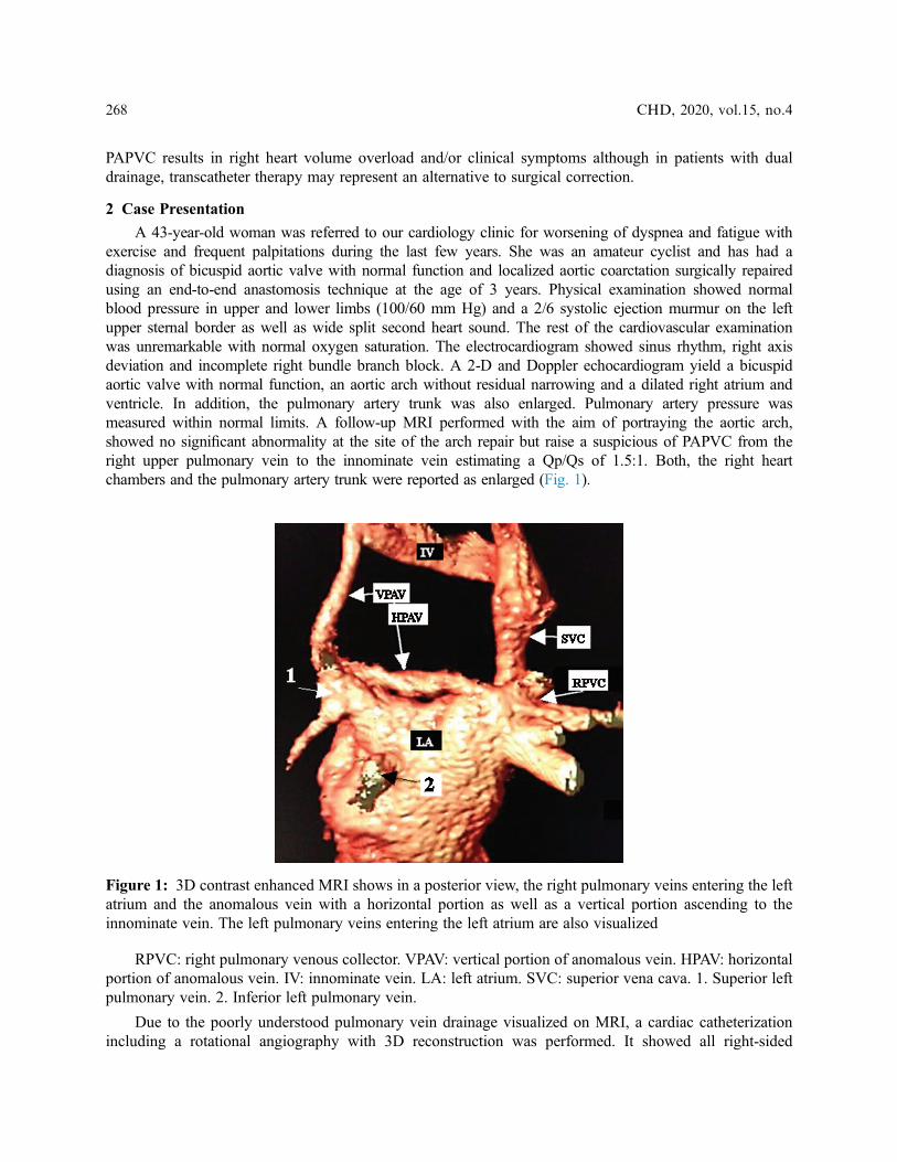
A 43-year-old woman was referred to our cardiology clinic for worsening of dyspnea and fatigue withexercise and frequent palpitations during the last few years. She was an amateur cyclist and has had adiagnosis of bicuspid aortic valve with normal function and localized aortic coarctation surgically repairedusing an end-to-end anastomosis technique at the age of 3 years. Physical examination showed normalblood pressure in upper and lower limbs (100/60 mm Hg) and a 2/6 systolic ejection murmur on the leftupper sternal border as well as wide split second heart sound. The rest of the cardiovascular examinationwas unremarkable with normal oxygen saturation. The electrocardiogram showed sinus rhythm, right axisdeviation and incomplete right bundle branch block. A 2-D and Doppler echocardiogram yield a bicuspidaortic valve with normal function, an aortic arch without residual narrowing and a dilated right atrium andventricle. In addition, the pulmonary artery trunk was also enlarged. Pulmonary artery pressure wasmeasured within normal limits. A follow-up MRI performed with the aim of portraying the aortic arch,showed no significant abnormality at the site of the arch repair but raise a suspicious of PAPVC from theright upper pulmonary vein to the innominate vein estimating a Qp/Qs of 1.5:1. Both, the right heartchambers and the pulmonary artery trunk were reported as enlarged (Fig. 1).
RPVC: right pulmonary venous collector. VPAV: vertical portion of anomalous vein. HPAV: horizontalportion of anomalous vein. IV: innominate vein. LA: left atrium. SVC: superior vena cava. 1. Superior leftpulmonary vein. 2. Inferior left pulmonary vein.
Due to the poorly understood pulmonary vein drainage visualized on MRI, a cardiac catheterizationincluding a rotational angiography with 3D reconstruction was performed. It showed all right‑sided
Figure 1: 3D contrast enhanced MRI shows in a posterior view, the right pulmonary veins entering the leftatrium and the anomalous vein with a horizontal portion as well as a vertical portion ascending to theinnominate vein. The left pulmonary veins entering the left atrium are also visualized
268 CHD, 2020, vol.15, no.4
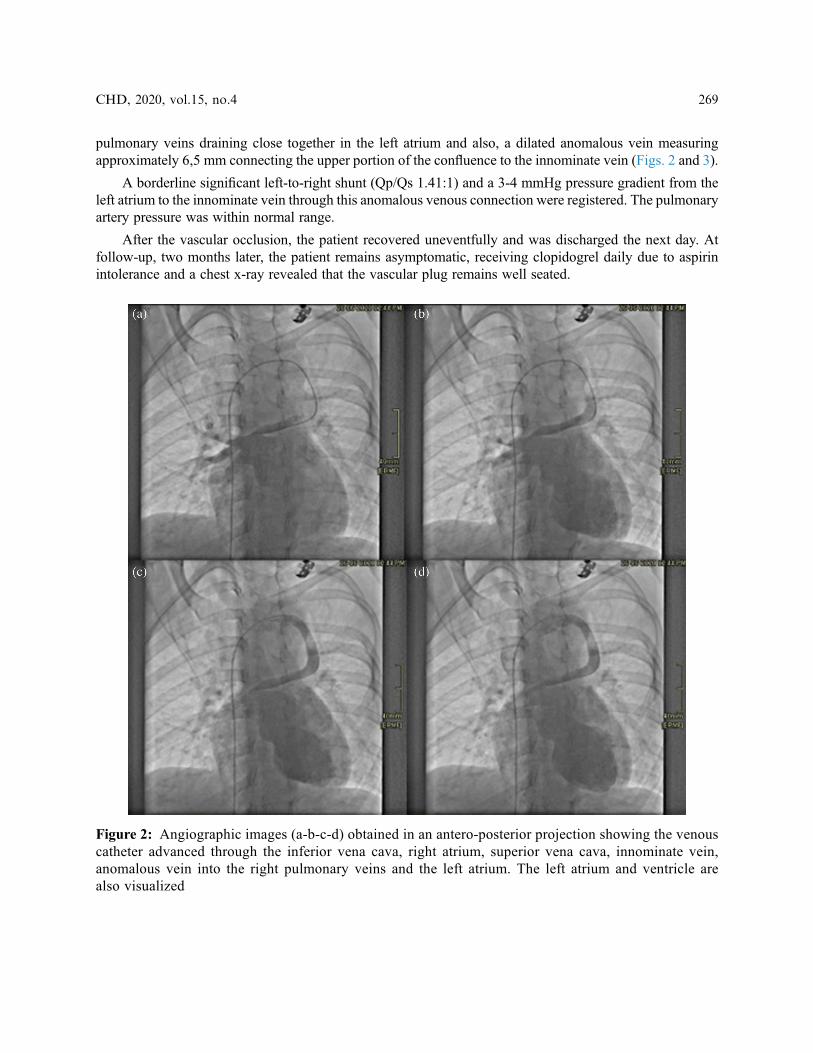
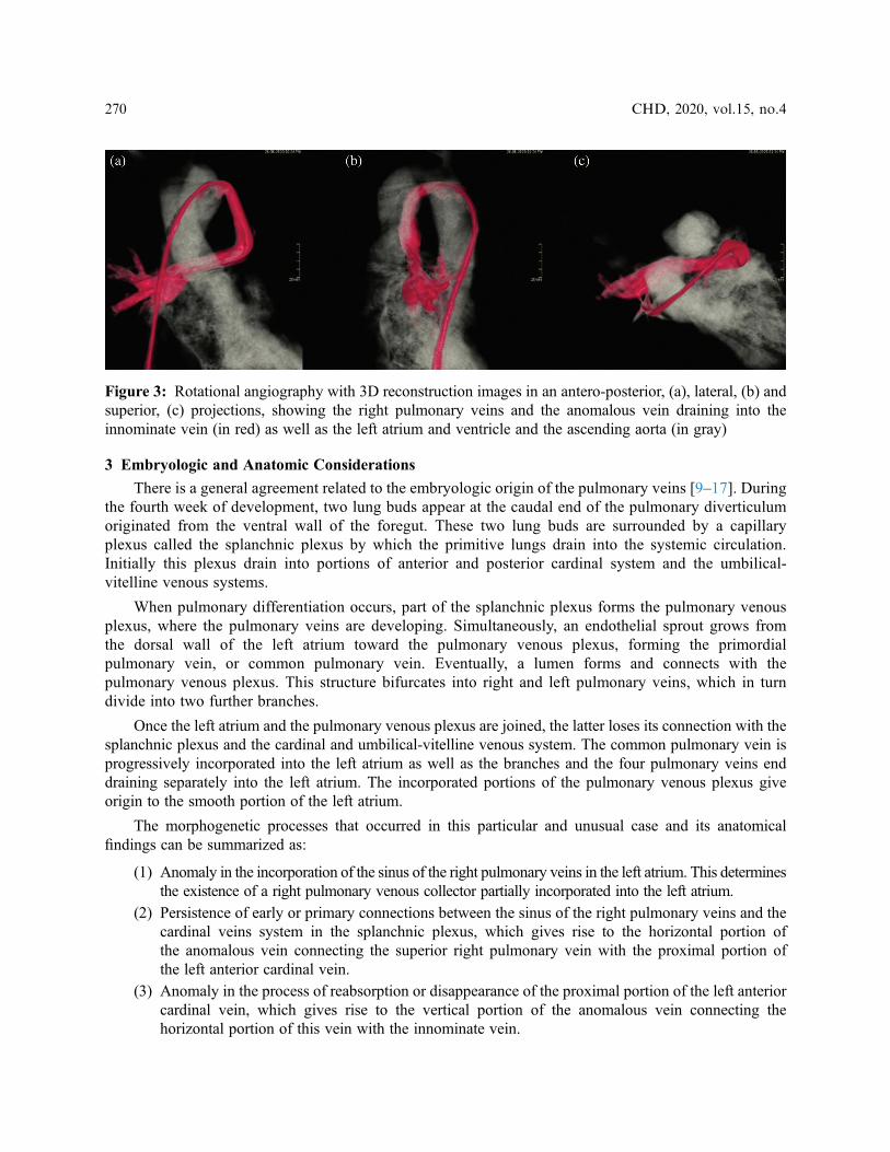
pulmonary veins draining close together in the left atrium and also, a dilated anomalous vein measuringapproximately 6,5 mm connecting the upper portion of the confluence to the innominate vein (Figs. 2 and 3).
A borderline significant left-to-right shunt (Qp/Qs 1.41:1) and a 3-4 mmHg pressure gradient from theleft atrium to the innominate vein through this anomalous venous connection were registered. The pulmonaryartery pressure was within normal range.
After the vascular occlusion, the patient recovered uneventfully and was discharged the next day. Atfollow‑up, two months later, the patient remains asymptomatic, receiving clopidogrel daily due to aspirinintolerance and a chest x-ray revealed that the vascular plug remains well seated.
Figure 2: Angiographic images (a-b-c-d) obtained in an antero-posterior projection showing the venouscatheter advanced through the inferior vena cava, right atrium, superior vena cava, innominate vein,anomalous vein into the right pulmonary veins and the left atrium. The left atrium and ventricle arealso visualized
CHD, 2020, vol.15, no.4 269
3 Embryologic and Anatomic Considerations
There is a general agreement related to the embryologic origin of the pulmonary veins [9–17]. Duringthe fourth week of development, two lung buds appear at the caudal end of the pulmonary diverticulumoriginated from the ventral wall of the foregut. These two lung buds are surrounded by a capillaryplexus called the splanchnic plexus by which the primitive lungs drain into the systemic circulation.Initially this plexus drain into portions of anterior and posterior cardinal system and the umbilical-vitelline venous systems.
When pulmonary differentiation occurs, part of the splanchnic plexus forms the pulmonary venousplexus, where the pulmonary veins are developing. Simultaneously, an endothelial sprout grows fromthe dorsal wall of the left atrium toward the pulmonary venous plexus, forming the primordialpulmonary vein, or common pulmonary vein. Eventually, a lumen forms and connects with thepulmonary venous plexus. This structure bifurcates into right and left pulmonary veins, which in turndivide into two further branches.
Once the left atrium and the pulmonary venous plexus are joined, the latter loses its connection with thesplanchnic plexus and the cardinal and umbilical-vitelline venous system. The common pulmonary vein isprogressively incorporated into the left atrium as well as the branches and the four pulmonary veins enddraining separately into the left atrium. The incorporated portions of the pulmonary venous plexus giveorigin to the smooth portion of the left atrium.
The morphogenetic processes that occurred in this particular and unusual case and its anatomicalfindings can be summarized as:
(1) Anomaly in the incorporation of the sinus of the right pulmonary veins in the left atrium. This determinesthe existence of a right pulmonary venous collector partially incorporated into the left atrium.
(2) Persistence of early or primary connections between the sinus of the right pulmonary veins and thecardinal veins system in the splanchnic plexus, which gives rise to the horizontal portion ofthe anomalous vein connecting the superior right pulmonary vein with the proximal portion ofthe left anterior cardinal vein.
(3) Anomaly in the process of reabsorption or disappearance of the proximal portion of the left anteriorcardinal vein, which gives rise to the vertical portion of the anomalous vein connecting thehorizontal portion of this vein with the innominate vein.
Figure 3: Rotational angiography with 3D reconstruction images in an antero-posterior, (a), lateral, (b) andsuperior, (c) projections, showing the right pulmonary veins and the anomalous vein draining into theinnominate vein (in red) as well as the left atrium and ventricle and the ascending aorta (in gray)
270 CHD, 2020, vol.15, no.4
(4) The different morphogenetic processes mentioned, determine that the right pulmonary veins havedual drainage in the left atrium and in the innominate vein.
4 Cardiac Catheterization
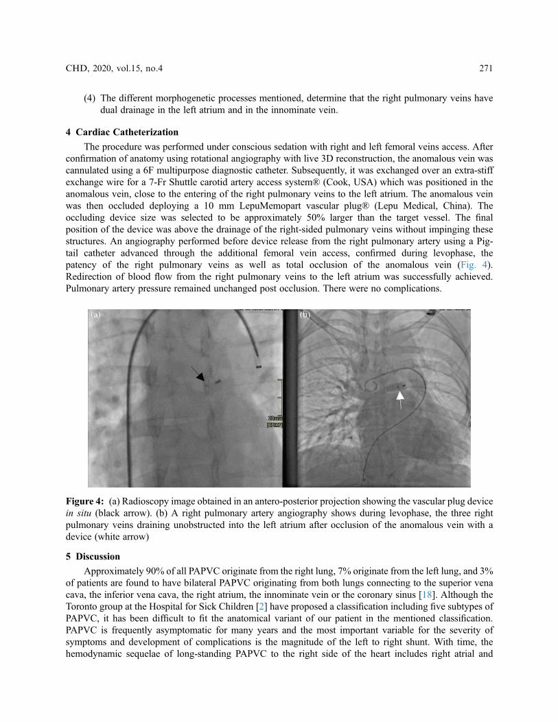
The procedure was performed under conscious sedation with right and left femoral veins access. Afterconfirmation of anatomy using rotational angiography with live 3D reconstruction, the anomalous vein wascannulated using a 6F multipurpose diagnostic catheter. Subsequently, it was exchanged over an extra-stiffexchange wire for a 7‑Fr Shuttle carotid artery access system® (Cook, USA) which was positioned in theanomalous vein, close to the entering of the right pulmonary veins to the left atrium. The anomalous veinwas then occluded deploying a 10 mm LepuMemopart vascular plug® (Lepu Medical, China). Theoccluding device size was selected to be approximately 50% larger than the target vessel. The finalposition of the device was above the drainage of the right‑sided pulmonary veins without impinging thesestructures. An angiography performed before device release from the right pulmonary artery using a Pig-tail catheter advanced through the additional femoral vein access, confirmed during levophase, thepatency of the right pulmonary veins as well as total occlusion of the anomalous vein (Fig. 4).Redirection of blood flow from the right pulmonary veins to the left atrium was successfully achieved.Pulmonary artery pressure remained unchanged post occlusion. There were no complications.
5 Discussion
Approximately 90% of all PAPVC originate from the right lung, 7% originate from the left lung, and 3%of patients are found to have bilateral PAPVC originating from both lungs connecting to the superior venacava, the inferior vena cava, the right atrium, the innominate vein or the coronary sinus [18]. Although theToronto group at the Hospital for Sick Children [2] have proposed a classification including five subtypes ofPAPVC, it has been difficult to fit the anatomical variant of our patient in the mentioned classification.PAPVC is frequently asymptomatic for many years and the most important variable for the severity ofsymptoms and development of complications is the magnitude of the left to right shunt. With time, thehemodynamic sequelae of long-standing PAPVC to the right side of the heart includes right atrial and
Figure 4: (a) Radioscopy image obtained in an antero-posterior projection showing the vascular plug devicein situ (black arrow). (b) A right pulmonary artery angiography shows during levophase, the three rightpulmonary veins draining unobstructed into the left atrium after occlusion of the anomalous vein with adevice (white arrow)
CHD, 2020, vol.15, no.4 271
ventricular dilation and pulmonary arterial hypertension leading to occurrence of atrial arrhythmias and right-sided heart failure. Exceptionally, paradoxical embolization may be consider with this type of anatomicalfinding in the view that any connection between a systemic vein and the left atrium may allowparadoxical embolism.
Two anatomic types of PAPVC with dual drainage have been encountered. The vertical vein type, withdual venous drainage from the upper lobe of the left lung to the innominate vein via a large vertical vein andto the left atrium via another more or less developed left pulmonary vein and the Scimitar vein type, with ananomalous venous connection from the middle and lower pulmonary lobes of the right lung to the inferiorvena cava below the level of the diaphragm, associated with an anomalous venous connection of the Scimitarvein to the left atrium and sequestration syndrome [19].
After confirmation of dual drainage or supply, transcatheter occlusion may be considered. The worldexperience reported by Luciano et al. indicates the after an adequate selection of patients, theinterventional procedures with the aim of diverting the flow exclusively to the left atrium have beensuccessful implanting both various coils and self- expandable devices. In their reported experience, nocomplications occurred, except for device embolization in one patient. Moreover, additional transcatheterprocedures were performed including secundum ASD closure, systemic arterial supply occlusion andstent implantation for aortic coarctation. Comparison between occlusion of a vertical vein type orScimitar vein type of PAPVC showed no difference. Interestingly, patients presenting after 40 year-oldtended to have more symptoms [19].
In the uncommon anatomical variant of TAPVC associated to dual drainage or supply, percutaneousclosure of the abnormal vertical vein implanting a self-expandable device or coils is possible. Thisstrategy leads to redirect the flow avoiding obstruction to the pulmonary venous circulation to the left atrium.
To our knowledge, there are only two previous reported cases [20,21] of PAPVC in association withcoarctation of aorta treated successfully with a percutaneous approach, thereby obviating the need forsurgery and its known potential risks. In both previous reports the abnormal veins were left-sided and aleft persistent vertical vein was occluded implanting a self-expandable device redirecting the flow to theleft atrium. The difference with our case relies on that the PAPVC was diagnosed many years afterthe initial aortic coarctation surgery; hence, both lesions were treated separately. Moreover, in our patientthe abnormality of the pulmonary veins was right-sided, on the contrary, in the other two previous casesreported, those were left-sided.
In summary, so far as we know, this is the first report of a patient suffering from PAPVC involving theright pulmonary veins associated to dual drainage via an anomalous vein with a horizontal portion due to apersistence of early connections between the sinus of the right pulmonary veins and the cardinal veins systemin the splanchnic plexus and also a persistence of the proximal portion of the left anterior cardinal vein. Theocclusion was planned to be performed proximately at the level of the connection of the anomalous vein tothe right pulmonary veins achieving successful diversion of the right pulmonary venous flow to the leftatrium. Diligent evaluation and knowledge during clinical diagnostic work-up of the occurrence ofPAPVC with dual supply, whether isolated or associated with other cardiac defects that may be amenableto additional interventional procedures, is mandatory to be able to plan a less-invasive transcatheterapproach to correct these cardiac defects. Percutaneous closure of the abnormal venous connection iscertainly the treatment of choice for PAPVC with dual drainage.
Funding Statement: The authors received no specific funding for this study.
Conflicts of Interest: The authors declare that they have no conflicts of interest to report regarding thepresent study.
272 CHD, 2020, vol.15, no.4
References1. Forbess, L. W., O’Laughlin, M. P., Harrison, J. K. (1998). Partially anomalous pulmonary venous connection:
demonstration of dual drainage allowing nonsurgical correction. Catheterization and CardiovascularDiagnosis, 44(3), 330–335. DOI 10.1002/(SICI)1097-0304(199807)44:3<330::AID-CCD19>3.0.CO;2-O.
2. Alsoufi, B., Cai, S., Van Arsdell, G. S., Williams, W. G., Caldarone, C. A. et al. (2007). Outcomes after surgicaltreatment of children with partial anomalous pulmonary venous connection. Annals of Thoracic Surgery, 84(6),2020–2026. DOI 10.1016/j.athoracsur.2007.05.046.
3. Dähnert, I., Riede, F. T., Kostelka, M. (2007). Partial anomalous pulmonary venous drainage of the left upperpulmonary vein–catheter interventional treatment is sometimes possible. Clinical Research in Cardiology,96(7), 511–513. DOI 10.1007/s00392-007-0518-8.
4. Gomez, J., Soledispa, C. (2012). Redirection of anomalous venous pulmonary flow to left atrium using a vascularplug II. Journal of Invasive Cardiology, 24(5), E96–E98.
5. Recto, M. R., Sadlo, H., Sobczyk, W. L. (2007). Rare case of persistent left superior vena cava to left upperpulmonary vein: pathway for paradoxical embolization and development of transient ischemic attack andsubsequent occlusion with an amplatzer vascular plug. Journal of Invasive Cardiology, 19(10), E313–E316.
6. Wilson, W., Horlick, E., Benson, L. (2015). Successful transcatheter occlusion of an anomalous pulmonary veinwith dual drainage to the left atrium. Catheterization and Cardiovascular Interventions, 85(7), 1212–1216. DOI10.1002/ccd.25734.
7. Kobayashi, D., Forbes, T. J., Delius, R. E., Aggarwal, S. (2012). Amplatzer vascular plug for transcatheter closureof persistent unligated vertical vein after repair of infracardiac total anomalous pulmonary venous connection.Catheterization and Cardiovascular Interventions, 80(2), 192–198. DOI 10.1002/ccd.23497.
8. Craig, J. M., Darling, R. C., Rothney, W. B. (1957). Total pulmonary venous drainage into the right side of theheart; report of 17 autopsied cases not associated with other major cardiovascular anomalies. LaboratoryInvestigation, 6(1), 44–64.
9. Lucas, R. V., Jr, Anderson, R. C., Amplatz, K., Adams, P., Jr, Edwards, J. E. (1963). Congenital causes of pulmonaryvenous obstruction. Pediatric Clinics of North America, 10(3), 781–836. DOI 10.1016/S0031-3955(16)31451-1.
10. Sadler, T. W. (1985). Langman’s medical embryology. 5th Edition. . Philadelphia: Lippincott Williams & WilkinsCompany.
12. Reller, M. D., McDonald, R. W., Gerlis, L. M., Thornburg, K. L. (1991). Cardiac embryology: basic review andclinical correlations. Journal of the American Society of Echocardiography, 4(5), 519–532. DOI 10.1016/S0894-7317(14)80388-X.
13. Valdés-Cruz, L. M., Cayre, R. O. (1999). Echocardiographic diagnosis of congenital heart disease. Philadelphia:Lippincot-Raven Publishers.
14. Blom, N. A., Gittenberger-de Groot, A. C., Jongeneel, T. H., DeRuiter, M. C., Poelmann, R. E. et al. (2001). Normaldevelopment of the pulmonary veins in human embryos and formulation of a morphogenetic concept for sinusvenosus defects. American Journal of Cardiology, 87(3), 305–309. DOI 10.1016/S0002-9149(00)01363-1.
15. Latson, L. A., Prieto, L. R. (2007). Congenital and acquired pulmonary vein stenosis. Circulation, 115(1), 103–108. DOI 10.1161/CIRCULATIONAHA.106.646166.
16. Murillo, H., Cutalo, M. J., Jones, R. P., Lane, M. J., Fleischmann, D. et al. (2012). Pulmonary circulation imaging:embryology and normal anatomy. Seminars in Ultrasound, CT and MRI, 33(6), 473–484. DOI 10.1053/j.sult.2012.08.001.
17. Poelmann, R. E., Gittenberger-de Groot, A. C., Jongbloed, M. R. M., DeRuiter, M. C. (2016). Congenital heartdiseases: the broken heart. Clinical features, human genetics and molecular pathways. Vienna: Springer-Verlag Wien.
18. Jongbloed, M. R. M., Poelmann, R. E., Gittenberger-de Groot, A. C. (2017). The complete reference for scimitarsyndrome. anatomy, epidemiology, diagnosis and treatment. London: Academic Press.
19. AboulHosn, J. A., Criley, J. M., Stringer, W. W. (2003). Partial anomalous pulmonary venous return: case report andreview of the literature. Catheterization and Cardiovascular Interventions, 58(4), 548–552. DOI 10.1002/ccd.10475.
CHD, 2020, vol.15, no.4 273

20. Luciano, D., Laux, D., Boudjemline, Y., Hascoët, S., Lusson, J. R. et al. (2013). Transcatheter therapy in partiallyabnormal pulmonary venous return with additional drainage to the left atrium. International Journal of Cardiology,170(2), 221–226. DOI 10.1016/j.ijcard.2013.10.061.
21. Mamas, M. A., Clarke, B., Mahadevan, V. S. (2010). Percutaneous treatment of dual pulmonary venous drainageand coarctation of the aorta in a single patient. Experimental and Clinical Cardiology, 15(1), 11–13.
22. Al Qbandi, M., Thinakar Vel, M. (2018). Transcatheter therapy of partial anomalous pulmonary venous connectionwith dual drainage and coarctation of the aorta in a single patient. Journal of the Saudi Heart Association, 30(4),311–315. DOI 10.1016/j.jsha.2018.06.003.
274 CHD, 2020, vol.15, no.4