Embed Size (px)
Citation preview
Vol. 7 No. 1, January-March 2005 �
JK SCIENCE
Pedicled Ileal Seromuscular Flap-A New Technique forProtection of Intestinal Anastomosis in Patients with Peritonitis
Nikhil Talwar, Romesh Lal, OP Pathania
ORIGINAL ARTICLE
From The Department of Surgery, Lady Hardinge Medical College & SSKH Connaught Place, New Delhi-110001.Correspondece to: Dr. Nikhil Talwar, CP-122, Maurya Enclave, Pitam Pura, Delhi 110088.
IntroductionIntestinal anastomotic leaks are a dreaded
complication for any surgeon. Few surgical proceduresthat reduce the risk of anastomotic leakage have beendescribed. A novel method of protecting intestinalanastomosis involves the use of a pedicled ilealseromuscular flap (1). To our knowledge, there are nopapers in literature, which have assessed the usefulnessof this technique in patients with peritonitis. Thus, weconducted a prospective study to ascertain the safety aswell as efficacy of this technique.Material and Methods
The study was carried out in a single surgicalunit in Department of Surgery, Lady Hardinge MedicalCollege, New Delhi, India between January 2001 andApril 2001. Informed consent was taken for theprocedure from all the patients. Twenty patients olderthan 16 years with peritonitis who required ileo-ileal orileo-colic anastomosis were included and all patientunderwent reinforcement of the anastomosis with a
AbstractPedicled ileal seromuscular flap- a new technique for protection of intestinal anastomosis in patientswith peritonitis. This method involves raising a seromuscular flap on a pedicle from the stump ofintestine to be anastomosed. The anastomosis is performed, and then covered with seromuscularflap. The submucosa due to its inherent properties, promotes better healing and reduces the tensionon the anastomosis. There has been no previous study to assess the usefulness of this technique. Weused a pedicled ileal seromuscular flap in twenty consecutive patients with peritonitis who requiredan ileo-ileal or an ileo-colic anastomosis. Anastomotic leak occurred in two out of twenty patients.The safety and efficacy of this technique involving autologous tissues to reinforce intestinalanastomosis has been demonstrated.Key WordsSeromuscular Flap; Anastomotic Dehiscence; Emergency Intestinal Surgery
pedicled ileal seromuscular flap. All patients wereadequately resuscitated before being operated upon. Theyreceived intravenous fluids, nasogastric decompressionand parenteral analgesics before surgery. Intravenousceftriaxone 1 gm was given every 8 hours to all thepatients. After the clinical diagnosis of peritonitis,abdominal surgery was performed as soon as operatingroom was available.Surgical Procedure
All surgeries were done under generalanaesthesia. Abdomen was opened by a midline incisionof adequate length. After identifying the site of the bowelpathology, necessary length of the bowel was resected.Following intestinal resection, a segment from theproximal intestinal stump, of appropriate length washarvested for the flap. To adequately cover theanastomosis, the segment was kept slightly longer thanthe circumference of the anastomosis. Approximately, 5cm of the intestine proximal to the intended flap was
� Vol. 7 No. 1, January-March 2005
JK SCIENCE
resected to achieve the mobility of the flap. Invertedanastomosis was carried out in 2 layers as is the standardpractice, inner layer consisting of through and throughcontinuous suture with 2/0 absorbable polyglycolic acidsutures, and the outer layer consisting of interruptedseromuscular sutures with 2/0 silk. Following theanastomosis, the flap was prepared by opening theintestinal segment longitudinally along the antimesentricborder. The submucosa was infiltrated by an injection ofnormal saline and the mucosa carefully removed withscissors, leaving the submucosal surface as thick aspossible. The submucosal surface of the flap wasapproximated to the anastomosis in such a manner thatthe mesentric axis of the flap was parallel to theanastomotic line. The flap and the anastomotic line werethen sutured with interrupted 4.0 polydioxanon (PDS) at1-cm intervals. Thorough peritoneal lavage wasperformed with pressurised warm normal saline. Twotubes drains, one in the subhepatic space and the otherin the pelvis were inserted and abdomen closed.
Postoperative Follow UpAn independent assessor visited every patient in themorning to record the clinical progress and return ofnormal bowel activity. A clinical leak was defined as ananastomotic dehiscence verified by reoperation, thedevelopment of an enterocutaneous fistula, or theevidence of bowel contents in the drainage fluid. Woundinfection was defined a purulent discharge from thelaparotomic incision.Results
From January 2001 to April 2001, 31 consecutivepatients with a preoperative diagnosis of peritonitis wereidentified, 20 of whom required an ileocolic or ileoilealanastomosis.The patient characteristics are listed in (Table1). Out of these twenty patients, eight patients (40%)had typhoid perforation of the ileum, six (30%) hadintestinal tuberculosis, five (25%) had perforations dueto abdominal trauma and one (5%) had a strangulatedincisional hernia. Fifteen patients (75%) patientsunderwent ileo-ileal anastomosis while five (25%)required an ileo-colic anastomosis.Table 1. Clinical and labarotory parameters of patients who
underwent emergency intestinal anastomosis
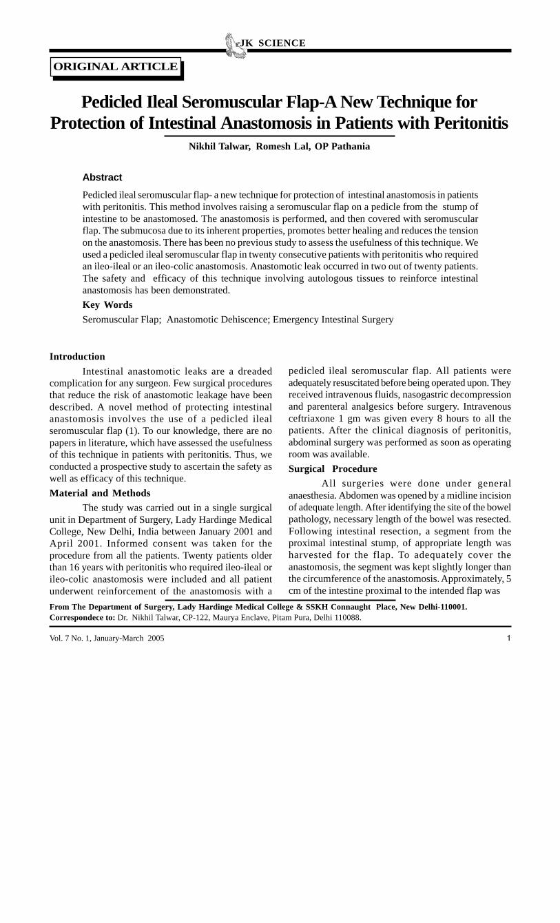
Fig. 1. Photograph taken after right hemicolectomy showingisolated ileal segment (arrow) with an intact pedicle(arrowhead) which was used to fashion a seromuscularflap. TI: Terminalileum; CO: Colon.
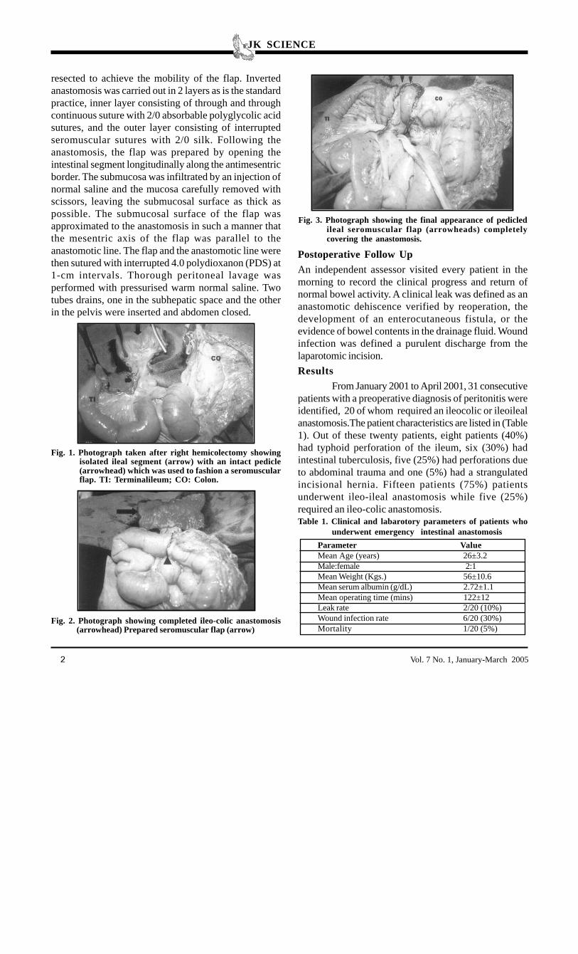
Fig. 2. Photograph showing completed ileo-colic anastomosis(arrowhead) Prepared seromuscular flap (arrow)
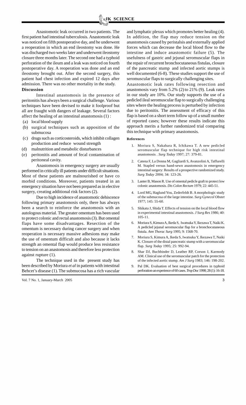
Fig. 3. Photograph showing the final appearance of pedicledileal seromuscular flap (arrowheads) completelycovering the anastomosis.
Parameter ValueMean Age (years) 26±3.2Male:female 2:1Mean Weight (Kgs.) 56±10.6Mean serum albumin (g/dL) 2.72±1.1Mean operating time (mins) 122±12Leak rate 2/20 (10%)Wound infection rate 6/20 (30%)Mortality 1/20 (5%)
Vol. 7 No. 1, January-March 2005 �
JK SCIENCE
Anastomotic leak occurred in two patients. Thefirst patient had intestinal tuberculosis. Anastomotic leakwas noticed on fifth postoperative day, and he underwenta reoperation in which an end ileostomy was done. Hewas discharged two weeks later and underwent ileostomyclosure three months later. The second one had a typhoidperforation of the ileum and a leak was noticed on fourthpostoperative day. A reoperation was done and an endileostomy brought out. After the second surgery, thispatient had chest infection and expired 12 days afteradmission. There was no other mortality in the study.Discussion
Intestinal anastomosis in the presence ofperitonitis has always been a surgical challenge. Varioustechniques have been devised to make it foolproof butall are fraught with dangers of leakage. Several factorsaffect the healing of an intestinal anastomosis (1) :(a) local blood supply(b) surgical techniques such as apposition of the
submucosa(c) drugs such as corticosteroids, which inhibit collagen
production and reduce wound strength(d) malnutrition and metabolic disturbances(e) peritonitis and amount of fecal contamination of
peritoneal cavity.Anastomosis in emergency surgery are usually
performed in critically ill patients under difficult situations.Most of these patients are malnourished or have comorbid conditions. Moreover, patients treated in anemergency situation have not been prepared as in electivesurgery, creating additional risk factors (2).
Due to high incidence of anastomotic dehiscencefollowing primary anastomosis only, there has alwaysbeen a search to reinforce the anastomosis with anautologous material. The greater omentum has been usedto protect colonic and rectal anastomosis (3). But omentalflaps have some disadvantages. Resection of theomentum is necessary during cancer surgery and whenreoperation is necessary massive adhesions may makethe use of omentum difficult and also because it lacksstrength an omental flap would produce less resistanceto tension on an anastomosis and therefore less protectionagainst rupture (1).
The technique used in the present study hasbeen described by Moriura et al in patients with intestinalBehcet’s disease (1). The submucosa has a rich vascular
and lymphatic plexus which promotes better healing (4).In addition, the flap may reduce tension on theanastomosis caused by peristalsis and externally appliedforces which can decrease the local blood flow to theintestine and induce anastomotic failure (5). Theusefulness of gastric and jejunal seromuscular flaps inthe repair of recurrent bronchocutaneous fistulas, closureof the pancreatic stump and infected aortic stumps iswell documented (6-8). These studies support the use ofseromuscular flaps to surgically challenging sites.Anastomotic leak rates following resection andanastomosis vary from 5.2% (2) to 21% (9). Leak ratesin our study are 10%. Our study supports the use of apedicled ileal seromuscular flap to surgically challengingsites where the healing process is perturbed by infectiondue to peritonitis. The assessment of efficacy of thisflap is based on a short term follow up of a small numberof reported cases; however these results indicate thisapproach merits a further randomized trial comparingthis technique with primary anastomosis.References
1. Moriura S, Nakahara R, Ichikawa T. A new pedicledseromuscular flap technique for high risk intestinalanastomosis . Surg Today 1997; 27: 379-81.
2. Catena F, La Donna M, Gagliardi S, Avanzolini A, TaffurelliM. Stapled versus hand-sewn anastomosis in emergencyintestinal surgery: Results of a prospective randomized study.Surg Today 2004; 34: 123-26.
3. Lanter B, Mason R. Use of omental pedicle graft to protect lowcolonic anastomosis. Dis Colon Rectum 1979; 22: 445-51.
4. Lord MG, Haglund You, Zederfeldt B. A morphologic studyof the submucosa of the large intestine. Surg Gynecol Obstet1977; 145: 55-60.
5. Shikata J, Shida T. Effects of tension on the local blood flowin experimental intestinal anastomosis. J Surg Res 1986; 40:105-11.
6. Moriura S, Kimura A, Ikeda S, Iwatsuka Y, Ikezawa T, Naiki K.A pedicled jejunal seromuscular flap for a bronchocutaneousfistula. Ann Thorac Surg 1995; 9: 1568-70.
7. Moriura S, Kimura A, Ikeda S, Iwatsuka Y, Ikezawa T, NaikiK. Closure of the distal pancreatic stump with a seromuscularflap. Surg Today 1995; 25: 992-94.
8. Shar DJ, Buchbinder D, Leather RP, Corson J, KarmodyAM. Clinical use of the seromuscular patch for the protectionof the infected aortic stump. Am J Surg 1983; 146: 198-202.
9. Pal DK. Evaluation of best surgical procedures in typhoidperforation-an experience of 60 cases. Trop Doc 1998; 28 (1): 16-18.