Embed Size (px)
Citation preview
CASE SERIES PEER REVIEWED | OPEN ACCESS
www.edoriumjournals.com
International Journal of Case Reports and Images (IJCRI)International Journal of Case Reports and Images (IJCRI) is an international, peer reviewed, monthly, open access, online journal, publishing high-quality, articles in all areas of basic medical sciences and clinical specialties.
Aim of IJCRI is to encourage the publication of new information by providing a platform for reporting of unique, unusual and rare cases which enhance understanding of disease process, its diagnosis, management and clinico-pathologic correlations.
IJCRI publishes Review Articles, Case Series, Case Reports, Case in Images, Clinical Images and Letters to Editor.
Website: www.ijcasereportsandimages.com
The distally pedicled peroneus brevis muscle and fasciocutaneous sural artery flap for reconstruction of the distal
third of lower leg
Ingo Schmidt
ABSTRACT
Introduction: The use of distally pedicled peroneus brevis muscle and fasciocutaneous sural artery flap for coverage of the distal end of lower leg is recommended for soft tissue defects with exposure of bones and/or tendons in patients who are not willing or healthy enough to undergo free microvascular tissue transplantation, and do not require microsurgical expertise. Case Series: A short presentation of six cases including a short review of literature will highlight current knowledge and complications of these procedures. Conclusion: The distally pedicled peroneus brevis muscle and fasciocutaneous sural artery flaps are useful for coverage of soft tissue defects of the distal third of lower leg. In our patients, the complication rate of distally pedicled neurofasciocutaneous sural artery flap is higher than the distally pedicled peroneus brevis muscle flap.
(This page in not part of the published article.)

International Journal of Case Reports and Images, Vol. 8 No. 1, January 2017. ISSN – [0976-3198]
Int J Case Rep Images 2017;8(1):17–21. www.ijcasereportsandimages.com
Schmidt 17
CASE REPORT OPEN ACCESS
The distally pedicled peroneus brevis muscle and fasciocutaneous sural artery flap for reconstruction of the
distal third of lower leg
Ingo Schmidt
ABSTRACT
Introduction: The use of distally pedicled peroneus brevis muscle and fasciocutaneous sural artery flap for coverage of the distal end of lower leg is recommended for soft tissue defects with exposure of bones and/or tendons in patients who are not willing or healthy enough to undergo free microvascular tissue transplantation, and do not require microsurgical expertise. Case Series: A short presentation of six cases including a short review of literature will highlight current knowledge and complications of these procedures. Conclusion: The distally pedicled peroneus brevis muscle and fasciocutaneous sural artery flaps are useful for coverage of soft tissue defects of the distal third of lower leg. In our patients, the complication rate of distally pedicled neurofasciocutaneous sural artery flap is higher than the distally pedicled peroneus brevis muscle flap.
Keywords: Distal third lower leg, Distally pedi-cled peroneus brevis muscle flap, Distally pedi-cled sural artery flap, Soft tissue defect
How to cite this article
Schmidt I. The distally pedicled peroneus brevis muscle and fasciocutaneous sural artery flap for reconstruction of the distal third of lower leg. Int J Case Rep Images 2017;8(1):17–21.
Ingo SchmidtAffiliation: SRH Poliklinik Gera GmbH, Straße des Friedens 122, 07548 Gera, Germany.Corresponding Author: Dr. Ingo Schmidt, SRH Poliklinik Gera GmbH, Straße des Friedens 122, 07548 Gera, Germany; Email: [email protected].
Received: 11 July 2016Accepted: 10 October 2016Published: 01 January 2017
Article ID: Z01201703CS10083IS
*********
doi:10.5348/ijcri-201704-CS-10083
INTRODUCTION
Anatomical features of the distal third of lower leg and heel like subcutaneous bone surrounded by tendons with no muscles, vessels in isolated compartments with little intercommunication between them make the coverage of the wounds in the region a challenging problem. Options for coverage of soft tissue defects are free flaps, perforator flaps, reverse flow flaps, muscle flaps, cross leg flaps, and axial pedicled fasciocutaneous flaps such as the distally pedicled sural artery flap [1–3]. Quality debridement is the key to success for the healing of wounds in this region. Negative-pressure vacuum assisted closure (VAC) therapy before soft tissue coverage provides a sterile and controlled environment that can lessen the duration of wound healing, promotes better capillary circulation, and decreases the bacterial load [4]. The use of distally pedicled peroneus brevis muscle and neurofasciocutaneous sural artery flap for coverage of the distal end of lower leg is recommended for soft tissue defects with exposure of bones and/or tendons in patients who are not willing or healthy enough to undergo free microvascular tissue transplantation, and do not require microsurgical expertise.
CASE SERIES
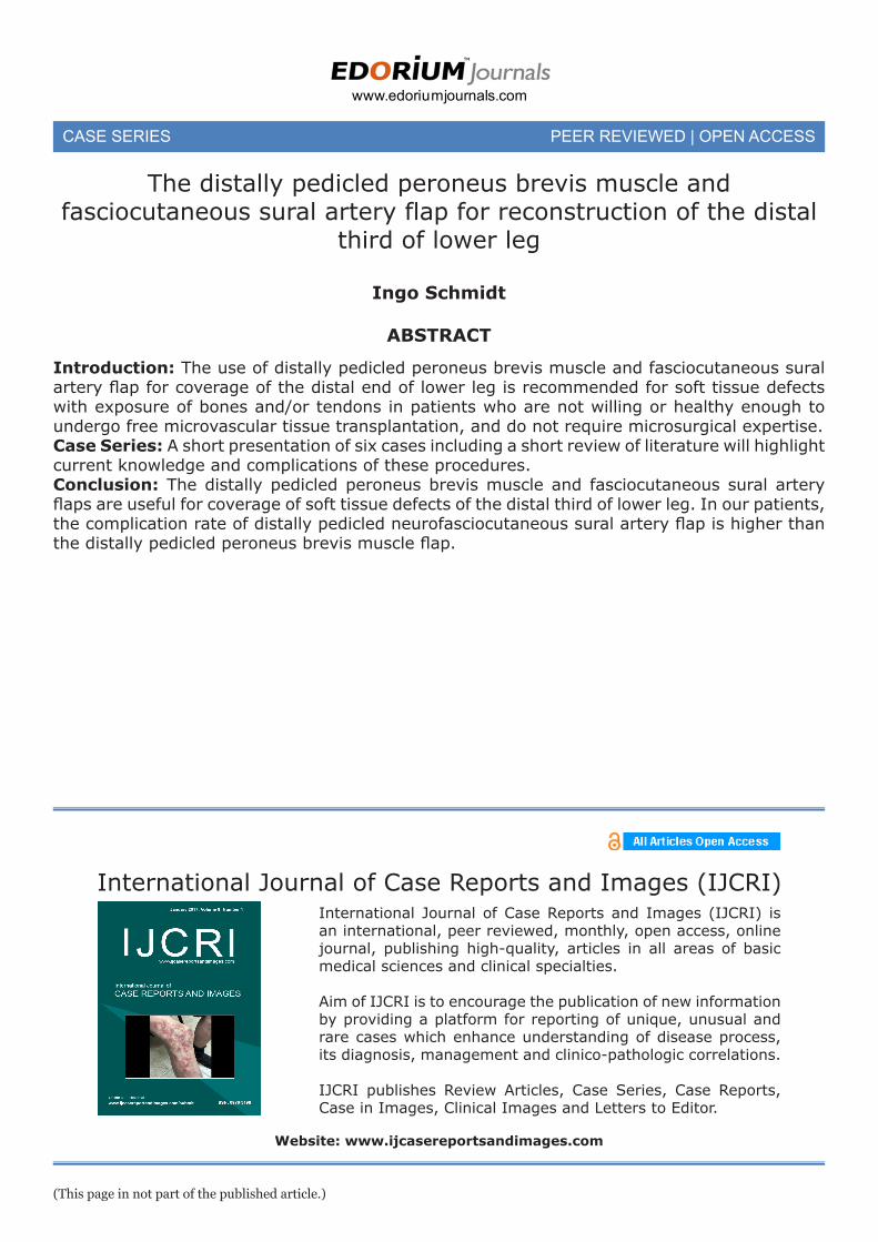
Case 1A 66-year-old female presented with chronically
destroyed left Achilles tendon (Figure 1A) that was treated with an open augmented repair (Figure 1B). The patient developed early wound healing problems
CASE SERIES PEER REVIEWED | OPEN ACCESS
International Journal of Case Reports and Images, Vol. 8 No. 1, January 2017. ISSN – [0976-3198]
Int J Case Rep Images 2017;8(1):17–21. www.ijcasereportsandimages.com
Schmidt 18
resulting in a large necrotizing soft tissue defect (Figure 1C). The defect was covered with the use of a distally pedicled peroneus brevis muscle flap and additional split-thickness skin grafts (Figure 1D-E). The wound healing was uncomplicated (Figure 1F).
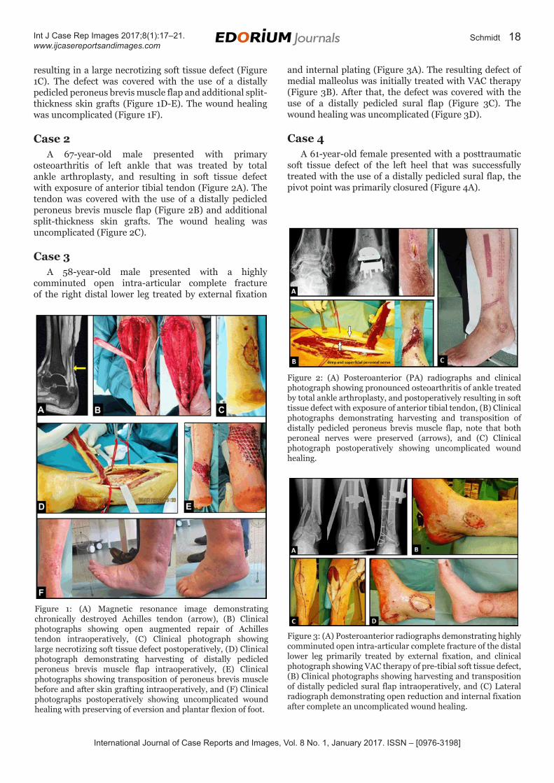
Case 2A 67-year-old male presented with primary
osteoarthritis of left ankle that was treated by total ankle arthroplasty, and resulting in soft tissue defect with exposure of anterior tibial tendon (Figure 2A). The tendon was covered with the use of a distally pedicled peroneus brevis muscle flap (Figure 2B) and additional split-thickness skin grafts. The wound healing was uncomplicated (Figure 2C).
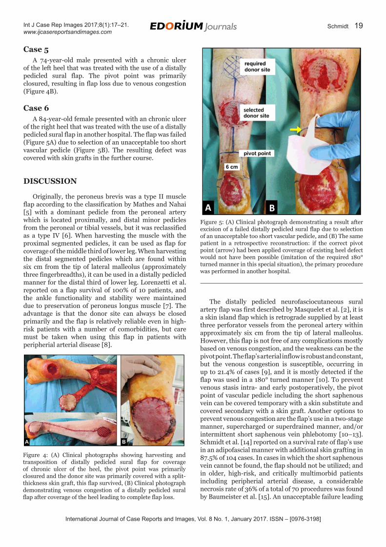
Case 3A 58-year-old male presented with a highly
comminuted open intra-articular complete fracture of the right distal lower leg treated by external fixation
Figure 2: (A) Posteroanterior (PA) radiographs and clinical photograph showing pronounced osteoarthritis of ankle treated by total ankle arthroplasty, and postoperatively resulting in soft tissue defect with exposure of anterior tibial tendon, (B) Clinical photographs demonstrating harvesting and transposition of distally pedicled peroneus brevis muscle flap, note that both peroneal nerves were preserved (arrows), and (C) Clinical photograph postoperatively showing uncomplicated wound healing.
Figure 3: (A) Posteroanterior radiographs demonstrating highly comminuted open intra-articular complete fracture of the distal lower leg primarily treated by external fixation, and clinical photograph showing VAC therapy of pre-tibial soft tissue defect, (B) Clinical photographs showing harvesting and transposition of distally pedicled sural flap intraoperatively, and (C) Lateral radiograph demonstrating open reduction and internal fixation after complete an uncomplicated wound healing.
Figure 1: (A) Magnetic resonance image demonstrating chronically destroyed Achilles tendon (arrow), (B) Clinical photographs showing open augmented repair of Achilles tendon intraoperatively, (C) Clinical photograph showing large necrotizing soft tissue defect postoperatively, (D) Clinical photograph demonstrating harvesting of distally pedicled peroneus brevis muscle flap intraoperatively, (E) Clinical photographs showing transposition of peroneus brevis muscle before and after skin grafting intraoperatively, and (F) Clinical photographs postoperatively showing uncomplicated wound healing with preserving of eversion and plantar flexion of foot.
and internal plating (Figure 3A). The resulting defect of medial malleolus was initially treated with VAC therapy (Figure 3B). After that, the defect was covered with the use of a distally pedicled sural flap (Figure 3C). The wound healing was uncomplicated (Figure 3D).
Case 4A 61-year-old female presented with a posttraumatic
soft tissue defect of the left heel that was successfully treated with the use of a distally pedicled sural flap, the pivot point was primarily closured (Figure 4A).
International Journal of Case Reports and Images, Vol. 8 No. 1, January 2017. ISSN – [0976-3198]
Int J Case Rep Images 2017;8(1):17–21. www.ijcasereportsandimages.com
Schmidt 19
Case 5A 74-year-old male presented with a chronic ulcer
of the left heel that was treated with the use of a distally pedicled sural flap. The pivot point was primarily closured, resulting in flap loss due to venous congestion (Figure 4B).
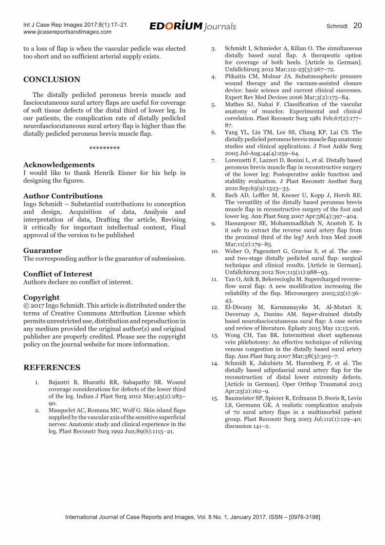
Case 6A 84-year-old female presented with an chronic ulcer
of the right heel that was treated with the use of a distally pedicled sural flap in another hospital. The flap was failed (Figure 5A) due to selection of an unacceptable too short vascular pedicle (Figure 5B). The resulting defect was covered with skin grafts in the further course.
DISCUSSION
Originally, the peroneus brevis was a type II muscle flap according to the classification by Mathes and Nahai [5] with a dominant pedicle from the peroneal artery which is located proximally, and distal minor pedicles from the peroneal or tibial vessels, but it was reclassified as a type IV [6]. When harvesting the muscle with the proximal segmented pedicles, it can be used as flap for coverage of the middle third of lower leg. When harvesting the distal segmented pedicles which are found within six cm from the tip of lateral malleolus (approximately three fingerbreadths), it can be used in a distally pedicled manner for the distal third of lower leg. Lorenzetti et al. reported on a flap survival of 100% of 10 patients, and the ankle functionality and stability were maintained due to preservation of peroneus longus muscle [7]. The advantage is that the donor site can always be closed primarily and the flap is relatively reliable even in high-risk patients with a number of comorbidities, but care must be taken when using this flap in patients with peripherial arterial disease [8].
The distally pedicled neurofasciocutaneous sural artery flap was first described by Masquelet et al. [2], it is a skin island flap which is retrograde supplied by at least three perforator vessels from the peroneal artery within approximately six cm from the tip of lateral malleolus. However, this flap is not free of any complications mostly based on venous congestion, and the weakness can be the pivot point. The flap’s arterial inflow is robust and constant, but the venous congestion is susceptible, occurring in up to 21.4% of cases [9], and it is mostly detected if the flap was used in a 180° turned manner [10]. To prevent venous stasis intra- and early postoperatively, the pivot point of vascular pedicle including the short saphenous vein can be covered temporary with a skin substitute and covered secondary with a skin graft. Another options to prevent venous congestion are the flap’s use in a two-stage manner, supercharged or superdrained manner, and/or intermittent short saphenous vein phlebotomy [10–13]. Schmidt et al. [14] reported on a survival rate of flap’s use in an adipofascial manner with additional skin grafting in 87.5% of 104 cases. In cases in which the short saphenous vein cannot be found, the flap should not be utilized; and in older, high-risk, and critically multimorbid patients including peripherial arterial disease, a considerable necrosis rate of 36% of a total of 70 procedures was found by Baumeister et al. [15]. An unacceptable failure leading
Figure 4: (A) Clinical photographs showing harvesting and transposition of distally pedicled sural flap for coverage of chronic ulcer of the heel, the pivot point was primarily closured and the donor site was primarily covered with a split-thickness skin graft, this flap survived, (B) Clinical photograph demonstrating venous congestion of a distally pedicled sural flap after coverage of the heel leading to complete flap loss.
Figure 5: (A) Clinical photograph demonstrating a result after excision of a failed distally pedicled sural flap due to selection of an unacceptable too short vascular pedicle, and (B) The same patient in a retrospective reconstruction: if the correct pivot point (arrow) had been applied coverage of existing heel defect would not have been possible (imitation of the required 180° turned manner in this special situation), the primary procedure was performed in another hospital.
International Journal of Case Reports and Images, Vol. 8 No. 1, January 2017. ISSN – [0976-3198]
Int J Case Rep Images 2017;8(1):17–21. www.ijcasereportsandimages.com
Schmidt 20
to a loss of flap is when the vascular pedicle was elected too short and no sufficient arterial supply exists.
CONCLUSION
The distally pedicled peroneus brevis muscle and fasciocutaneous sural artery flaps are useful for coverage of soft tissue defects of the distal third of lower leg. In our patients, the complication rate of distally pedicled neurofasciocutaneous sural artery flap is higher than the distally pedicled peroneus brevis muscle flap.
*********
AcknowledgementsI would like to thank Henrik Eisner for his help in designing the figures.
Author ContributionsIngo Schmidt – Substantial contributions to conception and design, Acquisition of data, Analysis and interpretation of data, Drafting the article, Revising it critically for important intellectual content, Final approval of the version to be published
GuarantorThe corresponding author is the guarantor of submission.
Conflict of InterestAuthors declare no conflict of interest.
Copyright© 2017 Ingo Schmidt. This article is distributed under the terms of Creative Commons Attribution License which permits unrestricted use, distribution and reproduction in any medium provided the original author(s) and original publisher are properly credited. Please see the copyright policy on the journal website for more information.
REFERENCES
1. Bajantri B, Bharathi RR, Sabapathy SR. Wound coverage considerations for defects of the lower third of the leg. Indian J Plast Surg 2012 May;45(2):283–90.
2. Masquelet AC, Romana MC, Wolf G. Skin island flaps supplied by the vascular axis of the sensitive superficial nerves: Anatomic study and clinical experience in the leg. Plast Reconstr Surg 1992 Jun;89(6):1115–21.
3. Schmidt I, Schmieder A, Kilian O. The simultaneous distally based sural flap. A therapeutic option for coverage of both heels. [Article in German]. Unfallchirurg 2012 Mar;112-25(3):267–72.
4. Plikaitis CM, Molnar JA. Subatmospheric pressure wound therapy and the vacuum-assisted closure device: basic science and current clinical successes. Expert Rev Med Devices 2006 Mar;3(2):175–84.
5. Mathes SJ, Nahai F. Classification of the vascular anatomy of muscles: Experimental and clinical correlation. Plast Reconstr Surg 1981 Feb;67(2):177–87.
6. Yang YL, Lin TM, Lee SS, Chang KP, Lai CS. The distally pedicled peroneus brevis muscle flap anatomic studies and clinical applications. J Foot Ankle Surg 2005 Jul-Aug;44(4):259–64.
7. Lorenzetti F, Lazzeri D, Bonini L, et al. Distally based peroneus brevis muscle flap in reconstructive surgery of the lower leg: Postoperative ankle function and stability evaluation. J Plast Reconstr Aesthet Surg 2010 Sep;63(9):1523–33.
8. Bach AD, Leffler M, Kneser U, Kopp J, Horch RE. The versatility of the distally based peroneus brevis muscle flap in reconstructive surgery of the foot and lower leg. Ann Plast Surg 2007 Apr;58(4):397–404.
9. Hassanpour SE, Mohammadkhah N, Arasteh E. Is it safe to extract the reverse sural artery flap from the proximal third of the leg? Arch Iran Med 2008 Mar;11(2):179–85.
10. Weber O, Pagenstert G, Gravius S, et al. The one- and two-stage distally pedicled sural flap: surgical technique and clinical results. [Article in German]. Unfallchirurg 2012 Nov;115(11):988–93.
11. Tan O, Atik B, Bekerecioglu M. Supercharged reverse-flow sural flap: A new modification increasing the reliability of the flap. Microsurgery 2005;25(1):36–43.
12. El-Diwany M, Karunanayake M, Al-Mutari S, Duvernay A, Danino AM. Super-drained distally based neurofasciocutaneous sural flap: A case series and review of literature. Eplasty 2015 May 12;15:e16.
13. Wong CH, Tan BK. Intermittent short saphenous vein phlebotomy: An effective technique of relieving venous congestion in the distally based sural artery flap. Ann Plast Surg 2007 Mar;58(3):303–7.
14. Schmidt K, Jakubietz M, Harenberg P, et al. The distally based adipofascial sural artery flap for the reconstruction of distal lower extremity defects. [Article in German]. Oper Orthop Traumatol 2013 Apr;25(2):162–9.
15. Baumeister SP, Spierer R, Erdmann D, Sweis R, Levin LS, Germann GK. A realistic complication analysis of 70 sural artery flaps in a multimorbid patient group. Plast Reconstr Surg 2003 Jul;112(1):129–40; discussion 141–2.

International Journal of Case Reports and Images, Vol. 8 No. 1, January 2017. ISSN – [0976-3198]
Int J Case Rep Images 2017;8(1):17–21. www.ijcasereportsandimages.com
Schmidt 21
ABOUT THE AUTHOR
Article citation: Schmidt I. The distally pedicled peroneus brevis muscle and fasciocutaneous sural artery flap for reconstruction of the distal third of lower leg. Int J Case Rep Images 2017;8(1):17–21.
Ingo Schmidt is Surgeon in the Department of Traumatology SRH Poliklinik, Waldklinikum Gera GmbH, Germany. From 1983 to 1989, he studied human medicine at the Friedrich-Schiller-University in Jena (Germany). From 1990 to 1999, Dr. Schmidt graduated his training for general surgery, traumatology, orthopaedics, and hand surgery at the University hospital in Jena. In 1994, he successfully defended his scientific work to gain the title as a medical doctor. He has published more than 20 scientific articles. His areas of interest include hip replacement, coverage of soft tissue defects, and hand surgery with special focus on total wrist replacement and arthroplasties of all other joints of the hand.
Access full text article onother devices
Access PDF of article onother devices
EDORIUM JOURNALS AN INTRODUCTION
Edorium Journals: On Web
About Edorium JournalsEdorium Journals is a publisher of high-quality, open ac-cess, international scholarly journals covering subjects in basic sciences and clinical specialties and subspecialties.
Edorium Journals www.edoriumjournals.com
Edorium Journals et al.
Edorium Journals: An introduction
Edorium Journals Team
But why should you publish with Edorium Journals?In less than 10 words - we give you what no one does.
Vision of being the bestWe have the vision of making our journals the best and the most authoritative journals in their respective special-ties. We are working towards this goal every day of every week of every month of every year.
Exceptional servicesWe care for you, your work and your time. Our efficient, personalized and courteous services are a testimony to this.
Editorial ReviewAll manuscripts submitted to Edorium Journals undergo pre-processing review, first editorial review, peer review, second editorial review and finally third editorial review.
Peer ReviewAll manuscripts submitted to Edorium Journals undergo anonymous, double-blind, external peer review.
Early View versionEarly View version of your manuscript will be published in the journal within 72 hours of final acceptance.
Manuscript statusFrom submission to publication of your article you will get regular updates (minimum six times) about status of your manuscripts directly in your email.
Our Commitment
Favored Author programOne email is all it takes to become our favored author. You will not only get fee waivers but also get information and insights about scholarly publishing.
Institutional Membership programJoin our Institutional Memberships program and help scholars from your institute make their research accessi-ble to all and save thousands of dollars in fees make their research accessible to all.
Our presenceWe have some of the best designed publication formats. Our websites are very user friendly and enable you to do your work very easily with no hassle.
Something more...We request you to have a look at our website to know more about us and our services.
We welcome you to interact with us, share with us, join us and of course publish with us.
Browse Journals
CONNECT WITH US
Invitation for article submissionWe sincerely invite you to submit your valuable research for publication to Edorium Journals.
Six weeksYou will get first decision on your manuscript within six weeks (42 days) of submission. If we fail to honor this by even one day, we will publish your manuscript free of charge.*
Four weeksAfter we receive page proofs, your manuscript will be published in the journal within four weeks (31 days). If we fail to honor this by even one day, we will pub-lish your manuscript free of charge and refund you the full article publication charges you paid for your manuscript.*
This page is not a part of the published article. This page is an introduction to Edorium Journals and the publication services.
* Terms and condition apply. Please see Edorium Journals website for more information.