Embed Size (px)
Citation preview

1
Pediatric Advanced Life Support
Preparatory Materials
National Certification Services 1/17 Review
www.CPRTrainingfast.com

2
PEDIATRIC ADVANCED LIFE SUPPORT (PALS) RECERTIFICATION
TABLE OF CONTENTS
CYCLIC APPROACH 03
PEDIATRIC ASSESSMENT FLOWCHART 04
MANAGEMENT OF RESPIRATORY EMERGENCIES FLOWCHART 05
MANAGEMENT OF SHOCK EMERGENCIES FLOWCHART 06
RECOGNITION OF SHOCK FLOWCHART 07
RECOGNITION OF RESPIRATORY PROBLEMS FLOWCHART 09
NORMAL VITAL SIGNS FOR PEDIATRIC PATIENTS 10
ALGORITHMS FOR PEDIATRICS
PULSELESS ARREST 11
BRADYCARDIA WITH A PULSE 13
TACHYCARDIA WITH ADEQUATE PERFUSION 14
TACHYCARDIA WITH PULSE AND POOR PERFUSION 15
AED TREATMENT ALGORITHM FOR PRE-HOSPITAL CRE OF CHILDREN >8 16
OVERVIEW OF RESUSCITATION IN THE DELIVERY ROOM 18
DRUGS USED IN PEDIATRIC ADVANCEDLIFE SUPPORT 19

3
www.CPRTrainingFast.com
I. Cyclic Approach
This is the cyclic approach used to assess and manage an ill or injured child. It is repeated frequently during
evaluation and management.
Assess: Evaluation starts with the general assessment and continues with the primary assessment, the
secondary assessment, and the tertiary assessment. If you recognize a life-threatening
condition at any time in any assessment, begin interventions.
Categorize: Attempt to categorize the child’s condition by type and severity.
Type Severity
Respiratory Upper airway obstruction
Lower airway obstruction
Lung tissue disease
Disordered control of breathing
Respiratory distress
Respiratory failure
Circulatory Hypovolemic Shock
Obstructive Shock
Distributive/Septic Shock
Cardiogenic Shock
Compensated Shock
Hypotensive Shock
The child’s condition may also be a combination of the two. As their condition deteriorates, one
category may lead to others.
Categorize
DecideAct
Assess

4
Decide: Now you need to decide on appropriate management based on your assessment and
categorization of the child’s condition. This is done based on your scope of practice.
Act: Start treatment appropriate for the clinical condition.
II. Pediatric Assessment Flowchart
General Assessment:
A – appearance B – work of breathing C – circulation
Primary Assessment:
A – airway B – breathing C – circulation D – disability E - exposure
Secondary Assessment:
Also:
S – signs and symptoms A – allergies M – medications P – past medical history L – last meal / liquids consumed E – events leading up to incident
Focused physical examination
Tertiary Assessment:
Labs X-Rays Other tests as needed
Categorize Illness by Type and Severity
Type
Severity
Respiratory
Upper airway obstruction
Lower airway obstruction
Lung tissue disease
Disordered control of breathing
Respiratory distress
Respiratory failure
Circulatory
Hypovolemic Shock
Obstructive Shock
Distributive/Septic Shock
Cardiogenic Shock
Compensated Shock
Hypotensive Shock

5
Respiratory + Circulatory = Cardiopulmonary failure
III. Management of Respiratory Emergencies Flowchart Airway positioning
Oxygen Pulse oximetry
ECG monitoring as needed
BLS as needed
Upper Airway Obstruction Specific Management for Selected Conditions
Croup
Anaphylaxis
Aspiration Foreign Body
Racemic epinephrine
Corticosteroids
IM epinephrine
Albuterol
Antihistamines
Corticosteroids
Allow position of comfort
Specialty consultation
Lower Airway Obstruction Specific Management for Selected Conditions
Bronchiolitis
Asthma
Nasal suctioning
Bronchodilator trial
Albuterol and/or ipratropium
Corticosteroids
SQ epinephrine
Magnesium sulfate
Terbutaline
Lung Tissue (Parenchymal) Disease Specific Management for Selected Conditions
Pneumonia / Pneumonitis
Infectious Chemical Aspiration
Pulmonary Edema Cardiogenic or ARDS
Albuterol
Antibiotics as needed
Consider noninvasive or invasive ventilator support with PEEP
Consider vasoactive support
Consider diuretic

6
Disordered Control of Breathing Specific Management for Selected Conditions
Increased ICP
Poisoning / Overdose
Neuromuscular Disease
Avoid hypoxemia
Avoid hypercarbia
Avoid hyperthermia
Antidote (if available)
Contact Poison Control
Consider noninvasive or invasive ventilator support
This chart does not include all respiratory emergencies.
IV. Management of Shock Emergencies Flowchart Oxygen
Pulse oximetry
ECG monitor
IV/IO access
BLS as needed
Bedside glucose
Hypovolemic Shock Specific Management for Selected Conditions
Non-hemorrhagic
Hemorrhagic
20 mL/kg NS/LR bolus, repeat as needed
Consider colloid after 3rd NS/RL bolus
Control external bleeding
20 mL/kg NS/RL bolus repeat 2 or 3x as needed
Transfuse PRBC’s as indicated
Distributive Shock Specific Management for Selected Conditions
Septic
Anaphylactic
Neurogenic
Management Algorithm
Septic Shock
IM epinephrine
Antihistamines
Corticosteroids
Epinephrine infusion
Albuterol
20 mL/kg NS/LR bolus, repeat PRN
Vasopressor
7
www.CPRTrainingFast.com
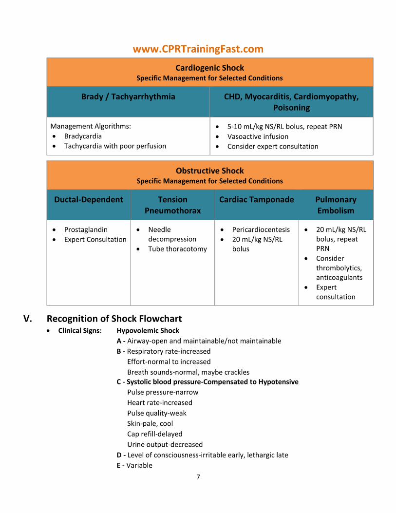
Cardiogenic Shock Specific Management for Selected Conditions
Brady / Tachyarrhythmia
CHD, Myocarditis, Cardiomyopathy, Poisoning
Management Algorithms:
Bradycardia
Tachycardia with poor perfusion
5-10 mL/kg NS/RL bolus, repeat PRN
Vasoactive infusion
Consider expert consultation
Obstructive Shock Specific Management for Selected Conditions
Ductal-Dependent
Tension Pneumothorax
Cardiac Tamponade
Pulmonary Embolism
Prostaglandin
Expert Consultation
Needle decompression
Tube thoracotomy
Pericardiocentesis
20 mL/kg NS/RL bolus
20 mL/kg NS/RL bolus, repeat PRN
Consider thrombolytics, anticoagulants
Expert consultation
V. Recognition of Shock Flowchart
Clinical Signs: Hypovolemic Shock
A - Airway-open and maintainable/not maintainable
B - Respiratory rate-increased
Effort-normal to increased
Breath sounds-normal, maybe crackles C - Systolic blood pressure-Compensated to Hypotensive
Pulse pressure-narrow
Heart rate-increased
Pulse quality-weak
Skin-pale, cool
Cap refill-delayed
Urine output-decreased
D - Level of consciousness-irritable early, lethargic late
E - Variable

8
Clinical Signs: Distributive Shock
A - Airway-open and maintainable/not maintainable
B - Respiratory rate-increased
Effort-normal to increased
Breath sounds-normal, maybe crackles
C - Systolic blood pressure-Compensated to Hypotensive
Pulse pressure-wide
Heart rate-increased
Pulse quality-bounding or weak
Skin-warm or cool
Cap refill-variable
Urine output-decreased D - Level of consciousness-irritable early, lethargic late E - Variable
Clinical Signs: Cardiogenic Shock
A - Airway-open and maintainable/not maintainable
B - Respiratory rate-increased
Effort-labored
Breath sounds-crackles, grunting
C - Systolic blood pressure-Compensated to Hypotensive
Pulse pressure-narrow
Heart rate-increased
Pulse quality-weak
Skin-pale, cool
Cap refill-delayed
Urine output-decreased D - Level of consciousness-irritable early, lethargic late E - Variable
Clinical Signs: Obstructive Shock
A - Airway-open and maintainable/not maintainable
B - Respiratory rate-increased
Effort-labored
Breath sounds-crackles, grunting
C - Systolic blood pressure-Compensated to Hypotensive
Pulse pressure-narrow
Heart rate-increased
Pulse quality-weak
Skin-pale, cool
Cap refill-delayed
Urine output-decreased D - Level of consciousness-irritable early, lethargic late

9
E - Variable
VI. Recognition of Respiratory Problems Flowchart
Clinical Signs: Upper Airway Obstruction
A - Airway-open and maintainable/not maintainable
B - Respiratory rate/effort-increased
Breath sounds-stridor (typically inspiratory)-seal like cough- hoarseness
Air movement-decreased
C - Heart rate-increased
Skin-pallor, cool skin (early) cyanosis (late)
D - Level of consciousness-anxiety, agitation (early) lethargy,
unresponsiveness (late)
E - Variable
Clinical Signs: Lower Airway Obstruction
A - Airway-open and maintainable/not maintainable
B - Respiratory rate/effort-increased
Breath sounds-wheezing (typically expiratory) prolonged expiratory phase
Air movement-decreased
C - Heart rate-increased Skin-pallor, cool skin (early) cyanosis (late)
D - Level of consciousness-anxiety, agitation (early) lethargy, unresponsiveness
(late)
E - Variable
Clinical Signs: Lung Tissue (Parenchymal) Disease
A - Airway-open and maintainable/not maintainable
B - Respiratory rate/effort-increased
Breath sounds-grunting, crackles, decreased breath sounds
Air movement-decreased
C - Heart rate-increased
Skin-pallor, cool skin (early) cyanosis (late)
D - Level of consciousness-anxiety, agitation (early) lethargy, unresponsiveness
(late)
E - Variable
Clinical Signs: Disordered Control of Breathing
A - Airway-open and maintainable/not maintainable
B - Respiratory rate/effort-variable
Breath sounds-normal
Air movement-variable
C - Heart rate-increased
Skin-pallor, cool skin (early) cyanosis (late)
D - Level of consciousness-anxiety, agitation (early) lethargy, unresponsiveness
(late)
E - Variable

10
www.CPRTrainingFast.com
VII. Normal Vital Signs for Pediatric Patients Normal Respiratory Rates
Age
Breaths / Minute
Infant (<1 year)
30 - 60
Toddler (1 – 3 years)
24 - 40
Preschooler (4 – 5 years)
22 - 34
School Age (6 – 12 years)
18 - 30
Adolescent (13 – 18 years)
12 - 18
* A respiratory rate more than 60 per minutes at any age is abnormal and should serve as a
“red Flag.”
Normal Heart Rates
Age
Awake
Sleeping
Newborn – 3 years
85 - 205
80 - 160
3 months – 2 years
100 - 190
75 - 160
2 years – 10 years
60 - 140
60 - 90
> 10 years
60 - 100
50 - 90
* Heart rate should be appropriate for the child’s age, activity level and clinical condition.
Heart rates vary in a sleeping or athletic child.
“red Flag.”
Minimum Systolic Blood Pressure Accepted (5th percentile)
Age
Systolic Blood Pressure (mm HG)
Birth (12h, < 1000g) Birth (12h, 3g) Neonate (96h) Infant (1-12mos)
39-59 60-76 67-84
72-104
Toddler (1 – 2 years)
86-106
Preschooler (3 – 5 years)
89-112
School Age (6 – 12 years)
97-115
Adolescent (10-11 years)
102-120

11
www.CPRTrainingFast.com
VIII. Algorithms for Pediatrics
Pulseless Arrest
Box 1 - Pulseless Arrest - BLS Algorithm: Continue CPR - Give Oxygen when available - Attach Monitor/defibrillator when available
Box 2 - Check Rhythm Is it a shockable rhythm?
Box 3 - VF / VT
Box 9 - Asystole / PEA
Box 4 - Give 1 Shock
Manual 2J/kg
AED: >1 year of age (use pediatric system. if available, for age 1 to 8 years of age
Resume CPR Immediately
Box 10 - Resume CPR Immediately
Give Epinephrine - IV/IO: 0.01 mg/kg (1:10 000:0.1 ml/kg - Endotracheal tube: 0.1 mg/kg
Repeat every 3 to 5 minutes
Give 5 cycles of CPR
Box 5 - Check Rhythm. Is it a Shockable rhythm?
Box 11 - Check Rhythm. Is it a Shockable rhythm?
Shockable Not Shockable Shockable
Box 6 - Continue CPR while defibrillator is charging. Give 1 shock
Manual 4J/kg
AED: >1 year of age (use pediatric system, if available, for age 1 to 8 years of age
Resume CPR immediately
Give Epinephrine - IV/IO: 0.01 mg/kg (1:10 000:0.1 ml/kg - Endotracheal tube: 0.1 mg/kg
Repeat every 3 to 5 minutes
Box 12 – - If asystole, go to Box 10
- If electrical activity, check pulse. If no pulse, go to Box 10
- If pulse is present, begin post resuscitation care.
Go to Box 4

12
YES, go to page 12
Box 7 - Check Rhythm. Is it a Shockable rhythm?
NO
YES, continued from page 11
Continue CPR while defibrillator is charging. Give 1 shock
Manual: 4J/kg
AED: >1 year of age Resume CPR immediately after the shock Consider antiarrhythmics (e.g. Amiodarone 5 mg/kg IV/IO once, or Lidocaine 1 mg/kg IV/VO) Consider Magnesium, 25 to 50 mg/kg. Max 2 g IV/VO for Torsades de Pointes After 5 cycles of CPR, go to Box 5
During CPR - Push hard and fast (100/min) - Ensure full chest recoil - Minimize interruptions in chest compressions - One cycle of CPR: 15 compressions then 2 breaths: 5 cycles = 2 min - Avoid hyperventilation - Secure airway and confirm placement - After an advanced airway is placed, rescuers no longer deliver “cycles” of CPR - Give continuous compressions without pauses for breaths - Give 8-10 breaths/min - Check Rhythm every 2 minutes - Rotate compressors every 2 minutes, with rhythm checks - Search for and treat possible contributing factors:
Hypovolemia, Hypoxia, Hydrogen Ion (acidosis) Hypo/Hyperkalemia, Hypoglycemia, Hypothermia, Toxins Tamponade (cardiac), Tension Pneumothorax, Thrombosis (coronary or pulmonary), Trauma (hypovolemia)

13
www.CPRTrainingFast.com Bradycardia with a Pulse
BRADYCARDIA with a pulse Causing Cardiorespiratory Compromise
Support ABCs as needed
Give oxygen
Attach Monitor/defibrillator
NO
Bradycardia still causing Cardiopulmonary compromise?
YES
- Support ABCs as needed - Give oxygen if needed - Observe - Consider expert consultation
NO
Perform CPR, if despite Oxygenation and ventilation HR <60, with poor perfusion
Persistent symptomatic bradycardia
Reminders
If Pulseless Arrest develops, go to Pulseless Arrest Algorithm
During CPR - Push hard and fast (100/min) - Ensure full chest recoil
Support ABCs
Secure airway if needed and confirm placement
Minimize interruptions in chest compressions
Search for and treat possible contributing factors: Hypovolemia Toxins Hypoxia Tamponade, Cardiac Hydrogen Ion (acidosis) Tension Pneumothorax Hypo-/Hyperkalemia Thrombosis (coronary or
pulmonary Hypoglycemia Trauma (hypovolemia,
increased ICP Hypothermia
YES
Give Epinephrine - IV/IO: 0.01 mg/kg (1:10 000:0.1 ml/kg - Endotracheal tube: 0.1 mg/kg
Repeat every 3 to 5 minutes
If increased vagal tome or Primary AV Block: - Give Atropine, first dose: 0.02 mg/kg.
May repeat (Minimum dose: 0.1 mg; Max dose 1 mg)
Consider cardiac pacing
If Pulseless Arrest develops go to Algorithm for Pulseless Arrest

14
www.CPRTrainingFast.com Tachycardia with Adequate Perfusion
BLS Algorithm: Assess and support ABCs as needed (assess signs of circulation and pulse; provide oxygen and ventilation
Provide O2
Attach monitor/defibrillator
Evaluate 12-lead ECG if practical
QRS Normal (≤0.08 sec) QRS Wide (≥0.08 sec)
Evaluate rhythm
What is the QRS duration?
Probable ventricular tachycardia
Consider alternative medications
Amiodarone, 5 mg/kg IV over 20 to 60 min
OR
Procainamide, 15 mg/kg IV over 30 to 60 min (Do not routinely administer Amiodarone and Procainamide together)
OR
Lidocaine, 1 mg/kg IV Bolus
Probable Sinus Tachycardia
History Compatible
P-waves present and normal
HR often varies with activity
Variable RR with constant PR
Infants: usually <220 bpm
Children: usually <180 bpm
Probable Supraventricular
Tachycardia
History incompatible with ST
P-waves absent/normal
HR not variable with activity
Abrupt rate changes
Infants: usually ≥220 bpm
Children: usually ≥180 bpm
Consider Vagal Maneuvers
During Evaluation
Provide Oxygen and ventilation as needed
Support ABCs
Confirm continuous monitor/pacer attachment
Consider expert consultation
Prepare for cardioversion 0.5 to 1J/kg (consider sedation)
Identify and treat possible causes:
Hypovolemia, Hypoxia, Hydrogen Ion (acidosis)
Hypo-/Hyperkalemia, Hypothermia, Toxins,
Establish vascular access
Consider Adenosine 0.1 mg/kg IV (maximum first dose 6 mg). May double or repeat one dose (maximum second dose: 12 mg). Use Rapid Bolus Technique.

15
Consult Pediatric cardiologist
Attempt cardioversion 0.5 to 1J/kg; may increase to 2J/kg if initial dose is ineffective
Sedate prior to cardioversion
Obtain 12-lead ECG
Tamponade (cardiac), Tension Pneumothorax,
Thrombosis (coronary or pulmonary), Trauma
(hypovolemia), increased ICP
www.CPRTrainingFast.com Tachycardia with Pulses and Poor Perfusion
Tachycardia with Pulses and Poor Perfusion
Assess and support ABCs as
Provide O2
Attach monitor/defibrillator
QRS Normal (≤0.08 sec) Symptoms Persists QRS Wide (≥0.08 sec)
Evaluate rhythm with monitor or 12-lead ECG
What is the QRS duration?
Probable ventricular tachycardia
Probable Sinus Tachycardia
History compatible
P-waves present and normal
HR often varies with activity
Variable RR with constant PR
Infants: usually <220 bpm
Children: usually <180 bpm
Probable Supraventricular Tachycardia
History incompatible with ST
P-waves absent/abnormal
HR not variable with activity
Abrupt rate changes
Infants: usually ≥220 bpm
Children: usually ≥180 bpm
Synchronized Cardioversion
0.5 to 1J/kg; may increase to 2J/kg if initial dose is ineffective
Sedate if possible, but do not delay cardioversion
Consider Adenosine, if it does not delay electrical cardioversion
Consider Vagal Maneuvers (no delays)
Expert Consultation Advised Amiodarone, 5 mg/kg IV over 20 to 60 min
OR
Procainamide 15 mg/kg IV over 30 to 60 min. (Do not routinely administer Amiodarone and Procainamide together).
If vascular access is available:
Consider Adenosine 0.1 mg/kg IV (maximum first dose 6 mg. May double or repeat one dose (maximum second dose 12 mg). Use Rapid Bolus Technique
Search for and treat cause!
16
OR
Attempt cardioversion 0.5 to 1 J/kg; may increase to 2J/kg if initial dose is ineffective
Sedate prior to cardioversion, if possible
During Evaluation
Provide Oxygen and ventilation as needed
Support ABC’s
Confirm continuous monitor/pacer attachment
Consider expert consultation
Prepare for cardioversion 0.5 to 1J/kg (consider sedation)
Identify and treat possible causes: Hypovolemia, Hypoxia, Hydrogen Ion (acidosis) Hypo-/Hyperkalemia, Hypothermia, Toxins, Tamponade (cardiac), Tension Pneumothorax, Thrombosis (coronary or pulmonary), Trauma (hypovolemia), increased ICP
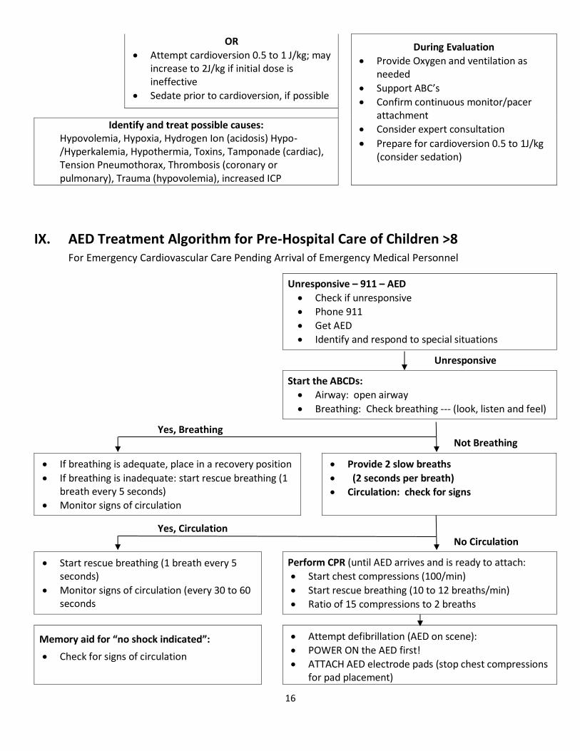
IX. AED Treatment Algorithm for Pre-Hospital Care of Children >8 For Emergency Cardiovascular Care Pending Arrival of Emergency Medical Personnel
Unresponsive – 911 – AED
Check if unresponsive
Phone 911
Get AED
Identify and respond to special situations
Unresponsive
Start the ABCDs:
Airway: open airway
Breathing: Check breathing --- (look, listen and feel)
Yes, Breathing Not Breathing
If breathing is adequate, place in a recovery position
If breathing is inadequate: start rescue breathing (1 breath every 5 seconds)
Monitor signs of circulation
Provide 2 slow breaths
(2 seconds per breath)
Circulation: check for signs
Yes, Circulation No Circulation
Start rescue breathing (1 breath every 5 seconds)
Monitor signs of circulation (every 30 to 60 seconds
Perform CPR (until AED arrives and is ready to attach:
Start chest compressions (100/min)
Start rescue breathing (10 to 12 breaths/min)
Ratio of 15 compressions to 2 breaths
Memory aid for “no shock indicated”:
Check for signs of circulation
Attempt defibrillation (AED on scene):
POWER ON the AED first!
ATTACH AED electrode pads (stop chest compressions for pad placement)

17
If signs of circulation present: check breathing
If inadequate breathing: start rescue breathing (1 breath every 5 seconds)
If adequate breathing: place in a recovery position
If no signs of circulation, analyze rhythm: repeat :shock indicated” or “no shock indicated” sequences
*Note: Signs of circulation: lay rescuers check for normal breathing, coughing or movement (typically assessed after 2 rescue breaths delivered to the unresponsive, non-breathing victim).
Analyze (“Clear!”)
Shock (“Clear!”) up to 3 times, if advised
After 3 shocks or after any “no shock indicated”
Check for signs of circulations
If no signs of circulations: perform CPR for 1 minute
Check for signs of circulation: if absent:
Press ANALYZE
Attempt defibrillation
Repeat up to 3 times
www.CPRTrainingFast.com
Post-arrest Treatment of Shock
And Maintenance Fluid Requirements
Estimation of Maintenance Fluid Requirements Infants <10 kg: Infusion of 0.2@ normal saline in 5% dextrose (d5/0.2% NaCl) at a rate of 4 mL/kg per hour. For example, the maintenance rate for an 8-kg baby is as follows:
4 mL/kg per hour x 8 kg = 32 mL/h
Children 10 to 20 kg: Infusion of d5/0.2% NaCl at a rate of 40 mL/h plus 2mL/kg per hour for each kilogram between 10 and 20 kg. For example, the maintenance rate for a 15-kg child is as follows:
40 mL/h + (2mL/kg per hour x 5 kg) = 50mL/h
Children >20 kg: Infusion of d5/0.2% NaCl at a rate of 60 mL/h plus 1 mL/kg per hour for each kilogram above 20 kg. For example the maintenance rate for a 30-kg child is as follows:
60 mL/h + (1 mL/kg per hour x 10 kg) = 70 mL/h
Post-arrest Stabilization
Post-arrest Shock
Fluid bolus (10-20 mL/kg NS or RL monitor response)
Reassess – Signs of shock continue
What is blood pressure?
Hypotensive (decompensated)
shock?
Normotensive (compensated)
shock?
Consider further fluid boluses Epinephrine (0.1 to 1 ug/kg per minute)
or
Consider further fluid boluses Dobutamine (2 to 20 ug/kg per minute)

18
Dopamine at higher doses (up to 20 ug/kg per minute) Norepinephrine (0.1 to 2 ug/kg per minute)
or Dopamine (1 to 20 ug/kg per minute)
or Low doses epinephrine (0.05 to 0.3 ug/kg per minute) Inamrinone: Load with 0.75 to 1 mg/kg over 5 minutes, may repeat up to 3 mg/kg. Infusion: 5 to 10 ug/kg per minute Milrinone: Load with 50 to 75 ug/kg. Infusion: 0.5 to .075 ug/kg per minute.
X. Overview of Resuscitation in the Delivery Room
Approximate Time Birth
30 Sec
Clear of meconium? Breathing or crying? Good muscle tone? Color pink? Term gestation?
YES
Routine Care
Provide warmth
Clear airway
Dry
NO
Provide warmth
Position, clear airway (as necessary)
Dry, stimulate, reposition
Give O2 (as necessary)
Evaluate respirations, heart rate, and color
Breathing
Supportive care
Apnea or <100
HR > 100 and pink
30 Sec
Provide positive pressure ventilation
Ventilating
Ongoing care
HR > 100
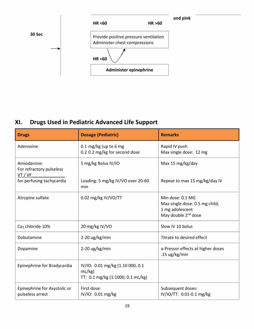
19
HR <60 HR >60
and pink
30 Sec
Provide positive pressure ventilation Administer chest compressions
HR <60
Administer epinephrine
XI. Drugs Used in Pediatric Advanced Life Support
Drugs
Dosage (Pediatric)
Remarks
Adenosine
0.1 mg/kg (up to 6 mg 0.2 0.2 mg/kg for second dose
Rapid IV push Max single dose: 12 mg
Amiodarone: For refractory pulseless VT / VF______________ for perfusing tachycardia
5 mg/kg Bolus IV/IO Loading: 5 mg/kg IV/VO over 20-60 min
Max 15 mg/kg/day Repeat to max 15 mg/kg/day IV
Atropine sulfate
0.02 mg/kg IV/VO/TT
Min dose: 0.1 MG Max single dose: 0.5 mg child, 1 mg adolescent May double 2nd dose
Ca2 chloride 10%
20 mg/kg IV/VO
Slow IV 10 bolus
Dobutamine
2-20 ug/kg/min
Titrate to desired effect
Dopamine
2-20 ug/kg/min
a-Pressor effects at higher doses .15 ug/kg/min
Epinephrine for Bradycardia
IV/IO: 0.01 mg/kg (1.10 000, 0.1 mL/kg) TT: 0.1 mg/kg (1:1000, 0.1 mL/kg)
Epinephrine for Asystolic or pulseless arrest
First dose: IV/IO: 0.01 mg/kg
Subsequent doses: IV/IO/TT: 0.01-0.1 mg/kg

20
(1:10 000, 0.1 mL/kg)
(1:1000, 0.1 mL/kg. IV/VO doses as high as 0.2 mg/kg of 1:1000 may be effective Repeat q 3-5 min
Epinephrine Infusion
Initial at 0.1 ug/kg/min
Titrate to desired effect (0.1-1 ug/kg/min
Glucose
0.5-1 g/kg IV/VO Max dose: 2-4 mL/kg Of 25% solution
5% = 10-20 mL/kg 10% = 5-10 mL/kg, 25% = 2-4 mL/kg (in large vein)
Lidocaine __________________ Infusion
1 mg/kg 20-50 ug/kg/min
IV/IO/TT
Magnesium Sulfate
25-50 mg/kg/min over 10-20 min
Max dose: 2 g
Drugs
Dosage (Pediatric)
Remarks
Milrinone
Loading dose 50-70 ug/kg IV/IO over 10-60 min Infusion dose 0.5–0.75 ug/kg/min IV/IO
Monitor BP, ECG
Naloxone
If <5 years old or <20 kg: 0.1 mg/kg If <5 years old or >20 kg: 2 mg
Titrate to desired effect
Prostaglandin E1
0.05-0.1 ug/kg/min
Titrate, monitor for apnea, hypotension, hypoglycemia, hypocalcemia
Sodium bicarbonate
1 mq/kg per dose
Infuse slowly and only if ventilation is adequate
For TT administration, dilute medication with NS to a volume of 3-5 mL and follow with several positive-
pressure ventilations.

21
www.CPRTrainingFast.com