Embed Size (px)
Citation preview
1484
The Origin and Distribution of Vasa Vasorumat the Bifurcation of the Common Carotid
Artery With AtherosclerosisWalter J. Bo, PhD, William M. McKinney, MD, and Robert L. Bowden
The purpose of our study was to determine the origin and relation of vasa vasorum to atheroscleroticplaque at the bifurcation of the common carotid artery. We randomly selected 12 unembalmedadult human cadavers, 40-96 years of age. We prepared luminal casts of the arteries from eightcadavers and cleared the arteries from the remaining four cadavers. A network of vasa vasorumsurrounding atherosclerotic plaque was observed in five luminal casts and in two cleared specimens;the vasa vasorum originated from the superior thyroid and ascending pharyngeal arteries. Three ofthe five luminal casts also demonstrated vasa vasorum arising directly from the internal carotidartery distal to the plaque. An extensive network of vasa vasorum was not observed in specimensfrom the five cadavers relatively free of gross atherosclerotic plaque. Our findings demonstrate theimportance of the external carotid artery in giving rise to the vasa vasorum that supply the areas ofatherosclerotic plaque. (Stroke 1989;20:1484-1487)
Nutrients are provided to large arteries byvasa vasorum, which supply the adventi-tia and outer media; the intima and inner
media are supplied by diffusion from the lumen.Following the injection of lead chromate, Higgin-botham et al1 described a superficial plexus of vasain the adventitia and deep plexus in the medioad-ventitial layer of the monkey aorta; the media andintima were free of vessels. Anastomoses betweenthe plexuses were observed. Song et al2 studied thevasa vasorum of the thoracic aortas of sheep, dogs,and pigs and observed that vasa vasorum extendedinto the internal elastic lamina. However, it wasimpossible to determine if the subintimal vesselscame from the lumen of those vessels.
With the development of atherosclerotic plaquethere is a thickening of the intima, which precipi-tates a change in the vascular pattern of the area.From histologic studies of the human aorta andcoronary arteries, Geiringer3 proposed that vesselsfrom the adventitia and the lumen extend into theplaque. However, vascularization from the lumenwas observed only when a portion of the arterial
From the Departments of Anatomy (W.J.B., R.L.B.) andNeurology (W.M.M.), Bowman Gray School of Medicine, WakeForest University, Winston-Salem, North Carolina.
Supported by United States Public Health Service GrantNS-06655.
Address for reprints: William M. McKinney, MD, Professor ofNeurology, Department of Neurology, Bowman Gray School ofMedicine, 300 South Hawthorne Road, Winston-Salem, NC27103.
Received December 5, 1988; accepted May 16, 1989.
intima exceeded a critical depth (0.5 mm for theaorta and 0.35 mm for the first portion of theanterior descending branch of the left coronaryartery). Barger et al4 studied the vasa of carotidarteries by cinematography of silicone polymer-injected, cleared human hearts and observed adense network of vasa in the area of atheroscleroticinjury. However, vasa vasorum were rarely seen inthe walls of normal coronary arteries.
The bifurcation of the common carotid artery ispredisposed to the development of atheroscleroticlesions. The plaques can exhibit intramuralhemorrhage,5 which may give rise to thromboemboli,resulting in transient ischemic attacks and/or stroke.A knowledge of the vascularization of carotid athero-sclerotic plaque may assist in the understanding of itssequela. Therefore, our study was designed to deter-mine the origin of the vasa vasorum and their relationto atherosclerotic plaque at the bifurcation of thecommon carotid artery. This was accomplished bypreparing luminal casts of the arteries and by visual-izing vasa vasorum in the walls of cleared arteries.
Materials and MethodsWe randomly selected 12 unembalmed adult
human cadavers, 40-96 years of age. The commoncarotid arteries of each cadaver were cannulatedand flushed with saline. Each artery was injectedwith methyl methacrylate resin, which was allowedto solidify. In specimens from eight cadavers, lumi-nal casts of the arteries were prepared by com-pletely corroding the tissue with NaOH. The injected
by guest on May 10, 2018
http://stroke.ahajournals.org/D
ownloaded from

Bo et al Vasa Vasorum and Carotid Atherosclerosis 1485
arteries from the remaining four cadavers weredissected free from the surrounding tissue, fixed in10% formalin for 12 hours, dehydrated in a series ofgraded alcohols, and cleared with a 1:1 mixture ofmethyl salicylate and methyl benzoate. To trace thedistribution of the vasa, the cleared specimens wereexamined with a dissecting microscope.
ResultsThe gross pathology in the region of the bifurca-
tion of the common carotid artery varied from aslight thickening to extensive calcification of theartery wall. Of the 12 cadavers, seven had extensivegross atherosclerotic plaques and five were rela-tively free of atherosclerotic pathology.
Luminal casts from five and cleared specimensfrom two cadavers demonstrated a network of vasavasorum in the area of atherosclerotic plaque (Fig-ures 1 and 2); the vasa vasorum originated from thesuperior thyroid and ascending pharyngeal arteriesin all seven. Luminal casts of three of these cadav-ers also demonstrated vasa vasorum arising from
FIGURE 1. Luminal cast demonstrates common (CC),internal (IC), and external (EC) carotid arteries. Networkof vasa vasorum (arrows) is seen in areas of atheroscle-rotic plaque. Branches from superior thyroid artery (ST)contribute to network of vessels.
FIGURE 2. Cleared specimen shows extensive networkof vasa vasorum (arrows) in area of marked atheroscle-rotic pathology of common (CC), internal (IC), andexternal (EC) carotid arteries.
the internal carotid artery distal to the plaque (Fig-ure 3). In one cast specimen a branch from theexternal carotid artery was observed contributing tothe network of vessels.
No extensive network of vasa vasorum wasobserved in arteries of the five cadavers that wererelatively free of gross atherosclerotic pathology(Figures 4 and 5).
DiscussionOur data show that the vasa vasorum are promi-
nent in areas of marked atherosclerosis. The vasavasorum originated from the superior thyroid andascending pharyngeal arteries as well as directlyfrom the lumen of the internal carotid artery distalto the plaque.
There are four stages in the development of grossatherosclerotic plaque, which have been described byMoossy6:1) fatty streaks, 2) fibrous plaques, 3) fibrous
by guest on May 10, 2018
http://stroke.ahajournals.org/D
ownloaded from
1486 Stroke Vol 20, No 11, November 1989
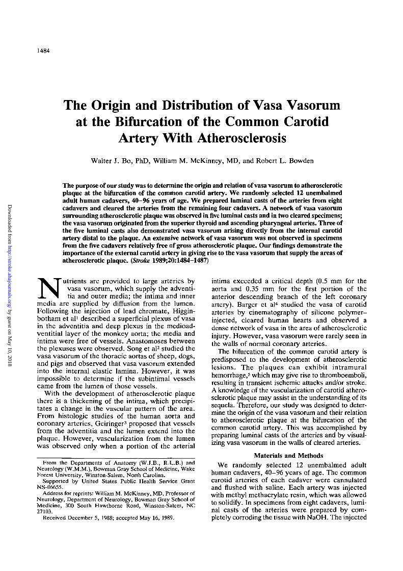
FIGURE 3. Luminal cast demonstrating small vessels(arrows) arising from lumen of internal carotid artery (IC)distal to atherosclerotic plaque and passing inferiorlyinto network of vasa vasorum.
fibrous plaques with hemorrhage and ulceration,and 4) calcified lesions. Whether there is a corre-lation between proliferation of the vasa vasorum andthe stage of atherosclerotic plaque development isnot clear. We observed vasa vasorum entering theadventitia in arteries relatively free of gross athero-sclerotic plaques and from the lumen of internalcarotid arteries containing marked atheroscleroticplaques. However, using the alkaline phosphatasetechnique to demonstrate endothelial cells, Peter-son et al7 reported that vascularization of the intimaand media of the abdominal aorta occur prior to thedevelopment of gross, recognizable atheroscleroticlesion. According to Geiringer,3 the intima is vas-cularized by channels from the lumen only when theintima reaches a critical thickness.
The proliferation of vasa vasorum into atheroscle-rotic plaque suggests neovascularization and indicatesthat angiogenic factors may be involved. Macrophages8
and platelets9 are involved at some stage during thedevelopment of atherosclerotic plaque and have angio-genic properties. Therefore, these cells may have arole in precipitating neovascularization.
AP
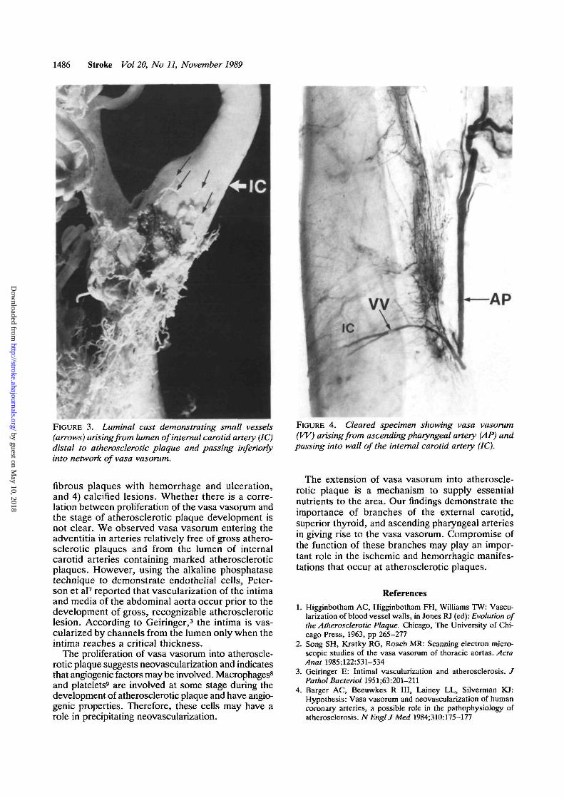
FIGURE 4. Cleared specimen showing vasa vasorum(W) arising from ascending pharyngeal artery (AP) andpassing into wall of the internal carotid artery (IC).
The extension of vasa vasorum into atheroscle-rotic plaque is a mechanism to supply essentialnutrients to the area. Our findings demonstrate theimportance of branches of the external carotid,superior thyroid, and ascending pharyngeal arteriesin giving rise to the vasa vasorum. Compromise ofthe function of these branches may play an impor-tant role in the ischemic and hemorrhagic manifes-tations that occur at atherosclerotic plaques.
References1. Higginbotham AC, Higginbotham FH, Williams TW: Vascu-
larization of blood vessel walls, in Jones RJ (ed): Evolution ofthe Atherosclerotic Plaque. Chicago, The University of Chi-cago Press, 1963, pp 265-277
2. Song SH, Kratky RG, Roach MR: Scanning electron micro-scopic studies of the vasa vasorum of thoracic aortas. ActaAnat 1985;122:531-534
3. Geiringer E: Intimal vascularization and atherosclerosis. JPathol Bacterial 1951;63:201-211
4. Barger AC, Beeuwkes R III, Lainey LL, Silverman KJ:Hypothesis: Vasa vasorum and neovascularization of humancoronary arteries, a possible role in the pathophysiology ofatherosclerosis. NEnglJMeet 1984;310:175-177
by guest on May 10, 2018
http://stroke.ahajournals.org/D
ownloaded from

Bo et al Vasa Vasorum and Carotid Atherosclerosis 1487
FIGURE 5. Vasa vasorum (W) are few at bifurcation ofcleared common carotid artery (CC) relatively free ofgross atherosclerotic pathology. EC, external carotidartery; IC, internal carotid artery.
5. Bluth El, Kay D, Merritt CRB, Sullivan M, Farr G, Mills NL,Foreman M, Sloan K, Schlater M, Stewart J: Sonographiccharacterization of carotid plaques: Detection of hemorrhage.AJR 1986;146:1061-1065
6. Moossy J: Morphology, sites and epidemiology of cerebralatherosclerosis, in Association for Research in Nervous andMental Disease. Baltimore, Williams & Wilkins Co, 1966,pp 1-22
7. Peterson JC, Mills J, Moffatt T: Vascularization of earlyatherosclerotic plaques. Arch Pathol 1957;64:127-136
8. Polverini PJ, Cotra RS, Gimbroue MA Jr, Unanue ER:Activated macrophages induce vascular proliferation. Nature1977;269:804-806
9. Ross R, Glomset J, Kariya B, Harker L: A platelet dependentserum factor that stimulates the proliferation of arterial smoothmuscle cells in vitro. Proc Natl Acad Sci USA 1974;71:1207-1210
KEY WORDSvasa vasorum
arteriosclerosis • carotid artery diseases
by guest on May 10, 2018
http://stroke.ahajournals.org/D
ownloaded from
W J Bo, W M McKinney and R L Bowdenwith atherosclerosis.
The origin and distribution of vasa vasorum at the bifurcation of the common carotid artery
Print ISSN: 0039-2499. Online ISSN: 1524-4628 Copyright © 1989 American Heart Association, Inc. All rights reserved.
is published by the American Heart Association, 7272 Greenville Avenue, Dallas, TX 75231Stroke doi: 10.1161/01.STR.20.11.1484
1989;20:1484-1487Stroke.
http://stroke.ahajournals.org/content/20/11/1484World Wide Web at:
The online version of this article, along with updated information and services, is located on the
http://stroke.ahajournals.org//subscriptions/
is online at: Stroke Information about subscribing to Subscriptions:
http://www.lww.com/reprints Information about reprints can be found online at: Reprints:
document. Permissions and Rights Question and Answer available in the
Permissions in the middle column of the Web page under Services. Further information about this process isOnce the online version of the published article for which permission is being requested is located, click Request
can be obtained via RightsLink, a service of the Copyright Clearance Center, not the Editorial Office.Stroke Requests for permissions to reproduce figures, tables, or portions of articles originally published inPermissions:
by guest on May 10, 2018
http://stroke.ahajournals.org/D
ownloaded from