Embed Size (px)
Citation preview

Rinaldi et al. BMC Veterinary Research 2014, 10:236http://www.biomedcentral.com/1746-6148/10/236
CASE REPORT Open Access
Angiostrongylus vasorum: epidemiological, clinicaland histopathological insightsLaura Rinaldi1,2*, Laura Cortese1, Leonardo Meomartino1, Teresa B Pagano1, Paola Pepe1, Giuseppe Cringoli1,2
and Serenella Papparella1
Abstract
Background: Canine angiostrongylosis is a nematode infection in domestic dogs and wild carnivores. The present reportfocuses on epidemiological, clinical and histopathological findings in a case of fatal disseminated angiostrongylosis in adog living in southern Italy and provides data on the extent of the spread of Angiostrongylus vasorum in the same area.
Case presentation: A 4-year-old female English Setter from the Campania region of southern Italy was referredwith a 2-week history of cough and severe respiratory distress that did not respond to antimicrobial therapy.Based on clinical, radiological, echographical and cytological findings (including the presence of larvae), a suspectdiagnosis of lungworm infection was performed. After few days the dog died due to progressive clinical aggravation.Complete postmortem examination was conducted within 24 hours from death and samples from lungs, heart, liver,kidney, spleen, stomach and small intestine were fixed in 10% buffered formalin. Grossly, several hemorrhagicfoci were observed mostly in the lungs, liver, kidney. Microscopically, the lungs contained numerous, multifocal tocoalescing granulomas composed of epitheliod macrophages, multinucleated giant cells and some neutrophils,frequently associated with parasite eggs and larvae. The lungs contained many firm nodules, many adult nematodesapproximately 1.5 to 2 cm in length were observed in cut sections and identified as A. vasorum. A subsequentparasitological survey performed with FLOTAC on stray dogs living in the same area showed the presence ofA. vasorum larvae in 17 of 1639 stray dogs examined (1.04%).
Conclusion: This survey provides new data on distribution of A. vasorum and underlines that canine angiostrongylosisshould be considered as differential diagnosis in dogs.
Keyword: Angiostrongylus vasorum, Dog, Pathology, Diagnosis
BackgroundAngiostrongylus vasorum commonly known as the “Frenchheartworm” is a metastrongyloid nematode of dogs andother canids, including various species of fox, wolf, coyoteand jackal (definitive hosts) [1]. The adults reside in theright side of the heart and in the pulmonary arteries, withpotentially severe consequences for the host [2]. The in-fection can cause a wide range of disease outcomes, whichare most often characterized by respiratory dysfunction,but it can also manifest as bleeding, neurological, cardio-vascular or gastrointestinal disorders, with or without re-spiratory involvement [3].
* Correspondence: [email protected] of Veterinary Medicine and Animal Productions, University ofNaples Federico II, Via Della Veterinaria 1, 80137 Naples, Italy2Inter-University Center for Research in Parasitology (CIRPAR), Naples, Italy
© 2014 Rinaldi et al.; licensee BioMed CentralCommons Attribution License (http://creativecreproduction in any medium, provided the orDedication waiver (http://creativecommons.orunless otherwise stated.
The geographic distribution of the parasite includesvarious countries of Europe, North and South Americaas well as Africa [1]. The parasite appears to be quitecommon in well-isolated endemic foci, with few occa-sional reports occurring outside them [4]. However, recentreports challenge this traditional view, as A. vasorum hasappeared in several new geographical areas [5]. Italy is oneof the countries where A. vasorum is undoubtedlyspreading, offering ideal environmental and epidemio-logical conditions for the expansion of A. vasorum andthe establishment of further new endemic foci [6,7].The reasons for this emergence are unclear and poorlyunderstood, but may involve global changes leading tospread of a variety of definitive, intermediate and para-tenic hosts and consequent modification of molluskphenology [8]. Due to this spread and the increasing clin-ical relevance of canine angiostrongylosis, the interest in
Ltd. This is an Open Access article distributed under the terms of the Creativeommons.org/licenses/by/4.0), which permits unrestricted use, distribution, andiginal work is properly credited. The Creative Commons Public Domaing/publicdomain/zero/1.0/) applies to the data made available in this article,

Rinaldi et al. BMC Veterinary Research 2014, 10:236 Page 2 of 7http://www.biomedcentral.com/1746-6148/10/236
A. vasorum is growing, particularly with respect to itstreatment and control [9].At present, diagnosis relies on clinical manifestations,
diagnostic imaging, bronchial washings and on the detec-tion of the first-stage larvae (L1) in faecal samples. Theparasite detection in faecal samples is usually performedusing conventional coprological examination such as dir-ect faecal smears, flotation and Baermann (gold standard)[10]. Also, the FLOTAC techniques [11] have been dem-onstrated as very sensitive for the diagnosis of A. vasorumin dogs [12]. Furthermore, serological [13,14] and molecu-lar [15,16] methods have been developed for the diagnosisof canine angiostrongylosis.With the aim to provide further insights on this para-
site, the present paper describes post mortem gross andhistopathological observation in a dog with a fatal infectionby A. vasorum. Subsequently to this case, a coprologicalsurvey was performed to evaluate the extent of A. vasoruminfection in stray dogs living in the city of Naples (southernItaly), using the FLOTAC techniques.
Case presentationA 4-year-old female English Setter from the Campaniaregion of southern Italy was referred with a 2-weeks historyof cough and severe respiratory distress that did not re-spond to antimicrobial therapy. On physical examination,the dog was alert, dyspneic and tachypneic. Mucous mem-branes and capillary refill time were normal. Cardiac aus-cultation revealed tachycardia.Hematological abnormalities included anemia and leu-
kocytosis. Routine biochemical analyses were unremarkable.An electrocardiogram demonstrated right axis deviation.Thoracic radiographs in standard lateral-lateral projectionand toracic ultrasound were carried out and an ultrasound-guided fine needle aspiration was performed on selectedpleural lesions. Cytological evaluation showed mixed inflam-matory cells occasionally admixed with full nematode larvae.Based on clinical, radiological, ultra sonographical and cyto-logical findings (including the presence of larvae), a suspectdiagnosis of lungworm infection was made; however, nolarvae were detected in faecal samples using the Baermanntechnique [17]. A therapeutic protocol including antibiotics,corticosteroid and milbemycin oxime was administered.Ten days later the dog was hospitalized following a severepneumothorax. Supplemental oxygen therapy was adminis-tered and a chest tube was placed. Subsequently pneumo-thorax developed again; thoracic surgery was necessary andled to its resolution. One month after surgery, the dyspneaworsened. After a few days, the dog suddenly died despitesupplemental oxygen therapy. Complete postmortem exam-ination was carried out within 24 hours of the death andsamples from lungs, heart, liver, kidney, spleen, stomach andsmall intestine were fixed in 10% buffered formalin. Pul-monary arteries were opened and examined in order to
identify intravascular nematodes. Some cytological smearswere performed from tracheobronchial lymphnode. Manyadult worms were collected and morphologically identifiedas adult A. vasorum based on morphology of the adult malebursa and the length of the spicules [18]. Formalin-fixedsamples were processed for sectioning, embedded in paraf-fin, sectioned at 5 micron, and stained with hematoxylinand eosin (HE), and Periodic Acid Shiff (PAS) stain.
Radiological findingsThoracic radiographs showed a multifocal to coales-cing bronchial and alveolar pattern and enlargement oftracheobronchial lymph nodes (Figure 1). Thoracic ultra-sound showed multiple sub pleural nodules (Figure 2) fromwhich an ultrasound-guided fine needle aspiration was per-formed. B-mode echocardiography showed a mild dilationof the right atrial, main pulmonary trunk and right pul-monary artery. Doppler echocardiography revealed tricus-pid regurgitation (maximal velocity: 243 cm/s; maximalpressure gradient, 23,6 mmHg).
Pathological findingsMacroscopically, pathological findings of the lungs con-sisted in multiple, poorly defined dark-red foci of necrotic-hemorrhagic areas. Firmer areas of broncopneumonia werealso present. Several adult parasites, approximately 2–2.5 cm in length, were collected from pulmonary arteries(Figure 3). Alveolar septa were mildly expanded with anincreased amount of dense fibrous tissue. The overlyingpleura were moderately thickened too.Microscopically, large areas of pulmonary parenchyma
were replaced by numerous, multifocal to coalescinggranulomas composed of macrophages, multinucleatedgiant cells, eosinophils and neutrophils granulocytes. Thecenter of these granulomas contained unembryonated andembryonated parasite eggs, coiled larvae and deposits ofamorphous, pale, eosinophilic necrotic debris, while lym-phocytes and plasma cells were present at the periphery(Figure 4). Fibroblastic proliferation with septal thick-ening and lymphoid hyperplasia were also observed.Numerous organized thrombi (Figure 5), often con-taining large adult nematodes were present in lumenof medium-sized pulmonary arteries, causing eosinophilicvasculitis. Occasionally, adult parasites were seen withinthe alveoli, which were affected by productive alveolitiswith cell esfoliation and presence of luminal suppurativeexudate. Wide hemorrhagic areas associated with hemosid-erin deposits, hemosiderin-laden macrophages, coagulativenecrosis foci, edema and hypersecrection of mucous glandswhere found in the remaining parenchyma.Many adult worms were found in right ventricular
cavity. Macroscopically, the right ventricular wall was thick-ened and trabeculae carnae muscles were hypertrophied.
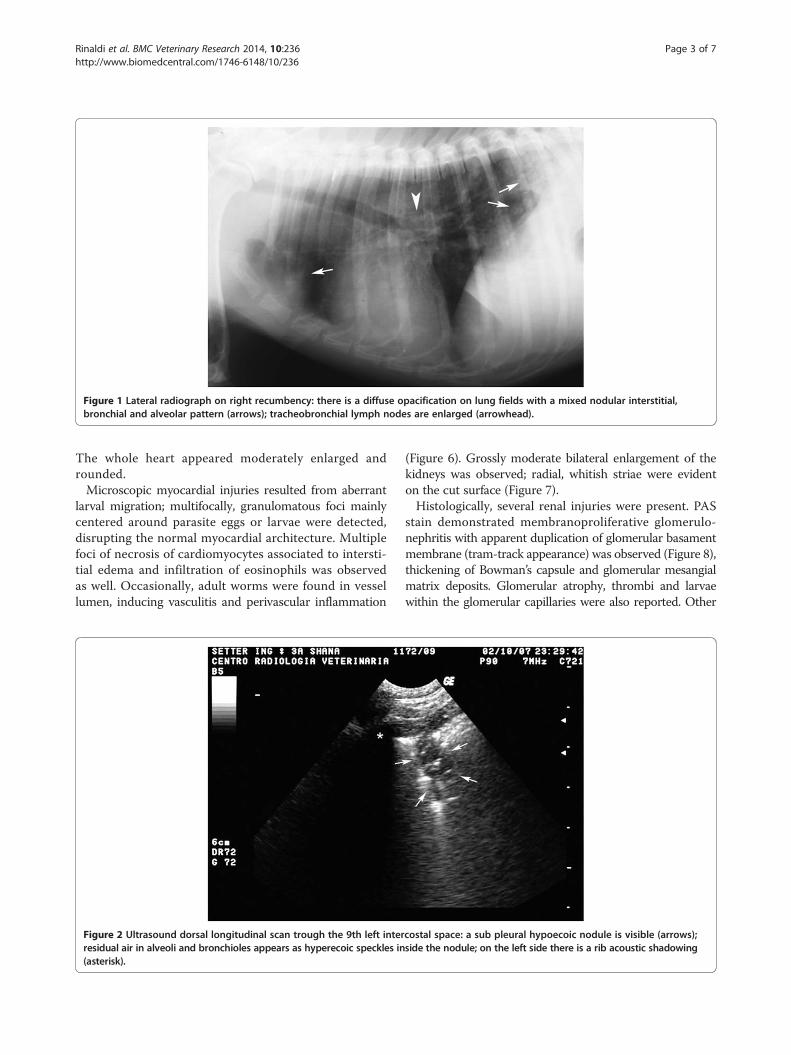
Figure 1 Lateral radiograph on right recumbency: there is a diffuse opacification on lung fields with a mixed nodular interstitial,bronchial and alveolar pattern (arrows); tracheobronchial lymph nodes are enlarged (arrowhead).
Rinaldi et al. BMC Veterinary Research 2014, 10:236 Page 3 of 7http://www.biomedcentral.com/1746-6148/10/236
The whole heart appeared moderately enlarged androunded.Microscopic myocardial injuries resulted from aberrant
larval migration; multifocally, granulomatous foci mainlycentered around parasite eggs or larvae were detected,disrupting the normal myocardial architecture. Multiplefoci of necrosis of cardiomyocytes associated to intersti-tial edema and infiltration of eosinophils was observedas well. Occasionally, adult worms were found in vessellumen, inducing vasculitis and perivascular inflammation
Figure 2 Ultrasound dorsal longitudinal scan trough the 9th left interresidual air in alveoli and bronchioles appears as hyperecoic speckles in(asterisk).
(Figure 6). Grossly moderate bilateral enlargement of thekidneys was observed; radial, whitish striae were evidenton the cut surface (Figure 7).Histologically, several renal injuries were present. PAS
stain demonstrated membranoproliferative glomerulo-nephritis with apparent duplication of glomerular basamentmembrane (tram-track appearance) was observed (Figure 8),thickening of Bowman’s capsule and glomerular mesangialmatrix deposits. Glomerular atrophy, thrombi and larvaewithin the glomerular capillaries were also reported. Other
costal space: a sub pleural hypoecoic nodule is visible (arrows);side the nodule; on the left side there is a rib acoustic shadowing
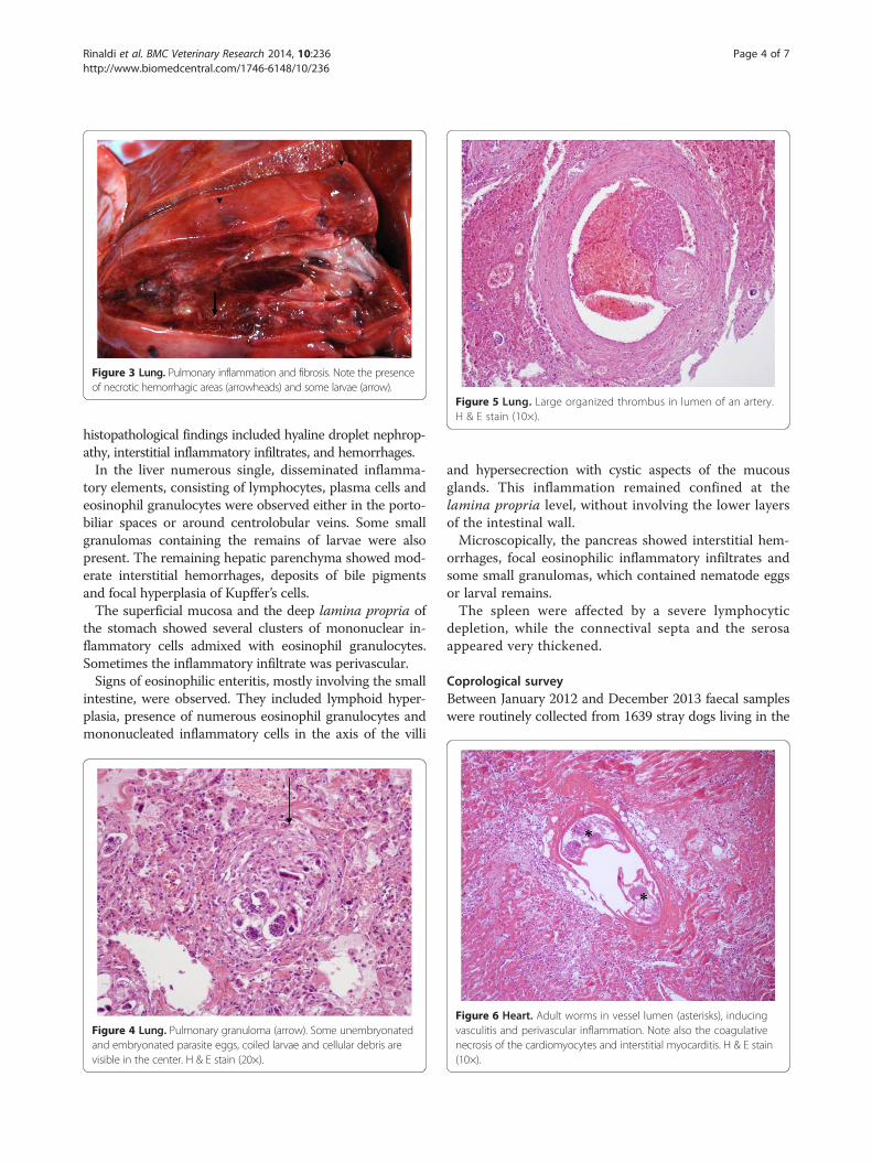
Figure 5 Lung. Large organized thrombus in lumen of an artery.H & E stain (10×).
Figure 3 Lung. Pulmonary inflammation and fibrosis. Note the presenceof necrotic hemorrhagic areas (arrowheads) and some larvae (arrow).
Rinaldi et al. BMC Veterinary Research 2014, 10:236 Page 4 of 7http://www.biomedcentral.com/1746-6148/10/236
histopathological findings included hyaline droplet nephrop-athy, interstitial inflammatory infiltrates, and hemorrhages.In the liver numerous single, disseminated inflamma-
tory elements, consisting of lymphocytes, plasma cells andeosinophil granulocytes were observed either in the porto-biliar spaces or around centrolobular veins. Some smallgranulomas containing the remains of larvae were alsopresent. The remaining hepatic parenchyma showed mod-erate interstitial hemorrhages, deposits of bile pigmentsand focal hyperplasia of Kupffer’s cells.The superficial mucosa and the deep lamina propria of
the stomach showed several clusters of mononuclear in-flammatory cells admixed with eosinophil granulocytes.Sometimes the inflammatory infiltrate was perivascular.Signs of eosinophilic enteritis, mostly involving the small
intestine, were observed. They included lymphoid hyper-plasia, presence of numerous eosinophil granulocytes andmononucleated inflammatory cells in the axis of the villi
Figure 4 Lung. Pulmonary granuloma (arrow). Some unembryonatedand embryonated parasite eggs, coiled larvae and cellular debris arevisible in the center. H & E stain (20×).
and hypersecrection with cystic aspects of the mucousglands. This inflammation remained confined at thelamina propria level, without involving the lower layersof the intestinal wall.Microscopically, the pancreas showed interstitial hem-
orrhages, focal eosinophilic inflammatory infiltrates andsome small granulomas, which contained nematode eggsor larval remains.The spleen were affected by a severe lymphocytic
depletion, while the connectival septa and the serosaappeared very thickened.
Coprological surveyBetween January 2012 and December 2013 faecal sampleswere routinely collected from 1639 stray dogs living in the
Figure 6 Heart. Adult worms in vessel lumen (asterisks), inducingvasculitis and perivascular inflammation. Note also the coagulativenecrosis of the cardiomyocytes and interstitial myocarditis. H & E stain(10×).

Figure 7 Kidney. Interstitial nephritis.
Rinaldi et al. BMC Veterinary Research 2014, 10:236 Page 5 of 7http://www.biomedcentral.com/1746-6148/10/236
city of Naples (Campania region, southern Italy) andbrought to the veterinary hospital of the Departmentof Veterinary Medicine for sterilization. Informationregarding sex, age and clinical signs was collected atthe time of arrival to the veterinary hospital. The age ofeach animal was recorded based on dental examination. Aminimum of 2 g of faeces was collected from each animal,immediately placed into a container, and fixed 1:4 withformalin 5% before being analyzed.Each sample was examined by the FLOTAC basic
technique [12] (using zinc sulphate s.g. 1.20 as flotationsolution). A differential diagnosis was also performed todiscriminate A. vasorum first stage larvae (L1) from thoseof Crenosoma vulpis, Oslerus osleri or Filaroides spp. onthe basis of morphological finding at the tail according to
Figure 8 Kidney. Membranoproliferative glomerulonephritis: apparentduplication of the glomerular basement membrane (tram-trackappearance) S. A larva within the glomerulus is also visible(arrowhead). PAS stain (40×).
Helm et al. [19]: the tapered tip of the tail of A. vasorumL1 has a kink with a dorsal spine (Figure 9).The presence of A. vasorum was detected in 17 of 1639
stray dogs examined (1.04%; 95% CI = 0.63-1.69). Amongthe others lungworms, only O. osleri was detected in 2 ofthe 1639 dogs examined (0.12%; 95% CI = 0.02-0.49).
ConclusionsThis report documents a case of fatal canine angiostron-gylosis in southern Italy. The clinical, pathological, andparasitological findings are consistent with a severe anddiffuse angiostrongylosis. In fact, the clinical manifesta-tions associated with this disease may vary greatly fromsubclinical state (with no or minor sign) to fatal condition.The cardiorespiratory distress due to the inflammatoryresponse to eggs and migrating larvae [20,21], bleedingdisorder such as hemorrhagic diatheses and coagulationdefects, cardiac failure and other nonspecific miscellan-eous systemic and gastro-enteric signs such as gagging,coughing, vomiting, oedema, anorexia, weight loss, stuntedgrowth as well as decreased exercise tolerance are the mostcommon clinical symptoms [1,22,23]. As described above,the definitive diagnosis requires demonstration of the firststage larvae in the faeces, tracheal wash or bronchoalveolar
Figure 9 First-stage larva of Angiostrongylus vasorum. The L1larvae when examined under a light microscope are approximately350 μm in length (range 310–400 μm), with a characteristic kinkedtail and a dorsal spine and notch.
Rinaldi et al. BMC Veterinary Research 2014, 10:236 Page 6 of 7http://www.biomedcentral.com/1746-6148/10/236
lavage. Several case reports, including this one, show thatfaecal Baermann examination can be false negative, due tointermittent shedding of larvae, a high variation in thenumber of shedded larvae and the long pre-patent period[2]. Thus, examination of faecal samples from three con-secutive days is recommended and tracheal wash shouldbe considered [2].The respiratory syndrome observed in the antemortem
case is typical of that reported for A. vasorum infectionin dog. The pulmonary lesions in dog angiostrongylosisare well described in the literature [24] and the presentfindings are in accordance with current knowledge [25].The most consistent finding in our dog was interstitialpneumonia with prominent vascular changes. Pneumonia,in this case, was generally granulomatous with variableamounts of suppurative and eosinophilic inflammation.Small granulomatous foci of inflammation, associated withnematodes eggs and larvae, were found in many organssuch as kidney, liver, pancreas. This is in accordance withthe recent findings that showed the presence of the lesionsin different organs [2], not only in the cardiovascular andrespiratory systems. Thus, the absence of the typical signsshould not preclude consideration of angiostrongylosis asa differential diagnosis [26]. The pathological findings inour case are in according with this propensity and shouldbe considered during the diagnosis of suspected cases.The findings of the present study confirm that the French
heartworm A. vasorum should be considered in the differ-ential diagnosis of pulmonary disease in dogs in Italy [27].The results of this research demonstrated also the oc-
currence of A. vasorum L1 in fecal samples of dogs fromthe Campania region, as reported in other areas of cen-tral and southern Italy with an infection rate similar tothat recently reported also in other studies [28-32]. Inaddition, they also show that FLOTAC can be utilized fordiagnosis of A. vasorum infection, as already demonstratedfor other lungworms as Crenosoma vulpis [33] in dogs andAelurostrongylus abstrusus [34] in cats. Furthermore, giventhe lack of specificity of clinical signs, these infections areoften not included in differential diagnosis, and animalsremain infected and untreated. The FLOTAC has the ad-vantage to be multivalent and therefore also other para-sites (protozoa, nematoda, trematoda and cestoda) can bedetected, which may be important in determining thecause of non-specific symptoms. A valid and affordablediagnostic method for the detection of A. vasorum –infected animals before the appearance of clinical signscould avoid the onset of severe pathological changes inearly anthelmintic-treated animals [35].The emergence of A. vasorum as an important agent of
respiratory disease in dogs, and apparent ongoing expan-sion of its range, must underline the need of an enhancedawareness concerning the knowledge of its epidemiologyand biology [4].
EthicsThe animals used in the present study were sampled fol-lowing approval by the animal ethics and welfare com-mittee of the University of Naples Federico II (in Italian,Comitato Etico-scientifico per la Sperimentazione Animaledell’ Università di Napoli Federico II; protocol number0075262).
Competing interestGC is the inventor of the FLOTAC apparatus. The method is licensed free ofcharge to universities and interested public non-commercial research centers.All other authors have no competing interest.
Authors’ contributionsLR, LC, SP– Participated in the study design and prepared the manuscript.LC and LM – Performed the clinical investigations. LR, PP and GC – Performedthe parasitological investigations. TBP and SP - Carried out the histopatologicalinvestigations. GG and SP - Conceived the study and participated in its designand coordination. All authors read and approved the final manuscript.
AcknowledgmentsThe authors acknowledge Doctors Maria Paola Maurelli and VincenzoMusella for their participation in the study.
Received: 22 April 2014 Accepted: 23 September 2014
References1. Ferdushy T, Hasan MT: Angiostrongylus vasorum: the “French Heartworm”.
Parasitol Res 2010, 107:765–771.2. Denk D, Matiasek K, Just FT, Hermanns W, Baiker K, Herbach N, Steinberg T,
Fischer A: Disseminated angiostrongylosis with fatal cerebralhaemorrhages in two dogs in Germany: a clinical case study. Vet Parasitol2009, 160:100–108.
3. Morgan ER, Shaw S: Angiostrongylus vasorum infection in dogs:continuing spread and developments in diagnosis and treatment.J Small Anim Pract 2010, 51:616–621.
4. Morgan ER, Shaw SE, Brennan SF, de Waal TD, Jones BR, Mulcahy G:Angiostrongylus vasorum: a real heartbreaker. Trends Parasitol 2005,21:49–51.
5. Conboy GA: Canine angiostrongylosis: the French heartworm: anemerging threat in North America. Vet Parasitol 2011, 176:382–389.
6. Morgan ER, Jefferies R, Krajewski M, Ward P, Shaw SE: Canine pulmonaryangiostrongylosis: the influence of climate on parasite distribution.Parasitol Int 2009, 58:406–410.
7. Traversa D, Di Cesare A, Conboy G: Canine and feline cardiopulmonaryparasitic nematodes in Europe: emerging and underestimated.Parasit Vectors 2010, 3:62.
8. Traversa D, Guglielmini C: Feline aelurostrongylosis and canineangiostrongylosis: a challenging diagnosis for two emerging verminouspneumonia infections. Vet Parasitol 2008, 157:163–174.
9. Gasser RB, Jabbar A, Mohandas N, Schnyder M, Deplazes P, Littlewood DTJ,Jex AR: Mitochondrial genome of Angiostrongylus vasorum: comparisonwith congeners and implications for studying the population geneticsand epidemiology of this parasite. Infect Genet Evol 2012, 12:1884–1891.
10. Koch J, Willesen JL: Canine pulmonary angiostrongylosis: an update.Vet J 2009, 179:348–359.
11. Cringoli G, Rinaldi L, Maurelli MP, Utzinger J: FLOTAC: new multivalenttechniques for qualitative and quantitative copromicroscopic diagnosisof parasites in animals and humans. Nat Protoc 2010, 5:503–515.
12. Schnyder M, Maurelli MP, Morgoglione ME, Kohler L, Deplazes P, Torgerson P,Cringoli G, Rinaldi L: Comparison of faecal techniques including FLOTAC forcopromicroscopic detection of first stage larvae of Angiostrongylusvasorum. Parasitol Res 2011, 109:63–69.
13. Verzberger-Epshtein I, Markham RJ, Stryhn H, Sheppard JA, Whitney H,Conboy GA: Serologic detection of Angiostrongylus vasorum infection indogs. Vet Parasitol 2008, 151:53–60.

Rinaldi et al. BMC Veterinary Research 2014, 10:236 Page 7 of 7http://www.biomedcentral.com/1746-6148/10/236
14. Schnyder M, Stebler K, Naucke TJ, Lorentz S, Deplazes P: Evaluation of arapid device for serological in-clinic diagnosis of canine angiostrongylosis.Parasit Vectors 2014, 7:72.
15. Al-Sabi MN, Deplazes P, Webster P, Willesen JL, Davidson RK, Kapel CM: PCRdetection of Angiostrongylus vasorum in faecal samples of dogs andfoxes. Parasitol Res 2010, 107:135–140.
16. Jefferies R, Morgan ER, Helm J, Robinson M, Shaw SE: Improved detectionof canine Angiostrongylus vasorum infection using real-time PCR andindirect ELISA. Parasitol Res 2011, 109:1577–1583.
17. Hendrix CM: Diagnostic Veterinary Parasitology. 2nd edition. St. Louis: Mosby;1998.
18. Rosen L, Ash LR, Wallace GD: Life history of canine lungwormAngiostrongylus vasorum (Baillet). Am J Vet Res 1970, 31:131–141.
19. Helm JR, Morgan ER, Jackson MW, Wotton P, Bell R: Canineangiostrongylosis: an emerging disease in Europe. J Vet Emerg Crit Care(San Antonio) 2010, 20:98–109.
20. Lepri E, Veronesi F, Traversa D, Conti MB, Marchesi MC, Miglio A, MandaraMT: Disseminated angiostrongylosis with massive cardiac and cerebralinvolvement in a dog from Italy. Parasitol Res 2011, 109:505–550.
21. Zarelli M, Shiel R, Gallagher B, Skelly C, Cahalan S: Imaging diagnosis: CTfindings in a dog with intracranial hemorrhage secondary toangiostrongylosis. Vet Radiol Ultrasound 2012, 53:420–423.
22. Chapman PS, Boag AK, Guitian J, Boswood A: Angiostrongylus vasoruminfection in 23 dogs (1999–2002). J Small Anim Pract 2004, 45:435–440.
23. Traversa D, Di Cesare A, Meloni S, Frangipane Di Regalbono A, Milillo P,PAMPURINI F, Venco L: Canine angiostrongylosis in Italy: occurrence ofAngiostrongylus vasorum in dogs with compatible clinical pictures.Parasitol Res 2013, 112:2473–2480.
24. Yamakawa Y, Mcgarry JW, Denk D, Dukes-Mcewan J, Macdonald N, MAS A,Mcconnell F, Tatton B, Valentine EG, Wayne J, Williams JM, Hetzel U:Emerging canine angiostrongylosis in Northern England: five fatalcases. Vet Rec 2009, 164:149–152.
25. Bolt G, Monrad J, Koch J, Jensen AL: Canine angiostrongylosis: a review.Vet Rec 1994, 135:447–452.
26. Gallagher B, Brennan SF, Zarelli M, Mooney CT: Geographical, clinical,clinicopathological and radiographic features of canineangiostrongylosis in Irish dogs: a retrospective study. Ir Vet J 2012, 65:5.
27. Traversa D, Torbidone A, Malatesta D, Guglielmini C: Occurrence of fatal canineAngiostrongylus vasorum infection in Italy. Vet Parasitol 2008, 152:162–166.
28. Sasanelli M, Paradies P, Otranto D, Lia RP, de Caprariis D: Haemothoraxassociated with Angiostrongylus vasorum infection in a dog. J Small AnimPract 2008, 49:417–420.
29. Di Cesare A, Castagna G, Meloni S, Milillo P, Latrofa S, Otranto D, Traversa D:Canine and feline infections by cardiopulmonary nematodes in Centraland Southern Italy. Parasitol Res 2011, 109:S87–S96.
30. Tieri E, Pomilio F, Di Francesco G, Saletti MA, Totaro P, Troilo M, Menna S,Tampieri MP, Morelli D: Angiostrongylus vasorum in 20 dogs in theprovince of Chieti, Italy. Vet Ital 2011, 47:77–88.
31. Guardone L, Schnyder M, Macchioni F, Deplazes P, Magi M: Serologicaldetection of circulating Angiostrongylus vasorum antigen and specificantibodies in dogs from Central and Northern Italy. Vet Parasitol 2013,192:192–198.
32. Pipia AP, Varcasia A, Tosciri G, Seu S, Manunta ML, Mura MC, Sanna G,Tamponi C, Brianti E, Scala A: New insights onto cardiopulmonarynematodes of dogs in Sardinia, Italy. Parasitol Res 2014, 113:1505–1509.
33. Rinaldi L, Calabria G, Carbone S, Carrella A, Cringoli G: Crenosoma vulpis indog: first case report in Italy and use of the FLOTAC technique forcopromicroscopic diagnosis. Parasitol Res 2007, 101:1681–1684.
34. Gaglio G, Cringoli G, Rinaldi L, Brianti E, Giannetto S: Use of the FLOTACtechnique for the diagnosis of Aelurostrongylus abstrusus in the cat.Parasitol Res 2008, 103:1055–1057.
35. Schnyder M, Tanner I, Webster P, Barutzki D, Deplazes P: An ELISA forsensitive and specific detection of circulating antigen of Angiostrongylusvasorum in serum samples of naturally and experimentally infecteddogs. Vet Parasitol 2011, 179:152–158.
doi:10.1186/s12917-014-0236-1Cite this article as: Rinaldi et al.: Angiostrongylus vasorum: epidemiological,clinical and histopathological insights. BMC Veterinary Research 2014 10:236.
Submit your next manuscript to BioMed Centraland take full advantage of:
• Convenient online submission
• Thorough peer review
• No space constraints or color figure charges
• Immediate publication on acceptance
• Inclusion in PubMed, CAS, Scopus and Google Scholar
• Research which is freely available for redistribution
Submit your manuscript at www.biomedcentral.com/submit