Embed Size (px)
Citation preview
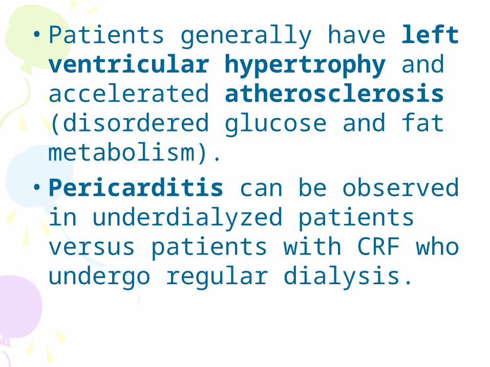
• Patients generally have left ventricular hypertrophy and accelerated atherosclerosis (disordered glucose and fat metabolism).
•Pericarditis can be observed in underdialyzed patients versus patients with CRF who undergo regular dialysis.
• A unique form of pulmonary congestion and edema may occur even in the absence of volume overload and
• is associated with normal or mildly elevated intracardiac and pulmonary capillary wedge pressure.
• This entity, characterized radiologically by peripheral vascular congestion giving rise to a “butterfly wing” distribution, is due to increased permeability of alveolar capillary membranes.
• This “low-pressure” pulmonaryedema and the cardiopulmonary
abnormalities associated with circulatory overload usually respond
• promptly to vigorous dialysis.
Hematologic Hematologic ManifestationsManifestations
• CRF usually causes a normochromic, normocytic anemia.
• Anemia is generally observed when the GFR decreases to less than 30 mL/min and is due to insufficient production of erythropoietin by the diseased kidneys.
• Other factors are iron deficiency, either related to or independent of blood loss from repeated laboratory testing, blood retention in the dialyzer, or gastrointestinal bleeding.
• Treatment of anemia with iron, darbepoetin alfa, and human recombinant erythropoietin normalizes the hematocrit,
• avoids repetitive red blood cell transfusion, reduces the requirement for hospitalization, and decreases cardiovascular mortality by about 30%.
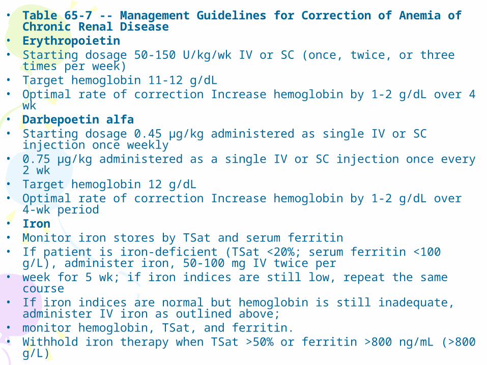
• Table 65-7 -- Management Guidelines for Correction of Anemia of Chronic Renal Disease
• Erythropoietin• Starting dosage 50-150 U/kg/wk IV or SC (once, twice, or three times per
week)• Target hemoglobin 11-12 g/dL• Optimal rate of correction Increase hemoglobin by 1-2 g/dL over 4 wk• Darbepoetin alfa• Starting dosage 0.45 μg/kg administered as single IV or SC injection once
weekly• 0.75 μg/kg administered as a single IV or SC injection once every 2 wk• Target hemoglobin 12 g/dL• Optimal rate of correction Increase hemoglobin by 1-2 g/dL over 4-wk
period• Iron• Monitor iron stores by TSat and serum ferritin• If patient is iron-deficient (TSat <20%; serum ferritin <100 g/L),
administer iron, 50-100 mg IV twice per• week for 5 wk; if iron indices are still low, repeat the same course• If iron indices are normal but hemoglobin is still inadequate, administer IV
iron as outlined above;• monitor hemoglobin, TSat, and ferritin.• Withhold iron therapy when TSat >50% or ferritin >800 ng/mL (>800 g/L)
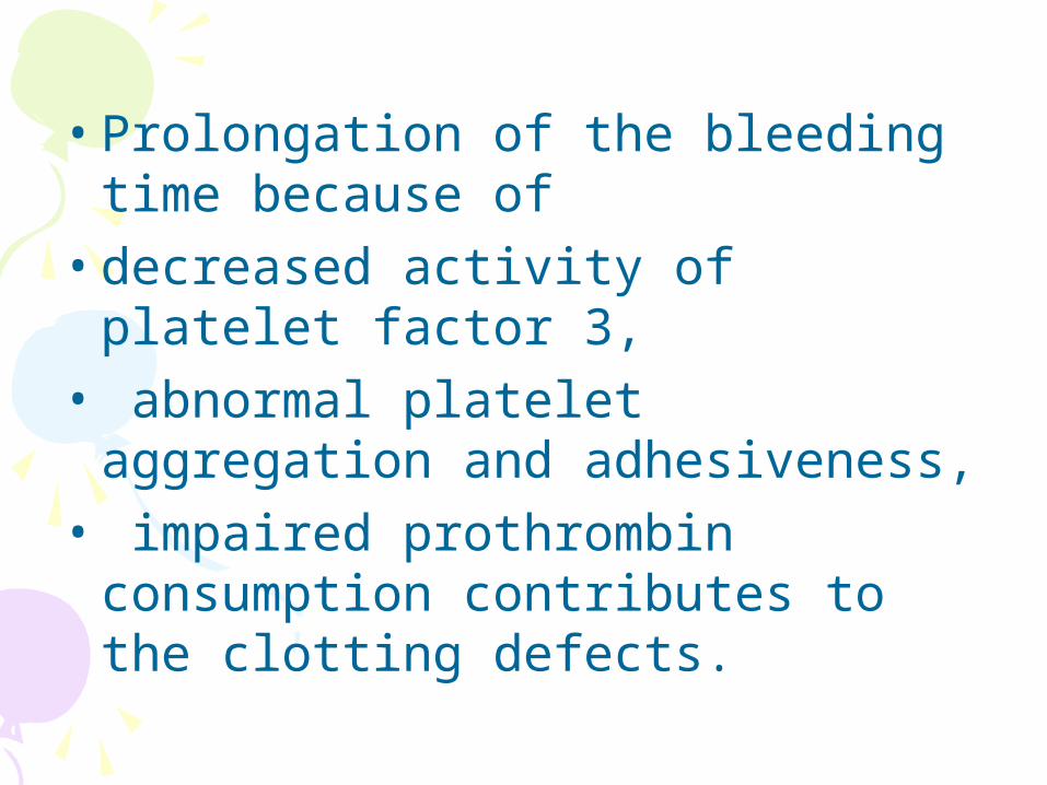
• Prolongation of the bleeding time because of
• decreased activity of platelet factor 3,
• abnormal platelet aggregation and adhesiveness,
• impaired prothrombin consumption contributes to the clotting defects.

•Abnormal bleeding times and coagulopathy in patients with renal failure may be managed with desmopressin, cryoprecipitate, conjugated estrogens, blood transfusions, and erythropoietin use.

Effects of Drugs in Patients with
Reduced Renal Function
OpioidsOpioids
• Renal failure has implications of major clinical importance with respect to morphine and meperidine
• For the fentanyl congeners, the clinical importance of renal failure is less marked.
• As with fentanyl, sufentanil pharmacokinetics are not altered in any consistent fashion by renal disease, although greater variability exists in the clearance and elimination half-life of sufentanil when patients have impaired renal function.
• An increased clinical effect is likely with alfentanil in renal failure because of a decreased initial volume of distribution and an increased free fraction of alfentanil.
• No delay in recovery after alfentanil administration should be expected

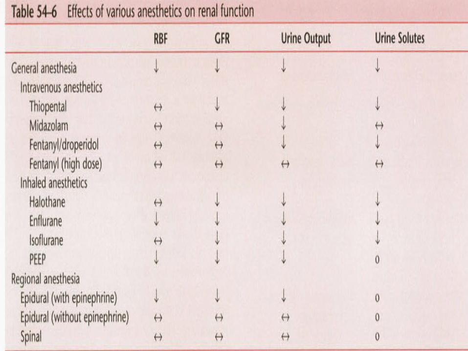
Inhaled AnestheticsInhaled Anesthetics• All inhaled anesthetics are
biotransformed to some extent, with the nonvolatile products of metabolism eliminated almost entirely by the kidney.
• Reversal of the CNS effects of inhaled anesthetics depends on pulmonary excretion, however, so impaired kidney function would not alter the response to these anesthetics.
• Sevoflurane is not very stable. Soda lime causes it to decompose, and it is biotransformed by the liver.
• Plasma inorganic fluoride concentrations approaching nephrotoxic levels (50 μmol/L) have been reported after prolonged inhalation of sevoflurane.
No evidence of gross changes in renal function was found in humans, however.
• Inhaled anesthetics cause a transient reversible depression in renal function.
• GFR, renal blood flow, urineoutput, and urinary excretion of sodium
are decreased . Probable mechanisms include
reduced renal blood flow, loss of renal autoregulation, neurohumoral factors (e.g., antidiuretic hormone, vasopressin,renin), and neuroendocrine responses.
Intravenous AnestheticsIntravenous Anesthetics• Reversal of CNS effects after the
administration of ultrashort-acting barbiturates such as thiopental and methohexital occurs as a result of redistribution, and hepatic metabolism is the sole route of elimination of these drugs .
• Thiopental is 75% to 85% bound to albumin, the concentration of which
• may be markedly reduced in uremia.
• In addition, thiopental is a weak• acid, with its pKa in the
physiologic range; acidosis results in more un-ionized, nonbound, active thiopental.
• In combination, these changes produce an increase in the free fraction of thiopental from 15% in normal patients to 28% in patients with CRF.
• Propofol does not adversely affect renal function as reflected by measurements of creatinine concentration.
• Prolonged infusions of propofol may result in the excretion of green urine because of the presence of phenols in the urine.
• This discoloration does not affect renal function.
• Urate excretion is increased after the• administration of propofol and is usually
manifested as cloudy urine when urate crystallizes under conditions
• of low pH and temperature.

Muscle Relaxants and Muscle Relaxants and Their AntagonistsTheir Antagonists
• Succinylcholine has been used without difficulty in patients with decreased or absent renal function .
• Its metabolism is catalyzed by pseudocholinesterase to yield the nontoxic end products succinic acid and choline.
• . Large doses of succinylcholine, which might result from prolonged infusion, should be avoided in patients with renal failure.
• Renal failure influences the• pharmacology of nondepolarizing
muscle relaxants by producing either decreased elimination of the drug or
• its metabolites by the kidney or decreased activity of enzymes that metabolize the drug, such as in the case of mivacurium .
• the duration of action of muscle relaxants may be prolonged in patients with renal failure.
• Pharmacokinetics data for the cholinesterase inhibitors neostigmine, pyridostigmine, and edrophonium for
• normal, anephric, and renal transplant patients are presented in Table 65-11 ; there are no major differences
• among the three drugs.
• Renal excretion is of major importance for the elimination of all three agents, with approximately 50% of neostigmine and 70% of pyridostigmine and edrophonium excreted in urine.
• Excretion of all the cholinesterase inhibitors is delayed in patients with impaired renal function to the same or perhaps to a slightly greater extent than is elimination of muscle relaxants.

Vasopressors and Vasopressors and Antihypertensive DrugsAntihypertensive Drugs
Acute renal failure
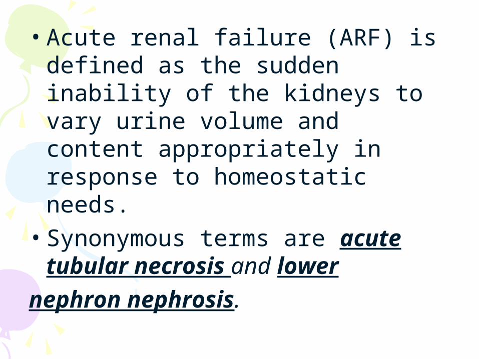
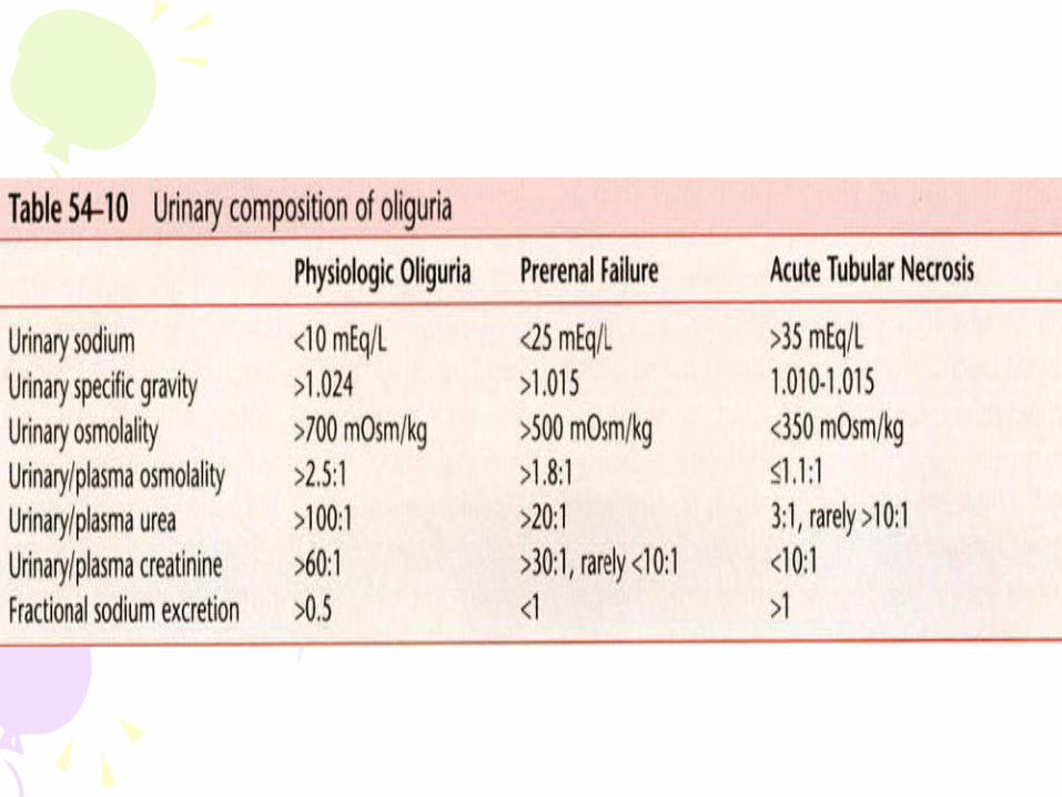
• Acute renal failure (ARF) is defined as the sudden inability of the kidneys to vary urine volume and content appropriately in response to homeostatic needs.
• Synonymous terms are acute tubular necrosis and lower
nephron nephrosis.
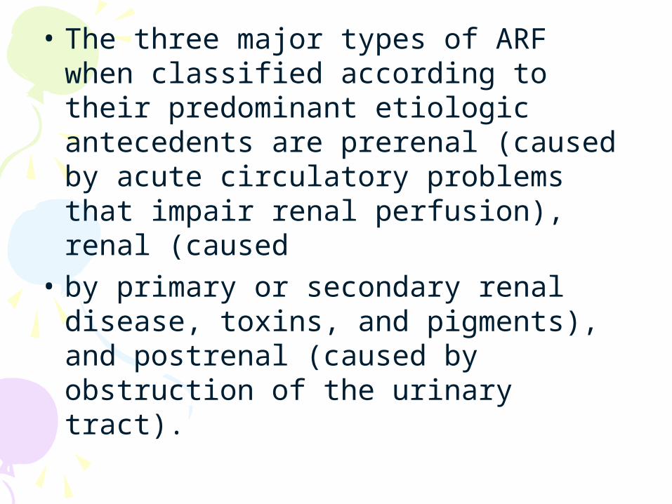
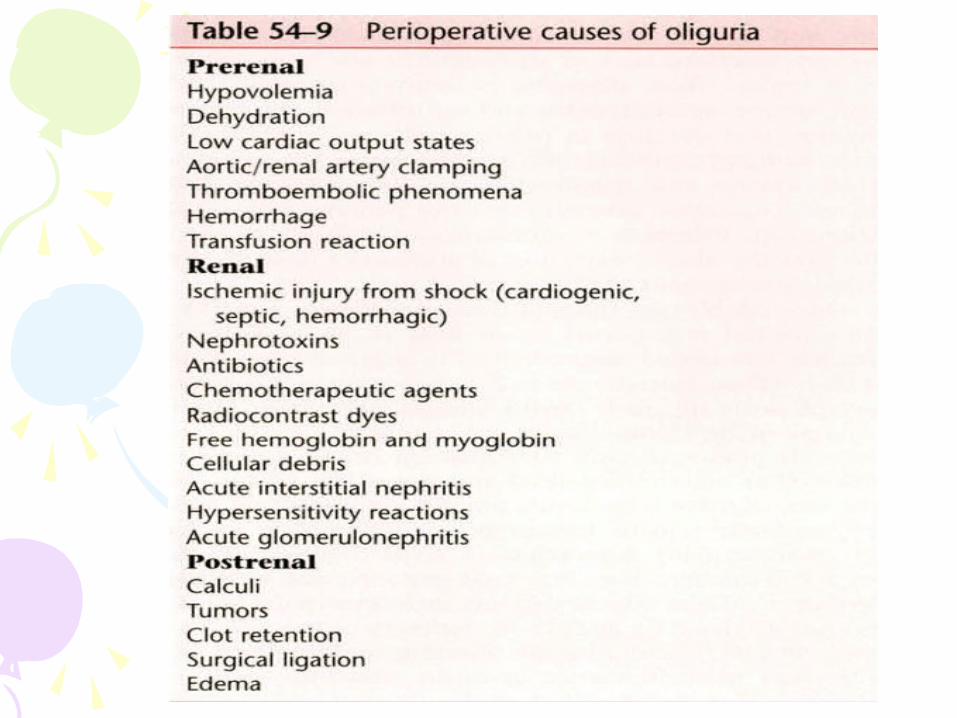
• The three major types of ARF when classified according to their predominant etiologic antecedents are prerenal (caused by acute circulatory problems that impair renal perfusion), renal (caused
• by primary or secondary renal disease, toxins, and pigments), and postrenal (caused by obstruction of the urinary tract).
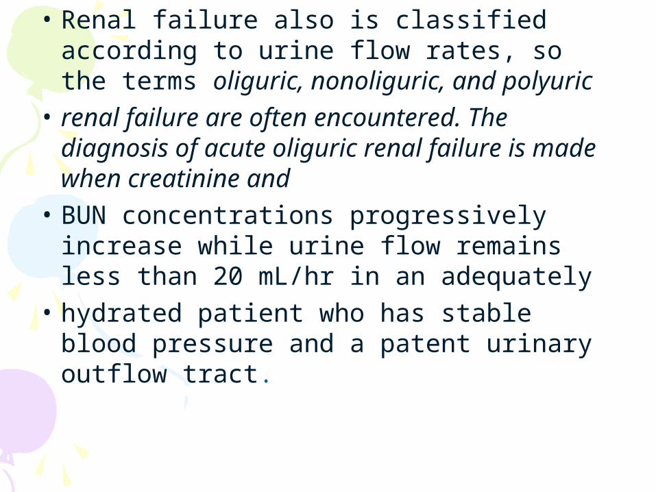
• Renal failure also is classified according to urine flow rates, so the terms oliguric, nonoliguric, and polyuric
• renal failure are often encountered. The diagnosis of acute oliguric renal failure is made when creatinine and
• BUN concentrations progressively increase while urine flow remains less than 20 mL/hr in an adequately
• hydrated patient who has stable blood pressure and a patent urinary outflow tract.

Hemodialysis
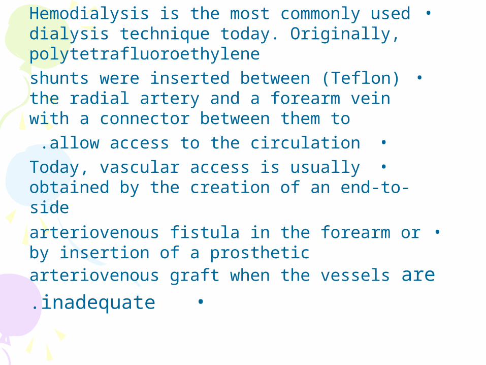
•Hemodialysis is the most commonly used dialysis technique today. Originally, polytetrafluoroethylene
•(Teflon )shunts were inserted between the radial artery and a forearm vein with a connector between them to
•allow access to the circulation .•Today, vascular access is usually obtained
by the creation of an end-to-side•arteriovenous fistula in the forearm or by
insertion of a prosthetic arteriovenous graft when the vessels are
•inadequate.
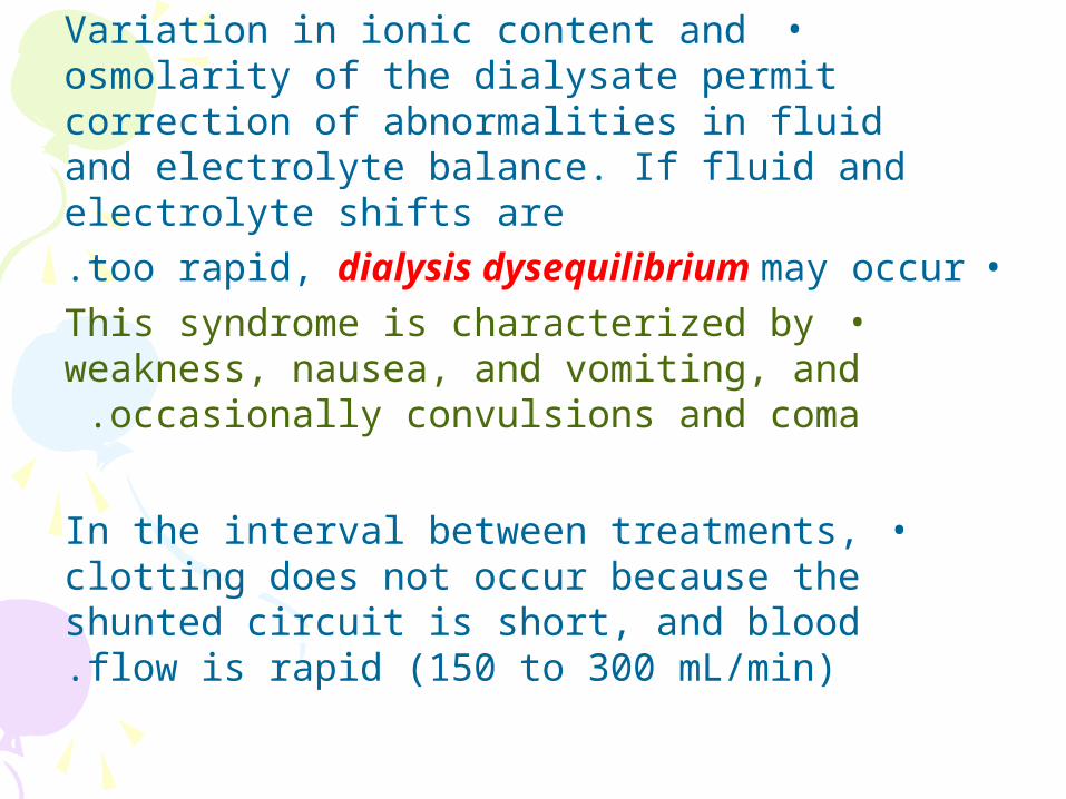
•Variation in ionic content and osmolarity of the dialysate permit correction of abnormalities in fluid and electrolyte balance. If fluid and electrolyte shifts are
•too rapid, dialysis dysequilibrium may occur .
•This syndrome is characterized by weakness, nausea, and vomiting, and occasionally convulsions and coma .
•In the interval between treatments, clotting does not occur because the shunted circuit is short, and blood flow is rapid (150 to 300 mL/min).
• Local infiltration, axillary block, and general anesthesia have been used successfully for creation of arteriovenous fistulas .
• Several anesthetic precautions• are appropriate.
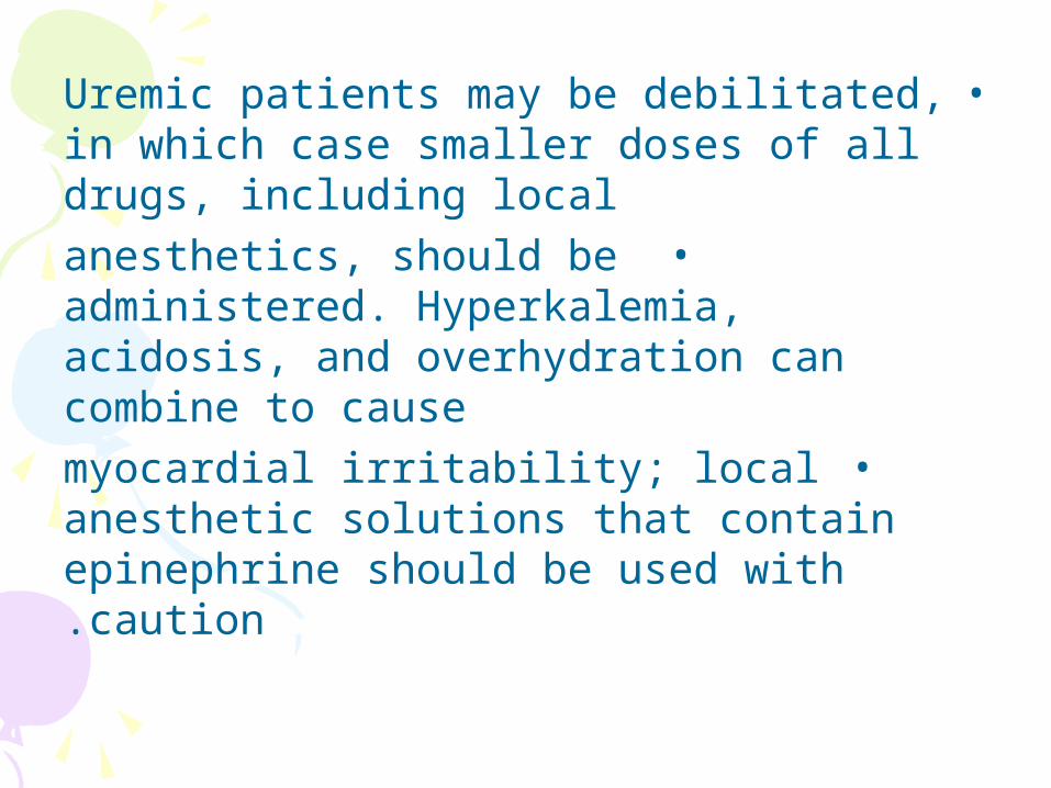
•Uremic patients may be debilitated, in which case smaller doses of all drugs, including local
•anesthetics, should be administered. Hyperkalemia, acidosis, and overhydration can combine to cause
•myocardial irritability; local anesthetic solutions that contain epinephrine should be used with caution.
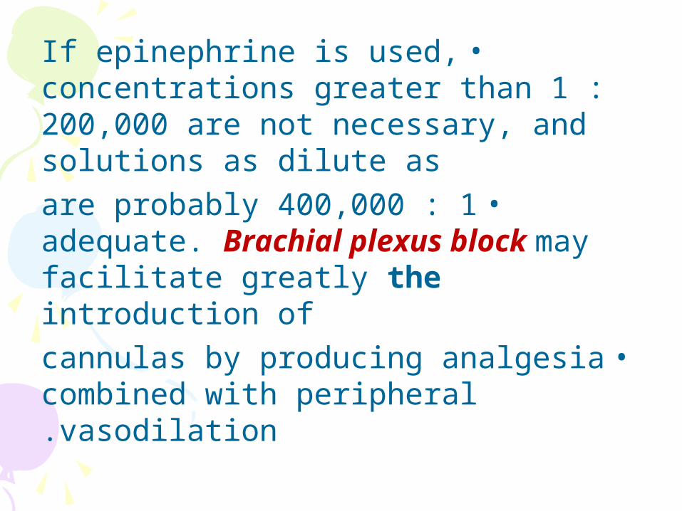
•If epinephrine is used, concentrations greater than 1 : 200,000 are not necessary, and solutions as dilute as
•1 : 400,000 are probably adequate. Brachial plexus block may facilitate greatly the introduction of
•cannulas by producing analgesia combined with peripheral vasodilation.
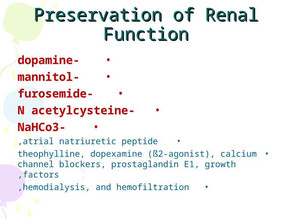
Preservation of Renal Preservation of Renal FunctionFunction
•-dopamine•-mannitol
•-furosemide•-N acetylcysteine
•-NaHCo3•atrial natriuretic peptide,
•theophylline, dopexamine (ß2-agonist), calcium channel blockers, prostaglandin E1, growth factors,
•hemodialysis, and hemofiltration,

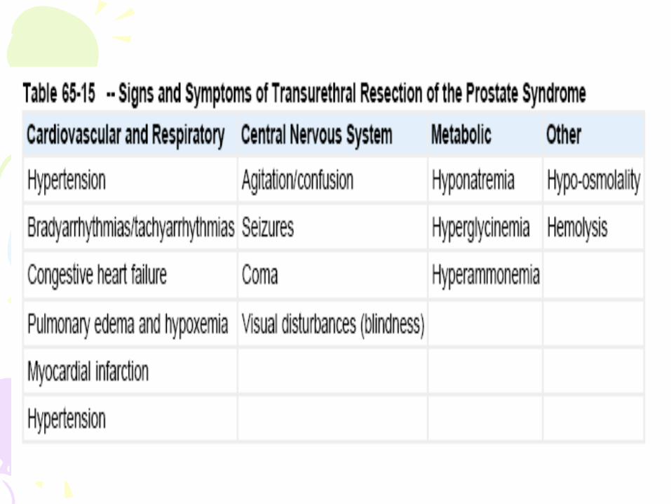
Transurethral Resection of the Prostate

Laser Prostatectomy, Laser Prostatectomy, Cryosurgery, and Microwave Cryosurgery, and Microwave
AblationAblation•Laser prostatectomy has found
renewed interest among urologists and is being conducted in several
•centers. Based on initial experience, it promises to replace conventional TURP in the near future.
•The main advantages over conventional TURP include minimal blood loss (50 to 70 mL) and minimalfluid absorption, which should nearly eliminate these two major complications of TURP; however, other
•potential complications are introduced, including coagulation through the prostatic fossa and sloughing of
•prostatic debris in the postoperative period, with subsequent urinary obstruction and urinary retention

Laser LithotripsyLaser Lithotripsy• Laser lithotripsy is used for ureteral
stones that are low in the ureter and not amenable to extracorporeal
shock wave lithotripsy (ESWL).• A pulsed dye laser is generated with
a laser beam of 504-nm wavelengthpassing through an organic green dye.
• These lasers are not well absorbed by red blood cells or
• other tissues, however, which provides safety against tissue coagulation or thermal injury.
• Because the laser beam is reflective, the user, other personnel, and the patient should wear protective eyeglasses.

Laparoscopic Surgery in Laparoscopic Surgery in UrologyUrology
• Although all the conventional complications and concerns associated with laparoscopy are applicable to
• urologic procedures , two unique problems also are identified.
• First, because the urogenital• system is mainly retroperitoneal, the large
retroperitoneal space and its communications with the thorax and
• subcutaneous tissue are exposed to the insufflated carbon dioxide.
• Subcutaneous emphysema occurs• frequently in these patients and may
extend all the way up to the head and neck.
• The upper airway is at risk for compromise in the most severe cases because of pharyngeal swelling secondary to submucous carbon dioxide.
• This complication should be kept in mind before extubation of these patients.
• Second, the procedures tend to be lengthy, allowing for sufficient absorption of carbon dioxide to result in acidemia and marked acidosis.
• Because of significant increases in intra-abdominal and intrathoracic pressure as a
• result of insufflated carbon dioxide, a steep Trendelenburg position, and lengthy procedures, general anesthesia with controlled ventilation is the method of choice.

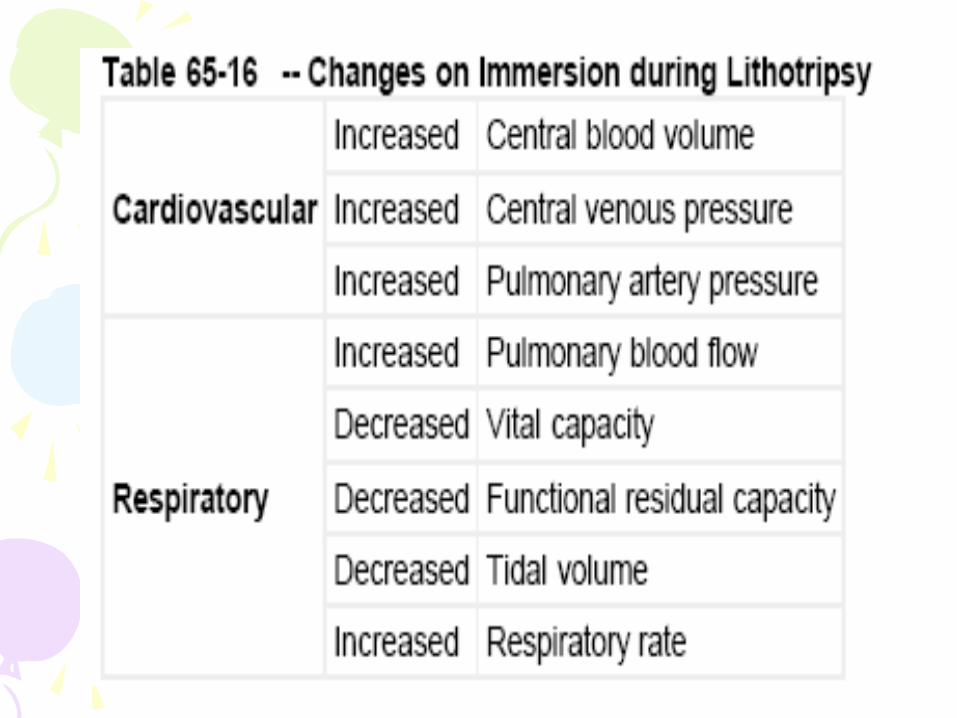
Extracorporeal Shock Wave Extracorporeal Shock Wave LithotripsyLithotripsy
• All lithotripters share similar technologic principles in having three main components: (1) an energy source, most commonly a spark plug (alternatively, an electromagnetic membrane or piezoelectric elements); (2) a system to focus the shock wave, such as ellipsoid or reflecting mirrors; and (3) fluoroscopy or ultrasound to visualize and localize the stone in focus.
• Although shock waves pass through most tissues relatively unimpeded, they do cause tissue injury, the extent of which depends on the tissue exposed and the shock wave energy at the tissue level. Skin bruising
•and flank ecchymoses are common at the entry site.
• Painful hematoma in the flank muscles may occur.
• Hematuria is almost always present at the end of the procedure and results from shock wave–induced
endothelial injury to the kidney and ureter.
• Adequate hydration is necessary to prevent clot retention.
• A decrease in postoperative hematocrit should arouse suspicion of a large perinephric hematoma.
• Punctate hemorrhages have been observed in the stomach and bowel, and this injury might be responsible for abdominal distention, nausea, and vomiting in some patients.

• Shock wave–induced cardiac arrhythmias previously reported in 10% to 14% of patients undergoing lithotripsy are extremely rare nowadays.
• These arrhythmias are believed to be due to mechanical
• stress on the conduction system exerted by the shock waves.
• Atrial and ventricular premature• complexes, atrial fibrillation, and
supraventricular and ventricular tachycardia have been reported.
• Artifacts on electrocardiogram also are common.
• Artifacts and arrhythmias usually disappear when the lithotripsy is stopped.
• Arrhythmias occasionally may persist and require treatment

Anesthetic Choices for Anesthetic Choices for LithotripsyLithotripsy
• Anesthetic regimens used successfully for lithotripsy include general anesthesia, epidural anesthesia, spinal
• anesthesia, flank infiltration with or without intercostal blocks, and analgesia-sedation, including patientcontrolled
• analgesia. • General anesthesia offers the advantages
of rapid onset and control of patient movement.
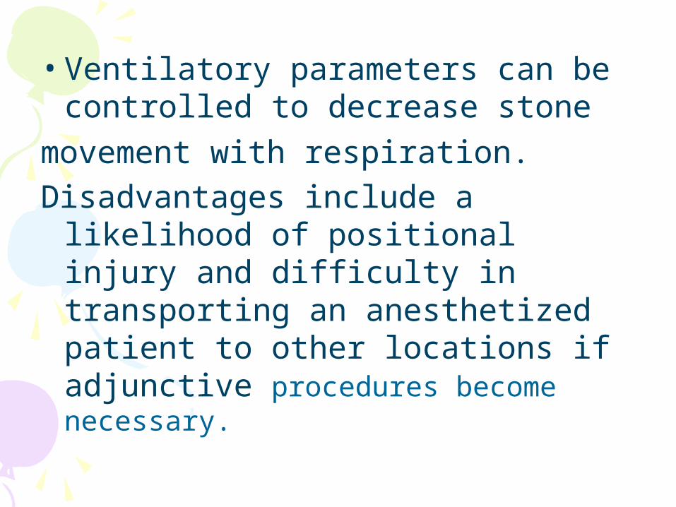
• Ventilatory parameters can be controlled to decrease stone
movement with respiration. Disadvantages include a likelihood
of positional injury and difficulty in transporting an anesthetized patient to other locations if adjunctive procedures become necessary.
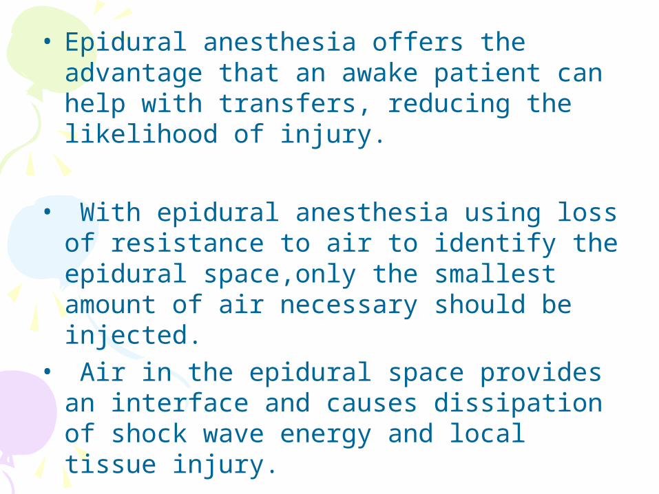
• Epidural anesthesia offers the advantage that an awake patient can help with transfers, reducing the likelihood of injury.
• With epidural anesthesia using loss of resistance to air to identify the epidural space,only the smallest amount of air necessary should be injected.
• Air in the epidural space provides an interface and causes dissipation of shock wave energy and local tissue injury.

ContraindicationsContraindications• Pregnancy and untreated bleeding
disorders are the only contraindications to lithotripsy.
• Women of childbearing age must have a pregnancy test that is documented to be negative before lithotripsy. Standard
• tests of coagulation, such as the platelet count, prothrombin time, and partial thromboplastin time, should be
• obtained.
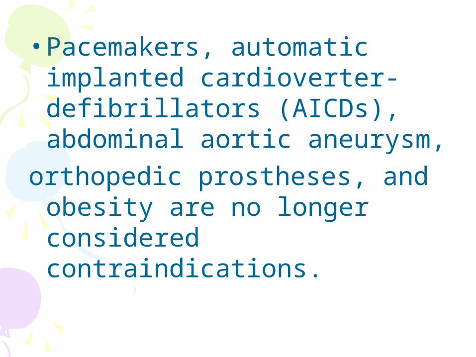
•Pacemakers, automatic implanted cardioverter-defibrillators (AICDs), abdominal aortic aneurysm,
orthopedic prostheses, and obesity are no longer considered contraindications.