Embed Size (px)
Citation preview
Rom J Morphol Embryol 2016, 57(1):237–242
ISSN (print) 1220–0522 ISSN (online) 2066–8279
CCAASSEE RREEPPOORRTT
Tuberculous constrictive pericarditis complicated with tuberculous mediastinitis – case report
MILENA ADINA MAN1), MIMI FLOAREA NIŢU2), LELIA STRÂMBU3), CRISTINA FLORESCU4), COSTIN TEODOR STREBA2), ANTIGONA-CARMEN TROFOR5)
1)Department of Pulmonary Diseases, “Iuliu Haţieganu” University of Medicine and Pharmacy, Cluj-Napoca, Romania
2)Department of Pneumology, University of Medicine and Pharmacy of Craiova, Romania 3)Department of Cardiology, “Niculae Stăncioiu” Heart Institute, Cluj-Napoca, Romania 4)Department of Cardiology, University of Medicine and Pharmacy of Craiova, Romania 5)Department of Pulmonary Diseases, Faculty of Medicine, “Grigore T. Popa” University of Medicine and Pharmacy, Iassy, Romania
Abstract Constrictive pericarditis is a rare and severe disease. A 37-year-old patient was admitted in the hospital for dyspnea, precordial pain, right-sided cardiac failure. Chest X-ray showed cardiac enlargement and an opacity suggestive for pleural effusion. Echocardiography revealed an adhesive–effusive–constrictive pericarditis, a very thickened pericardium and bilateral pleural effusion. After a pericardiectomy done to restore cardiac compensation and to identify etiological factors, a tuberculous pericarditis (TBP) was diagnosed. After surgery and starting anti-TB treatment, the patient presented altered clinical status, dyspnea, dry cough, fever and delayed callus formation at sternum level. Thoracic scan revealed mediastinal air collections, pericarditis and pleurisy. Thus, the TBP diagnosis was extended to mediastinal TB and anti-TB therapy was continued. After four months of treatment, another thoracic scan showed disappearance of the mediastinal air-leakage bubbles, multiple new micronodules in both lungs and lymph nodes of up to 15 mm; also increasing pericardial and pleural effusions. This case was interpreted as a TB treatment failure situation. A retreatment regimen was started, resulting in a slow favorable outcome. Pericardial TB is a rare condition, usually with delayed diagnosis and poor treatment benefits. Whenever possible, earlier diagnostic can contribute to better management of these cases.
Keywords: tuberculosis constrictive pericarditis, mediastinitis, pleuritis, Mycobacterium tuberculosis.
Introduction
Tuberculosis (TB), one of the oldest diseases that have plagued mankind, remains today a major cause of morbidity and mortality worldwide [1]. Due to therapeutic measures, in the past 100 years in rich industrialized countries there has been a significant decline in TB; however, in the last 20 years, we have witnessed a resurgence of the disease, the estimated number of new cases worldwide growing steadily from 8.0 million in 1997 to 8.3 million in 2000, 8.7 million in 2011 and is expected to reach 10.2 million at the end of 2015 [1–3]. Most cases of active TB (about 95%) were found in Africa, Asia and Latin America [2]. An increasing number of new cases of TB are due on one hand to migration of populations in poor areas of the world [4, 5], and on the other hand to the infection with human immunodeficiency virus (HIV), which dramatically increased both the morbidity and mortality of TB in some areas of the world [6–9].
Pericardial tuberculosis or tuberculous pericarditis (TBP) is a rare but particularly severe complication, with a mortality of 80–90% when not handled properly and 12–17% with appropriate treatment [1]. TB is responsible for about 4% of cases of acute pericarditis, 7% of cases of cardiac tamponade and about 6% of cases of constrictive pericarditis [10–12]. In a few underdeveloped countries, TB is the leading cause of pericarditis [3, 13, 14].
TBP incidence in developed countries is only 4% of all cases of TB [15] but in some areas of South Africa, tuberculous pericarditis represented 69.5% of pericarditis that required pericardial puncture for diagnosis [16]. In sub-Saharan Africa, the incidence of TBP is increasing due to the HIV epidemic, and this trend is likely to occur in other parts of the world due to HIV infection [17, 18].
Tuberculous pericarditis diagnosis is difficult to establish in the absence of personal or family history suggesting a TB infection history or contact with another person with TB. In this paper, we present a case of TB pericarditis, which raised issues of etiological diagnosis and treatment.
Case presentation
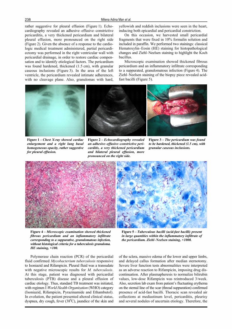
A 37-year-old male patient, HIV-negative, with a medical history of pleuritis and pericarditis of unknown origin, interpreted by rheumatologist in the context of a polyserositis secondary to a systemic disease and treated with Prednisone (0.5 mg/kg) and immunosuppressive (four months before), was admitted in the Department of Cardiology, “Niculae Stăncioiu” Heart Institute, Cluj-Napoca, Romania, for dyspnea on exertion, precordial pain, right-sided cardiac failure with massive lower limbs edema and hepatic disorder. Chest X-ray showed cardiac enlargement and a right lung basal homogeneous opacity,
R J M ERomanian Journal of
Morphology & Embryologyhttp://www.rjme.ro/
Milena Adina Man et al.
238
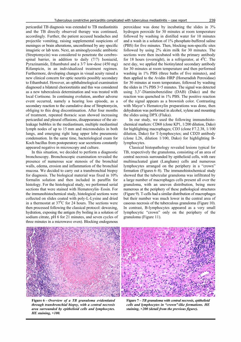
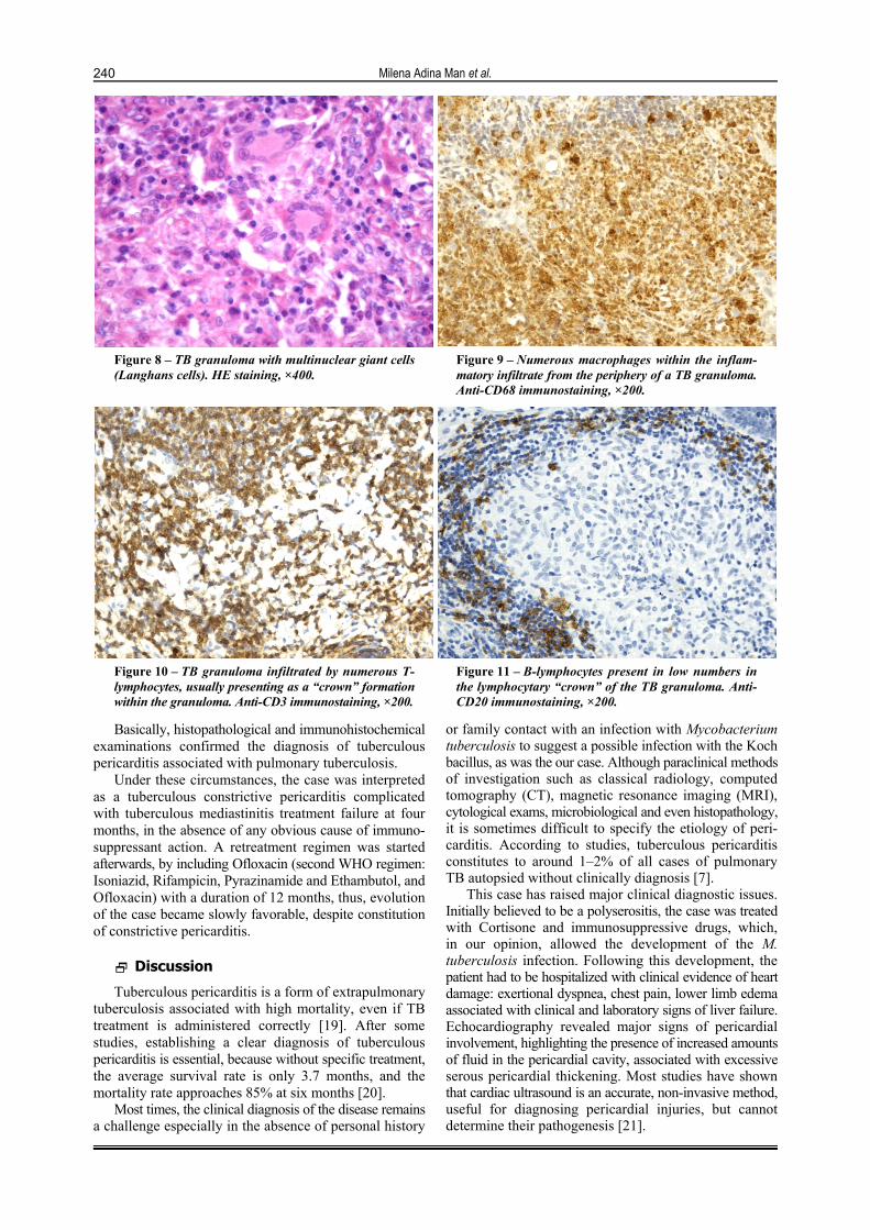
rather suggestive for pleural effusion (Figure 1). Echo-cardiography revealed an adhesive–effusive–constrictive pericarditis, a very thickened pericardium and bilateral pleural effusion, more pronounced on the right side (Figure 2). Given the absence of a response to the cardio-logic medical treatment administered, partial pericardi-ectomy was performed in the right ventricular wall with pericardial drainage, in order to restore cardiac compen-sation and to identify etiological factors. The pericardium was found hardened, thickened (1.5 cm), with granular caseous inclusions (Figure 3). In the area of the left ventricle, the pericardium revealed intimate adherences, with no cleavage plane. Also, granulomas with hard,
yellowish and reddish inclusions were seen in the heart, inducing both epicardial and pericardial constriction.
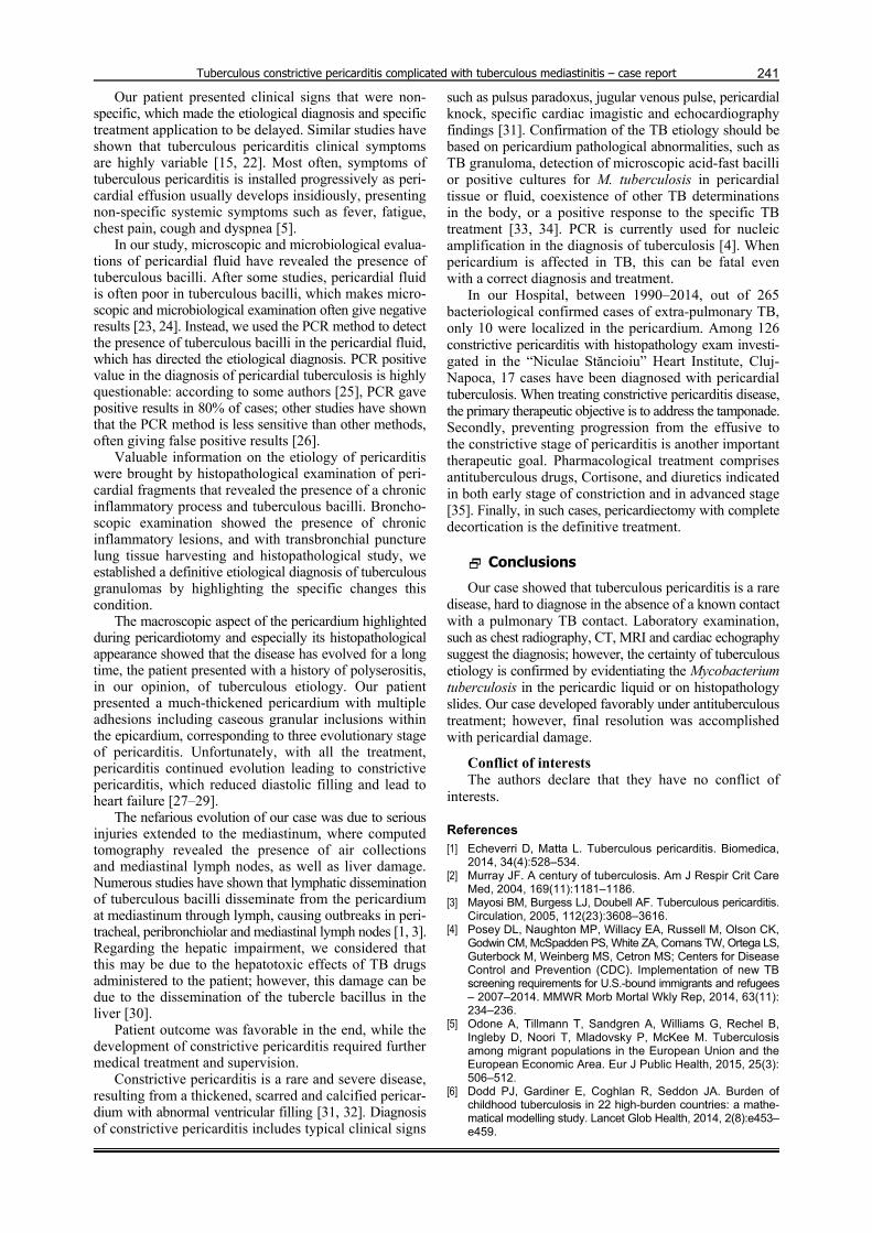
On this occasion, we harvested small pericardial fragments that were fixed in 10% formalin solution and included in paraffin. We performed two stainings: classical Hematoxylin–Eosin (HE) staining for histopathological changes and Ziehl–Neelsen staining to highlight the Koch bacillus.
Microscopic examination showed thickened fibrous pericardium and an inflammatory infiltrate corresponding to a suppurated, granulomatous infection (Figure 4). The Ziehl–Neelsen staining of the biopsy piece revealed acid-fast bacilli (Figure 5).
Figure 1 – Chest X-ray showed cardiac enlargement and a right lung basal homogeneous opacity, rather suggestive for pleural effusion.
Figure 2 – Echocardiography revealed an adhesive–effusive–constrictive peri-carditis, a very thickened pericardium and bilateral pleural effusion, more pronounced on the right side.
Figure 3 – The pericardium was found to be hardened, thickened (1.5 cm), with granular caseous inclusions.
Figure 4 – Microscopic examination showed thickened fibrous pericardium and an inflammatory infiltrate corresponding to a suppurative, granulomatous infection, without histological criteria for a tuberculosis granuloma. HE staining, ×100.
Figure 5 – Tuberculous bacilli (acid-fast bacilli) present in large quantities within the inflammatory infiltrate of the pericardium. Ziehl–Neelsen staining, ×1000.
Polymerase chain reaction (PCR) of the pericardial
fluid confirmed Mycobacterium tuberculosis responsive to Isoniazid and Rifampicin. Pleural fluid was a transudate with negative microscopic results for M. tuberculosis. At this stage, patient was diagnosed with pericardial tuberculosis (PTB) disease and a pleural effusion of cardiac etiology. Thus, standard TB treatment was initiated, with regimen I World Health Organization (WHO) category (Isoniazid, Rifampicin, Pyrazinamide and Ethambutol). In evolution, the patient presented altered clinical status, dyspnea, dry cough, fever (380C), jaundice of the skin and
of the sclera, massive edema of the lower and upper limbs, and delayed callus formation after median sternotomy. Severe liver function tests abnormalities were interpreted as an adverse reaction to Rifampicin, imposing drug dis-continuation. After plasmapheresis to normalize bilirubin values, low-dose Rifampicin was reintroduced 3/week. Also, secretion lab exam from patient’s fluctuating erythema on the sternal line of the scar (thread suppuration) confirmed presence of acid-fast bacilli. Thoracic scan revealed air collections at mediastinum level, pericarditis, pleurisy and several nodules of uncertain etiology. Therefore, the
Tuberculous constrictive pericarditis complicated with tuberculous mediastinitis – case report
239
pericardial TB diagnosis was extended to TB mediastinitis and the TB directly observed therapy was continued, accordingly. Further, the patient accused headaches and projectile vomiting, raising supplemental suspicions of meninges or brain alterations, unconfirmed by any specific imagistic or lab tests. Next, an aminoglycosidic antibiotic (Streptomycin) was considered to penetrate the cerebro-spinal barrier, in addition to daily (7/7) Isoniazid, Pyrazinamide, Ethambutol and a 3/7 low-dose (450 mg) Rifampicin, in an individualized treatment regimen. Furthermore, developing changes in visual acuity raised a new clinical concern for optic neuritis possibly secondary to Ethambutol. However, an ophthalmological examination diagnosed a bilateral chorioretinitis and this was considered as a new tuberculosis determination and was treated with local Cortisone. In continuing evolution, another adverse event occurred, namely a hearing loss episode, as a secondary reaction to the cumulative dose of Streptomycin, obliging to this drug discontinuation. After four months of treatment, repeated thoracic scan showed increasing pericardial and pleural effusions, disappearance of the air-leakage bubbles in the mediastinum, multiple mediastinal lymph nodes of up to 15 mm and micronodules in both lungs, and emerging right lung upper lobe pneumonic condensation. In the same time, bacteriological exam for Koch bacillus from postoperatory scar secretions constantly appeared negative in microscopy and culture.
In this situation, we decided to perform a diagnostic bronchoscopy. Bronchoscopic examination revealed the presence of numerous scar stenosis of the bronchial walls, edema, erosion and inflammation of the bronchial mucosa. We decided to carry out a transbronchial biopsy for diagnosis. The biological material was fixed in 10% formalin solution and then included in paraffin for histology. For the histological study, we performed serial sections that were stained with Hematoxylin–Eosin. For the immunohistochemical study, histological sections were collected on slides coated with poly-L-Lysine and dried in a thermostat at 370C for 24 hours. The sections were then processed following the classical protocol: dewaxing, hydration, exposing the antigen (by boiling in a solution of sodium citrate, pH 6 for 21 minutes, and seven cycles of three minutes in a microwave oven). Blocking endogenous
peroxidase was done by incubating the slides in 3% hydrogen peroxide for 30 minutes at room temperature followed by washing in distilled water for 10 minutes and a wash in a solution of 1% phosphate-buffered saline (PBS) for five minutes. Then, blocking non-specific sites followed by using 2% skim milk for 30 minutes. The sections were then incubated with the primary antibody for 18 hours (overnight), in a refrigerator, at 40C. The next day, we applied the biotinylated secondary antibody for 30 minutes at room temperature and then performed washing in 1% PBS (three baths of five minutes), and then applied to the Avidin–HRP (Horseradish Peroxidase) for 30 minutes at room temperature, followed by washing the slides in 1% PBS 3×5 minutes. The signal was detected using 3,3’-Diaminobenzidine (DAB) (Dako) and the reaction was quenched in 1% PBS. The positive reaction of the signal appears as a brownish color. Contrasting with Mayer’s Hematoxylin preparations was done, then dehydration was performed in alcohol, xylene and mounting the slides using DPX (Fluka).
In our study, we used the following immunohisto-chemical markers: CD68 (clone KP1, 1/200 dilution, Dako) for highlighting macrophages; CD3 (clone F7.2.38, 1/100 dilution, Dako) for T-lymphocytes; and CD20 antibody (clone L26, dilution 1/100, Dako) for highlighting B-lymphocytes.
Classical histopathology revealed lesions typical for TB, respectively the granuloma, consisting of an area of central necrosis surrounded by epithelioid cells, with rare multinucleated giant (Langhans) cells and numerous lymphocytes arranged on the periphery in a “crown” formation (Figures 6–8). The immunohistochemical study showed that the tubercular granuloma was infiltrated by a large number of macrophages cells present all over the granuloma, with an uneven distribution, being more numerous at the periphery of these pathological structures (Figure 9). T-cells had a similar distribution of macrophages but their number was much lower in the central area of caseous necrosis of the tuberculous granuloma (Figure 10). In contrast, B-lymphocytes appeared as a very small lymphocytic “crown” only on the periphery of the granuloma (Figure 11).
Figure 6 – Overview of a TB granuloma evidentiated through transbronchial biopsy, with a central necrosis area surrounded by epithelioid cells and lymphocytes. HE staining, ×100.
Figure 7 – TB granuloma with central necrosis, epithelioid cells and lymphocytes in “crown”-like formations. HE staining, ×200 (detail from the previous figure).
Milena Adina Man et al.
240
Figure 8 – TB granuloma with multinuclear giant cells (Langhans cells). HE staining, ×400.
Figure 9 – Numerous macrophages within the inflam-matory infiltrate from the periphery of a TB granuloma. Anti-CD68 immunostaining, ×200.
Figure 10 – TB granuloma infiltrated by numerous T-lymphocytes, usually presenting as a “crown” formation within the granuloma. Anti-CD3 immunostaining, ×200.
Figure 11 – B-lymphocytes present in low numbers in the lymphocytary “crown” of the TB granuloma. Anti-CD20 immunostaining, ×200.
Basically, histopathological and immunohistochemical examinations confirmed the diagnosis of tuberculous pericarditis associated with pulmonary tuberculosis.
Under these circumstances, the case was interpreted as a tuberculous constrictive pericarditis complicated with tuberculous mediastinitis treatment failure at four months, in the absence of any obvious cause of immuno-suppressant action. A retreatment regimen was started afterwards, by including Ofloxacin (second WHO regimen: Isoniazid, Rifampicin, Pyrazinamide and Ethambutol, and Ofloxacin) with a duration of 12 months, thus, evolution of the case became slowly favorable, despite constitution of constrictive pericarditis.
Discussion
Tuberculous pericarditis is a form of extrapulmonary tuberculosis associated with high mortality, even if TB treatment is administered correctly [19]. After some studies, establishing a clear diagnosis of tuberculous pericarditis is essential, because without specific treatment, the average survival rate is only 3.7 months, and the mortality rate approaches 85% at six months [20].
Most times, the clinical diagnosis of the disease remains a challenge especially in the absence of personal history
or family contact with an infection with Mycobacterium tuberculosis to suggest a possible infection with the Koch bacillus, as was the our case. Although paraclinical methods of investigation such as classical radiology, computed tomography (CT), magnetic resonance imaging (MRI), cytological exams, microbiological and even histopathology, it is sometimes difficult to specify the etiology of peri-carditis. According to studies, tuberculous pericarditis constitutes to around 1–2% of all cases of pulmonary TB autopsied without clinically diagnosis [7].
This case has raised major clinical diagnostic issues. Initially believed to be a polyserositis, the case was treated with Cortisone and immunosuppressive drugs, which, in our opinion, allowed the development of the M. tuberculosis infection. Following this development, the patient had to be hospitalized with clinical evidence of heart damage: exertional dyspnea, chest pain, lower limb edema associated with clinical and laboratory signs of liver failure. Echocardiography revealed major signs of pericardial involvement, highlighting the presence of increased amounts of fluid in the pericardial cavity, associated with excessive serous pericardial thickening. Most studies have shown that cardiac ultrasound is an accurate, non-invasive method, useful for diagnosing pericardial injuries, but cannot determine their pathogenesis [21].
Tuberculous constrictive pericarditis complicated with tuberculous mediastinitis – case report
241
Our patient presented clinical signs that were non-specific, which made the etiological diagnosis and specific treatment application to be delayed. Similar studies have shown that tuberculous pericarditis clinical symptoms are highly variable [15, 22]. Most often, symptoms of tuberculous pericarditis is installed progressively as peri-cardial effusion usually develops insidiously, presenting non-specific systemic symptoms such as fever, fatigue, chest pain, cough and dyspnea [5].
In our study, microscopic and microbiological evalua-tions of pericardial fluid have revealed the presence of tuberculous bacilli. After some studies, pericardial fluid is often poor in tuberculous bacilli, which makes micro-scopic and microbiological examination often give negative results [23, 24]. Instead, we used the PCR method to detect the presence of tuberculous bacilli in the pericardial fluid, which has directed the etiological diagnosis. PCR positive value in the diagnosis of pericardial tuberculosis is highly questionable: according to some authors [25], PCR gave positive results in 80% of cases; other studies have shown that the PCR method is less sensitive than other methods, often giving false positive results [26].
Valuable information on the etiology of pericarditis were brought by histopathological examination of peri-cardial fragments that revealed the presence of a chronic inflammatory process and tuberculous bacilli. Broncho-scopic examination showed the presence of chronic inflammatory lesions, and with transbronchial puncture lung tissue harvesting and histopathological study, we established a definitive etiological diagnosis of tuberculous granulomas by highlighting the specific changes this condition.
The macroscopic aspect of the pericardium highlighted during pericardiotomy and especially its histopathological appearance showed that the disease has evolved for a long time, the patient presented with a history of polyserositis, in our opinion, of tuberculous etiology. Our patient presented a much-thickened pericardium with multiple adhesions including caseous granular inclusions within the epicardium, corresponding to three evolutionary stage of pericarditis. Unfortunately, with all the treatment, pericarditis continued evolution leading to constrictive pericarditis, which reduced diastolic filling and lead to heart failure [27–29].
The nefarious evolution of our case was due to serious injuries extended to the mediastinum, where computed tomography revealed the presence of air collections and mediastinal lymph nodes, as well as liver damage. Numerous studies have shown that lymphatic dissemination of tuberculous bacilli disseminate from the pericardium at mediastinum through lymph, causing outbreaks in peri-tracheal, peribronchiolar and mediastinal lymph nodes [1, 3]. Regarding the hepatic impairment, we considered that this may be due to the hepatotoxic effects of TB drugs administered to the patient; however, this damage can be due to the dissemination of the tubercle bacillus in the liver [30].
Patient outcome was favorable in the end, while the development of constrictive pericarditis required further medical treatment and supervision.
Constrictive pericarditis is a rare and severe disease, resulting from a thickened, scarred and calcified pericar-dium with abnormal ventricular filling [31, 32]. Diagnosis of constrictive pericarditis includes typical clinical signs
such as pulsus paradoxus, jugular venous pulse, pericardial knock, specific cardiac imagistic and echocardiography findings [31]. Confirmation of the TB etiology should be based on pericardium pathological abnormalities, such as TB granuloma, detection of microscopic acid-fast bacilli or positive cultures for M. tuberculosis in pericardial tissue or fluid, coexistence of other TB determinations in the body, or a positive response to the specific TB treatment [33, 34]. PCR is currently used for nucleic amplification in the diagnosis of tuberculosis [4]. When pericardium is affected in TB, this can be fatal even with a correct diagnosis and treatment.
In our Hospital, between 1990–2014, out of 265 bacteriological confirmed cases of extra-pulmonary TB, only 10 were localized in the pericardium. Among 126 constrictive pericarditis with histopathology exam investi-gated in the “Niculae Stăncioiu” Heart Institute, Cluj-Napoca, 17 cases have been diagnosed with pericardial tuberculosis. When treating constrictive pericarditis disease, the primary therapeutic objective is to address the tamponade. Secondly, preventing progression from the effusive to the constrictive stage of pericarditis is another important therapeutic goal. Pharmacological treatment comprises antituberculous drugs, Cortisone, and diuretics indicated in both early stage of constriction and in advanced stage [35]. Finally, in such cases, pericardiectomy with complete decortication is the definitive treatment.
Conclusions
Our case showed that tuberculous pericarditis is a rare disease, hard to diagnose in the absence of a known contact with a pulmonary TB contact. Laboratory examination, such as chest radiography, CT, MRI and cardiac echography suggest the diagnosis; however, the certainty of tuberculous etiology is confirmed by evidentiating the Mycobacterium tuberculosis in the pericardic liquid or on histopathology slides. Our case developed favorably under antituberculous treatment; however, final resolution was accomplished with pericardial damage.
Conflict of interests The authors declare that they have no conflict of
interests.
References [1] Echeverri D, Matta L. Tuberculous pericarditis. Biomedica,
2014, 34(4):528–534. [2] Murray JF. A century of tuberculosis. Am J Respir Crit Care
Med, 2004, 169(11):1181–1186. [3] Mayosi BM, Burgess LJ, Doubell AF. Tuberculous pericarditis.
Circulation, 2005, 112(23):3608–3616. [4] Posey DL, Naughton MP, Willacy EA, Russell M, Olson CK,
Godwin CM, McSpadden PS, White ZA, Comans TW, Ortega LS, Guterbock M, Weinberg MS, Cetron MS; Centers for Disease Control and Prevention (CDC). Implementation of new TB screening requirements for U.S.-bound immigrants and refugees – 2007–2014. MMWR Morb Mortal Wkly Rep, 2014, 63(11): 234–236.
[5] Odone A, Tillmann T, Sandgren A, Williams G, Rechel B, Ingleby D, Noori T, Mladovsky P, McKee M. Tuberculosis among migrant populations in the European Union and the European Economic Area. Eur J Public Health, 2015, 25(3): 506–512.
[6] Dodd PJ, Gardiner E, Coghlan R, Seddon JA. Burden of childhood tuberculosis in 22 high-burden countries: a mathe-matical modelling study. Lancet Glob Health, 2014, 2(8):e453–e459.
Milena Adina Man et al.
242
[7] Venturini E, Turkova A, Chiappini E, Galli L, de Martino M, Thorne C. Tuberculosis and HIV co-infection in children. BMC Infect Dis, 2014, 14(Suppl 1):S5.
[8] Belay M, Bjune G, Abebe F. Prevalence of tuberculosis, HIV, and TB-HIV co-infection among pulmonary tuberculosis suspects in a predominantly pastoralist area, northeast Ethiopia. Glob Health Action, 2015, 8:27949.
[9] Efsen AM, Schultze A, Post FA, Panteleev A, Furrer H, Miller RF, Losso MH, Toibaro J, Skrahin A, Miro JM, Caylà JA, Girardi E, Bruyand M, Obel N, Podlekareva DN, Lundgren JD, Mocroft A, Kirk O; TB:HIV study group in EuroCoord. Major challenges in clinical management of TB/HIV coinfected patients in Eastern Europe compared with Western Europe and Latin America. PLoS One, 2015, 10(12):e0145380.
[10] Fowler NO. Tuberculous pericarditis. JAMA, 1991, 266(1):99–103.
[11] Suwan PK, Potjalongsilp S. Predictors of constrictive peri-carditis after tuberculous pericarditis. Br Heart J, 1995, 73(2): 187–189.
[12] Yoon SA, Hahn YS, Hong JM, Lee OJ, Han HS. Tuberculous pericarditis presenting as multiple free floating masses in pericardial effusion. J Korean Med Sci, 2012, 27(3):325–328.
[13] Gibbs CR, Watson RD, Singh SP, Lip GY. Management of pericardial effusion by drainage: a survey of 10 years’ experience in a city centre general hospital serving a multi-racial population. Postgrad Med J, 2000, 76(902):809–813.
[14] Mayosi BM, Wiysonge CS, Ntsekhe M, Gumedze F, Volmink JA, Maartens G, Aje A, Thomas BM, Thomas KM, Awotedu AA, Thembela B, Mntla P, Maritz F, Blackett KN, Nkouonlack DC, Burch VC, Rebe K, Parrish A, Sliwa K, Vezi BZ, Alam N, Brown BG, Gould T, Visser T, Magula NP, Commerford PJ. Mortality in patients treated for tuberculous pericarditis in sub-Saharan Africa. S Afr Med J, 2008, 98(1): 36–40.
[15] Sagristà-Sauleda J, Permanyer-Miralda G, Soler-Soler J. Tuberculous pericarditis: ten-year experience with a prospective protocol for diagnosis and treatment. J Am Coll Cardiol, 1988, 11(4):724–728.
[16] Reuter H, Burgess LJ, Doubell AF. Epidemiology of pericardial effusions at a large academic hospital in South Africa. Epidemiol Infect, 2005, 133(3):393–399.
[17] Cegielski JP, Ramiya K, Lallinger GJ, Mtulia IA, Mbaga IM. Pericardial disease and human immunodeficiency virus in Dar es Salaam, Tanzania. Lancet, 1990, 335(8683):209–212.
[18] Maher D, Harries AD. Tuberculous pericardial effusion: a prospective clinical study in a low-resource setting – Blantyre, Malawi. Int J Tuberc Lung Dis, 1997, 1(4):358–364.
[19] Mayosi BM, Ntsekhe M, Bosch J, Pandie S, Jung H, Gumedze F, Pogue J, Thabane L, Smieja M, Francis V, Joldersma L, Thomas KM, Thomas B, Awotedu AA, Magula NP, Naidoo DP, Damasceno A, Chitsa Banda A, Brown B, Manga P, Kirenga B, Mondo C, Mntla P, Tsitsi JM, Peters F, Essop MR, Russell JB, Hakim J, Matenga J, Barasa AF, Sani MU, Olunuga T, Ogah O, Ansa V, Aje A, Danbauchi S, Ojji D, Yusuf S; IMPI Trial Investigators. Prednisolone and Mycobacterium indicus pranii in tuberculous pericarditis. N Engl J Med, 2014, 371(12):1121–1130.
[20] Desai HN. Tuberculous pericarditis. A review of 100 cases. S Afr Med J, 1979, 55(22):877–880.
[21] Corey GR, Campbell PT, Van Trigt P, Kenney RT, O’Connor CM, Sheikh KH, Kisslo JA, Wall TC. Etiology of large pericardial effusions. Am J Med, 1993, 95(2):209–213.
[22] Sagristà-Sauleda J, Angel J, Sánchez A, Permanyer-Miralda G, Soler-Soler J. Effusive-constrictive pericarditis. N Engl J Med, 2004, 350(5):469–475.
[23] Reuter H, Burgess L, van Vuuren W, Doubell A. Diagnosing tuberculous pericarditis. QJM, 2006, 99(12):827–839.
[24] Pandie S, Peter JG, Kerbelker ZS, Meldau R, Theron G, Govender U, Ntsekhe M, Dheda K, Mayosi BM. Diagnostic accuracy of quantitative PCR (Xpert MTB/RIF) for tuberculous pericarditis compared to adenosine deaminase and unsti-mulated interferon-γ in a high burden setting: a prospective study. BMC Med, 2014, 12:101.
[25] Cegielski JP, Devlin BH, Morris AJ, Kitinya JN, Pulipaka UP, Lema LEK, Lwakatare JL, Reller LB. Comparison of PCR, culture, and histopathology for diagnosis of tuberculous peri-carditis. J Clin Microbiol, 1997, 35(12):3254–3257.
[26] Lee JH, Lee CW, Lee SG, Yang HS, Hong MK, Kim JJ, Park SW, Chi HS, Park SJ. Comparison of polymerase chain reaction with adenosine deaminase activity in pericardial fluid for the diagnosis of tuberculous pericarditis. Am J Med, 2002, 113(6):519–521.
[27] Yetkin U, Ilhan G, Calli AO, Yesil M, Gurbuz A. Severe calcific chronic constrictive tuberculous pericarditis. Tex Heart Inst J, 2008, 35(2):224–225.
[28] Liu YW, Tsai HR, Li WH, Lin LJ, Chen JH. Tuberculous constrictive pericarditis with concurrent active pulmonary tuberculous infection: a case report. Cases J, 2009, 2:7010.
[29] Fatimi SH, Faheem-ul-Haq, Jalil F, Muzaffar M, Hanif HM. Tuberculous pericardial abscess with impending pericardial effusion and cardiac tamponade. J Pak Med Assoc, 2011, 61(3):286–287.
[30] Petrescu IO, Gheonea C, Voican CS, Ciobanu D, Niţu M, Petrescu F. Disseminated tuberculosis presenting as febrile seizures with fatal evolution in an infant. Rom J Morphol Embryol, 2014, 55(4):1483–1489.
[31] Hoit BD. Management of effusive and constrictive pericardial heart disease. Circulation, 2002, 105(25):2939–2942.
[32] Szabó G, Schmack B, Bulut C, Soós P, Weymann A, Stadtfeld S, Karck M. Constrictive pericarditis: risks, aetiologies and outcomes after total pericardiectomy: 24 years of expe-rience. Eur J Cardiothorac Surg, 2013, 44(6):1023–1028; discussion 1028.
[33] Trautner BW, Darouiche RO. Tuberculous pericarditis: optimal diagnosis and management. Clin Infect Dis, 2001, 33(7): 954–961.
[34] Cherian G. Diagnosis of tuberculous aetiology in pericardial effusions. Postgrad Med J, 2004, 80(943):262–266.
[35] Schwefer M, Aschenbach R, Heidemann J, Mey C, Lapp H. Constrictive pericarditis, still a diagnostic challenge: compre-hensive review of clinical management. Eur J Cardiothorac Surg, 2009, 36(3):502–510.
Corresponding author Mimi Floarea Niţu, Professor, MD, PhD, Department of Pneumology, University of Medicine and Pharmacy of Craiova, 2 Petru Rareş Street, 200349 Craiova, Romania; Phone +40722–491 034, e-mail: [email protected] Received: May 4, 2015
Accepted: March 1, 2016