Embed Size (px)
Citation preview

PATHOLOGY OF STOMACH AND INTESTINE.
DISEASES OF LIVER, GALL BLADDER AND PANCREAS.
(gastric ulcer, chronic gastritis,
ulcerous colitis, hepatitis, liver cirhosis, chronic cholecystitis,
acute hemoragic necrosis of pancreas)
Dentistry
MUDr. Peter GregorMUDr.Michal Palkovič
Prof. MUDr. Ľudovít Danihel, PhD.

Peptic ulcer (64)
• ulcer = defect extending deeper than lamina
muscularis mucosae
• erosion = more shallow defect
• 70% prox. duodenum (young, ♂), dist.stomach (A)
• disbalance between protective and aggressive
factors
Protective factors
Mucus
HCO3 –
epithelium regeneration
neutralisation with saliva,
bile, pancreatic fluid
PGE
good blood supply
Agressive factors
acid
Pepsin
H.pylori
Nicotin
Alcohol
Drugs
Stress
• G: decreased
mucosal protection
• D: increased gastric
acid and pepsin
secretion

Peptic ulcer (64)
1. ACUTE
• severe stress – shock, trauma, sepsis, extensive burns, intracranial
lesions, NSAIDs…
• multiple, more common in the stomach, smaller, more shallow
2. CHRONIC
• H. pylori gastritis, NSAIDs, local irritants, psychological stress,
genetic factors, hormones (gastrin – gastrinoma – Zollinger-Ellison sy)
• solitary, flat or slightly elevated firm margins, mucosal folds converge
towards the ulcer, superficial / deep

Peptic ulcer (64)
clinics: epigastric pain (G – after meal, bad appetite - weight loss, D – on empty
stomach, good appetite - weight gain),
DG: GFS, H.pylori
TH: H.pylori, decreasing of acidity (inhibitor PP, H2, antacids), surgery
CHRONIC G. ULCER
1. necrotic zone
2. spfc exsudative z.
3. granulation tissue
4. fibrous conn.tissue

Chronic gastritis (216)
•chronic inflammation of gastric mucosa
•E: infection with H. pylori, reflux of duodenal contents, associated
disease of stomach, AutoAb, chemical and physical factors
•--- cytotoxic effect on mucosa – inflammatory response
•type A – rare, corpus and fundus, AutoAb – gastric atrophy,
hypo/achlorhydria
•type B – more frequent, antrum, H.pylori, associated ulcer ... Ca!!!
•type AB (mixed gastritis) – both areas, many factors
•superficial, atrophic, hypertrophic, eosinophilic, hemorrhagic,
granulomatous

Inflammatory bowel disease – unknown ethiology
• idiopathic inflammatory disease, 2 forms:
Morbus Crohn (Crohn disease)
• ileitis terminalis (whole GIT, extraGIT)
• segmental transmural inflammation
Ulcerative colitis (250)
• rectum and colon (extraGIT)
• continuous superficial inflammation

Inflammatory bowel disease – unknown ethiology
Clinics:change exacerbation and remission
abdominal pain
diarrhea
extraintestinal manifestation
COMPLICATIONS:
mimics appendicitis
rarely bloody
rare
above symphysis, tenesmus
blood and mucus
often
perianal fistulas,
abscesses
stenosis, ileus,
malabsorption,
perforation
Ca
toxic megacolon
massive bleeding
Ca!!!
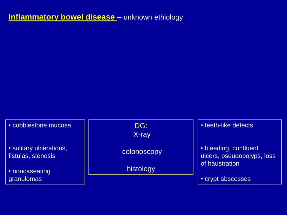
DG:
X-ray
colonoscopy
histology
• cobblestone mucosa
• solitary ulcerations,
fistulas, stenosis
• noncaseating
granulomas
• teeth-like defects
• bleeding, confluent
ulcers, pseudopolyps, loss
of haustration
• crypt abscesses
Inflammatory bowel disease – unknown ethiology

Ulcerative colitis
• continuous
involvement by spfc
ulcers
• inflammatory
pseudopolyps
• mucosa is reddish
and friable
• ulcers – not
penetrating into the
muscle layer
• nonspecific inf.inf.
in lamina propria
• goblet cells
diminished
• congestion of
mucosal capillaries
- hemorrhages
• crypts – deformed
• with accumulated
Neu – crypt
abscesses

Adenocarcinoma of the colon (47)
•E: genetic factors (FAP, HNPCC), environmental (alcohol, smoking, diet
low in fiber and rich in fat), adenomatous polyp, ulcerous colitis
• CP: asympt. ... alteration of diarrhea and obstipation, blood in
stool, abdominal pain, meteorism, weight loss, palpable Tu, ileus
• DG:
• colonoscopy, imaging techniques (X-ray, CT), staging
• prevention: per rectum + OB (yearly after 45 years of age)

Adenocarcinoma of the colon (47)
• variously differentiated tubular structures with atypia and mitosis
• infiltration of muscle and fat tissue
• grading
• mucus production (ec / ic)

Chronic hepatitis (136, 218)
• chronic / relapsing hepatic disease lasting over 6 months with
evidence of inflammation and necrosis
• E: viruses (HCV, HBV, HDV), drugs, AOI
• CP: fatigue, malaise, loss of appetite – hepatomegaly and
splenomegaly, hepatic tenderness
DG (in HBV hepatitis):
• elevated serum levels of HBsAg and HBcAg
• imunohistochemical evidence of HBV infection
• + elevated liver enzymes, bilirubin, decreased coagulation factors
and proteins

Chronic hepatitis
• necrosis of
hepatocytes
• piecemeal necrosis
• apoptotic bodies
• lymphoplasmocytic
infiltration
• steatosis
• fibrosis
• Mallory´s hyalin
• regressive changes of
bile ducts,
• activation of Kuppfer
cells,
• necrotic lobule with
collapsed reticulin
framework

Liver cirrhosis (62, 63)
• progressive irreversible destruction of hepatic architecture by fibrous septa
that encompass regenerative nodules of hepatocytes
• alcohol, HBV, HCV, AOI diseases, metabolic, biliary occlusion, cardiac
failure, toxic agents
• clinics:
• skin changes (icterus, spider nevi...)
• endocr. (gynecomastia, menstruation disorders)
• hypocoagulative condition, oedema, ascites
• COMPLICATIONS:
• portal hypertension
(esophageal varices, ascites, splenomegaly)
• hepatic failure, encephalopathy, hepatorenal
syndrome, Ca
• DG:
• lab. tests
• USG, liver biopsy

Liver cirrhosis (62, 63)
• multiple nodules: macro (>3mm) / micronodular (<3mm)
• liver more firm, larger / normal / shrunk, colour variable
• normal lobular structure not present – replaced by nodules
(disorganised proliferation of HC, no CV)
• nodules divided by fibrous septa with chronic inf.inf.
• steatosis
• necrosis (in active form)
• pseudotubules in portal regions

Chronic cholecystitis (99)
• E: gallstones, bacterial infection (streptococcus, Escherichia coli),
repeated attacks of acute CHC
• fibrous thickening of the wall of the gallbladder
• shrinking of the gallbladder
• calcification of the wall (porcelain gallbladder)
• thickening of the muscle layer
• fibrosis of the wall
• chronic inf.inf. of the mucosa and subserosal layer
• invagination of epithelial cells into musculature (Rokitansky-Aschoff
sinuses)

Acute hemorrhagic necrosis of pancreas (130)
• severe acute inflammation of pancreatic tissue
• autodigestion by pancreatic enzymes (acinic cell damage / duct
obstruction / increased exocrine production of enzymes)
• E: idiopathic, gallstone, alcohol, fatty foods, trauma, steroids, mumps,
autoimmune disease, hyperlipidemia, hypercalcemia, drugs
• P:
• enzymes digest the pancreas and surrounding tissues
• proteases – proteolysis
• elastases – destroy the elastic fibres of the vessel walls
• lipases and phospolipases – lipolysis – fat necrosis – combine with Ca –
saponification (Balser necrosis)

• clinics:
• severe epigastric pain with belt-like irradiation below the ribs
• fever, nausea, vomiting
• ascites, peritoneal irritation (defens, meteorism, subileus)
• COMPLICATIONS:
• peritonitis (chemical and bacterial) – shock – acute renal failure
• DIC
• pancreatic abscess / pseudocyst (formed after clearing of necrotic
debris)
• obstruction of duodenum
• thrombosis v.portae, v.cava
• mortality is high!!! 20-30%

• pancreas larger, swollen,
edematous
• necrosis
• hemorrhage
• Balser necrosis
• blood stained ascitic fluid
• hyperemia
• oedema
• necrosis of pancreatic tissue
• necrosis of vessel walls - hemorrhage
• fat necrosis
• acute inf.inf.